UNIVERSITÀ DEGLI STUDI DI SASSARI UNIVERSITY OF SASSARI
Dipartimento di Scienze Biomediche Department of Biomedical Sciences
Scuola di Dottorato di Ricerca in Scienze Biomolecolari e Biotecnologiche International PhD School in Biomolecular and Biotechnological Sciences
Curriculum: Biochemistry, Physiology and Molecular Biology
Cycle XXVII
Director: Prof. Leonardo A. Sechi
Cellular and Molecular Study of Vascular Damage during
Systemic Sclerosis
Tutor/Supervisor: Prof.Gianfranco Pintus
PhD Student: Dr. Tulasigeri M.Totiger
ACADEMIC YEAR 2013-2014
Dr. Tulasigeri M.Totiger
“Cellular and Molecular Study of Vascular Damage during Systemic Sclerosis”
PhD Thesis in Biochemistry, Physiology and Molecular Biology of PhD School in Biomolecular and Biotechnological Sciences, University of Sassari
Dr. Tulasigeri M.Totiger
“Cellular and Molecular Study of Vascular Damage during Systemic Sclerosis”
PhD Thesis in Biochemistry, Physiology and Molecular Biology of PhD School in Biomolecular and Biotechnological Sciences, University of Sassari
Table of Contents
ABSTRACT ………1
ABBREVIATIONS………...2
CHAPTER 1. INTRODUCTION………3
1.1 Historical Background & Definition of Systemic Sclerosis……...4
1.2 Classification of Systemic Sclerosis………...5
1.3Pulmonary Hypertension………..5
1.4 Classification of Pulmonary Arterial Hypertension (PAH)………6
1.5 SSc-PAH………...7
1.6 Epidemiology of SSc-PAH………..8
1.7 Subtypes of PAH in SSc………9
1.8 Introduction and Biochemistry of Oxidative Stress (OS)……….10
1.9Role of OS in Vascular Damage of SSc-PAH……….11
1.10 Role of OS in Vascular Remodeling in SSc-PAH………14
1.11Involvement of NOX in SSc-PAH………..15
1.12 Involvement of ERK in Collagen Synthesis in SSc-PAH………..16
CHAPTER 2 AIM OF THE WORK………19
CHAPTER 3 MATERIALS AND METHODS………24
3.1 Materials………25
3.2 Cell culture and treatments……….25
Dr.Tulasigeri M.Totiger
“Cellular and Molecular Study of Vascular Damage during Systemic Sclerosis”
PhD Thesis in Biochemistry, Physiology and Molecular Biology of PhD School in Biomolecular and Biotechnological Sciences, University of Sassari
Table of Contents
3.4 Measurements of Intracellular ROS……….27
3.5 Production of Lentiviral Particle containing COL1A1………….28
3.6 Transduction of COL1A1 promoter containing Lentivirus…..31
3.7 Measurement of COL1A1 Promoter Activity………..32
3.8Study of NOX Involvement………..32
3.9Study of ERK Involvement………...32
3.10 Statistical Analysis……….33
CHAPTER 4 RESULTS……….34
4.1 Demographic Data and Clinical Characteristics of Subjects..35
4.2Exposure of subjects ‘sera to HPASMCs & Kinetic Measurement of Intracellular ROS……….39
4.3 Exposure of subjects ‘sera to HPASMCs & Kinetic Measurement of COL1A1 Promoter Activity……….42
4.4 Effect of N0X2 inhibitor gp91on Intracellular ROS………...45
4.5 Effect of N0X2 inhibitor gp91 on COLA1 promoter Activity…47 4.6 Effect of ERK inhibitor PD98059 on Intracellular ROS………..49
4.7 Effect of ERK inhibitor PD98059 on COLAA1 Activity………….51
CHAPTER 5 DISCUSSION & CONCLUSION……….53
CHAPTER 6 BIBLIOGRAFY………..62
1 Dr.Tulasigeri M.Totiger
“Cellular and Molecular Study of Vascular Damage during Systemic Sclerosis”
PhD Thesis in Biochemistry, Physiology and Molecular Biology of PhD School in Biomolecular and Biotechnological Sciences, University of Sassari
ABSTRACT
Systemic Sclerosis is a devastating vascular, connective and a multisystem autoimmune disease characterized by fibrosis of skin and internal organs, exhibiting various forms of disease conditions. Pulmonary arterial hypertension (PAH) is one of the clinical manifestations that arise as a result of cellular and molecular vascular damage in the vascular wall, and is one of the leading causes of death in systemic sclerosis (SSc). SSc-PAH is a progressive depleting condition that can lead to right-sided heart failure and death. Oxidative stress is largely evidenced in the development of arterial hypertension in SSc.This may give rise to unbalanced redox homeostasis that could further bring about vascular remodeling characterized by Vascular Smooth Muscle Cells (VSMC) hypertrophy and hyperplasia and ECM deposition leading to subsequent obliterative vasculopathy and vessel occlusion.So it is likely that circulating pro-oxidant factors may be involved in the pathogenesis of SSc-PAH by inducing VSMCs activation and phenotypic switch. So we speculate, that in SSc patients associated with Pulmonary Arterial Hypertension (PAH), circulating factors may drive an aberrant vascular remodeling by exerting pro-oxidant and pro-fibrotic effects on Human Pulmonary Arterial Smooth Muscle Cells (HPASMCs) and activate collagen I synthesis. To test this hypothesis, we exposed primary Human Pulmonary Artery Smooth Muscle Cells (HPASMCs) to serum obtained from SSc patients with or without PAH and healthy donors (HD) and looked for the production of reactive oxygen species (ROS) and collagen I synthesis. From our study, we found that SSc-PAH patients’ circulating factors exhibited pro-oxidant effect by inducing ROS generation through NOX2 in HPASMCs and we also found that, circulating factors exhibited pro-fibrotic effect by inducing activation of collagen I synthesis inHPASMCs via ERK signaling, thus asserting the vascular damage in SSc patients.
2 Dr. Tulasigeri M.Totiger
“Cellular and Molecular Study of Vascular Damage during Systemic Sclerosis”
PhD Thesis in Biochemistry, Physiology and Molecular Biology of PhD School in Biomolecular and Biotechnological Sciences, University of Sassari
ABBREVIATIONS
Systemic Sclerosis(SSc)
Limited cutaneous SSc (lcSSc) Diffuse cutaneous SSc (dcSSc)
Vascular smooth-muscle cells (VSMCs) Pulmonary Arterial Hypertension (PAH) Vascular remodeling (VR)
Oxidative Stress (OS)
Reactive Oxygen Species (ROS)
Nicotinamide Adenine Dinucleotide Phosphate-Oxidase (NADPH Oxidase or NOX)
Human Pulmonary Artery Smooth Muscle Cells (HPASMCs) Pulmonary Artery (PA)
Endothelin (ET)
Extracellular signal-regulated kinase (ERK) Right heart catheterization (RHC)
Healthy donors (HD)
Right ventricular systolic pressure (RVSP)
Dichlorodihydrofluorescein-diacetate (H2-DCFDA) Collagen type-I(COL1A1)
3 Dr. Tulasigeri M.Totiger
“Cellular and Molecular Study of Vascular Damage during Systemic Sclerosis”
PhD Thesis in Biochemistry, Physiology and Molecular Biology of PhD School in Biomolecular and Biotechnological Sciences, University of Sassari
4 Dr. Tulasigeri M.Totiger
“Cellular and Molecular Study of Vascular Damage during Systemic Sclerosis”
PhD Thesis in Biochemistry, Physiology and Molecular Biology of PhD School in Biomolecular and Biotechnological Sciences, University of Sassari
CHAPTER 1. INTRODUCTION
1.1 Historical Background and Definition of Systemic Sclerosis
Systemic Sclerosis (SSc) is also called as Scleroderma or Systemic Scleroderma or Familial Progressive Scleroderma or Progressive Scleroderma. The first recitation of skin diseases consubstantial to SSc were reported as early as Hippocrates (460 - 370 B.C.), that are cited by the detailed historical account of SSc by Rodnan and Benedek in 1962 [1, 2]. However, Italian physician and dermatologist Carlo Curzio in 1753 [3] was the first to describe this disease condition. He reported about a 17-year old woman who had excessive tension and hardness of skin and she could hardly move her limbs. Goetz gave the name progressive systemic sclerosis or systemic sclerosis in 1945 [4], and this has become widely accepted term for this disease. Scleroderma was recognized as a clinical entity in mid-19th century with its present name [5].
More than 20 descriptive names had been proposed in the 19th century before this disease condition was named as sclerodermie or scleroderma. Systemic Sclerosis is derived from the Greek words ‘scleros’ and ‘derma’, means thickened, hardened skin. It is integrated by two words Systemic and Sclerosis where in Systemic refers to affecting a particular body system, or circulating through the entire body and Sclerosis refers to abnormal thickening or hardening of a body part.It is a connective tissue and a chronic multisystem autoimmune disease characterized by vasculopathy, diffuse fibrosis of skin and various internal organs. Systemic sclerosis involves hardening of internal organs and stops them working normally. It is a rare, heterogeneous and a slow-motion disease, but can be very serious. It varies greatly from person to person and hence there are many symptoms and problems that may progress or evolve with systemic sclerosis. As of now, there is no cure for scleroderma but effective treatments for some phenotypes of the disease are available.
5 Dr. Tulasigeri M.Totiger
“Cellular and Molecular Study of Vascular Damage during Systemic Sclerosis”
PhD Thesis in Biochemistry, Physiology and Molecular Biology of PhD School in Biomolecular and Biotechnological Sciences, University of Sassari
Introduction
1.2 Classification of SSc
There are mainly two classes of SSc, named as Localized & Systemic
Scleroderma. Localized scleroderma does not involve internal organs. But
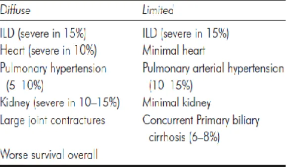
systemic scleroderma has been given more importance due to its involvement of internal organs. It has two subsets based on the extent of skin involvement (as shown in Fig. C) , namely Limited cutaneous SSc (lcSSc) with skin involvement distal to the elbows and knees, with or without involvement of face and Diffuse cutaneous SSc (dcSSc) with skin involvement of the proximal limbs and/or trunk [6, 7]. The distinguishing features between the both the subsets are explained in the table below [8].
Figure C: Skin involvement in LSSc & DSSc
. Table 1.Distinguishing Fetures:LSSc & DSSc
Baltimore 2002; Ferri C. et al (Medicine) 81(2):139-153. Dinesh Khanna. Indian Journal of Rheumatology June 2010
1.3 Pulmonary Hypertension
There are several heterogeneous clinical manifestations of SSc involving cutaneous, vascular, pulmonary, gastrointestinal, endocrine, neurologic, cardiac, renal and muscoskeletal. Pulmonary hypertension is one of the serious manifestations that develop in SSc patients. Actually, pulmonary hypertension is an abnormal increase of the pressure in the blood vessels of
6 Dr. Tulasigeri M.Totiger
“Cellular and Molecular Study of Vascular Damage during Systemic Sclerosis”
PhD Thesis in Biochemistry, Physiology and Molecular Biology of PhD School in Biomolecular and Biotechnological Sciences, University of Sassari
Introduction
the lungs. This is usually called as the “high blood pressure” of the lungs. In normal functioning of lungs, the pressure in the blood vessels is about one-quarter of the pressure in the arteries of the body and this can temporarily
adapt to increased pressures that occur during exercise [9]. But during pulmonary hypertension, in the lungs, small arteries are very much narrow,
that’s why the pressure rises in these vessels. Because of this, the right side of the heart, which pumps blood into the lungs, will have to pump against a higher resistance of blood flow. This would cause more difficult to pump the blood through the lungs, exactly when increased flow is needed, in a patient when exercises [9].Pulmonary arterial hypertension (PAH) is described as a progressive condition characterized by elevated pulmonary arterial pressures causing right ventricular (RV) failure [10]. In medical terms it is defined as an increase in mean pulmonary arterial pressure (mPAP) ≥ 25 mm Hg at rest as assessed by right heart catheterization [11,12]. Different hemodynamic definitions of PHare described based on pulmonary capillary wedge pressure, pulmonary vascular resistance (PVR) and cardiac output (CO).Clinical groups 1, 3, and 4 are of pre-capillary PH, group 2 compose post-capillary PH [13-19] and group 5 linked to PH with unclear or multifactorial etiologies.
1.4 Classification of Pulmonary Arterial Hypertension (PAH)
The revised classification comprises of 5 different types of PAH, with the inclusion of subclasses & their subtypes [13, 16-19]. They are as follows.
1. Pulmonary arterial hypertension
2. Pulmonary hypertension due to left heart disease
7 Dr. Tulasigeri M.Totiger
“Cellular and Molecular Study of Vascular Damage during Systemic Sclerosis”
PhD Thesis in Biochemistry, Physiology and Molecular Biology of PhD School in Biomolecular and Biotechnological Sciences, University of Sassari
Introduction
4. Chronic thromboembolic pulmonary hypertension (CTEPH)
5. Pulmonary hypertension with unclear multifactorial mechanisms
PAH or Group 1 PH include PAH having different etiologies. The pathological characteristics of this group involve pulmonary arterial endothelial cell (EC)
dysfunction, pulmonary artery EC and smooth muscle cell (SMC) proliferation, vasoconstriction and in situ thrombosis [20]. The sub-groups of PAH include
common clinical characteristics and share similarities in terms of
management [17]. PAH remains an incurable disease process, even though there are many new available therapies from the last two decades. If PAH not
interrupted, would leads to right heart failure and death [22, 23, 24].
1.5 Systemic Sclerosis-associated Pulmonary Arterial
Hypertension (SSc -PAH)
The existence of a small vessel vasculopathy is one of the distinguishing features of SSc & hence SSc patients are at greater risk of developing & arriving at disease condition called PAH. This disorder is now admitted as the major cause of morbidity and mortality. So the newest (2013) American College of Rheumatology / European League Against Rheumatism (ACR/EULAR) classification criteria has given importance & recognition to it and provided equal weight and consideration to SSc-PAH as similar to other manifestations of SSc. PAH is a life-threatening ailment, which could rapidly progress to severe right heart failure [13, 14]. The prevalence of PH in connective tissue diseases is usually estimated on the basis of echocardiographic examinations, however most of the recent studies show that for systemic sclerosis (SSc), PH assessment depends on the basis of strict hemodynamic criteria [25,26,27]. Pulmonary arterial hypertension (PAH) is described as a progressive pulmonary vascular disease that involves
8 Dr. Tulasigeri M.Totiger
“Cellular and Molecular Study of Vascular Damage during Systemic Sclerosis”
PhD Thesis in Biochemistry, Physiology and Molecular Biology of PhD School in Biomolecular and Biotechnological Sciences, University of Sassari
Introduction
narrowing or tightening or constriction of the pulmonary arteries that connect the right side of the heart to the lungs. PAH is characterized by an increase in mean pulmonary arterial pressure (PAP) to ≥25 mmHg at rest, and a mean primary capillary wedge pressure of ≤15 mmHg [15]. As PAH progresses, the blood flow through the pulmonary arteries is restricted and because of this, there occurs an enhanced strain of pumping blood through the lungs, resulting in the enlargement of the right side of the heart. As a result of this strain on the heart and also due to the reduction in blood to the left heart and also to the systemic circulation through the lungs lead to usual symptoms of PAH, such as Dyspnea (breathlessness), fatigue,weakness,Exertional syncope, angina, and abdominal distension [12].The parameter that indicates a high risk of future development of PAH in patients with SSc is the existence of ‘borderline’ pressures aligning between 21 and 24mmHg [28, 29].
1.6 Epidemiology of SSc-PAH
PAH is caused by the pulmonary vascular remodeling and can either occur alone (SSc-PAH), or may advance secondary to pulmonary parenchymal involvement resulting as interstitial lung diseases (ILD-PH). ILD-PAH is the most frequent and a serious complication of SSc. It occurs in 75% of casesand is more frequent in diffuse cutaneous SSc (DcSSc) [30-33]. Based on right heart catheterization (RHC), the prevalence of PAH other than ILD-PH is between 7.85 and 12% [30, 31] and more common in later stage of limited cutaneous SSc (LcSSc). Isolated PAH (SSc-PAH) is more common in LcSSc, when compared to DcSSc,however fewrecent studies [30-38] suggest that the prevalence of PAH is similar in both limited and diffuse cutaneous SSc patients. SSc-PAH has worse prognosis than that of IPAH [39] and patients are at higher risk of death than IPAH patients. Even though both types share
9 Dr. Tulasigeri M.Totiger
“Cellular and Molecular Study of Vascular Damage during Systemic Sclerosis”
PhD Thesis in Biochemistry, Physiology and Molecular Biology of PhD School in Biomolecular and Biotechnological Sciences, University of Sassari
Introduction
similar histopathological & haemodynamic characteristics but estimated one-year survival rates are 55% and 84%, respectively [40].PAH among patients with SSc is estimated to be 0.61 cases per 100 patient-years [41]. Median survival time of SSc patients with PAH is between 1 to 3 years [40, 42] and SSc patients with ILD alone have a median survival of 5–8 years [43].
1.7 Subtypes of PAH in SSc
Pulmonary hypertension, defined by mean pulmonary arterial pressure greater than 25 mm Hg, can be isolated in SSc, occurring as PAH
(SSc-associated PAH or SSc-PAH), and this one is due to pulmonary vascular remodeling. The other subtype is in combination with ILD [pulmonary
hypertension (PH)-ILD] or (ILD-PH) [44,45], which may develop secondary due to parenchymal involvement.
A. Isolated Pulmonary Arterial Hypertension {IPAH} (in the
absence of significant pulmonary fibrosis or Interstitial Lung
Disease)
PAH in SSc patients without pulmonary fibrosis or ILD is a severe complication due to narrowing or occlusion of small pulmonary arteries caused by smooth muscle hypertrophy, intimal hyperplasia, inflammation, thrombosis in situ. The breathlessness (dyspnea) progression rate from normal exercise tolerance to oxygen dependency is about 6–12 months & the mean duration of survival is 2 years. Unlikely, SSc patients with PAH with respect to interstitial lung disease have a same degree of disability, but the difference is that they progress more slowly for a period more than 2, up to 10 years [46-50].The prevalence ofIsolated PAH in SSc (without significant
10 Dr. Tulasigeri M.Totiger
“Cellular and Molecular Study of Vascular Damage during Systemic Sclerosis”
PhD Thesis in Biochemistry, Physiology and Molecular Biology of PhD School in Biomolecular and Biotechnological Sciences, University of Sassari
Introduction
pulmonary fibrosis) is between 7 and 12% of patients [11].However, the most frequent type of pulmonary hypertension in SSc patients isGroup I PAH [51].
B. PAH associated with pulmonary fibrosis
With either of both the (diffuse or limited) forms of the disease,more than one third of SSc patients havepulmonary fibrosis. The clinical tests have revealed alveolar, interstitial, peribronchial and pleural fibrosis. This kind of PAH has relatively slow progression & has gradual elevation in the resistance of pulmonary vasculature resulting in the widespread pulmonary fibrosis [47]. ILD is prevalent in up to 75% of SSc cases and is more frequent in diffuse cutaneous SSc (DcSSc), beingoften complicated with pulmonary hypertension, plays significant role in PAH development [45].
1.8 Introduction and Biochemistry of Oxidative Stress (OS).
From long time it has been known that Oxidative stress plays a major role in the pathogenesis and vascular damage of SSc-PAH, which is elucidated by the abnormal redox state in SSc patients [52]. It is well documented that ROS mediates vascular damage in the vascular wall [53]. “Any species which is capable of independent existence and contains one or more unpaired electron in atomic or molecular orbitals” are termed as free radicals [54] and also called by the name Reactive Oxygen Species (ROS).Since free radicals possess atomic structure that has an unpaired electron in the outer orbital, gives them a special configuration of great instability. This provides it very un-stable state, making it short-lived and extremely reactive, with a tremendous capacity to interact nonspecifically with the diverse set of molecules of the cell structure such as carbohydrates, lipids, proteins and nucleic acids [55].Pro-oxidant denotes to any endobiotic or xenobiotic that induces oxidative stress
11 Dr. Tulasigeri M.Totiger
“Cellular and Molecular Study of Vascular Damage during Systemic Sclerosis”
PhD Thesis in Biochemistry, Physiology and Molecular Biology of PhD School in Biomolecular and Biotechnological Sciences, University of Sassari
Introduction
either by generation of ROS or by inhibiting antioxidant systems. It can include all reactive, free radical containing molecules in cells or tissues [56]. Pro-oxidants may be broadly classified into two categories as Exogenous and
Endogenous. Exogenous pro-oxidants include pro-oxidants derived from
dietary ingredients, toxicants, drugs, pathogens, environmental pollutions and climate. Endogenous pro-oxidants include pro-oxidants derived from endogenous metabolites, drug metabolites, cellular metabolism, ion flux, anxiety, pathophysiology and ischemia [56].These pro-oxidants may be present in the circulatory fluid (CF) which can induce OS in the vascular wall leading to vascular morbidities such as SSc-PAH and (CF) can be used to study the OS or vascular damage related pathophysiology of the vascular diseases.
1.9 Role of Oxidative stress in Cellular and Molecular Vascular
Damage of SSc-PAH
Oxidative stress (OS) has been recognized as an important triggering agent in inflammatory processes within the vascular wall, especially in the setting of systemic arterial disease [57]. OS may mediate through activation of mononuclear cells [58, 59] with infiltration of inflammatory cells in pulmonary perivascular spaces within and around plexiform lesions [60–62]. In the vasculature of HT patients, ROS are formed in high concentrations that cannot be balanced by the normal protective antioxidant mechanisms employed by the cells, resulting in oxidative stress [64]. As a result, •O2− reacts
with nitric oxide (NO) to produce a dramatic concentration of the toxic peroxynitrite (ONOO−) which stimulates a variety of negative effects on cellular function involving alteration in the functioning of kinases, protein synthesis, and redox-sensitive genes [65-67]. There exists a strong relationship between BP and some parameters related to OS [68].It has been
12 Dr. Tulasigeri M.Totiger
“Cellular and Molecular Study of Vascular Damage during Systemic Sclerosis”
PhD Thesis in Biochemistry, Physiology and Molecular Biology of PhD School in Biomolecular and Biotechnological Sciences, University of Sassari
Introduction
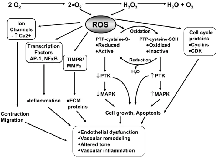
shown that ROS production is elevated with the redox-dependent signaling amplified and antioxidant activity reduced in cultured vascular smooth muscle cells of arteries from hypertensive rats and humans [69, 70].Reactive oxygen species (as depicted in Figure D) are known to play a significant role in intracellular signal transduction and trigger many growth-associated signaling pathways in vascular smooth muscle cells, comprising phosphorylation-mitogen activated protein arterial pressure (MAP) kinases (p38 and ERK5) and others [71-84]. ROS stimulate STATs, activate Akt by Ang II and activate tyrosine kinases and tyrosine phosphatases, and activate ras as well [71, 83]. These signaling events would lead to redox-sensitive growth in vascular smooth muscle cells, proliferation, hypertrophy, which eventually would confer to vascular wall thickening and remodeling (as depicted in Figure D) in hypertension [71-99]. The organization of membrane myofilaments poses a great impact on the integrity of vascular smooth muscle cell morphology and ROS can cause severe morphologic and structural alterations resulting in the vast damage of the cellular cytoskeleton. Thus ROS are involved in endothelial dysfunction, increased reactivity, and vascular remodeling which are the characteristic features of vascular damage in
13 Dr. Tulasigeri M.Totiger
“Cellular and Molecular Study of Vascular Damage during Systemic Sclerosis”
PhD Thesis in Biochemistry, Physiology and Molecular Biology of PhD School in Biomolecular and Biotechnological Sciences, University of Sassari
Figure D. Redox-dependent signaling pathways in vascular smooth muscle cells. Intracellular reactive oxygen species (ROS) modify the activity of protein tyrosine kinases (PTK), such as Src, Ras, JAK2, Pyk2, PI3K, and EGFR, as well as mitogen-activated protein kinases (MAPK), particularly p38MAPK, JNK and ERK5. These processes probably occur through oxidation/reduction of protein tyrosine phosphatases (PTP), which are susceptible to oxidation and inactivation by ROS. ROS also influence gene and protein expression by activating transcription factors, such as NF-nB, activator protein-1 (AP-1) and hypoxia-inducible factor-1 (HIF-1). ROS stimulate ion channels, such as plasma membrane Ca2+ and K+ channels, leading to changes in cation concentration. Activation of these redox-sensitive pathways results in numerous cellular responses which, if uncontrolled, could contribute to hypertensive vascular damage.ECM, extracellular matrix; MMPs, matrix metalloproteinases; TIMP, tissue inhibitor of matrix metalloproteinase.Tamara M and R.M.Touyz .Redox signaling in hypertension. Cardiovascular Research 71 (2006) 247 – 258.
14 Dr. Tulasigeri M.Totiger
“Cellular and Molecular Study of Vascular Damage during Systemic Sclerosis”
PhD Thesis in Biochemistry, Physiology and Molecular Biology of PhD School in Biomolecular and Biotechnological Sciences, University of Sassari
Introduction
1.10 Role of Oxidative Stress in Vascular Remodeling in
SSc-PAH
Vascular remodeling is an active process of structural change that relies on active interactions between local growth factors, vasoactive substances, and hemodynamic stimuli and is an active process that arises in retort to longstanding changes in hemodynamic conditions and leads to the pathophysiology of vascular diseases. It is well documented that OS plays significant role in Vascular Remodeling in SSc-PAH. During this process, the arterial system endures structural remodeling that involves hypertrophy of the arterial wall and increased wall-to lumen ratio and accompanying decreased arterial distensibility. The stage when the remodeling process develops maladaptive, it carries further vascular damage followed by impaired endothelial function, enhanced reactivity, and vascular inflammation. In the condition of hypertension, OS promotes VSMC proliferation and hypertrophy, collagen deposition, and alterations in activity of MMPs, which results in thickening of the vascular media and arterial remodeling [103]. It has reported that Superoxide anion and H2O2 excite several growth factor-like cellular responses, such as intracellular alkalinization, MAP kinase phosphorylation, and tyrosine kinase activation [71]. In hypertension, OS not only influences arterial structure, media thickening but also eventually influences & affects complete vessel redox state. So vascular wall thickening increases the distance required for diffusion of oxygen from the lumen. With this, there would be a reduced pO2, which results in incomplete oxidation and increased concentrations of free radicals and abnormalities of the oxidant state. With this overall excess of OS state in hypertension, would further lead to vascular smooth muscle cell growth, endothelial dysfunction, and over-all vascular damage [104]. This would lead to progressive increase in pulmonary vascular resistance, pulmonary arterial pressure, and right ventricular (RV) pressure overload.Initially,
15 Dr. Tulasigeri M.Totiger
“Cellular and Molecular Study of Vascular Damage during Systemic Sclerosis”
PhD Thesis in Biochemistry, Physiology and Molecular Biology of PhD School in Biomolecular and Biotechnological Sciences, University of Sassari
Introduction
compensatory mechanisms in the right ventricle preserve the stroke volume and cardiac index, but as the limits are crossed, cardiac failure and death follow.
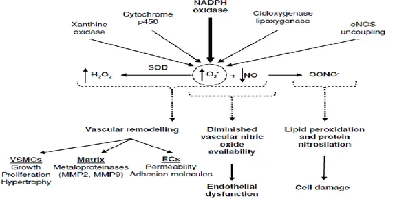
1.11 Involvement of NOX in SSc-PAH
The activation of vascular NAD (P) H Oxidase plays a pivotal role in vascular functional and structural changes for the development of hypertension [67]. The NOX1 (NADPH oxidase 1) and NOX2 oxidases are the major sources of
Figure E. NADPH oxidase(NOX) a major component involved in generation of ROS, leading to OS, Vascular Remodeling & Vascular Damage in SSc-PAH {Ana Fortuno et al. Oxidative stress and vascular remodeling. Exp Physiol 90.4 pp 457–462(2005)}.
16 Dr. Tulasigeri M.Totiger
“Cellular and Molecular Study of Vascular Damage during Systemic Sclerosis”
PhD Thesis in Biochemistry, Physiology and Molecular Biology of PhD School in Biomolecular and Biotechnological Sciences, University of Sassari
Introduction
ROS in the artery wall in hypertension as well as in SSc-PAH, and thus are formed to be important contributors [63] to the oxidative stress that would lead to vascular damage[105-108]even in the premature stages[109, 110] of
vascular disease. The abnormal vasoconstriction in the vascular wall by hormones, such as endothelin-1, angiotensin II, or urotensin II is mediated by ROS [111] produced, majorly by NOX2.ROS derived from NOX isoforms, in particular NOX2 and NOX4, are evident to be involved in long-term responses of the pulmonary vasculature to hypoxia [112-115]. NOX2 is involved in hypoxia induced endothelial dysfunction in the intrapulmonary arteries [115].Increased level of NOX was documented in the pulmonary artery smooth muscle cells (PASMCs) in hypoxia dependent development of PAH in mice [113]. As illustrated in the above Figure E, NOX more specifically NOX2, by generating ROS, contribute to the narrowing of the arterial lumen and consequently to increased peripheral resistance and blood pressure, thus
leading to Hypertension in SSc patients.
1.12 Involvement of ERK in Collagen Synthesis in SSc-PAH
There are many signaling pathways involved in the regulation & synthesis of Collagen. The MAPK/ERK pathway is one of the predominant one among them [116,117].In both the vasoconstriction and vascular smooth muscle cell growth, extracellular signal-regulated kinase (ERK) is involved, which is a member of the mitogen-activated protein kinase family [118]. From the reports of hypertension in animal models, it has been showed that ERK activity is raised in vascular smooth muscle cells, and inhibition of ERK activation reduces both vascular smooth muscle cell growth and vasoconstriction [118,119]. ERK1 and ERK2 are the extensively explored, even though other isozymes of ERK are present. Activation of ERK can happen through stimulation of either a G protein-coupled receptor or a growth factor
17 Dr. Tulasigeri M.Totiger
“Cellular and Molecular Study of Vascular Damage during Systemic Sclerosis”
PhD Thesis in Biochemistry, Physiology and Molecular Biology of PhD School in Biomolecular and Biotechnological Sciences, University of Sassari
Introduction
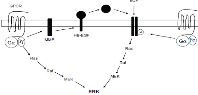
receptor, and this can be followed by activation of the Ras, Raf, mitogen-activated protein kinase pathway [118, 119]. There are evidences which reveals that, activation of ERK through G protein-coupled receptors can happen (as shown in Fig: F) through direct activation of the Ras, Raf, or mitogen-activated protein kinase pathway, or through transactivation of a growth factor receptor, such as EGFR. This might happen through activation of a matrix metalloprotease and succeeding cleavage of a membrane-bound ligand such as heparin-binding epidermal growth factor (EGF), leading to release of the ligand and activation of the receptor. In other way, activation of the G protein-coupled receptor could lead to tyrosine phosphorylation of the epidermal growth factor receptor [118]. However, there are few evidences suggesting that ERK activation occurs via ROS, but the mechanism needed to be elucidated [120].
Figure F. Schematic diagram summarizing the potential mechanisms of extracellular signal-regulated kinase activation. Richard E Roberts. The extracellular signal-regulated kinase (ERK) pathway: a potential therapeutic
18 Dr. Tulasigeri M.Totiger
“Cellular and Molecular Study of Vascular Damage during Systemic Sclerosis”
PhD Thesis in Biochemistry, Physiology and Molecular Biology of PhD School in Biomolecular and Biotechnological Sciences, University of Sassari
Introduction
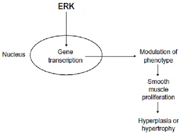
It has been shown that activation of ERK is linked with changes in gene
transcription and cell proliferation [118]. Activated (as shown in Fig: F) ERK is known to be involved in Collagen synthesis and has been reported in various types of cells including VSMCs.
Figure G: Schematic diagram summarizing the effect of extracellular signal-regulated kinase on vascular smooth muscle cell growth and proliferation through effects on gene transcription. Richard E Roberts 2012.
Undeniably, changes of primary vascular smooth muscle cells from a contractile to a proliferating phenotype are associated with prolonged ERK activation [118] and the accompanying synthesis of collagen. This could progress the disease conditions such as IPAH, SSc-PAH and stenosis in which there is elevation in vascular smooth muscle cell growth, collagen synthesis
and subsequent aberrant vascular remodeling (hypertrophy or hyperplasia).
19 Dr. Tulasigeri M.Totiger
“Cellular and Molecular Study of Vascular Damage during Systemic Sclerosis”
PhD Thesis in Biochemistry, Physiology and Molecular Biology of PhD School in Biomolecular and Biotechnological Sciences, University of Sassari
CHAPTER 2. AIM OF THE WORK
20 Dr. Tulasigeri M.Totiger
“Cellular and Molecular Study of Vascular Damage during Systemic Sclerosis”
PhD Thesis in Biochemistry, Physiology and Molecular Biology of PhD School in Biomolecular and Biotechnological Sciences, University of Sassari
CHAPTER 2. AIM OF THE WORK
An important note to assign SSc straight to vascular morbidity is because the vascular bed is a primary site of injury [32, 38, 44, 48, 121], and diffuse devascularization of multiple tissues is the major consequence. Hence, SSc is termed as Vascular Disease. Even, the course of the pathogenesis clearly shows the mode of vascular complications that line from the onset of the disease till late clinical manifestations. Raynaud phenomenon (RP) is observed in all most all SSc patients & is one of the earliest symptoms seen in SSc patient. When looked at cellular level, it is the clinical manifestation that involves abnormal functioning of cutaneous vessels that happens due to the thermal regulation of blood flow [122-124]. RP occurs before the onset of clinical signs of tissue fibrosis. The vascular abnormalities that begin & participate in this disease condition have prompted to better understand the disease course from the vascular point of view, which could help in finding out cellular & molecular hints that set the vascular damage during systemic sclerosis. Pulmonary involvement occurs in at least two thirds of systemic sclerosis patients and about 10-15% of them will develop severe lung disease during the course of their illness. Pulmonary disease has surpassed renal disease and is now the leading cause of death amongst patients with scleroderma [34, 44, 45]. It is estimated that 80% of patients with SSc have some evidence of pulmonary disease. The estimated mortality of pulmonary disease from all causes is said to be 33% [34, 35, 41]. Moreover, pulmonary involvement has a poorer prognosis [39]. Pulmonary arterial hypertension (PAH) is a grimly progressive life-threatening & a serious condition which prematurely takes a toll on many lives. Systemic sclerosis-related pulmonary arterial hypertension (SSc-PAH) is a major complication of both limited and diffuse systemic sclerosis that leads to substantial morbidity & mortality. SSc-PAH has emerged as a leading cause of death [38, 44]. These are some of the reasons that have prompted us take up this study. In vascular smooth muscle
21 Dr. Tulasigeri M.Totiger
“Cellular and Molecular Study of Vascular Damage during Systemic Sclerosis”
PhD Thesis in Biochemistry, Physiology and Molecular Biology of PhD School in Biomolecular and Biotechnological Sciences, University of Sassari
Aim of the work
activity and tyrosine phosphorylation in response to cooling and are linked to excessive alpha2-adrenergic response and shown that increased level ROS activate of Rho/Rho kinase pathway and up-regulates alpha2c-adrenergic receptors on the surface of vascular smooth muscle cells, thus determining an excessive vasoconstrictive response to cooling [122-124] and clearly showing that ROS[52] are involved in the early pathogenesis. Structural alterations of small and medium-sized arteries contribute to RP involving Smooth muscle cells & endothelial cells, thus leading to Vascular remodeling as result of deposition of extracellular matrix (ECM) and fibrosis. These above mentioned participations of SMCs prompted us to use this type of cells as an ideal cell model to study SSc-PAH. In hypertension, OS besides influencing arterial structure, media thickening, also eventually influences & affects complete vessel redox state [125,126]. Hence, vascular wall thickening would lead to increase in the distance required for diffusion of oxygen from the lumen thus resulting in reduced pO2, which leads to incomplete oxidation and increased concentrations of free radicals and abnormalities of the oxidant state [52]. This overall excess of OS state in hypertension, would further lead to vascular smooth muscle cell growth, endothelial dysfunction, and over-all vascular damage [125,126].Many circulating factors have been shown to be associated in the pathogenesis of Raynaud’s phenomenon related to SSc, involving in platelet activation, impaired fibrinolysis, white blood cell activation, reduced red blood cell deformability and oxidative stress [122,127]. Due to OS, there occurs an unbalanced redox homeostasis, and circulatory factors may contain the endogenous derived factors which can exhibit the pathophysiology state that is caused by the OS. Thus the circulatory factors play very significant roles in the assessment of SSc-PAH, wherein they could be used to study the pathophysiology that has been exerted by OS. In such condition the blood as the circulatory fluid may have ROS or free radicals or pro-oxidant factors, activated growth factors, cytokines or molecules in it, which may direct in
22 Dr. Tulasigeri M.Totiger
“Cellular and Molecular Study of Vascular Damage during Systemic Sclerosis”
PhD Thesis in Biochemistry, Physiology and Molecular Biology of PhD School in Biomolecular and Biotechnological Sciences, University of Sassari
Aim of the work
activating the pulmonary arterial smooth muscle cells, endothelial cells or the complete vascular bed with the wrong signals formed by the abnormality
caused by OS or other factors. From previous reports, it is well evident that NOX are the major contributors of ROS in the vascular wall. It has been shown
that NOX2 subunit involved in generation of large amounts of ROS, and so inhibiting or deleting NOX2 or p47phox subunits significantly reduced vascular oxidative stress in several disease models [106, 129-136]. So there is a strong rationale for therapeutically targeting NOX2 oxidase in the arterial wall for the treatment of vascular disease such as SSc-PAH, so as to combat the oxidative stress and prevent the progression of vascular disease such as SSc-PAH.It is has been suggested that the high mortality in SSc-PAH may be due to excessive collagen content in the heart and the pulmonary arteries [137]. Vascular collagen content has impact on hemodynamics of SSc-PAH, which is due to the overproduction of collagen throughout the body. However, it has been reported that excessive PA collagen accumulation is linked to increased PA stiffening and has been shown that excessive PA(Pulmonary Artery)collagen accumulation elevates (pulmonary vascular impedance) PVZ or right ventricular afterload [138-142]. So, whether sera of SSc-PAH stimulate collagen synthesis has become one of the objectives of this study. From the reports it is known that MAPK/ERK is the predominant pathway involved in the regulation & synthesis of Collagen and there are evidences reporting that ERK activation occurs via ROS [120] but the mechanism is not clearly understood. ERK signaling contributing to the overexpression of pro-fibrotic proteins in scleroderma fibroblasts as well as VSMCs is unclear. Hence, targeting ROS and as well as collagen synthesis by inhibiting ERK or ROS-mediated ERK could potentially be an important pharmacological treatment of hypertension in SSc. By gathering the above background, it can be explained collectively, that OS is involved in the Vascular Damage of SSc-PAH. This leads to the state of unbalanced redox homeostasis and generate large amount of ROS by stimulating growth factors & cytokines. These factors
23 Dr. Tulasigeri M.Totiger
“Cellular and Molecular Study of Vascular Damage during Systemic Sclerosis”
PhD Thesis in Biochemistry, Physiology and Molecular Biology of PhD School in Biomolecular and Biotechnological Sciences, University of Sassari
Aim of the work
may be present in the circulatory fluid and can be detected and their effect can be analyzed in in-vitro. This ligand stimulated change in the cellular redox state could lead to vascular remodeling characterized by VSMC hypertrophy & hyperplasia and ECM deposition leading to subsequent Obliterative Vasculopathy. Hence, it can be hypothesized that in SSc patients associated with Pulmonary Arterial Hypertension (PAH), circulating factors may drive an aberrant vascular remodeling by exerting pro-oxidant & Pro-fibrotic effects on Pulmonary Arterial Smooth Muscle Cells (PASMCs) and activate collagen I synthesis.
24 Dr. Tulasigeri M.Totiger
“Cellular and Molecular Study of Vascular Damage during Systemic Sclerosis”
PhD Thesis in Biochemistry, Physiology and Molecular Biology of PhD School in Biomolecular and Biotechnological Sciences, University of Sassari
25 Dr. Tulasigeri M.Totiger
“Cellular and Molecular Study of Vascular Damage during Systemic Sclerosis”
PhD Thesis in Biochemistry, Physiology and Molecular Biology of PhD School in Biomolecular and Biotechnological Sciences, University of Sassari
CHAPTER 3. MATERIALS AND METHODS
3.1. MaterialsSmooth Cell medium was purchased from ScienCell Research Labs, Fetal Bovine Serum (FBS) purchased from Invitrogen, rat tail Collagen I coating agent, ERK Inhibitor PD98059, DMEM were purchased from Sigma-Aldrich, GFP-based Lentiviral Particle (harboring the COL1A1 promoter having COL1A1-LV-tGFP and EF1α-LV-FP602) was initially purchased from Innoprot- Innovative Technologies In Biological Systems, S.L. But the production of the same Lentiviral particles was done by purchasing second generation packaging systems from Addgene,Inc. 2‟,7‟-dichlorodihydrofluorescein diacetate (H2DCF-DA) (Molecular Probe) was purchased from Eugene, OR.NOX NOX2ds-tat was purchased from Anaspec (Fremont, CA, USA) .
3.2 Cell culture and treatments.
Human Pulmonary Artery Smooth Muscle Cells (HPASMCs) purchased from Innoprot-Innovative Technologies In Biological Systems, S.L. The cell line was supplemented with specific medium(Smooth Cell medium, ScienCell). The cell culture was done by using rat tail Collagen I as a matrix or coating agent to promote adherence and attachment to provide proper cell growth. When cultured HPASMCs reach confluent stage, they were split in to 1:2 ratio and the cells were used for experiments with passage numbers below six. In routine maintenance of HPASMCs, during splitting of cells, the neutralization is done to stop trypsinization by using Neutralization Solution and Fetal Bovine Serum (FBS) in the ratio of 1:1.Experiments were done by plating required number of HPASMCs in 96-well black plates and white plates (BD Falcon, Franklin Lakes, NJ.
26 Dr. Tulasigeri M.Totiger
“Cellular and Molecular Study of Vascular Damage during Systemic Sclerosis”
PhD Thesis in Biochemistry, Physiology and Molecular Biology of PhD School in Biomolecular and Biotechnological Sciences, University of Sassari
Materials and Methods
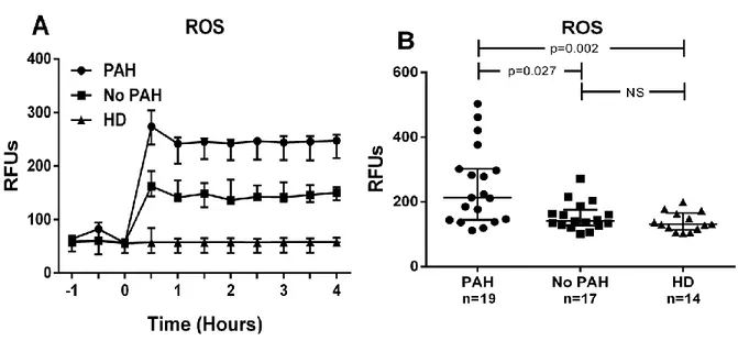
For measuring intra cellular ROS 10 μM of H2-DCFDA (Figures 1A & 1B) was used prior to stimulation by sera and the intra cellular ROS was measured kinetically for 4hrs.
In the experiments for measuring the collagen promoter activity(Figures 2A,2B,4,6), the cells were transduced with lentiviral particles obtained from the COL1A1-LV-tGFP and EF1α-LV-FP602 lentivectors, and then cultured in basal medium for stimulating the cells with sera and COL1A1 promoter activity were kinetically measured for 10 hours.
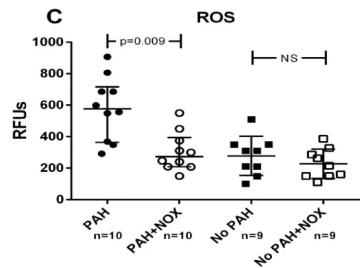
In the experiments (Figures 3, 4) for studying NOX involvement, HPASMCs were incubated for 1 hour with 5 μM NADPH oxidase specific inhibitor NOX2ds-tat (NOX) before treatment with SSc sera.
In the experiments (Figures 5, 6) for studying ERK involvement, HPASMCs were incubated for 1 hour with 15 μM of PD98059, a specific inhibitor of ERK before treatment with SSc sera.
3.3 Study Subjects.
Study subjects were enrolled by signing the informed consent and the collected data of clinical, serologic, and diagnostic criteria were according to the protocol approved by the Johns Hopkins Scleroderma Center Baltimore, Johns Hopkins University’s Institutional Review Board (IRB). Through posted flyers, Healthy donors (HD) were recruited and enrolled after going through such a screening questionnaire that aimed at excluding the presence of any underlying vascular or autoimmune disease. Each SSc patient met the American College of Rheumatology criteria or had 3 of 5 features of the CREST (Calcinosis, Raynaud’s syndrome; Esophageal dysmotility; Sclerodactyly; Telangiectasia) syndrome [143]. EachPatient’s demographic profile, SSc
27 Dr. Tulasigeri M.Totiger
“Cellular and Molecular Study of Vascular Damage during Systemic Sclerosis”
PhD Thesis in Biochemistry, Physiology and Molecular Biology of PhD School in Biomolecular and Biotechnological Sciences, University of Sassari
Materials and Methods subtype, autoantibody status, Medsger severity scores, pulmonary function tests, echocardiography, and right heart catheterization parameters,modified Rodnan skin scores(mRSS) were assessed. Demographic data comprising sex, race, smoking status (never, past current) were also evaluated. Calculation of SSc disease duration was done at the time of serum sampling, by counting the years from the onset of RP or from the first non-RP symptom. RP, heart and lung severity scores were evaluated as previously defined by Medsger et al [144].
3.4 Measurements of Intracellular ROS
Intracellular ROS levels were measured by using the ROS molecular probe 2‟,7‟-dichlorodihydrofluorescein diacetate (H2-DCFDA). Esterases cleave the acetate groups on H2-DCFDA within the cell, consequently trapping the reduced form of the probe (H2DCF). Intracellular ROS oxidize H2DCF, yielding the fluorescent product, DCF. When HPASMCs reach sub-confluent were loaded with 10 μM of H2-DCFDA. Then washed with PBS1X buffer followed by treatment with 5% (V/V) of sera from scleroderma (SSc) patients with pulmonary arterial hypertension (PAH), without PAH(No PAH) and healthy donors (HD)respectively in basal medium. Intracellular ROS levels were kinetically measured using GENios plus microplate reader (Tecan, Männedorf, CH) with excitation at 485 & emission at 535 respectively in a 4 hour time-course experiment (Figure 1A) and values at 2 hours (steady state) used for comparison (Figure 1B). Fluorescence measurements were corrected for background fluorescence and protein concentration.
28 Dr. Tulasigeri M.Totiger
“Cellular and Molecular Study of Vascular Damage during Systemic Sclerosis”
PhD Thesis in Biochemistry, Physiology and Molecular Biology of PhD School in Biomolecular and Biotechnological Sciences, University of Sassari
Materials and Methods
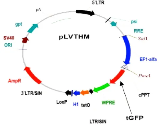
3.5 GFP-based Lentiviral Particle (harboring the COL1A1 promoter) Production.
Lentiviral vectors (LV) are very efficient (can exceed 90%) at transferring viral DNA into host cells and integrate stably into the host-cell genome of dividing and non-dividing cells and can provide long-term expression of the vectored transgene in target cells and thus provide great potential for gene therapeutic applications [145-148 ]. Because of these advantages we employed lentiviral vectors as shown below (Fig .H & I) in our study. The 2nd generation system plasmids are less bulky to use and highly efficient and have ease of use over third-generation system led us to use these in our study.
29 Dr. Tulasigeri M.Totiger
“Cellular and Molecular Study of Vascular Damage during Systemic Sclerosis”
PhD Thesis in Biochemistry, Physiology and Molecular Biology of PhD School in Biomolecular and Biotechnological Sciences, University of Sassari
Materials and Methods
Description of Each Component of the Vector Map:
5’LTR 5’ Long terminal repeat, contains cis regulatory regions which recruit
proteins that promote transcription of full length lentivirus.Sequences here facilitate proviral integration into the host genome; an intact 5’LTR indicates that this is a 2nd generation transfer plasmid
Psi Sequence that facilitates packaging of viral RNA
RRE Rev response element: sequence to which the Rev protein binds to
facilitate nuclear export of viral RNA
EF1α Promoter for elongation factor 1α, used to the expression of a marker
gene such as GFP
cPPT Central poly purine tract allows nuclear import of lentivirus in host cells GFP Green fluorescent protein used to mark successfully infected cells
WPRE The woodchuck hepatitis virus Posttranscriptonal response element
augments shRNA expression
TetO Binding site for the tet repressor protein or a variant thereof which can
be used to turn on or off shRNA expression
H1 Histone H1 promoter used to drive shRNA expression
LoxP Site which is recombined by the Cre recombinase in order to
conditionally remove the lentiviral provirus from the host genome after successful infection and incorporation
3’LTR/SIN 3’LTR with a large deletion in the U3 region, makes the virus
replication incompetent
Amp R Ampicillin resistance gene for clonal selection of transfer vector in
30 Dr. Tulasigeri M.Totiger
“Cellular and Molecular Study of Vascular Damage during Systemic Sclerosis”
PhD Thesis in Biochemistry, Physiology and Molecular Biology of PhD School in Biomolecular and Biotechnological Sciences, University of Sassari
Materials and Methods
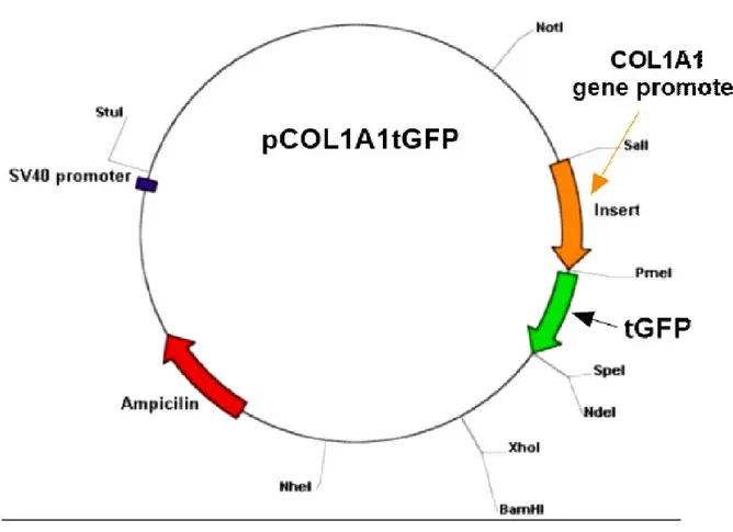
C) Map of Human α(I) Procollagen Gene Promoter-Lentiviral vector-tGFP LentiFP602: These lentiviral particles express FP602 red fluorescent protein
under the EF1a promoter. This promoter is not tissue specific and highly expressed in all cell types.
LentipCOL1A1tGFP: These lentiviral particles express tGFP green fluorescent
protein under the collagen type1 promoter (-804+42). This promoter is tissue specific and expressed in smooth muscle cell types.
31 Dr. Tulasigeri M.Totiger
“Cellular and Molecular Study of Vascular Damage during Systemic Sclerosis”
PhD Thesis in Biochemistry, Physiology and Molecular Biology of PhD School in Biomolecular and Biotechnological Sciences, University of Sassari
Materials and Methods By using the above shown 2nd generation plasmids containing COL1A1-LV-tGFP and EF1α-LV-FP602 lentivectors, the co-transfection into human embryonic kidney 293T cells was done , and then the medium was collected 3 times at 24 hour intervals beginning 24 hours after changing the medium post-transfection. Thus lentiviral particle productionwas done by using 293T cells [149, 150] as described. Later purification process was performed. In this step, every sample was filtered immediately through a 0.22 mm cellulose acetate filter and stored at 40 C. The virus pellet was collected by carrying out 2hrs of ultracentrifugation at 19.4K rpm and at room temperature. Since lentiviral particles contain GFP as a reporter system, transfection was confirmed by using Fluorescent microscopy and then titration of lentiviral vectors was performed.
3.6 GFP-based Lentivirus (harboring the COL1A1 promoter) Transduction in HPASMCs.
The lentivirus transduction was done in HPASMCs by following the company protocol (Innoprot- Innovative Technologies In Biological Systems, S.L.). The determined number of HPASMCs were seeded in Smooth Muscle Cell complete medium and incubated overnight. When 50%-75% confluent,the lentiviral stock was thawed at room temperature. The culture medium was then removed and appropriate amount of this virus stock was loaded to cells and the required volume of the medium was bought by using OptiMEM. The cells were then placed in the CO2 incubator maintained at 5% CO2 and 37oC and the plate was carefully rotated for every 15 min for 1 hour. Later Smooth Muscle Cell complete medium was added to bring to the required volume.At around 48-72h after transduction, the Green Florescent protein production or expression was checked under fluorescence microscope.
32 Dr. Tulasigeri M.Totiger
“Cellular and Molecular Study of Vascular Damage during Systemic Sclerosis”
PhD Thesis in Biochemistry, Physiology and Molecular Biology of PhD School in Biomolecular and Biotechnological Sciences, University of Sassari
Materials and Methods
3.7 Measurement of COL1A1 Promoter Activity
HPASMCs transduced with lentiviral vector were treated with of 5% (V/V) of
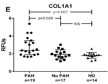
sera from PAH, no PAH and HD subjects in basal medium. The kinetic measurement COL1A1 promoter activation was performed for 10 hours (Figure2A) and any deviations occurred during this course of time was studied. The steady state in the promoter activation was attained at 8hrs and the values at this time point were used for comparison (Figure 2A, 2B, 4, 6) and data were normalized for transduction efficiency by reporting the ratio of COL1A1-LV-tGFP to EF1α-LV-FP602 Relative Fluorescence Units (RFU).
3.8 Study of NOX Involvement.
HPASMCs were pretreated with 5 μM NADPH oxidase specific inhibitor (NOX2ds-tat, formerly gp91ds-tat) [151] for 1hr before stimulating with sera from PAH, no PAH and HD subjects and Intracellular ROS was then measured( as shown in Fig.3) as previously described to observe the NOX inhibitor effect.HPASMCs transduced with lentiviral vector (obtained from the COL1A1-LV-tGFP and EF1α-LV-FP602) were incubated for 1 hour with 5μM of NOX before treatment with SSc sera from the three subjects (PAH, no PAH and HD). Then collagen type-I (COL1A1) promoter activity was measured (as shown in Fig.4) as previously described to determine whether NOX inhibitor has effect on collagen promoter activity.
3.9 Study of ERK Involvement
For studying the effect of ERK inhibitor PD98059 on Intracellular ROS, HPASMCs were pretreated with 15μM of ERK inhibitor PD98059 for 1hr before stimulating with sera from PAH, no PAH and HD subjects and
33 Dr. Tulasigeri M.Totiger
“Cellular and Molecular Study of Vascular Damage during Systemic Sclerosis”
PhD Thesis in Biochemistry, Physiology and Molecular Biology of PhD School in Biomolecular and Biotechnological Sciences, University of Sassari
Materials and Methods
Intracellular ROS was then measured as described earlier. In another set of experiments for studying the effect of ERK inhibitor
PD98059 on COL1A1 promoter activity,HPASMCs were pre-incubated with 15 μM of ERK inhibitor PD98059 for 1hr before stimulating with sera from PAH, no PAH and HD subjects and collagen I synthesis was assessed as described earlier to observe the effect of this inhibitor.
3.10 Statistical Analysis
Healthy donors were matched for gender, race and smoking status. Horizontal lines indicate the median with interquartile range (Fig1A,2A). Kruskall–Wallis one-way analyses of variance followed by post-hoc Dunn’s test for multiple comparisons were used to detect differences among studied groups in (Fig 1B, 2B). For studying subjects’ characteristics, P values were determined by Fisher’s exact test or the Wilcoxon rank-sum test, as appropriate. Wilcoxon matched-pairs signed rank test was used to determine meaningful differences between pre- and post-NOX & ERK inhibitor treatment pairs in Figures 3, 4, 5 & 6. All statistical analysis were performed using GraphPad Prism version 6.00 for Windows (GraphPad Software, San Diego, CA) and p-values <0.05 were considered to be statistically significant.
34 Dr. Tulasigeri M.Totiger
“Cellular and Molecular Study of Vascular Damage during Systemic Sclerosis”
PhD Thesis in Biochemistry, Physiology and Molecular Biology of PhD School in Biomolecular and Biotechnological Sciences, University of Sassari
35 Dr. Tulasigeri M.Totiger
“Cellular and Molecular Study of Vascular Damage during Systemic Sclerosis”
PhD Thesis in Biochemistry, Physiology and Molecular Biology of PhD School in Biomolecular and Biotechnological Sciences, University of Sassari
CHAPTER 4.RESULTS
4.1 Demographic Data and Clinical Characteristics of Subjects:
Mainly the middle age, white women were the SSc patients enrolled in this study. SSc subjects were concluded as “No PAH” (n = 17) if their right ventricular systolic pressure (RVSP) estimated by echocardiogram was ≤35 mm Hg, and they were defined as “PAH” (n = 19) if the RVSP was >35 mm Hg. They underwent right heart catheterization (RHC) showing a mean pulmonary artery pressure (mPAP) ≥ 25 mm Hg and pulmonary capillary wedge pressure (PCWP) ≤ 15 mm Hg. For this study,Healthy Donors [HD] (n = 14) were selected through such a screening questionnaire, so as to rule out any underlying autoimmune or vascular disease in the donors. In this study, patients with PAH were slightly older than No-PAH (64.0 ± 9.4 vs 53.3 ± 11.6; p = 0.009) and had longer disease duration (18.5 ± 9.5 vs 10.5 ± 7.3 years; p = 0.01). These observations matched with most of the reports saying that PAH occurs as late age manifestation in SSc [152-156]. It was also registered that in this study, there was slight elevation in modified Rodnan skin score( mRSS) in SSc-PAH patients as compared to No-PAH (7.3 ± 10.3 vs 5.5 ± 6.1;p=0.567) pin pointing there was no significant difference in the values of mRSS between the comparable subjects. RP severity scores (1.6 ± 0.8 2.0 ± 1.0; p= 0.194) and Heart severity scores (0.2 ± 0.7 1.2 ± 1.7; p=0.062) were little bit higher in SSc-PAH vs No-PAH, but not reaching to the significant difference between the subjects. But as anticipated,SSc-PAH subjects revealed relatively higher lung severity scores (3.1 ± 1.3 vs 1.1 ± 1.3; p < 0.001) against No-PAH subjects. Cardiac hemodynamic measurements by Right heart Catheterization showed that mean pulmonary arterial pressure (mPAP) values (35.2 ± 8.1mm Hg) and pulmonary capillary wedge pressure (PCWP) values (11.5 ± 4.0 mm Hg) were remained in the range as expected to be in SSc-PAH subjects as defined by mPAP should be ≥25 mm Hg, as well as PCWP should be ≤ 15 mm
36 Dr. Tulasigeri M.Totiger
“Cellular and Molecular Study of Vascular Damage during Systemic Sclerosis”
PhD Thesis in Biochemistry, Physiology and Molecular Biology of PhD School in Biomolecular and Biotechnological Sciences, University of Sassari
Results
Hg to diagnostically characterize the individuals having PAH according to the reports [17,157]. These diagnostic measurements clearly provided the proof
that severe abnormalities were present in the structural & functional mode of pulmonary vascular compartment. It was observed that there was significantly lower diffusion capacity of lung for carbon monoxide (DLCO) (48.7 ± 16.8 vs 78.2 ± 23.0; p < 0.001) with comparable forced vital capacity (FVC) with decreased values against No-PAH subjects, which provided the confirmed clues that there exists an underlying pulmonary vascular disease. Pulmonary function tests (PFT) revealed that values determining restrictive lung disease (RLD) remain equal in both the subjects. In this study, it was observed that SSc-PAH patients exhibited significantly higher estimated right ventricular systolic pressure (ERVSP) values (65.2 ± 19.9 vs 24.0 ± 6.3; p= <0.001) against those of Non-PAH subjects. This elevated value of ERVSP further clarified the devastating damage of pulmonary vascular function of this ailment and also the severity of the underlying pulmonary vascular disease. ERVSP values >30–45mmHg are often considered abnormal even though there are no consensus about the elevated levels of healthy individuals but ERVSP values generally does not increase in normal individuals. Studies have been reported that SSc patients having ERVSP >35mmHg showed elevated pulmonary pressures [158,159], thus proving the underlying impairment in pulmonary vascular compartment. Another study also confirmed that combination of eRVSP on TTE and PFT parameters provided in identifying of up to 97–100% of SSc patients with RHC confirmed PAH [160].The antibody status in this study revealed that SSc-PAH subjects showed not though significant but slight higher level of Anti centromere antibodies {(ACA) 10 (53) vs 4 (24); P= 0.07} which have been put very relevant to the underlying cause of PAH in SSc by the earlier reports. Demographic Studies have shown ACA is prevalent in older, female Caucasians having SSc, so the same case in this study as well. It has been reported that the presence of ACAs linked with higher risk of development of
37 Dr. Tulasigeri M.Totiger
“Cellular and Molecular Study of Vascular Damage during Systemic Sclerosis”
PhD Thesis in Biochemistry, Physiology and Molecular Biology of PhD School in Biomolecular and Biotechnological Sciences, University of Sassari
Results PAH [161,162] in SSc patients particularly the lSSc patients [161]. Anti-Scl-70 (anti-topoisomerase I) antibodies in this study were found lower in SSc-PAH against the No-PAH subjects as this study includes SSc-PAH subjects mostly from lcSSc subjects and Anti-Scl-70 are mostly found in patients with dcSSc having pulmonary involvement, more specifically linked to pulmonary fibrosis & interstitial lung disease [161,163,164]. The medications used by this study’s subjects were vasodilators (i.e. endothelin receptor antagonists and phosphodiesterase 5 inhibitors) which were significantly higher in SSc-PAH subjects.
Variables No PAH (N = 17) PAH (N = 19) HD (N = 14) p value• Age at serum sampling (years)* 53.3 ± 11.6 64.0 ± 9.4 54.1 ± 10.4 0.009 Female 15 (88) 16 (84) 15 (85) 0.727 Race White 14 (82) 16 (84) 12 (80) 0.881 Black 3 (18) 3 (16) 3 (20) Smoking status Never 9 (53) 10 (53) 8 (53) 0.280 Past 6 (35) 9 (47) 5 (33) Current 2 (12) 0 2 (13) SSc types Limited 11 (65) 16 (84) 0.177 Diffuse 6 (35) 3 (16) mRSS* (range 0–51) 5.5 ± 6.1 7.3 ± 10.3 0.567 SSc duration (RP onset)*, years 14.0 ± 12.6 21.7 ± 9.4 0.008