INTRODUCTION
Amalgam is predicted to have an increasingly limited role in dentistry. Class II resin-based composite restorations continue to grow in use and popularity as many patients prefer that their restorations match the color of their teeth. Many dentists no longer include amalgam restorations in their practices, but the use of posterior tooth-colored restorations varies by country and the economic status of the patient. One survey reported that 68 percent of respondents in the United States and Canada still used amalgam at least part of the time in their practices1),and other surveys have also indicated that amalgam is still a major treatment alternative2-4). The use of amalgam is not rapidly declining because it is a reliable and durable restoration. The strength of the material (particularly high-copper dental amalgam alloys), the longevity of the restorations, and the low cost justify the use of amalgam restorations, particularly in situations that do not demand a more sensitive technique5). Amalgam has a number of drawbacks including color, lack of adhesion to tooth structure, marginal microleakage, and the need for mechanical retention in the form of undercuts, box forms, or grooves which require removal of healthy tooth structure and weaken the prepared tooth5-7). It is now well accepted that restorations must offer a conservative approach to dental hard tissues, disinfection of the enamel-dentin surfaces, and adhesion to the tooth substrate to reduce microleakage. A restoration combining the reliability of amalgam with increased fracture resistance through firm bonding to the tooth is desirable. Bonded amalgam restorations employ an amalgam filling bonded to the tooth using an adhesive. The adhesive bonds to the enamel and dentin after the tooth is etched and primed, similar to composite bonding
procedures. The amalgam is condensed onto the viscous adhesive, forming an interlocking mechanical joint when the amalgam and adhesive harden8). In vitro test results suggest that these restorations provide reduced microleakage, lower incidence of postoperative sensitivity9) and degree of marginal fracture10), increased strength of the prepared tooth11), and better retention of restorations, with the potential for conserving tooth structure12). Several studies have evaluated the adhesion of amalgam alloy to tooth structures using techniques such as shear bond strength13), dye leakage14), microscopic evaluation13), and cuspal fracture resistance15). The aim of the present study was to evaluate the in vitro shear bond strength (SBS), the mode of failure, and the interfacial micromorphology of two amalgam alloys (spherical and admixed) to dentin using five different adhesive systems.
MATERIALS AND METHODS
The study was performed with the approval of the Ethics in Research Committee of the Centre of Health Sciences of the University of Rome “Tor Vergata.” The samples consisted of 120 freshly-extracted human third molars devoid of caries, cracks, endodontic treatments, or restorations. The teeth were cleaned of gross debris and stored at room temperature in a distilled-water solution of 0.2% thymol prior to and throughout the preparation phase. The teeth were transferred to pure distilled water 24 hours prior to bonding. Two regular-set high-copper dental amalgam alloys were tested: Amalcap Plus (Ivoclar-Vivadent AG, Schaan, Liechtenstein), which is composed of spherically shaped particles; and Valiant Ph.D (Ivoclar-Vivadent) which is admixed. The adhesives were (1) Prime & Bond NT Dual Cure (Dentsply Caulk, Milford, DE, USA), (2) Amalgambond Plus (Parkell Inc,
Shear bond strength, failure modes, and confocal microscopy of bonded
amalgam restorations
Luigi CIANCONI1, Gabriele CONTE2 and Manuele MANCINI2
1Affiliate professor of Restorative Dentistry and Endodontics, University of Rome “Tor Vergata”, Rome, Italy
2Former Resident in the Department of Restorative Dentistry and Endodontics, University of Rome “Tor Vergata”, Rome, Italy
Corresponding author, Manuele MANCINI; E-mail: [email protected]
This study evaluated the shear bond strength, failure modes, and confocal microscopy of two different amalgam alloy restorations lined with five adhesive systems. Two regular-set high-copper dental amalgam alloys, Amalcap Plus and Valiant Ph.D, and five commercially available adhesive systems were selected. One hundred and twenty freshly-extracted human third molars were used for the study. The results were statistically evaluated using two-factor analysis of variance (ANOVA). The shear bond strength (SBS) of amalgam to dentin was significantly affected by both the adhesive (p<0.0001) and amalgam alloy (p<0.0002). Regarding mode of failure (MF), among samples restored with Valiant Ph.D, 31 of 50 exhibited adhesive failure, and 19 displayed mixed failure. Laser optical microscopy (OM) of the bonded interface revealed the presence of a good hybrid layer was evident in all experimental groups. Higher bond strengths were measured for four of the five adhesives when used in combination with the spherical alloy.
Keywords: Adhesive systems, Amalgam alloy, Confocal microscopy, Failure mode
Received Jul 30, 2010: Accepted Nov 26, 2010
Edgewood, NY, USA), (3) Amalgambond Plus with HPA Powder (Parkell Inc.), (4) Adper Scotchbond Multi-Purpose Plus (3M Dental Products, St Paul, MN, USA), and (5) All-Bond 2 (Bisco, Schaumburg, IL, USA).
The teeth were mounted in phenolic rings (Buehler Ltd, Lake Bluff, IL, USA) using autocuring acrylic resin (Lang Dental Mfg, Wheeling, IL, USA). The occlusal surfaces were ground on a water-cooled abrasive wheel (Buehler Ltd) using 180-grit followed by 600-grit silicon carbide paper (3M Dental Products). The specimens were thoroughly washed under a stream of tap water. The ground samples were examined at 30× magnification using a stereomicroscope (Carl Zeiss, Oberkochen, Germany) to ensure that no enamel remained on the dentine surfaces. The teeth were randomly assigned to one of ten treatment groups (n=12) as indicated in Table 1.
The dentin surface conditioning and adhesive system application were performed according to the manufacturers’ instructions. Prior to adhesive application dentin was treated with 36% phosphoric etching gel (Conditioner 36, Dentsply Caulk) for 20 s, copiously flushed with water for 30 s and then gently dried to remove excess water. Adhesives of Ab, SMP, A2 and HPA groups were then used strictly following manufacturer instructions for amalgam bonding procedures. Unlike the other products in this study, Prime & Bond NT Dual Cure is not marketed as an amalgam adhesive. As a result, the manufacturer includes no amalgam bonding instructions. For this study, the instructions for the dual cure technique for large restorations were followed. Therefore in the P&B groups, the adhesive liner was light cured with an Optilux 501 unit (KerrHawe SA, Bioggio, Switzerland) prior to amalgam condensation as directed by the manufacturer. Prior to the study, the curing light was tested with an Optilux radiometer (KerrHawe SA) to confirm the light output exceeded 800 mW/cm2. Immediately after the application of adhesive to the dentin surface, each specimen was secured in a Teflon split mold, the upper aspect of which contained a round opening with a diameter of 3 mm and 2 mm thick to
serve as a matrix for amalgam placement. Double spill (600 mg) amalgam capsules were triturated in an SDS Kerr 4000 amalgamator (KerrHawe SA) according to each manufacturer’s instructions and condensed into the preparations using a flat, smooth-faced, circular condenser (2mm diameter) and hand pressure of 1 kg± 250 g. One operator completed all preparations and restorations. The alloy specimens were allowed to set for 30 minutes prior to mold removal. The specimens were stored in distilled water at 37°C for 7 days, thermocycled in distilled water (5°± 5°C/50°± 5°C; 300 cycles; 30-second immersion time), and stored in 37°C distilled water for an additional 36 hours prior to shear testing.
Shear bond strengths and modes of failure
A 0.5 mm thick knife-edged rod was applied to the base of the bonded amalgam cylinder. Each specimen was locked in a fixture attached to the compression load cell of an Instron testing machine (Instron Corp., Canton, MA, USA) and the shear bond strength was determined at a crosshead speed of 0.5 mm/min. The shear forces were recorded in MPa and obtained directly from the Instron computer software. The debonded dentin surfaces were viewed under a stereomicroscope (Carl Zeiss) at 30× magnification to determine the mode of failure. The mean shear bond strength and the standard deviation were calculated for each treatment group. The category means were compared using two-factor analysis of variance. These calculations indicated statistically significant F-ratios (p<0.01), so the data were further analyzed using Duncan’s multiple range tests.
Optical microscopy
Two specimens from each experimental group were sectioned for morphological analysis of the adhesive interface. The specimens were fixed in 4% paraformaldehyde. In order to avoid artefacts, the samples were not embedded in resin. The sections were cut in a bucco-lingual direction using a high-speed diamond saw (Buehler Ltd) equipped with a 100×0.3 mm regular concentration diamond blade and water-spray cooling. The resulting sections were approximately 120
Group Adhesive Liner Amalgam alloy
P&B Prime & Bond NT Dual Cure Amalcap Plus
P&Bβ Prime & Bond NT Dual Cure Valiant Ph.D
Ab Amalgam bond Plus Amalcap Plus
Abβ Amalgam bond Plus Valiant Ph.D
HPA Amalgam bond Plus + HPA* Amalcap Plus
HPAβ Amalgam bond Plus + HPA* Valiant Ph.D
SMP Scotchbond Multi-Purpose Plus Amalcap Plus
SMP β Scotchbond Multi-Purpose Plus Valiant Ph.D
A2 All-Bond 2 Amalcap Plus
A2β All-Bond 2 Valiant Ph.D
*HPA: High Performance Additive
µm thick. The cut surfaces were finished using 2000-grit silicon carbide paper and then treated with 32% phosphoric acid for 10 seconds. The cross-sections were examined using a Laser Optical Microscope (Carl Zeiss) both before and after staining with a pH 4.42% toluidine blue solution. The final magnification of the images was 320×.
The specimens were fixed only after completion of the restoration in order to avoid any possible interaction between dentin collagen and the adhesive systems. Resin encapsulation requires dehydration with alcohol or acetone which could alter the resin components of the adhesives or the hybrid layer morphology. Resin infiltration could also interfere with dye penetration and could fill gaps and voids at the dentine-adhesive or adhesive-amalgam interfaces. Therefore to avoid artefacts due to resin immersion the specimens were freshly sectioned. Dye staining with 2% toluidine blue solution was used to selectively stain the primer to enable discrimination between the various components of the hybrid layer and to evaluate resin infiltration into the dentin.
RESULTS
Shear bond strength
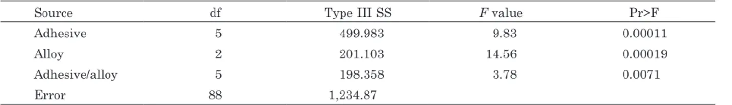
The results of the two-factor Analysis of Variance (ANOVA) are listed in Table 2. The shear bond strengths of amalgam to dentin were significantly affected by both the adhesive(p=0.0001) and amalgam alloy (p=0.0002). In addition, a statistically significant interaction was noted between the two variables (p=0.0071). Table 3
contains the mean shear bond strengths between both amalgam materials and the five resins. Except in the case of All-bond 2 resin, higher bond strengths were achieved with Amalcap Plus. The difference in bond strength using All-bond 2 was not statistically significant (p<0.38). Among the resins for which Amalcap Plus exceeded Valiant Ph.D in bond strength, the differences were statistically significant for Prime & Bond NT Dual Cure (p=0.01) and Amalgambond Plus (p<0.05), but not for Scotch Bond Multipurpose (p>0.46) or Amalgambond Plus with HPA powder (p>0.18).
Modes of failure
The failure modes for each group are listed in Table 4. No samples failed adhesively between the adhesive and dentin or cohesively within the adhesive. Therefore, the failures were categorized as adhesive (failure between the adhesive and amalgam, with the adhesive attached to dentin and no visible amalgam particles on the adhesive/dentin surface), cohesive (within the amalgam), or mixed (some amalgam remaining on both adhesive and dentin surfaces). Among samples restored with Valiant Ph.D, 31 of 50 exhibited adhesive failure, and 19 displayed mixed failure. Among samples restored with Amalcap Plus, 26 of 50 displayed adhesive failures, while 22 had mixed failures; two samples (Amalcap Plus/ All-Bond 2) failed cohesively within the amalgam. Optical microscopy
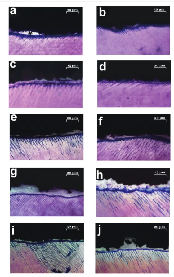
Laser optical microscopy of the bonded interface revealed the relative thickness and uniformity of the amalgam liners (Figure 1). The presence of a good hybrid layer
Source df Type III SS F value Pr>F
Adhesive 5 499.983 9.83 0.00011
Alloy 2 201.103 14.56 0.00019
Adhesive/alloy 5 198.358 3.78 0.0071
Error 88 1,234.87
Adhesive Amalcap Plus Valiant Ph.D
Mean SB (±SD)(MPa) Mean SB (±SD)(MPa) P value
Prime & Bond NT Dual Cure 14.17 (±4.48) 7.13 (±2.63) p=0.01
Amalgambond Plus 8.42 (±1.93) 3.89 (±2.50) p<0.05
Amalgambond/HPA 12.06 (±3.69) 9.91 (±3.11) p>0.18
Scotchbond Multi-Purpose Plus 10.82 (±3.56) 9.19 (±3.98) p>0.46
All-Bond 2 4.98 (±4.25) 6.36 (±2.45) p<0.38
By Alloy Adhesive Mixed Cohesive
Amalcap Plus 26 22 2
Valiant Ph.D 31 19 0
Table 2 Two-way analysis of variance (ANOVA)
Table 3 Shear bond strengths of amalgam to dentin
Fig. 1 Laser optical microscopy images of amalgam-dentin interface.
a) P&B 160× ;b) P&Bβ 160×; c) Ab 320×; d) Abβ 240×; e) HPA 240×; f) HPAβ 240×; g) SMP 240×; h) SMPβ 320×; i) A2 160×; j)A2β 240×.
* presence of amalgam adhesive gap. In any experimental group is evident the presence of a well defined hybrid layer. Within the hybrid layer except in the P&B specimens, where primer and bonding are mixed, primer was stained by toluidine blue, while bonding remained clear. Resin tags, mainly constituted by primer, where present in any experimental group. The Valiant Ph. D. produced a significantly less uniform adhesive/amalgam interface than the spherical alloy.
was evident in all experimental groups with little difference in adhesive penetration into the dentinal structure, hybrid layer thickness, or amalgam/adhesive interface morphology. Staining with 2% toluidine blue enabled discrimination between the primer and bonding agent at the adhesive interface. The primer absorbed the dye well due to its hydrophilic nature, while the bonding agent remained unstained. The bonding layers were relatively homogeneous with an average thickness of 2–4 µm for all groups except the HPA and P&B groups in which the adhesive layer was approximately 6–7 µm thick, irregular, and porous. Resin tags, where present, were always constituted by primer. The Valiant Ph. D. produced a significantly less uniform adhesive/amalgam interface than the spherical alloy. The Valiant Ph. D. yielded a higher degree of interlocking between the adhesive and the amalgam core. Few gaps were seen in the SMP and P&B groups with an average thickness of 1 µm. In the A2, HPA, and Ab groups any gaps between the dentin and amalgam core were completely sealed by the adhesive system.
DISCUSSION
Although the use of resin composite materials is increasingly widespread, amalgam restorations can still be considered suitable in non-aesthetically demanding regions of the oral cavity. Adhesive systems have been recommended for multiple applications including bonding porcelain, resin composite, alloy, and amalgam to tooth dentin and enamel. One reported benefit of these agents is their ability to seal dentin, resulting in a reduction in interfacial microleakage and pulpal sensitivity. Failure to seal the restoration against the ingress of bacteria and oral fluids at the cavosurface margin may contribute to staining, adverse pulpal response, postoperative sensitivity, and recurrent caries16). An additional advantage of adhesive techniques is the increased bond strength, producing greater structural integrity of both the tooth and the restoration17,18). A pilot study was conducted to determine the feasibility of the experimental design and to identify potential problems. Test samples prepared using Copalite (Cooley & Cooley Ltd, Houston, TX, USA) as a liner consistently fractured during matrix removal and were therefore not included in the final study.
The manufacturer of Amalgambond plus recommends using the HPA powder only in “virtually impossible situations” where mechanical retention is lacking and additional adhesion is needed. The powder consists of polymethylmethacrylate fibers that form a thick layer when mixed with the Amalgambond plus base and catalyst liquids. With All-bond 2, Amalgambond plus, Amalgambond Plus with HPA and Scotchbond Multi-Purpose Plus, the objective was to condense the amalgam into the unset adhesive, thus facilitating formation of a mechanical union between the amalgam and adhesive as the adhesive sets. In the P&B group the dual cure components were mixed and light cured for 30 seconds immediately prior to amalgam condensation.
Previous trials using Prime & Bond NT DC without light curing resulted in failure of the setting reaction and no bonding between the amalgam and dentin. These results were confirmed in our pilot study. Light curing initiates the setting reaction and enables the bottom layer of adhesive to bond to dentin. The surface layer, however, remains unpolymerized in the presence of oxygen19). As the amalgam is condensed against this air-inhibited surface layer oxygen is excluded, allowing the adhesive to polymerize completely and mechanically bond to the amalgam20). Vargas et al. compared the amalgam-to-dentin bond strengths of five adhesives20). They reported mean shear bond strengths (MPa) for All-Bond 2 (6.23 MPa), Clearfil liner bond (6.82 MPa), Imperva Dual Bond (7.23 MPa), Optibond (8.24 MPa), and Amalgambond plus with HPA powder (11.97 MPa). Amalgam Bond Plus with HPA powder was significantly stronger than the other four adhesives. In the present study, the Amalcap Plus/Prime & Bond NT DC combination produced a considerably higher bond strength (14.17 MPa) than that reported by Vargas et al.20). Possible explanations for this are the present study’s longer storage time (7–10 days) prior to thermocycling and shear testing, which may have permitted the adhesive to more fully cure, or the different matrices used for amalgam placement. In our pilot study, the removal of plastic matrices similar to those used in Vargas et al. resulted in a high number of bond failures, while the Teflon split mold did not. It is possible that matrix removal creates stresses at the adhesive-amalgam interface insufficient to cause debonding but large enough to lower bond strength. With no significant chemical adhesion between the amalgam and liner, interlocking projections providing mechanical retention are the predominant means of retention for adhesive liners. Greater mixing of the amalgam and liners would seemingly occur with chemically-cured liners in which the amalgam alloy is condensed on thick layer of uncured adhesive. Surprisingly, light-cured materials performed better than the chemically-cured liners, as previously reported by Winkler et al.21). In our research, Prime&Bond NT Dual Cure (the only liner cured prior to amalgam condensation) performed better than chemically-cured liners, especially in the case of spherical alloy amalgam.
Differences in adhesive retention have been previously explained in terms of variations in liner thickness21,22). The liners presenting the thickest interfaces provided the greatest retention. The adhesive interfaces of the light-cured material (Prime & Bond NT DC) were relatively thick, while, except for the HPA groups, the chemically-cured liners were relatively thin. The influence of liner thickness reported by Ramos and Perdigão and Winkler et al. supports our findings21,22). Liners providing a thicker adhesive layer such as Prime & Bond NT Dual Cure or Amalgam Bond Plus with HPA performed better than thinner liners.
Ruzickova et al.23), reported increased shear bond strengths to amalgam when more viscous highly-filled adhesives were used; x-ray microanalysis revealed that amalgam particles were entrapped within the adhesive.
The differences in the performance of the two amalgam alloys in this study were surprising and are difficult to explain. Cooley et al.24) found no difference between a spherical and an admixed alloy bonded with Amalgambond. However, Morril et al.25), also using Amalgambond, reported that a spherical alloy (Artalloy) provided significantly higher shear bond strength to both composite and dentin than did an admixed alloy (Luxalloy). Diefenderfer & Reinhardt26) also described better adhesive performance for a spherical amalgam. These investigators speculated that the improved performance may have been caused by differences in the chemical components of the two amalgam alloys or because the slowly-setting admixed alloy was more affected by removal of the mold used during condensation of the amalgam. A spherical amalgam particle also has more surface area available for contact with the adhesive. However, one might expect the adhesive entrapment exhibited by admixed alloys to produce higher bond strengths than spherical alloys. The micromorphological analysis of the present study seems to support this speculation as the admixed amalgam/adhesive interface appeared more favorable to micromechanical retention. Apparently, this does not occur. In the present study, Valiant Ph.D consistently exhibited both lower bond strengths and less residual amalgam on debonded dentin surfaces.
CONCLUSIONS
Within the limits of this study, higher bond strengths were measured for four of the five adhesives when used in combination with the spherical alloy. To achieve better adhesion, a thicker adhesive layer is favorable. Therefore a dual cured adhesive system is preferred to obtain higher amalgam bonding. The combination of a thicker layer of adhesive with a spherical amalgam alloy appears promising as an acceptable definitive restoration.
REFERENCES
1) Clinical Research Associates. Clinicians’ Preferences 2005: survey data from 1652 random CRA Newsletter subscribers. CRA Newsletter 2005; 29: 3.
2) Guelmann M, Mjör IA, Jerrell GR. The teaching of Class I and II restorations in primary molars: a survey of North American dental schools. Pediatr Dent 2001; 23: 410-414. 3) Pair RL, Udin RD, Tanbonliong T. Materials used to restore
class II lesions in primary molars: a survey of California pediatric dentists. Pediatr Dent 2004; 26: 501-507.
4) Lynch CD, McConnell RJ, Wilson NH. Trends in the placement of posterior composites in dental schools. J Dent Educ 2007; 71: 430-434.
5) Charlton DG, Moore BK, Swartz ML. In vivo evaluation of the use of resin liner to reduce microleakage and improve
retention of amalgam restorations. Oper Dent 1992; 17: 112-119.
6) Varga J, Matsumura H, Masuhara E. Bonding of amalgam filling to tooth cavity with adhesive resin. Dent Mater 1986; 5: 158-164.
7) Lacy AM, Staninec M. The bonded amalgam restoration. Quintessence Int 1989; 20: 521-524.
8) Mccomb D, Brown J, Forman M. Shear bond strength of resin-mediated amalgam dentin attachment after cyclic loading. Oper Dent 1995; 20: 236-240.
9) Staninec M, Holt M. Bonding of amalgam to tooth structure: tensile adhesion and microleakage tests. J Prosthet Dent 1988; 59: 397-402.
10) Tarim B, Suzuki S, Suzuki S, Cox CF. Marginal integrity of bonded amalgam restorations. Am J Dent 1996; 9: 72-76. 11) Eakle WS, Staninec M, Lacy AM. Effect of bonded amalgam
on the fracture resistance of teeth. J Prosthet Dent 1992; 68: 257-260.
12) Staninec M. Retention of amalgam restorations: undercuts
versus bonding. Quintessence Int 1989; 20: 347-351.
13) Dhanasomboon S, Nikaido T, Shimada Y, Tagami J. Bonding amalgam to enamel: Shear bond strength and SEM morphology. J Prosthet Dent 2001; 86: 297-303.
14) Ziskind D, Venezia E, Kreisman I, Mass E. Amalgam type, adhesive system, and storage period as influencing factors on microleakage of amalgam restorations. J Prosthet Dent 2003; 90: 255-260.
15) Rasheed AA. Effect of bonding amalgam on the reinforcement of teeth. J Prosthet Dent 2005; 93: 51-55.
16) Duke ES. Adhesion and its application with restorative materials. Dent Clin North Am 1993; 37: 329-340.
17) Boyer DB, Roth L. Fracture resistance of teeth with bonded amalgams. Am J Dent 1994; 7: 91-94.
18) Neme AL, Evans DB, Maxson BB. Evaluation of dental adhesive systems with amalgam and resin composite restorations: comparison of microleakage and bond strength results. Oper Dent 2000; 25: 512-519.
19) Ruyter IE. Monomer systems and polymerization. In “Posterior composite resin dental restorative materials”. Eds Vanherle G & Smith DC, Peter Szulc Press, Utrecht, the Netherlands 1985: 109-137.
20) Vargas MA, Denehy GE, Ratananakin T. Amalgam shear bond strength to dentin using different bonding agents. Oper Dent 1994; 19: 224-227.
21) Winkler MM, Moore BK, Allen J, Rhodes B. Comparison of retentiveness of amalgam bonding agent types. Oper Dent 1997; 22: 200-208.
22) Ramos JC, Perdigão J. Bond strengths and SEM morphology of dentin-amalgam adhesives. Am J Dent 1997; 10: 152-158. 23) Ruzickova T, Staninec M, Marshall GW. SEM analysis of
resin-amalgam adhesion after debonding. J Dent Res 1994; 73: 388.
24) Cooley RL, Tseng EY, Barkmeier WW. Dentinal Bond strengths and microleakage of a 4-META adhesive to amalgam and composite resin. Quintessence Int 1991; 22: 979-983.
25) Morril F, Galburt R, Kugel G, Zive M, Habib C. Comparison of different shaped particles of amalgam alloy bonded to composite and dentin with 4-methacryloxyethyl trimellitate anhydride. J Dent Res 1994: 73: 221.
26) Diefenderfer KE, Reinhardt JW. Shear bond strengths of 10 adhesive resin/amalgam combinations. Oper Dent 1997; 22: 50-56.