Dottorato in Scienze Mediche Specialistiche
DOTTORATO DI RICERCA IN MEDICINA DEL SONNO
XXIV Ciclo
SPINAL CORD INJURY:
ASSESSMENT OF AUTONOMIC STATE-DEPENDENT
CONTROL OF CARDIOVASCULAR SYSTEM AND
BODY CORE TEMPERATURE
Presentata da: Dr. Pietro Guaraldi
Coordinatore Dottorato: Relatori:
Prof. Pietro Cortelli Prof. Pietro Cortelli
Dr.ssa Federica Provini
Alma Mater Studiorum – Università di Bologna
Alma Mater Studiorum – Università di Bologna
DOTTORATO DI RICERCA IN SCIENZE MEDICHE SPECIALISTICHE
Progetto n°2 “MEDICINA DEL SONNO”
XXIV Ciclo
Settore Concorsuale: 06/D6 - NEUROLOGIA
Settore Scientifico Disciplinare di afferenza: MED 26
SPINAL CORD INJURY:
ASSESSMENT OF AUTONOMIC STATE-DEPENDENT
CONTROL OF CARDIOVASCULAR SYSTEM AND
BODY CORE TEMPERATURE
Presentata da: Dr. Pietro Guaraldi
(n° matr. 376057)
Coordinatore Dottorato: Relatori:
Prof. Pietro Cortelli Prof. Pietro Cortelli
Dr.ssa Federica Provini
Alma Mater Studiorum – Università di Bologna
ad Achira (Chiara)
“To whom I owe the leaping delight That quickens my senses in our wakingtime And the rhythm that governs the repose of our sleepingtime,
the breathing in unison” T. S. Eliot
INDEX OF CONTENTS
INTRODUCTION 1
Section I
SLEEP STRUCTURE IN SPINAL CORD INJURY PATIENTS
Section II
ANALYSIS OF CARDIOVASCULAR PARAMETERS
Autonomic control of the cardiovascular system during wakefulness
The circadian rhythm of blood pressure and heart rate in spinal cord injury patients
Section III
BODY CORE TEMPERATURE REGULATION IN SPINAL CORD INJURY PATIENTS
CONCLUSIONS BIBLIOGRAPHY APPENDIX Section I Section II.1 Section II.2 Section III 5 19 41 55 67 69 79 86 101 106
LIST OF ABBREVIATIONS
∆ (delta): Change of any variable quantity
∆BcT: changes in BcT ∆DBP: changes in DBP ∆HR: changes in HR ∆SBP: changes in SBP
ACR: acrophase
AD: autonomic dysreflexia
AHI: apnea hypopnea index
AMP: amplitude
ANOVA: analysis of variance
ANS: autonomic nervous system
ASIA: American Spinal Injury Association
BcT: body core temperature
BMI: body mass index BP: blood pressure
C4-7: cervical SCI
CF: cold face
CHD: coronary heart disease
CNT: control subjects
CPG: central pattern generators
CVD: cardiovascular disease
D2-12: thoracic SCI
DB: deep breathing
DBP: diastolic blood pressure
EEG: electroencephalogram
HF: high frequency
HR: heart rate
HRV: heart rate variability
HUTT: head-up tilt test
IE: isometric exercise
LF: low frequency
N1: stage 1 NREM sleep N2: stage 2 NREM sleep N3: stage 3 NREM sleep
NREM: non REM
NREM: non-rapid eye-movement sleep
OSA: obstructive sleep apnea
OV: overshoot
PLMS: periodic limb movement during sleep
PS: parasympathetic
PSA: power spectral analysis
PSG: polysomnographic
REM: rapid eye-movement sleep
SBP: systolic blood pressure
SCI: spinal cord injury
SE: sleep efficiency
SNA: sympathetic nerve activity
SRBD: sleep related breathing disorders
SSR: sympathetic skin response
SWS: slow wave sleep
TP: Total Power
TPR: total peripheral resistance
TST: total sleep time
V-PSG: Video-Polysomnography
VLF: very low frequency
VM: Valsalva manoeuvre
VR: Valsalva ratio
INTRODUCTION
“The frog instantly dies when the spinal cord is pierced, and previous to this it lived without head, heart or any bowels or skin, and here, therefore, it would seem lies the foundation of movement and life.”
Leonardo da Vinci
INTRODUCTION
Scientific Background and Object of the Study:
Few survivable injuries have as much impact on a patient’s life as acute spinal cord trauma and its associated human and social cost. Spinal cord injury (SCI) results not only in paralysis; but it is also associated with a range of autonomic dysregulation that can interfere with cardiovascular, bladder, bowel, temperature, and sexual function. The entity of the autonomic dysfunction is related to the level and severity of injury to descending
autonomic (sympathetic) pathways[1-3].
For many years there was limited awareness of these issues and the attention given to them by the scientific and medical community was scarce. Yet, even if a new system to document the impact of SCI on autonomic function has recently been proposed [4], the current standard of assessment of SCI (American Spinal Injury Association (ASIA) examination) evaluates motor and sensory pathways, but not severity of injury to autonomic pathways [5].
Beside the severe impact on quality of life, autonomic dysfunction in persons with SCI is associated with
increased risk of cardiovascular disease and mortality [3,6,7]. Therefore, obtaining information regarding autonomic function in persons with SCI is pivotal and clinical examinations and laboratory evaluations to detect the presence of autonomic dysfunction and quantitate its severity are mandatory.
Furthermore, previous studies demonstrated that there is an intimate relationship between the autonomic nervous system and sleep from anatomical, physiological, and neurochemical points of view [8]. Although, even if previous epidemiological studies demonstrated that sleep problems are common in SCI [9-12], so far only limited polysomnographic (PSG) data are available [13,14]. Finally, until now, circadian and state dependent autonomic regulation of blood pressure (BP), heart rate (HR) and body core temperature (BcT) were never assessed in SCI patients.
Aim of the current study was to establish the association between the autonomic control of the cardiovascular function, thermoregulation, sleep parameters and increased cardiovascular risk in SCI patients.
Study Population and Main Parameters:
Thanks to the collaboration with the Spinal Unit of Azienda Ospedaliera Carreggi, Florence, ITALY, we assessed 5 cervical SCI with tetraplegia (C4-C7 lesions), 7 thoracic SCI with paraplegia (D2-D12 lesions). All patients underwent the following studies:
I. Nocturnal cardio-respiratory monitoring for detection of sleep related breathing disorders;
II. Assessment of daytime autonomic control of the cardiovascular system through cardiovascular reflexes;
III. Assessment of heart rate variability (HRV) during supine rest and head-up tilt;
IV. Evaluation of Sleep structure and efficiency by means of video-PSG;
V. Assessment of circadian rhythm and state and time dependent regulation of BP and HR;
VI. Assessment of circadian rhythm and state and time dependent regulation of BcT.
Introduction
Patients results were compared to controls, whose number varied according to the different assessments.
Inclusion Criteria:
• Male sex;
• Complete spinal cord lesion (Frankel A);
• Clinically stable (at least 6 months after the spinal cord lesion);
• Absence of symptoms or signs of diabetes, cardiorespiratory disease or of other pathological conditions that might affect autonomic cardiovascular control;
• Patient provided informed consent to participate to the study.
Study Protocol
Patients and controls were admitted to hospital and studied following a 3-days protocol (Fig 0.1). Subjects were admitted on Monday morning; in the afternoon they received an enema to prepare for rectal temperature monitoring and at night they underwent nocturnal cardio-respiratory monitoring for detection of sleep related breathing disorders;.
On the following morning, between 8:00 am and 11:00 am, subjects underwent cardiovascular reflexes tests in a temperature controlled (23±1°C) clinical investigation room according to standard procedures [15].
After the assessment of cardiovascular reflexes (between 11:00 am and 12:00 am), arterial BP, HR, BcT and sleep-wake cycle were continuously monitored for 24 hours under controlled conditions (recording ending at 11:00 am of the following day).
Fig 0.1: study protocol
Day 1 Mon Day 2 Tue Day 3 Wed
- stanza con variabili fisiche controllate;
- monitoraggio continuo di pressione arteriosa, frequenza cardiaca, respiro, ciclo
sonno-veglia con sistema di acquisizione del segnale COLLEAGUE (1000 Hz/canale)
- monitoraggio della T° corporea interna (rettale) mediante mini-logger (ogni 2
minuti)
1:00 pm: Enema 2:00 pm: Nocturnal cardio-respiratory monitoring 9:00 am: Cardiovascular reflexes11:00 am: Circadian rhythm of BP and HR and BcT
11:00 am: Stop recording
Guaraldi P. Introduction
Thesis Structure
The thesis is organized in Sections originating from the original study protocol.
In each module it is specified the included population of patients and controls, the methods of data collection and analysis, results, discussion and conclusions.
The sections are structured as follow:
I. Sleep structure in spinal cord injury patients II. Analysis of cardiovascular parameters
1.Autonomic control of the cardiovascular system during wakefulness
2. The circadian rhythm of blood pressure and heart rate in spinal cord injury patients
III. Body core temperature regulation in spinal cord injury patients
In the Appendix figures, which were reduced in size to facilitate reading in each section, are reported
in higher magnification, together with additional supplementary material.
The thesis is written in English to allow international diffusion and to favour its publication on peer reviewed medical journal.
Introduction
Bibliography:
1. Claydon VE, Krassioukov AV (2006) Orthostatic hypotension and autonomic pathways after spinal cord injury. Journal of neurotrauma 23 (12):1713-1725.
2. Furlan JC, Fehlings MG, Shannon P, Norenberg MD, Krassioukov AV (2003) Descending vasomotor pathways in humans: correlation between axonal preservation and cardiovascular dysfunction after spinal cord injury. Journal of
neurotrauma 20 (12):1351-1363.
3. Krassioukov A, Claydon VE (2006) The clinical problems in cardiovascular control following spinal cord injury: an overview. Progress in brain research 152:223-229.
4. Alexander MS, Biering-Sorensen F, Bodner D, Brackett NL, Cardenas D, Charlifue S, Creasey G, Dietz V, Ditunno J, Donovan W, Elliott SL, Estores I, Graves DE, Green B, Gousse A, Jackson AB, Kennelly M, Karlsson AK, Krassioukov A, Krogh K, Linsenmeyer T, Marino R, Mathias CJ, Perkash I, Sheel AW, Schilero G, Schurch B, Sonksen J, Stiens S, Wecht J, Wuermser LA, Wyndaele JJ (2009) International standards to document remaining autonomic function after spinal cord injury. Spinal Cord 47 (1):36-43.
5. Marino RJ, Barros T, Biering-Sorensen F, Burns SP, Donovan WH, Graves DE, Haak M, Hudson LM, Priebe MM (2003) International standards for neurological classification of spinal cord injury. J Spinal Cord Med 26 Suppl 1:S50-56. 6. Myers J, Lee M, Kiratli J (2007) Cardiovascular disease in spinal cord injury: an overview of prevalence, risk, evaluation,
and management. Am J Phys Med Rehabil 86 (2):142-152.
7. Garshick E, Kelley A, Cohen SA, Garrison A, Tun CG, Gagnon D, Brown R (2005) A prospective assessment of mortality in chronic spinal cord injury. Spinal Cord 43 (7):408-416.
8. Cortelli P, Lombardi C (2007) Autonomic Dysfunctions in Sleep Disorders. In: Culebras A (ed) Sleep disorders and neurologic diseases. vol 2, 2nd edn. Informa Healthcare, New York, pp 337-348.
9. Biering-Sorensen F, Biering-Sorensen M (2001) Sleep disturbances in the spinal cord injured: an epidemiological questionnaire investigation, including a normal population. Spinal Cord 39 (10):505-513.
10. Norrbrink Budh C, Hultling C, Lundeberg T (2005) Quality of sleep in individuals with spinal cord injury: a comparison between patients with and without pain. Spinal Cord 43 (2):85-95.
11. Jensen MP, Hirsh AT, Molton IR, Bamer AM (2009) Sleep problems in individuals with spinal cord injury: frequency and age effects. Rehabil Psychol 54 (3):323-331.
12. Biering-Sorensen F, Jennum P, Laub M (2009) Sleep disordered breathing following spinal cord injury. Respir Physiol Neurobiol 169 (2):165-170.
13. Adey WR, Bors E, Porter RW (1968) EEG sleep patterns after high cervical lesions in man. Arch Neurol 19 (4):377-383. 14. Scheer FA, Zeitzer JM, Ayas NT, Brown R, Czeisler CA, Shea SA (2006) Reduced sleep efficiency in cervical spinal cord
injury; association with abolished night time melatonin secretion. Spinal Cord 44 (2):78-81.
15. Mathias CJ, Bannister R (1999) Investigation of autonomic disorders. In: Mathias CJ, Bannister R (eds) Autonomic failure : a textbook of clinical disorders of the autonomic nervous system. 4th edn. Oxford University Press, Oxford; New York, pp 169-195.
Guaraldi P. Introduction
Section I
SLEEP STRUCTURE IN SPINAL CORD INJURY PATIENTS
“Something for the rag and bone man Over my dead body
Something big is gonna happen Over my dead body
Someone saw someone's daughter Over my dead body
This is how I ended up sucked in Over my dead body
I'm gonna go to sleep And let this wash all over me
We don't really want a monster taking over Tip toeing, tying down
We don't want the loonies takin' over Tip toeing, tying down our arms May pretty horses
Come to you as you sleep I'm gonna go to sleep And let this wash over me” Radiohead
Section I: SLEEP STRUCTURE IN SPINAL CORD INJURY PATIENTS:
A 24-h study under bed rest controlled conditions
Background:
Previous cross-sectional descriptive studies and epidemiological reviews [1-4] demonstrated that sleep problems are common in spinal cord injury (SCI). These studies, which are based on sleep scales and self-administered questionnaire, demonstrated that patients with SCI, in comparison to the normal population, have greater difficulty falling asleep, have more frequent awakenings, sleep subjectively less well, sleep more hours, take more and longer naps, and snore more [4]. The elevated 15–40% prevalence of sleep disorders in SCI
individuals [1,3,5,6] is higher than the ~4% prevalence typically reported in middle-aged men [7] and were
related to: a) direct consequences of the injury, like pain and muscle spasm; b) concomitant sleep disorders, such as sleep related breathing disorders (SRBD) or periodic limb movement during sleep (PLMS); or c) to less specific associated condition as anxiety and depression.
Most of the study on sleep in SCI patients focused on SRBD demonstrating an increased incidence of sleep apnea [5,8-13]. Back in 1995, McEvoy et al. demonstrated, by means of a overnight cardio-respiratory monitoring on 40 tetraplegic patients, that the prevalence of SRBD is more than twice that observed in normal population [8]. Eleven out of 40 patients had an apnea hypopnea index (AHI) >15 per hour. More than 80% of apneas were obstructive or mixed, with no sustained hypoventilation. The AHI was found to correlate with neck circumference and sleeping position, but not with respiratory function, level of lesion or dose of antispastic medications. Further studies confirmed that SCI and tetraplegics in particular, have a higher incidence of SRBD [4,14,15], but provided conflicting results on the mechanisms contributing to sleep apnea [13]. The pathophysiology of SRBD in SCI is yet not fully understood. Several different predisposing factors may be involved: circumstances related to the injury, like paralyzed intercostal and abdominal muscles, impaired activation of diaphragm (in lesions above C5), or generic risk factors for SRBD, such as obesity, neck circumference, increased upper airways resistance and supine sleep posture. Furthermore, medications, such as benzodiazepines and baclofen, having relaxant effect on breathing muscle and upper airways together with a depressant effect on the central nervous system, may be involved [8-12,16-18].
Despite the previously mentioned epidemiological studies and the plethora of studies on SRBD, so far only limited data are available on sleep architecture in SCI. In 1968 Adey et al. [19] assessed by means of polysomnography (PSG) 8 upper cervical and 2 upper thoracic SCI patients in the early post-lesion period and 6 upper cervical and 1 lower thoracic SCI patients in a late post-lesion period. SCI patients in early post lesion period had a reduced total sleep time (TST) and an abnormal representation of sleep phases (a reduction in slow wave sleep (SWS) & REM sleep). Instead, SCI patients in the late post lesion period, demonstrated only a high prevalence of light sleep. No relation between age at onset and sleep distortion was found, but the patient with thoracic SCI, had a proportion of non REM (NREM) sleep and REM sleep similar to controls. Similarly, in a more recent polysomnographic case-controlled preliminary study by Scheer et al. [20] on a small sample of patients (3 cervical and 2 thoracic SCI), tetraplegics had a significantly lower (83%) sleep efficiency (SE) compared to thoracic SCI (93%). The SE of thoracic SCI was not different from healthy control subjects (94%). Moreover, there was no difference in the proportion of the different sleep stages, but cervical SCI had a significantly increased REM-onset latency compared to subjects with thoracic SCI.
Section I
Objective:
Aim of the study was to assess with a 24-h Video-Polysomnography (V-PSG) the sleep structure in a population of 5 cervical SCI with tetraplegia, 7 thoracic SCI with paraplegia and 7 age and sex matched control subjects.
Materials and Methods: Subjects
Five cervical SCI with tetraplegia (C4-7) and 7 thoracic SCI with paraplegia (D2-12) and 7 control subjects (CNT)
underwent a 24-h V-PSG under bed-rest controlled conditions (Table I.1). One thoracic SCI (Pt 12) and all
controls prolonged the V-PSG recording for further 24-h. Prior to data collection, all subjects were questioned regarding SRBD symptoms, and underwent a clinical examination to exclude pathological conditions that might affect sleep-wake cycle. All patients (excluding patient 1 and 4) and all controls were assessed for sleep related breathing disorders by means of a previous nocturnal cardio-respiratory monitoring.
The investigation conformed with the principles outlined in the Declaration of Helsinki [21]. The protocol was approved by the Institutional Review Board of the University of Bologna and all participants provided informed consent.
Study protocol
Wake-sleep cycle were continuously monitored for 24-h by an ambulatory polygraphic recorder (Albert Grass Heritage®, Colleague TM PSG Model PSG16P-1, Astro-Med, Inc, West Warwick, RI, USA or Neurofax Electroencephalograph EEG-1200, Nihon-Kohden Corp., Tokyo, Japan) recording electroencephalogram (EEG:
C3-A2, C4-A1, O2-A1), right and left electrooculogram, electrocardiogram, and electromyogram of the
mylohyoideus, left and right anterior tibialis muscles and thoraco-abdominal breathing. Simultaneous video
monitoring was performed throughout the recording to assist in off-line data interpretation. During the study subjects were allowed to sleep ad libitum, living in a temperature (24±1°C) and humidity (40-50%) controlled room, lying in bed except when eating, in a light-dark schedule (light-off period: 11:00 pm-7:00 am). The subjects were placed on a 1.800 kcal/day diet divided into three meals (8:00 am, 12:00 am, 6:00 pm) and three snacks (10:00 am, 4:00 pm, 11:00 pm). From midnight preceding the monitoring, subjects were instructed to avoid alcohol and caffeinated beverages and to abstain from smoking.
Data analyses
Patients were considered “underweight” if BMI was below 18,5; “normoweight” if BMI was between 18,5 and 24,9; “overweight” if BMI was between 25 and 29,9; “obese” if BMI was 30 or greater.
Sleep-related breathing disorders were diagnosed according to the criteria of the American Academy of Sleep Medicine Task Force [22] and rated as “mild” if the AHI was between 5 and 15; “moderate”, if AHI was between15 and 30 or “severe” if the AHI was greater than 30.
The 24-h sleep-wake cycle starting from 11:00 am on the first day of PSG recording was visually scored in 30 s epochs according to the standardized criteria of Rechtschaffen and Kales [23] as light (stages N1 and N2) NREM sleep, deep (stage N3) NREM sleep, and REM sleep. The total sleep time (TST), the sleep efficiency (SE: time spent asleep/time in bed x 100), the duration (min) and the percentage (%) of TST of each NREM sleep stage (N1, N2, N3) and of REM sleep, were calculated for every subject over the light-off period (from 11:00 pm to 7:00 am).
Guaraldi P. Sleep structure in spinal cord injury patients
The arousal index (AI: number of arousals/hour of sleep), the periodic limb movements in sleep index (PLMS-I: number of PLMS/hour of sleep) and the PLMS-arousal index (PLMS-AI: number of PLM associated with arousal/hour of sleep) were computed over the light-off period according to the scoring rules of the Sleep Disorders Atlas Task Force of the American Sleep Disorders Association [24] and the American Academy of Sleep Medicine Manual for the scoring of Sleep [25] respectively. An AI>15 and a PLMS-I>5 were considered as abnormal.
Wake-sleep fragmentation was determined by calculating the frame shifts index indicating the number of 30 s sleep stage shifts occurring every 15 minutes throughout the light-off period (from 11:00 pm to 7:00 am).
Statistical analysis
Data from sleep variables not normally distributed, were compared between groups using the Kruskal–Wallis test. All statistical analyses were performed with IBM SPSS Statistics version 20.0 and a p<0,05 was considered significant.
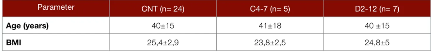
Table I.1 Patients clinical characteristics and medical therapy Table I.1 Patients clinical characteristics and medical therapy Table I.1 Patients clinical characteristics and medical therapy Table I.1 Patients clinical characteristics and medical therapy Table I.1 Patients clinical characteristics and medical therapy Table I.1 Patients clinical characteristics and medical therapy Table I.1 Patients clinical characteristics and medical therapy Table I.1 Patients clinical characteristics and medical therapy Table I.1 Patients clinical characteristics and medical therapy Table I.1 Patients clinical characteristics and medical therapy
Pt n° Group Lesion Site Age (yrs) Dis Hx (yrs)
BMI SRB symptoms Therapy / dayTherapy / dayTherapy / day
1 C4-7 C4 32 15 24 habitual snoring, apneas, daytime sleepiness - baclofen 100 mg - tamsulosin hydrochl 0,4 mg - baclofen 100 mg - tamsulosin hydrochl 0,4 mg - baclofen 100 mg - tamsulosin hydrochl 0,4 mg 2 C4-7 C4-C5 44 11 32 nil - risperidone 2mg - bisacodyl 10 mg - triazolam 125 mcg - lactulose 3 C4-7 C5-C6 60 25 23 habitual snoring,
apneas - dantrolene sodium 75 mg- valproic acid 500 mg
- lorazepam 2,5 mg
- baclofen 75 mg - terazosin 5 mg
4 C4-7 C5-C6 16 2 23 nil - sertraline hydrochl 50 mg
5 C4-7 C6-C7 54 1 22 habitual snoring, apneas, daytime sleepiness - dihydroergotamine 2mg/ml 15 - tamsulosin hydrochl 0,4 mg - desmopressin 60 mcg - dalteparin sodium 5000 U - gabapentin 600 mg - baclofen 50 mg - oxybutynin 10 mg - citalopram 20 mg - lansoprazole 15 mg 6 D2-12 D2-D3 24 4 18 nil - baclofen 75 mg - pregabalin 300 mg - trospium chloride 40 mg - lansoprazole 30 mg - clomipramine 25 mg 7 D2-12 D4 59 1 29 habitual snoring in
supine position - hydroclorotiazide 25 mg- carvedilol 12,5 mg
- amlodipine 5 mg - ramipril 5 mg - amiloride 2,85 mg - citalopram 10 mg 8 D2-12 D4-D5 34 1 20 nil - baclofen 75 mg - oxybutynin 15 mg - pregabalin 150 mg - tizanidine 3 mg 9 D2-12 D7-D8 26 0,5 23 nil - oxybutynin 10 mg - etidronic acid 1200 mg 11 D2-12 D9 61 1 23 habitual snoring,
apneas - triazolam 125 mcg- paroxetine 20 mg
- enoxaparin sodium 6000 U
- oxybutynin 15 mg - diltiazem 30 mg - lansoprazole 30 mg
- tizanidine 18 mg
12 D2-12 D10 31 3 31 nil - intrathecal baclofen 0,5 mg
- intrathecal morphine 1,5 mg
13 D2-12 D12 44 1,5 30 habitual snoring in
supine position - nil
BMI=body mass index; Dis Hx=disease history; Pt n°=patient number; SRB symptoms=symptoms of sleep related breathing disorders; yrs=years. BMI=body mass index; Dis Hx=disease history; Pt n°=patient number; SRB symptoms=symptoms of sleep related breathing disorders; yrs=years. BMI=body mass index; Dis Hx=disease history; Pt n°=patient number; SRB symptoms=symptoms of sleep related breathing disorders; yrs=years. BMI=body mass index; Dis Hx=disease history; Pt n°=patient number; SRB symptoms=symptoms of sleep related breathing disorders; yrs=years. BMI=body mass index; Dis Hx=disease history; Pt n°=patient number; SRB symptoms=symptoms of sleep related breathing disorders; yrs=years. BMI=body mass index; Dis Hx=disease history; Pt n°=patient number; SRB symptoms=symptoms of sleep related breathing disorders; yrs=years. BMI=body mass index; Dis Hx=disease history; Pt n°=patient number; SRB symptoms=symptoms of sleep related breathing disorders; yrs=years. BMI=body mass index; Dis Hx=disease history; Pt n°=patient number; SRB symptoms=symptoms of sleep related breathing disorders; yrs=years. BMI=body mass index; Dis Hx=disease history; Pt n°=patient number; SRB symptoms=symptoms of sleep related breathing disorders; yrs=years. BMI=body mass index; Dis Hx=disease history; Pt n°=patient number; SRB symptoms=symptoms of sleep related breathing disorders; yrs=years.
Results:
Subjects characteristics
Patients clinical characteristics and medical therapy are listed in Table I.1. All subjects were males. Cervical SCI ranged from C4 to C7. Thoracic SCI ranged from T2 to T12. Mean age±SD was 41±18 years for tetraplegics, 41±15 years for paraplegics and 46±11 years for controls (Table I.2). All patients had a complete Frankel A spinal cord lesion and they were clinically stable (the traumatic event occurred more than 6 months before the
Section I
time of the study). All SCI patients had a traumatic lesion, with the exception of patient 7 who suffered a spinal cord ischemic lesion. Cervical SCI had a disease history of 11±10 years (min-max=1-25), while in thoracic SCI ranged from 0,5 to 5 years with a mean of 2±1 years.
There were no age statistically significant differences between groups (Table I.2).
Among the 5 cervical SCI patients 4 were normoweight and 1 patient (Pt 2) was obese. In the thoracic SCI, 1 patient was underweight, 3 normoweight, 2 overweight and 1 obese. In the controls 1 patient was normoweight, 2 were overweight and 4 obese. Even if controls compared to SCI patient had a higher BMI, there were no statistically significant differences in BMI between groups (Table I.2).
Three (Pt 1, 3 and 5) out of 5 cervical SCI patients reported apneas during sleep, 2 thoracic patients reported habitual snoring in supine position (Pt 7 and 13) and 1 thoracic patient (Pt 11) reported sleep apneas with no daytime sleepiness (Table I.1). The nocturnal cardio-respiratory monitoring conformed that 2 (Pt 2 and 3) out of the 3 cervical SCI patients had a severe obstructive sleep apnea (OSA), while 1 (Pt 5) had a moderate OSA (Fig I.1). Due to the limitations deriving from the many complex issues related to SCI patient handling and time constraint, patient 1, who was symptomatic for SRBD, and patient 4 did not perform the nocturnal cardio-respiratory monitoring. Among the thoracic SCI group the nocturnal cardio-cardio-respiratory monitoring revealed that 2 patients (Pt 7 and 13) had a moderate OSA (Fig I.1). The remaining paraplegic patients had a AHI<5. All controls, despite their higher BMI, had a AHI<10 and reported no SRBD disturbances (with the exception of positional snoring). Overall, cervical SCI had a significantly higher AHI and a higher prevalence of obstructive sleep apnea compared to thoracic SCI and controls.
Table I.2: Age and body mass index (BMI) in SCI patients and controls Table I.2: Age and body mass index (BMI) in SCI patients and controls Table I.2: Age and body mass index (BMI) in SCI patients and controls Table I.2: Age and body mass index (BMI) in SCI patients and controls
CNT (n=7) C4-7 (n=5) D2-12 (n=7)
Age (years) 46±11 41±18 40±15
BMI 28,97±4,04 23,77±2,51 24,84±4,97
Values are means ± SD. Values are means ± SD. Values are means ± SD. Values are means ± SD.
Fig I.1 Apnea hypopnea index (AHI) in SCI patients
C4-C7: cervical SCI; D2-D12: thoracic SCI; SCI: spinal cord injury. Numbers on axis of abscissas correspond to patients’ numbers. 2" 3" 5" 6" 7" 8" 9" 11" 12" 13" 0" 10" 20" 30" 40" 50" 60" 70" 80" 90" 0" 2" 4" 6" 8" 10" 12" 14" AH I$( n° $a pn ea /h )$ Pa/ents'$n°$ AHI$ 1" 2" 3" 4" 5" 6" 7" 8" 9" 11" 12" 12,5" 13" 14" 15" 16" 17" 18" 19" 20" 14,5" 15,5" 16,5" 17,5" 18,5"19,5" 20,5" 0" 10" 20" 30" 40" 50" 60" 70" 80" 0" 5" 10" 15" 20" 25" Pa#ents')n°) Arousal)Index) C4.7" D2.12" CNT" 1" 2" 3" 4" 5" 6" 7" 8" 9" 11" 12" 12,5" 13" 14" 15" 16" 17" 18" 19" 20" 14,5" 15,5"16,5" 17,5" 18,5"19,5"20,5" 0" 20" 40" 60" 80" 100" 120" 140" 160" 180" 0" 5" 10" 15" 20" 25" Pa#ents')n°) PLMS)Index) C4.7" D2.12" CNT" 1" 2" 3" 4" 5" 6" 7" 8" 9" 11" 12" 12,5" 13" 14" 15" 16" 17" 18" 19" 20" 14,5" 15,5" 16,5" 17,5"18,5" 19,5" 20,5" 0,00" 10,00" 20,00" 30,00" 40,00" 50,00" 60,00" 70,00" 0" 5" 10" 15" 20" 25" Pa#ents')n°) Sleep)Fragmenta#on)Index) C4.7" D2.12" CNT" Arousal Index PLMS Index Sleep Fragm. Index
Guaraldi P. Sleep structure in spinal cord injury patients
Sleep parameters
Sleep variables over the night-time (from 11:00 pm to 7:00 am) are shown in Table I.3, Fig I.2, Fig I.3 and Fig I.4. There were no significant differences in TST and SE between the three groups (Table I.3).
Cervical SCI compared to controls had a significantly lower amount of N1 NREM sleep (p=0,01), while no differences were observed between thoracic SCI and CNT (Table I.3). Cervical and thoracic SCI had a significantly higher amount of N2 NREM sleep compared to controls (p=0,008 and p=0,000, respectively) while the amount of stage 3 NREM seep was significantly reduced in tetraplegics and paraplegics compared to controls (p=0,039 and p=0,002, respectively) (Table I.3). Noteworthy, 1 cervical SCI patient (Pt 2) and two thoracic SCI (Pt 11 and 12) did not reach stage 3 NREM sleep during the night-time. REM sleep was equally present in all the 3 groups, but one cervical SCI (Pt 2) and one thoracic SCI (Pt 8) presented no REM sleep stages during the light-off period (Fig I.3). The analysis of the percentages of sleep phases out of TST (Fig I.2) confirmed a higher prevalence of N2 NREM sleep and a reduced percentage of SWS in cervical and thoracic SCI compared to controls.
Table I.3: sleep parameters over the night-time in SCI patients and controls Table I.3: sleep parameters over the night-time in SCI patients and controls Table I.3: sleep parameters over the night-time in SCI patients and controls Table I.3: sleep parameters over the night-time in SCI patients and controls
CNT (n=14) C4-7 (n=5) D2-12 (n=8) AHI <10* 51±25 7±10* TST (min) 345±84 388±97 386±92 Sleep Efficiency (%) 72±17 74±17 71±16 N1 (min) 22±20* 5±5 9±5 N2 (min) 157±48*,§ 275±56 283±62 N3 (min) 89±30*,§ 49±28 34±42 REM (min) 80±26 59±39 59±34 N° of Arousals 56±34* 177±95 115±117 Arousal index 10±5 32±24 18±18 N° PLMS 29±48 176±393 210±312 PLMS index 5±7 25±56 38±60 N° of arousals related to leg movement 9±13 22±43 34±61
Arousal related to leg
movement index 2±2 3±6 6±10
N° of phase shift 99±24 135±102 152±58
Phase shift index 18±6 25±23 25±14
Values are means ± SD. =p<0,05 vs. cervical SCI. §=p<0,05 vs. thoracic SCI.
Values are means ± SD. =p<0,05 vs. cervical SCI. §=p<0,05 vs. thoracic SCI.
Values are means ± SD. =p<0,05 vs. cervical SCI. §=p<0,05 vs. thoracic SCI.
Values are means ± SD. =p<0,05 vs. cervical SCI. §=p<0,05 vs. thoracic SCI.
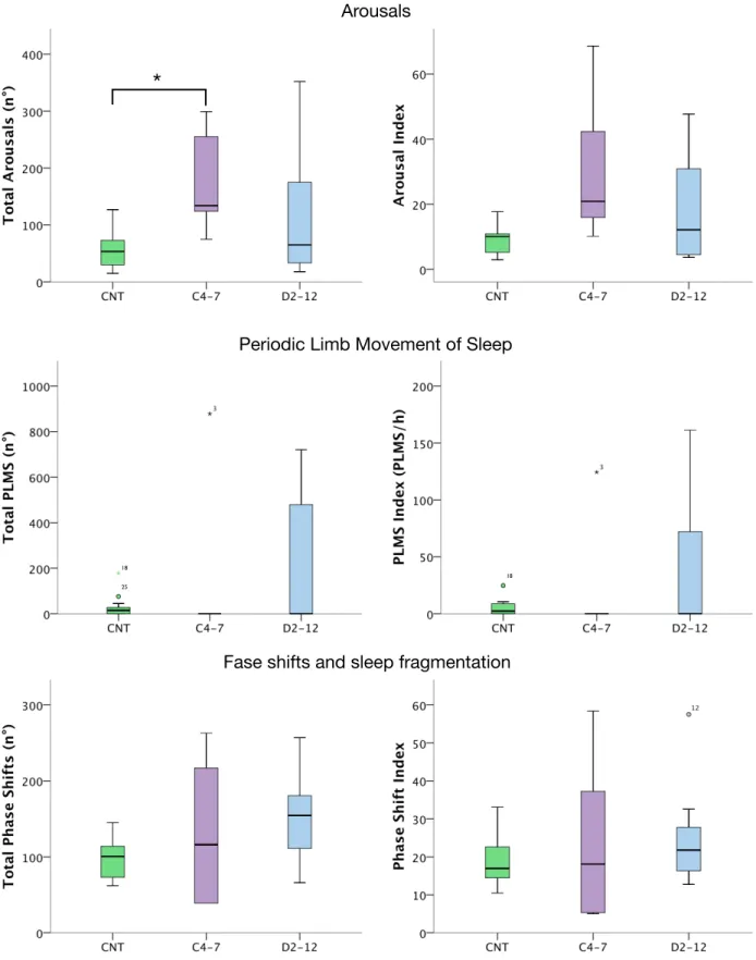
The total number of arousal was significantly higher in cervical SCI compared to controls (p=0,022) but no statistically significant differences were observed between thoracic SCI and controls, nor in the arousal index between the 3 groups (Table I.3, Fig I.4).
The analysis wake-sleep fragmentation (based on the number of phase shift and phase shift index) over the night-time-period, due marked intra-subjects variability, disclosed no statistically significant differences between the 3 groups (Table I.3; Fig I.4).
Only 1 cervical SCI (Pt 3), 2 thoracic SCI (Pt 11 and 12) and 2 controls (subjects 17 and 19) had a PLMS Index > 5 (Fig I.5). The PLMS index was markedly elevated in Pt 3, 11 and 12 (PLMS-I=124; 66; 80, respectively) while it was 13 and 9,5 in subjects 17 and 19 (Fig I.5). Due to this intra-subjects variability, despite the mean value of PLMS and PLMS-I appeared higher in SCI subjects, no statistically significant differences were observed between the 3 groups (Table I.3; Fig I.4).
Section I
Fig I.2: Percentage of TST occupied by NREM and REM sleep phases in SCI patients and controls
Fig I.1 Percentage of sleep phases in SCI patients and controls
*
*
*
*
*
19 27 27*
=p<0,05C4-7: cervical SCI; CNT: controls; D2-12: thoracic SCI; SCI: spinal cord injury, N1: stage 1 NREM sleep; N2: stage 2 NREM sleep; N3: stage 3 NREM sleep; REM: REM sleep; SCI: spinal cord injury;TST: total sleep time.
Fig I.3: minutes of NREM and REM sleep phases in SCI patients and controls
N1 N2 N3 REM 1" 2" 3" 4" 5" 6" 7" 8" 9" 11" 12" 12,5" 13" 14" 15"16" 17" 18" 19" 20" 14,5" 15,5" 16,5" 17,5" 18,5" 19,5" 20,5" 0" 10" 20" 30" 40" 50" 60" 70" 80" 90" 0" 5" 10" 15" 20" 25" N1#(m in)# Subjects'#n°# N1# C4.7" D2.12" CNT" 1" 2" 3" 4" 5" 6" 7" 8" 9" 11" 12" 12,5" 13" 14" 15" 16" 17" 18" 19" 20" 14,5" 15,5" 16,5" 17,5" 18,5" 19,5" 20,5" 0" 50" 100" 150" 200" 250" 300" 350" 400" 450" 0" 5" 10" 15" 20" 25" N2#(m in)# Subjects'#n°# N2# C4.7" D2.12" CNT" 1" 2" 3" 4" 5" 6" 7" 8" 9" 11" 12"12,5" 13" 14" 15" 16" 17" 18" 19" 20" 14,5" 15,5" 16,5" 17,5" 18,5" 19,5" 20,5" 0" 20" 40" 60" 80" 100" 120" 140" 160" 0" 5" 10" 15" 20" 25" N3#(m in)# Subjects'#n°# N334# C4.7" D2.12" CNT" 1" 2" 3" 4" 5" 6" 7" 8" 9" 11" 12" 12,5" 13" 14" 15" 16"17" 18" 19" 20" 14,5" 15,5" 16,5" 17,5" 18,5" 19,5" 20,5" 0" 20" 40" 60" 80" 100" 120" 140" 0" 5" 10" 15" 20" 25" RE M$ (m in )$ Subjects'$n°$ REM$ C4.7" D2.12" CNT" 4" 5" 6" 7"8" 9" 11" 12" 13" 14" 15" 16" 17"18" 19" 20" 21" 22" 23" 24" 25" 27" 28" 29" 30" 31" 32" 33" 34" 35" 36" 37" 5" 10" 15" Pa#ents')n°)20" 25" 30" 35" 40"
C4-7" D2-12" CNT"Numbers on axis of abscissas correspond to subjects’ numbers. C4-7: cervical SCI; CNT:
controls; D2-12: thoracic SCI; SCI: spinal cord injury..
Guaraldi P. Sleep structure in spinal cord injury patients
Fig I.4: arousals, periodic limb movement and phase shifts in SCI patients and controls Arousals
Periodic Limb Movement of Sleep
Fase shifts and sleep fragmentation
Fig I.2 Arousals, periodic limb movement and sleep fragmentation in SCI patients and controls
*
18 25
18
*
=p<0,05. C4-7: cervical SCI; CNT: controls; D2-12: thoracic SCI; SCI: spinal cord injury, PLMS: periodic limb movement during sleep.Section I
Fig I.5: arousals, periodic limb movement and phase shifts in SCI patients and controls 1" 2" 3" 4" 5" 6" 7" 8" 9" 11" 12" 12,5" 13" 14" 15" 16" 17" 18" 19" 20" 14,5" 15,5" 16,5" 17,5" 18,5"19,5" 20,5" 0" 10" 20" 30" 40" 50" 60" 70" 80" 0" 5" 10" 15" 20" 25" Pa#ents')n°) Arousal)Index) C4.7" D2.12" CNT" 1" 2" 3" 4" 5" 6" 7" 8" 9" 11" 12" 12,5" 13" 14" 15" 16" 17" 18" 19" 20" 14,5" 15,5"16,5" 17,5" 18,5"19,5"20,5" 0" 20" 40" 60" 80" 100" 120" 140" 160" 180" 0" 5" 10" 15" 20" 25" Pa#ents')n°) PLMS)Index) C4.7" D2.12" CNT" Arousal Index PLMS Index Phase Shift Index 1" 2" 3" 4" 5" 6" 7" 8" 9" 11" 12" 12,5" 13" 14" 15" 16" 17" 18" 19" 20" 14,5" 15,5" 16,5" 17,5"18,5" 19,5" 20,5" 0" 10" 20" 30" 40" 50" 60" 70" 0" 5" 10" 15" 20" 25" Subjects'*n°* Phase*Shi1*Index* C4.7" D2.12" CNT" 4" 5" 6" 7"8" 9" 11" 12" 13" 14" 15" 16" 17"18" 19" 20" 21" 22" 23" 24" 25" 27" 28" 29" 30" 31" 32" 33" 34" 35" 36" 37" 5" 10" 15" Pa#ents')n°)20" 25" 30" 35" 40"
C4-7" D2-12" CNT" C4-7: cervical SCI; CNT: controls; D2-12: thoracic SCI; SCI: spinal cord injury, PLMS: periodic
limb movement during sleep. Numbers on axis of abscissas correspond to subjects’ numbers.
Guaraldi P. Sleep structure in spinal cord injury patients
Discussion:
This study investigated the sleep structure and the phase shifts in 5 cervical SCI, 7 thoracic SCI and 7 healthy control subjects following a 24-h schedule of bed-rest under controlled condition, at least 6 month after the SCI. So far, this study represent the largest V-PSG study in chronic SCI patients ever published.
The results showed that there were no significative differences in TST and SE among the 3 groups. Although, cervical and thoracic SCI patients presented a significantly higher amount of N2 NREM sleep and a significant reduction in SWS.
Our results are in line with previous findings form Adey et al. [19] who found that in the late post-lesion period 6
upper cervical patients demonstrated a high prevalence of light sleep (78±9,6% of TST), but no other
abnormalities. In this previous study only 1 patients with chronic lower thoracic SCI was assessed, and presented a proportion of NREM sleep and REM sleep similar to heathy subjects [19]. Instead, the 7 thoracic SCI we assessed had an increase in light seep and a reduction in SWS similar to tetraplegics. Compared to the preliminary results by Scheer et al. [20] on 3 cervical and 2 thoracic SCI, they did not find any differences in the proportion of the different sleep stages in tetraplegics, paraplegics and controls as we did. Furthermore, they observed a significantly lower sleep efficiency in cervical SCI compared to thoracic SCI and controls. However, the number of patients they assessed is too exiguous to draw any conclusion, and the small sample size could explain the discrepancies between our results and theirs.
No differences were found in the amount of REM sleep between the three groups. In humans, high cervical lesions interrupting continuity between the medulla and spinal cord have been shown to disrupt spinal innervated REM atonia, but to preserve other characteristics of REM sleep, localizing the neural structures generating REM sleep rostral to the spinal cord, but caudal to the midbrain [26]. Even though, all our SCI patients (including high cervical SCI patients, i.e. Pt 1 who had a C4 lesion) who reached REM sleep, preserved the standard atonia.
Noteworthy, 1 cervical (Pt 2) and 2 thoracic SCI patients (Pt 11 and 12) did not reach stage 3 NREM sleep. Patient 2 and 12 were obese (BMI=32 and 31 respectively). Pt 2 had a severe OSA (AHI=78), while Pt 11 and 12 had no SRBD, but a markedly elevated PLMS index (respectively 66 and 80) which could justify why they could not reach SWS stages. One cervical SCI patient (Pt 2), and one thoracic SCI (Pt 8) did not present REM sleep throughout the night-time recording. As previously stated Pt 2 was obese with severe OSA, while Pt 8 was normoweight, had no SRBD and no PLM, but had a prolonged nap lasting 2 h and 21 min during the light-on period (around 11:00 am) in which presented 15 min of REM (data not shown).
The significant reduction in SWS we observed in tetraplegics and paraplegics could be due to several factors: SRBD may exert an important role in determining an increase proportion of light sleep stages due to the sleep fragmentation and the difficulty to reach deep sleep caused by the recurrent apneas. Therefore the higher incidence of OSA and the significantly increased number of arousals may explain the alteration in sleep architecture observed in cervical SCI. Although, SRBD could not explain why thoracic SCI presented a reduction in SWS similar to tetraplegic and singificantly different form controls, since thoracic SCI AHI was similar to controls. Therefore, as demonstrated by previous studies, the sleep disturbances observed in patients with SCI cannot be explained solely by disruptions in breathing during sleep [1,8,19].
Previous studies reported a high prevalence of PLMS in SCI patients [27-32], and suggested that PLMS causing frequent arousals may alter the sleep architecture. Although, in our study PLMS were present only in one cervical SCI (Pt 3, normoweight with severe OSA) and two thoracic SCI (Pt 11 and 12, see above), and were not strictly related to the AI.
Section I
A further possible explanation of the observed alteration in the sleep pattern in SCI patients involves melatonin [20]. Since the neural pathway for the endogenous production of melatonin passes through the cervical spinal cord [33,34] and patients with cervical SCI have an absence of the normal endogenous melatonin secretion at night [20,33] it was suggested that the alteration in night time melatonin secretion could chronically reduced sleep quality in cervical SCI patients [20]. Even if we believe that the disturbance in melatonin secretion may play a role in determining the disturbed sleep in cervical SCI patients, this hypothesis could not explain the sleep alteration we observed in paraplegics, whose melatonin secretion is supposed to be unaffected.
Therefore, other possible mechanisms may be involved. These mechanisms could be either the direct consequence of the injury or due to concomitant conditions or non specific factors.
Interestingly, thermoregulatory and hypnic mechanisms are thought to be linked [15]. Dependent on the site and extent of the lesion, individuals with a spinal cord injury (SCI) depend on the region above the lesion for thermoregulation [35,36] and may present a thermal dysfunction [35,37,38]. Therefore, it is tempting to speculate that the impaired thermoregulatory function in SCI patient may explain the sleep alteration we observed in cervical and thoracic SCI (see Section III). Although, the reverse could be also possible, being the reduced SWS to interfere with body temperature changes.
Further studies will be necessary to shed light on the possible pathogenetic mechanism of the reduction of SWS in cervical and thoracic SCI. Ideally, studies should be designed to investigate the effects of various interventions (e.g. treatment of OSA, PLMS or correction of thermal dysfunction) on normalization of sleep pattern in individuals with SCI.
Limitations
The main limitation of our study is that during V-PSG, due to the limitations deriving from the many complex
issues related to SCI patients handling, we could record only a single respiratory channel and no SO2. Therefore
the occurrence of apneas during the V-PSG could not be assessed. Although, all subjects were questioned about SRBD disturbances during history taking and underwent a nocturnal cardio-respiratory monitoring (excluding Pt 1 and 4 and all controls). Furthermore, for ethical reasons we could not discontinue medications such as benzodiazepines and baclofen, which have a depressant effect on the central nervous system, increase the frequency and severity of SDB [39-41] but reduce PLMS [42].
Conclusions:
This study represent the largest V-PSG study in chronic SCI patients ever published and demonstrate a compromised sleep in patients with chronic cervical and thoracic SCI, characterized by an augmented proportion N2 NREM sleep and reduced SWS compared to controls. No statistical differences in any sleep parameter were found between cervical and thoracic SCI patients. While the sleep disturbances in cervical SCI could be related to a significantly higher AHI, and possibly to an alteration in melatonin secretion, the pathogenesis of the sleep disorder in thoracic SCI is jet to be determined.
Guaraldi P. Sleep structure in spinal cord injury patients
Patient 10
Several case reports have described PLMS in relation to various spinal cord lesion, including traumatic SCI
[27-32,43]. The temporal relationship between the lesion occurrence and the PLMS presentation, and the
reduction of PLMS in relation to the amelioration of the clinical conditions suggested a causal relationship between the two events. Furthermore, occurrence of PLMS in SCI led to formulation of pathogenetic mechanisms pointing to a possible participation of the Central Pattern Generators (CPG) and the flexor
afferent reflex in the physiopathology of PLMS as discussed by Paulus and Schomburg [43,44].
Interesting we had the chance to assess a patient (Pt 10) with a D8 Brown-Séquard syndrome. He was male, 47 years old, with a BMI=22,99. He was on Baclofen 50 mg/die and oxibutinin 30 mg/die.
Patient 10 was not included in the study (who was limited to complete Frankel A SCI patients) but underwent V-PSG, which demonstrated the occurrence of spontaneous leg movement during sleep on both legs, which did not fulfill the criteria for PLMS.
We believe that assessing by means of formal V-PSG further patients with Brown-Séquard syndromes may shed light on the intriguing physiopathology of PLMS.
Section I
Bibliography:
1. Biering-Sorensen F, Biering-Sorensen M (2001) Sleep disturbances in the spinal cord injured: an epidemiological questionnaire investigation, including a normal population. Spinal Cord 39 (10):505-513.
2. Norrbrink Budh C, Hultling C, Lundeberg T (2005) Quality of sleep in individuals with spinal cord injury: a comparison between patients with and without pain. Spinal Cord 43 (2):85-95.
3. Jensen MP, Hirsh AT, Molton IR, Bamer AM (2009) Sleep problems in individuals with spinal cord injury: frequency and age effects. Rehabil Psychol 54 (3):323-331.
4. Biering-Sorensen F, Jennum P, Laub M (2009) Sleep disordered breathing following spinal cord injury. Respir Physiol Neurobiol 169 (2):165-170.
5. Burns SP, Little JW, Hussey JD, Lyman P, Lakshminarayanan S (2000) Sleep apnea syndrome in chronic spinal cord injury: associated factors and treatment. Arch Phys Med Rehabil 81 (10):1334-1339.
6. Hyyppa MT, Kronholm E (1989) Quality of sleep and chronic illnesses. Journal of clinical epidemiology 42 (7):633-638. 7. Young T, Palta M, Dempsey J, Skatrud J, Weber S, Badr S (1993) The occurrence of sleep-disordered breathing among
middle-aged adults. The New England journal of medicine 328 (17):1230-1235.
8. McEvoy RD, Mykytyn I, Sajkov D, Flavell H, Marshall R, Antic R, Thornton AT (1995) Sleep apnoea in patients with quadriplegia. Thorax 50 (6):613-619.
9. Short DJ, Stradling JR, Williams SJ (1992) Prevalence of sleep apnoea in patients over 40 years of age with spinal cord lesions. J Neurol Neurosurg Psychiatry 55 (11):1032-1036.
10. Klefbeck B, Sternhag M, Weinberg J, Levi R, Hultling C, Borg J (1998) Obstructive sleep apneas in relation to severity of cervical spinal cord injury. Spinal Cord 36 (9):621-628.
11. Cahan C, Gothe B, Decker MJ, Arnold JL, Strohl KP (1993) Arterial oxygen saturation over time and sleep studies in quadriplegic patients. Paraplegia 31 (3):172-179.
12. Flavell H, Marshall R, Thornton AT, Clements PL, Antic R, McEvoy RD (1992) Hypoxia episodes during sleep in high tetraplegia. Arch Phys Med Rehabil 73 (7):623-627.
13. Tran K, Hukins C, Geraghty T, Eckert B, Fraser L (2009) Sleep-disordered breathing in spinal cord-injured patients: a short-term longitudinal study. Respirology 15 (2):272-276.
14. Ayas NT, Epstein LJ, Lieberman SL, Tun CG, Larkin EK, Brown R, Garshick E (2001) Predictors of loud snoring in persons with spinal cord injury. J Spinal Cord Med 24 (1):30-34.
15. Stockhammer E, Tobon A, Michel F, Eser P, Scheuler W, Bauer W, Baumberger M, Muller W, Kakebeeke TH, Knecht H, Zach GA (2002) Characteristics of sleep apnea syndrome in tetraplegic patients. Spinal Cord 40 (6):286-294. 16. Bonekat HW, Andersen G, Squires J (1990) Obstructive disordered breathing during sleep in patients with spinal cord
injury. Paraplegia 28 (6):392-398.
17. Atkinson G, Davenne D (2007) Relationships between sleep, physical activity and human health. Physiology & behavior 90 (2-3):229-235.
18. Star AM, Osterman AL (1988) Sleep apnea syndrome after spinal cord injury. Report of a case and literature review. Spine (Phila Pa 1976) 13 (1):116-117.
19. Adey WR, Bors E, Porter RW (1968) EEG sleep patterns after high cervical lesions in man. Arch Neurol 19 (4):377-383. 20. Scheer FA, Zeitzer JM, Ayas NT, Brown R, Czeisler CA, Shea SA (2006) Reduced sleep efficiency in cervical spinal cord
injury; association with abolished night time melatonin secretion. Spinal Cord 44 (2):78-81.
Guaraldi P. Sleep structure in spinal cord injury patients
21. Williams JR (2008) The Declaration of Helsinki and public health. Bulletin of the World Health Organization 86 (8): 650-652.
22. Sleep-related breathing disorders in adults: recommendations for syndrome definition and measurement techniques in clinical research. The Report of an American Academy of Sleep Medicine Task Force (1999). Sleep 22 (5):667-689. 23. Rechtschaffen A, Kales A (1968) A manual of standardized terminology, techniques and scoring system for sleep stages
of human subjects. . National Institutes of Health publication,, vol no 204. U. S. National Institute of Neurological Diseases and Blindness, Neurological Information Network, Bethesda, MD.
24. EEG arousals: scoring rules and examples: a preliminary report from the Sleep Disorders Atlas Task Force of the American Sleep Disorders Association (1992). Sleep 15 (2):173-184.
25. Iber C, Ancoli-Israel S, Chesson A, Quan S (eds) (2007) The AASM manual for the scoring of sleep and associated events: rules terminology and technical specifications. 1st edn. American Academy of Sleep Medicine, Westchester, IL
26. Smith HR (2008) The basic neurology of sleep. In: Smith HR, Comella C, Högl B (eds) Sleep medicine. Cambridge University Press, Cambridge ; New York, pp 1-8.
27. Yokota T, Hirose K, Tanabe H, Tsukagoshi H (1991) Sleep-related periodic leg movements (nocturnal myoclonus) due to spinal cord lesion. J Neurol Sci 104 (1):13-18.
28. Dickel MJ, Renfrow SD, Moore PT, Berry RB (1994) Rapid eye movement sleep periodic leg movements in patients with spinal cord injury. Sleep 17 (8):733-738.
29. Lee MS, Choi YC, Lee SH, Lee SB (1996) Sleep-related periodic leg movements associated with spinal cord lesions. Mov Disord 11 (6):719-722.
30. de Mello MT, Lauro FA, Silva AC, Tufik S (1996) Incidence of periodic leg movements and of the restless legs syndrome during sleep following acute physical activity in spinal cord injury subjects. Spinal Cord 34 (5):294-296.
31. Brown LK, Heffner JE, Obbens EA (2000) Transverse myelitis associated with restless legs syndrome and periodic movements of sleep responsive to an oral dopaminergic agent but not to intrathecal baclofen. Sleep 23 (5): 591-594.
32. Telles SC, Alves RC, Chadi G (2011) Periodic limb movements during sleep and restless legs syndrome in patients with ASIA A spinal cord injury. J Neurol Sci 303 (1-2):119-123.
33. Zeitzer JM, Ayas NT, Shea SA, Brown R, Czeisler CA (2000) Absence of detectable melatonin and preservation of cortisol and thyrotropin rhythms in tetraplegia. J Clin Endocrinol Metab 85 (6):2189-2196.
34. Moore RY (1996) Neural control of the pineal gland. Behavioural brain research 73 (1-2):125-130.
35. Mathias CJ, Frankel HL (2002) Autonomic disturbances in spinal cord lesions. In: Mathias CJ, Bannister R (eds) Autonomic failure : a textbook of clinical disorders of the autonomic nervous system. 4th edn. Oxford University Press, Oxford ; New York, pp 494–513.
36. Guttmann L, Silver J, Wyndham CH (1958) Thermoregulation in spinal man. J Physiol 142 (3):406-419.
37. Sawka MN, Latzka WA, Pandolf KB (1989) Temperature regulation during upper body exercise: able-bodied and spinal cord injured. Medicine and science in sports and exercise 21 (5 Suppl):S132-140.
38. Boot CR, Binkhorst RA, Hopman MT (2006) Body temperature responses in spinal cord injured individuals during exercise in the cold and heat. International journal of sports medicine 27 (8):599-604.
Section I
39. Khan S, Plummer M, Martinez-Arizala A, Banovac K (2007) Hypothermia in patients with chronic spinal cord injury. J Spinal Cord Med 30 (1):27-30.
40. Dolly FR, Block AJ (1982) Effect of flurazepam on sleep-disordered breathing and nocturnal oxygen desaturation in asymptomatic subjects. The American journal of medicine 73 (2):239-243.
41. Taasan VC, Block AJ, Boysen PG, Wynne JW (1981) Alcohol increases sleep apnea and oxygen desaturation in asymptomatic men. The American journal of medicine 71 (2):240-245.
42. Bensmail D, Quera Salva MA, Roche N, Benyahia S, Bohic M, Denys P, Bussel B, Lofaso F (2006) Effect of intrathecal baclofen on sleep and respiratory function in patients with spasticity. Neurology 67 (8):1432-1436.
43. Bensmail D, Marquer A, Roche N, Godard AL, Lofaso F, Quera-Salva MA (2012) Pilot study assessing the impact of intrathecal baclofen administration mode on sleep-related respiratory parameters. Arch Phys Med Rehabil 93 (1): 96-99.
44. Vetrugno R, Provini F, Plazzi G, Cortelli P, Montagna P (2005) Propriospinal myoclonus: a motor phenomenon found in restless legs syndrome different from periodic limb movements during sleep. Mov Disord 20 (10):1323-1329. 45. Paulus W, Schomburg ED (2006) Dopamine and the spinal cord in restless legs syndrome: does spinal cord physiology
reveal a basis for augmentation? Sleep medicine reviews 10 (3):185-196.
Guaraldi P. Sleep structure in spinal cord injury patients
Section II
ANALYSIS OF CARDIOVASCULAR PARAMETERS
‘‘Spinal cord injury is a ferocious assault on the body that leaves havoc in its wake. Paralysis is certainly part of its legacy, but there are other equally devastating consequences including autonomic dysfunction: compromised cardiovascular, bowel, bladder, and sexual function. Treatments and cures for these losses would greatly improve the quality of life for all of us living with spinal cord injury. I am hopeful that the multi-faceted and collaborative approach to spinal cord repair ... means that there will be effective therapies for autonomic dysfunction in the not too distant future.’’
Section II.1: Autonomic control of the cardiovascular system during wakefulness
Background:
Normal functioning of the autonomic nervous system is critically dependent on integrity of the spinal cord, as the entire sympathetic outflow (T1-L2/3) and the sacral parasympathetic outflow travel and synapse within the spinal cord, before supplying various target organs (Fig II.1.1).
Fig II.1.1: schematic outline of the major autonomic pathways controlling the circulation
From Mathias C and Low D (2012) Autonomic
Disturbances in Spinal Cord Injuries. In: Robertson D (ed) Primer on the autonomic nervous system. pp 505-509.
Schematic outline of the major autonomic pathways controlling the circulation. The major afferent input into the central nervous system is through the glossopharyngeal (CR,9) and vagus (CR,10) nerves by activation of baroreceptors in the carotid sinus and aortic arch. Chemoreceptors and low pressure receptors also influence the efferent outflow. The latter consists of the cranial parasympathetic (PS) outflow to the heart via the vagus nerves, and the sympathetic outflow from the thoracic and upper lumbar segments of the spinal cord. Activation of visceral, skin, and muscle receptors, in addition to cerebral stimulation influences the efferent outflow. In high spinal cord lesions, therefore, the input from chemoreceptors and baroreceptors is preserved along with the vagal efferent outflow, but there is no connection between the brain and the rest of the sympathetic outflow. The spinal sympathetic outflow may be activated through a range of afferents (visual, skin, muscle). This occurs through isolated spinal cord reflexes, not controlled by cerebral pathways, as seen normally.
X. AUTONOMIC DISORDERS
104. AUTONOMIC DISTURBANCES IN SPINAL CORD INJURIES
506
0.6 mg IV
20 min post atropine
(0.6mg IV)
6 hr post atropine
(0.6mg IV)
FIGURE 104.2 (a) The effect of disconnecting the respirator (as required for aspirating the airways) on the blood pressure (BP) and heart rate (HR) of a recently injured tetraplegic patient (C4/5 lesion) in spinal shock, 6 hours after the last dose of intravenous atropine. Sinus brady-cardia and brady-cardiac arrest, also observed in the electrocardiograph, were reversed by reconnection, intravenous atropine and external cardiac mas-sage (from Frankel et al. Lancet 1975;ii:1183–1185). (b) The effect of tra-cheal suction, 20 minutes after atropine in the same patient. Disconnection from the respirator and tracheal suction did not lower either heart rate or blood pressure. (from Mathias Eur J Int Care Med 1976;2:147–156).
FIGURE 104.3 (a) Blood pressure (BP) and heart rate (HR) in a tet-raplegic patient before and after head-up tilt, in the early stages of reha-bilitation, where there were few muscle spasms and minimal autonomic dysreflexia (from Mathias and Frankel Handbook of Clinical Neurology 1992;17:435–56). (b) Blood pressure (BP) and heart rate (HR) in a chronic tetraplegic patient before, during and after head-up tilt to 45°. Blood pressure promptly falls but with partial recovery, which in this case is linked to skeletal muscle spasms (S) inducing spinal sympathetic activ-ity. Some of the later oscillations may be due to the rise in plasma renin, which was measured where there were interruptions in the intra-arterial record. In the later phases of head-up tilt, skeletal muscle spasm occur more frequently and further elevate the blood pressure. On return to the horizontal, blood pressure rises rapidly above the previous level and then returns slowly to horizontal levels Heart rate usually moves in the opposite direction, except during muscle spasms when there is an initial increase. (from Mathias and Frankel Ann Rev Physiol 1999;50:577–592).
FIGURE 104.1 Schematic outline of the major autonomic pathways control-ling the circulation. The major afferent input into the central nervous system is through the glossopharyngeal (CR,9) and vagus (CR,10) nerves by activation of baroreceptors in the carotid sinus and aortic arch. Chemoreceptors and low pressure receptors also influence the efferent outflow. The latter consists of the cranial parasympathetic (PS) outflow to the heart via the vagus nerves, and the sympathetic outflow from the thoracic and upper lumbar segments of the spinal cord. Activation of visceral, skin, and muscle receptors, in addition to cerebral stimulation influences the efferent outflow. In high spinal cord lesions, therefore, the input from chemoreceptors and baroreceptors is preserved along with the vagal efferent outflow, but there is no connection between the brain and the rest of the sympathetic outflow. The spinal sympathetic outflow may be activated through a range of afferents (visual, skin, muscle). This occurs through isolated spinal cord reflexes, not controlled by cerebral pathways, as seen normally.
Therefore spinal cord injury (SCI) results not only in paralysis; but it is also associated with a range of autonomic
dysregulation that can interfere with cardiovascular, bladder, bowel, temperature, and sexual function. The entity of the autonomic dysfunction is related to the level and severity of injury to descending autonomic (sympathetic)
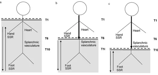
pathways [1-3]. For the sake of the comprehension, we can consider three types of SCI lesions that offer typical
autonomic reactions (tetraplegics, paraplegics at T6 and paraplegics at T10) (Fig II.1.2):
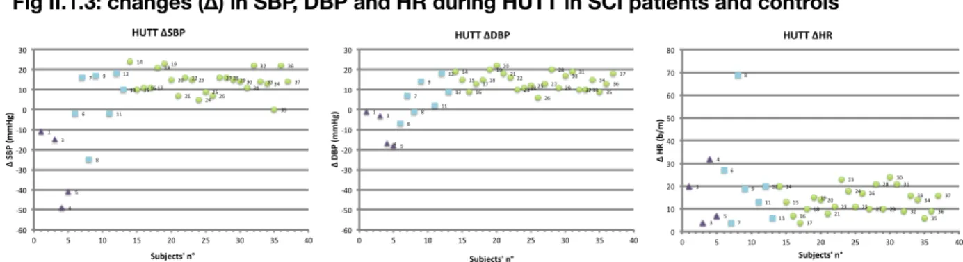
1. In cervical transection, the entire part of the sympathetic outflow, together with the sacral parasympathetic outflow, is totally disconnected from cerebral control (Fig II.1.2 a), causing an autonomic malfunction which may affect the cardiovascular, thermoregulatory, sudomotor, gastrointestinal, urinary and reproductive systems [4]. Conversely, all somatic and visceral afferents coming from below the lesion (starting from T1–T2 dermatomes) can reflexively activate the whole sympathetic column. Postural challenges cause an immediate fall in both systolic and diastolic blood pressure (SBP, DBP), due to the lack of rise in total peripheral resistance (TPR) [5,6], which is inversely related to a rapid rise in heart rate (HR), due to withdrawal of vagal tone in response to unloading of baroreceptor afferents [5-8]. Similar cardiovascular findings are observed during a static maximum inspiratory breath-hold [9] or the Valsalva manoeuvre (VM) [10]. After the VM, the blood pressure (BP) overshoot relative to baseline is absent [10]. Pressor stimuli that either originate in, or are modulated by, the brain (either cerebral or cervical afferents) will have no effect [11]. Pressor stimuli below the lesion will activate sympathetic reflex along the isolated spinal cord that can result in autonomic dysreflexia (AD).