UNIVERSITY OF PISA
Scuola di Dottorato in Fisiopatologia Clinica e Scienze del farmaco
D
OCTORATE OFP
HILOSOPHY INF
UNDAMENTAL ANDC
LINICALV
IROLOGYXXIV
CYCLEABOLITION OF THE JC VIRUS TRANSFORMING
CAPACITY BY SF2/ASF IN JCV–TRANSFORMED CELLS
RELATOR: Prof Antonina Dolei
COORDINATOR: PROF. LUCA CECCHERINI-NELLI SSD:MED 07
LIST OF CONTENTS
List of abbreviations 3
Abstract 4
CHAPTER 1: INTRODUCTION 6
1.1. JCV History and Classification 8
1.2. Genome organization 10
1.3. Viral Life Cycle 11
1.4. Natural infection of JCV 12
1.5. Reactivation and disease 16
1.6. JCV oncogenic features 19
1.6.1. Animal studies 20
1.6.2. Transgenic mouse models 22
1.6.3. JCV and associated human cancer 24 1.6.4. Molecular basis of JCV–T antigen–mediated transformation 28
1.7. SF2/ASF as new regulatory protein in JCV gene expression and
replication 31
Purpose of the thesis 34
CHAPTER 2: MATERIALS AND METHODS 36 2.1 Cell lines and cell culture 36
2.2 Plasmid constructs 36
2.3 Western blot analysis 37
2.4 Colony formation assay 38
2.5 Soft agar growth assay 38
2.6 MTT (3-(4,5-dymethylthiazo-2-yl)-2,5-diphenyltetrazolium bromide) assay for cell proliferation 38
2.7 Flow cytometry 39
2.8 Lentiviral infection and RNA interference 39
2.9 Reporter gene assay 40
CHAPTER 3: RESULTS 41
3.1 JCV tumor antigen expression is suppressed by SF2/ASF in viral–
transformed cell lines 42
3.2 SF2/ASF inhibits proliferation of JCV – transformed cells 43 3.3 Extintion of JCV tumor antigens by SF2/ASF results in loss of
viability and induction of apoptosis 46 3.4 The 76- to- 100 – amino acidic region of SF2/ASF is sufficient to
suppress growth of JCV – transformed cells 49
CHAPTER 4: DISCUSSION 54
CHAPTER 5: LIST OF FIGURES 58
LIST OF ABBREVIATIONS
aa amino acid
AIDS Acquired Immunodeficiency Syndrome AP1 Activator Protein 1
BKV BK virus
CAT Chloramphenicol Acetil Transferase CNS Central Nervous System
HAART Highly Active Anti-Retroviral Therapy HIV Human Immunodeficiency Virus IAP Inhibitors of Apoptosis Proteins
IRIS Immune Reconstitution Inflammatory Syndrome IRS-1 Insulin Receptor Substrate 1
JCV John Cunningam virus
MRI Magnetic Resonance Imaging
MS Multiple Sclerosis
NCCR Non Coding Control Region NF-B Nuclear Factor kB
NF – 1 Nuclear Factor 1
NF-1X Nuclear Factor 1 X-type NF2 Neurofibromin 2 (merlin)
NFAT Nuclear Factor of Activated T cells p21/WAF-1 cyclin-dependent kinase inhibitor
P53 Protein 53
PCR Polymerase Chain Reaction
PML Progressive Multifocal Leukoencephalophaty PNET Primitive Neuroectodermal Tumor
PP2A Protein Phosphatase 2 PBL Peripheral Blood Leukocytes pRB Retinoblastoma Protein RRM RNA Recognition Motif
RS Serine rich Region
SF2/ASF Splicing Factor 2/Alternative Splicing Factor shRNA short hairpin RNA
SV40 Simian Vacuolating virus 40 t-Ag small Tumor Antigen
T-Ag Large Tumor Antigen
VP Viral Protein
ABSTRACT
The human neurotropic polyomavirus JC (JCV) induces a broad range of neural–origin tumors in experimental animals and has been repeatedly detected in several human cancer, most notably neural crest– origin tumors including medulloblastomas and glioblastomas. The oncogenic activity of JCV is attributed to the viral early gene products, large T and small t antigens, as evident by results from in vitro cell culture and in vivo animal studies. The alternative splicing factor, SF2/ASF, has the capacity to exert a negative effect on transcription and splicing of JCV genes in glial cells through direct association with a specific DNA motif within the viral promoter region. In the present thesis is demonstrated that SF2/ASF suppresses large T antigen expression in JCV–transformed tumor cell lines, and the espression of SF2/ASF in such tumor cells thereby inhibits the transforming capacity of the viral tumor antigens. Moreover, down–regulation of SF2/ASF in viral–transformed tumor cell lines induces growth and proliferation of the tumor cells.
Mapping analysis of the minimal peptide domain of SF2/ASF responsible for JCV promoter silencing and tumor suppressor activity suggests that amino acid residues 76 to 100 of SF2/ASF are functionally sufficient to suppress the growth of the tumor cells. These observations demonstrate a role for SF2/ASF in JCV–mediated cellular transformation and provide a new avenue of research to pathogenic mechanisms of JCV– induced tumors.
1. INTRODUCTION
JCV is an ubiquitous human polyomavirus, very common in the general population, infecting 70 to 90 percent of humans; most people acquire JCV in childhood or adolescence. In individuals under immunosuppressive conditions, JCV may lead to the development of progressive multifocal leukoencephalopaty (PML). In addition to its role in the development of PML, JCV has also been shown to be associated with various tumors in laboratory animals and humans.
JCV can transform primary human fetal glial cells in a similar manner to SV40 (Mandl et al., 1987; Major et al., 1985). JCV–transformed primary human cells express viral–early genes and exhibit a transformed phenotype (Gallia et al., 1998). Inoculation of JCV into owl and squirrel monkeys induces glioblastomas, neuroblastomas, and astrocytomas (London WT. et al., 1978). Transgenic animals expressing the JCV–early genome under the control of the JCV promoter develop tumors of neural origin including adrenal neuroblastoma, medulloblastoma, malignant peripheral nerve sheath tumors, and pituitary adenomas (Franks et al., 1996; Shollar et al., 2004; Krynska et al., 1999; Gordon et al., 2000). The oncogenic potential of JCV is strongly related to the expression of viral large and small tumor antigens. Ample evidence suggests that the mechanism of JCV–mediated transformation relies on the sequestration and suppression of the tumor suppressor proteins, p53 and the pRB family, by the viral large T antigen (T Ag). Binding of these tumor suppressor proteins with large T antigen appears to interfere with the cell cycle regulatory properties of these proteins.
SF2/ASF (splicing factor 2/alternative splicing factor) is a member of the arginine/serine–rich splicing factor family and is one of the key regulators of alternative splicing of many genes (Manley et al., 1996;). Aside its role in the regulation of gene expression through the modulation of pre– mRNA alternative splicing, SF2/ASF has also been shown to be an inducer of translation initiation by suppressing the activity of 4E–BP1, an inhibitor of cap–dependent translation (Michlewski et al., 2008).
Recently it has been demostrated that SF2/ASF strongly regulates JCV transcription by directly targeting a double stranded DNA motif within the viral promoter region (Sariyer et al., 2011). In this study, the expression of SF2/ASF in glial cells suppresses the transcription of the JCV–early proteins (large T antigen and small t antigen), as well as the viral–late proteins (agnoprotein, VP1, VP2, and VP3), resulting in abrogation of JCV propagation. Here, it is investigated the impact of SF2/ASF on JCV–induced transformation of glial cells and its effect on the maintenance of a transformed phenotype mediated by the JCV tumor antigens. The results show that expression of SF2/ASF in tumor cell lines, transformed by JCV, strongly suppresses the expression of large T antigen, causes the growth arrest, and induces apoptosis. In contrast, down–regulation of SF2/ASF in such tumor cell lines increases the growth and expansion rates of the cells under anchorage indipendent conditions. Collectively, these observations may suggest a significant role of SF2/ASF in JCV–mediated cellular transformation and provide a novel approach to target JCV–induced tumors.
1.1 JCV History and Classification
Human polyomavirus JC is a DNA virus belonging to the taxonomic family Polyomavirinae that comprises 13 distinct viruses with a common ancestor that exhibit a limited range of host species to infect (Cole et al., 2001). Human polyomaviruses are JCV, BKV and the newly discovered polyomaviruses WUV (Washington University poliomavirus) (Gaynor et al., 2007), KIV (Karolinska Institute polyomavirus) (Allander et al., 2007) and MCV (Merkell cell carcinoma polyomavirus or MCPyV) (Feng et al., 2008), TSV (Trichodysplasia spinulosa-associated polyomavirus), HPyV6 e HPyV7 (correlated with KIV e WUV) e HPyV9 (correlated to the African green monkey-derived lymphotropic polyomavirus). While these eight polyomavirus are defined humans and ubiquitous, SV40 is a monkey virus inadvertently introduced into the human context between 1955 and 1963, through the administration of polio vaccine produced in cell lines of monkey kidney SV40 found infected. It is also unknown how widespread the virus was among humans before the 1950s, though one study found that 12% of a sample of German medical students in 1952 had SV40 antibodies. It is thought that exist a human variant of the SV40. Although horizontal transmission between people has been proposed, is not clear if this actually happens and if it does, how frequently it occurs (Martini et al., 2007). Although variations exist with respect to phylogenetic relatedness when different genes are used for analysis, JCV, BKV, KIV and WUV appear to group closely with SV40 (Gjoerup and Chang, 2010).
The name polyoma is derived from the greek terms: poly, meaning many, and oma, meaning tumors, and refers to the capacity of these viruses to cause tumors (Magginis et al., 2009) in non – permissive hosts.
Human Polyomavirus JC has been first identified in 1965 (Fig 1.1) by electron microscopy in a cases of PML (Silverman et al., 1965) and was first cultured and isolated in 1971 from PML brain tissue in a patient with Hodkin disease (Padgett et al., 1971; Gardner et al., 1971).
Figure 1.1. Electron micrography of JC virions. JC virions, uniform in bothsize and shape, display
perfect alignment patterns called crystalloid arrays (Yukiko Shishido-Hara, Acta Neuropathol, 2010). Brain tissue was used to inoculate primary cultures derived from the human fetal brain, represented mostly by glial cells, and the virus was isolated. The name of the patient involved was John Cunningam, whose initials were attributed to the virus JC. The prototype strain of JCV, mostly utilized in vitro and in vivo studies, is named Mad-1. 50-80% of adult individuals are seropositive for JCV (Major et al., 1992; Knowles et al., 2003). The mechanism of human-to-human transmission of JCV is unknown but is thought to be spread via a urine-oral route as JCV can be detected in untreated urban sewage. It is thought that the primary infection of JCV occur in the tonsils, then the virus can infect the epithelium of the kidney, where it established a life-long persistent infection (Dorries, 1998).
JCV primary infection in immunocompetent hosts is usually subclinical; the kidney is thought to be a major organ of JCV persistence during latency. Infections in humans by JCV are usually restricted by the actions of the immune system, particularly cell–mediated immunity and a defect in the generation of cytotoxic T–cells is associated with the emergence of JCV from latency to cause the disease PML (Koralnik, 2002).
1.2 Genome organization
JCV shares approximately 70% of DNA sequence similarity with the other polyomaviruses, SV40 and BK virus, and displays a similar tripartite genome organization with early and late coding regions and a regulatory region (Yukiko Shishido–Hara, 2010), as is shown in Figure 1.2.
JCV is a small, nonenveloped virus with an 40-45 nm icosahedral capsid containing 5- and 6-fold axes of symmetry; the viral capsid is comprised of the major capsid protein, VP1, arranged as 72 pentamers, each of which interacts with the C-terminus of one of the minor capsid proteins, VP2 or VP3 (Mengxi et al., 2009). The genome is a 3 x 10³ kd supercoiled
double-stranded circular DNA, closely associated with histones, and is packaged into chromatin (minichromosomes) which has structural similarities to cellular host chromatin (Gjoerup and Chang, 2010)
Likewise the other polyomaviruses, JCV studies has focused mainly on a prototypical or reference strain of virus; for JCV it is the Mad-1 strain, composed by 5,128 bp. The viral genome is divided into two protein-coding regions that are transcribed in opposite directions starting from a common noncoding control region (NCCR).
The viral genome is divided into three main regions: the early coding region, the late coding region, and the non-coding control region (NCCR) or regulatory region (RR). The early coding region encodes for small tumor antigen (t Ag) and large tumor antigen (T Ag) as well as T’ proteins (T’ 165,
Figure 1.2. Organization of the JC virus genome
(Strain Mad-1). A schematic representation showing the JCV genome is shown. Nucleotides are numbered relative to the Mad-1 reference strain (GenBank # NC_001699). Large-T antigen, small-t antigen, T'135, T'136 and T'165 – three additional alternatively spliced forms of T-antigen (Trowbridge and Frisque, 1995), Agno – the late auxiliary protein, agnoprotein, ELP – putative early leader protein.
T’135 and T’ 136), created by alternative splicing of the viral early pre-mRNA, and the late coding region encodes for the viral capsid proteins (or V antigens): VP1, VP2, and VP3, as well as the viral nonstructural protein agnoprotein. The NCCR separates the early and late coding and constitutes a bidirectional regulatory region in that it contains promoter/enhancer elements for the viral early and late genes and also contains the origin of viral DNA replication (Maginnis et al., 2009).
The regulatory region can be divided into two groups, according to the differences within the sequences, “archetype” and “neurotropic type”. The archetype is found mainly in the urine of healthy individuals. The viruses isolated from demyelinating brains is designated as the “neurotropic type” and is also referred “PML–type”. The neurotropic type NCCR appear to have evolved from the archetype through deletions and duplications, and these modifications may increase the restricted tissue tropism or virulence, thus leading to changes in the pathogenicity of the virus (Daniel et al., 1996).
1.3 Viral life cycle
The JC virus begins its life cycle through the recognition of cellular receptors, moving toward the nucleus, where viral transcription and genome replication can take place. Recent studies demonstrate that JCV use an N-linked glycoprotein with -2,3 – or -2,6-linked sialic acid (Dugan et al., 2008). Viral protein 1 (VP1) mediates the binding to sialic acid. Recent studies to identify a proteinaceus receptor for JCV revealed that serotonin receptor 5–hydroxytryptophan (5–HT)2A facilitates the JCV entry into host
cells. The serotonin receptor 5-HT2AR is a seven trans membrane–spanning
G-protein–coupled receptor (GPCR) that belongs to the family of 5–HT serotonin receptors. It is though that 5-HT2A R is a key host–cell determinant,
i.e correlated with the restricted JCV tropism, since is abundantly expressed in the brain and in the kidney (Maginnis et al., 2009).
the cell to the nucleus where the virus life cycle takes place (Ashok and Atwood, 2003). In the nucleus begins the transcription of viral genes occurs in a finely regulated manner. First, the early genes are transcribed. The promoter of the Mad-1 virus contains two 98–base pair repeats, which function in a bidirectional manner to regulate the expression of early and late genes. Many transcription factors have been identified including, AP1, NF – 1, NF-B, NFAT, Pur-alpha and YB -1; they can act as activator or silencer in JCV gene expression (Safak et al., 1999; Major et al., 1990). The JCV early mRNA is alternatively spliced into 5 transcripts: large T Ag, small t Ag, T’ (135), T’ (136), and T’(165) (Trowbridge PW. et al., 1995;). The splicing factor SF2/ASF, object of the studies shown in the present thesis, is a member of the splicing machinery (Sariyer and Khalili, 2011), moreover it is also involved in the post-transcriptional regulation of JCV early and late genes.
The T-Ag is the main regulatory protein from which depends all the viral functionality: T-Ag binds to the origin of DNA replication, unwinds the viral DNA through its helicase activity, and recruits the host–cell DNA polymerase to drive replication and this activity is implemented by the T’ proteins. Next, T Ag suppresses early genes transcription and initiates transcription of the late viral genes (the capsid viral is assembled in the nucleus), and Agno; agnoprotein is produced in JCV–infected cells and regulates viral transcription and replication by directly interacting with T Ag (Safak et al., 2001). After assembly of the virus in nuclear regions named ND10, the release of the viral particles can take place (Shishido – Hara Y. et al., 2004).
1.4 Natural infection of JCV
The polyomavirus JC has a limited host range; despite the fact that it can enter a variety of mammalian cell types, its replication is restricted to glial cells and B-cells precursors. Productive infection ends with cell lysis. JCV is able to establish a persistent infection in the kidney, that is thought to be the major site of persistence during latency but JCV rarely induces
diseases in immunocompetent individuals. The entire viral life cycle has been extensively studied in the context of PML, which JCV is the causative agent. The immunosuppressed individuals mostly prone to PML are those with human immunodeficiency virus (HIV) infection/AIDS (Hou et al., 2000), The occurrence of PML was very rare in the pre-HIV/AIDS era. Moreover, a strong cross-talk between HIV and JCV is thought to exist.
The major sites of persistence for JCV are the cells of the kidney and urinary tract; JCV can be detected in approximately 10 to 50% of normal kydney samples (Grinnel et al., 1983).
Numerous reports of JCV DNA in lymphocytes argue for the importance of these cells in persistence and dissemination via circulatory routes (Doerries et al., 2006; Chesters et al., 1983). It is not known whether the viruses enter a latent state or maintain a low level of viral gene expression and replication at these sites, although intermittent replication must occur as evidenced by periodic excretion of virus in the urine. Approximately 5% of immunocompetent individuals have BKV viruria while 20 to 30% are actively shedding JCV (Chesters et al., 1983). How the host innate, humoral, and cellular immune responses control these persistent infections remains unknown.
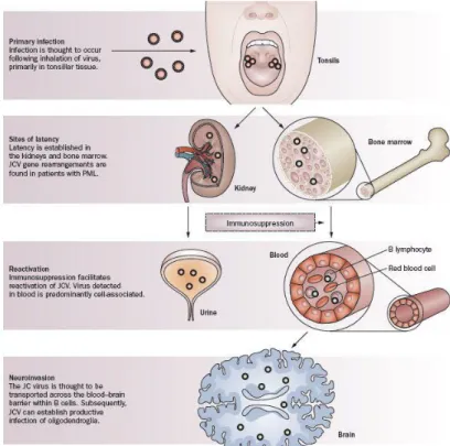
The infection by JCV occurs through inhalation or by ingestion of foods or beverages contaminated with the virus; after the primary infection, the virus is thought to localize in the kidneys or in the lymphoid organs such as the bone marrow, tonsils or spleen in a state of latency (White and Khalili, 2005). During immunosuppression, it is believed that the JC virus is reactivated, since it is recovered in urine and blood, mainly associated with lymphocytes B; in favor of this statement there is a recent work that shown intact JCV DNA sequences in bone marrow biopsies of patients obtained years before the development of PML (Leslie et al., 2010).
JC virus is ubiquitous in the human population and approximately 50 – 80 % of adults are seropositive, with asymptomatic infections by early
Figure 1.3. Possible mechanism of JCV infection (Nat Rev Neurol,
2010 Nature Publishing Group).
A variety of cell types have been reported to contain BKV and JCV sequences, and it has been suggested that infection of these cells may be involved in dissemination or persistence. There are rare reports of JCV sequence detection in various tissues and cells including the liver, lung, spleen, lymphonodes, and colorectal epithelium (Caldarelli et al., 1999; Laghi et al., 1999). While JCV reactivation leads primarily to infection of the brain, it is unclear whether normal brain tissue harbors the virus. Published studies have reported both the presence (Caldarelli SR. et al., 1999; Elsner C. et al., 1992) and the absence (Chesters et al., 1983; Quinlivan et al., 1992) of JCV sequences in the brains of patients without PML. The presence of JC viral DNA has been repeatedly detected in postmortem brain tissues from patients who died from non-neurologic diseases (Del Valle et al., 2001)
It is thought that JCV enters the host through infection of lymphoid cells and that these cells transfer the virus to B lymphocytes, and thereby to the sites of latency (Leslie et al., 2010), as the brain.
BJAB and Namalwa B cell lines have been successfully tested in vitro for the replication of JCV (Atwood et al., 1992); the replication of JCV in B cells
correlates directly to the expression of the nuclear proteins that bind the viral promoter similar to those found in the brain (Major et al., 1990). Sequence studies show that the JC virus found in the bone marrow belongs to the PML-type (Tan et al., 2009).
It is thought that infected B cells enter circulation and deliver the virus to the bone marrow, where CD34 + hematopoietic precursors become infected and
maintain latently the viral genome. The nuclear factor (NF)-1X, which is required for JCV replication in the brain, seems to be essential also for CD34+ cells susceptibility to JCV entry (Monaco et al., 2001). In bone marrow the viral DNA has been detected, but not intact infectious progeny virus, suggesting that JCV may be latent in these cells. If the control by immune defences dampens, the bone marrow latently infected CD34+ cells differentiate into mature B cells that
can cross the blood-brain-barrier and carry the virus to the brain. In the brain, infected B cells release infectious virus that infects oligodendrocytes, which in turn replicate massively the virus leading to the severe demyelination, peculiar of PML. To note that in each compartment where JCV sequences of the PML-type are detected (B cells, bone marrow, and brain), virus expression is restricted by the binding of host cell transcription factors (Leslie et al., 2010).
Of considerably importance the so-called “archetypal theory”, in which seems that archetypal virus and the neurotropic PML-type are two different JC viruses, that circulate in the human population; the archetypal virus can be detected in the urine of healthy individuals and in urban sewage. It has been proposed that it is the archetype that circulates in the human population and, after a primary infection, remains quiescent in the kidney or other organs ,with occasional excretion to the urine. Some time later, the neurotropic type (PML-type) would originate from the archetypal virus, through sequence rearrangements. How and where the JCV virus evolves from the archetype to the neurotropic type remains to be established. One study reports that the viruses in PBLs were mostly of the neurotropic type (Dorries et al., 1994), while Azzi and collegues have shown the presence of both archetype and neurotropic
populations and in different tissues from immunocompromised PML patients. Thus, it might be that the demyelinating disease could develop after primary infection, without requiring the period of latency.
1.5 Reactivation and disease
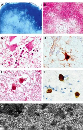
The PML is a fatal demyelinating disease of the central nervous system (CNS) that is characterized by multiple foci of demyelination caused by the lytic infection by JCV of oligodendrocytes, the cells of the CNS that produce myelin (Gordon et al., 1998). PML is characterized by a triad of findings: 1) oligodendrocytes having enlarged nuclei with viral inclusion bodies, 2) bizarre, atypical astrocytes, and 3) loss of myelin with accompanying phagocytic infiltrate (Fig 1.4).
Figure 1.4. Progressive multifocal leukoencephalopathy. (A) PML is characterized by multiple subcortical foci of demyelination highlighted using Luxol fast blue stain for myelin (original magnification: x20). (B) Higher magnification of several demyelinated lesions (H&E, original magnification: x100). (C) Bizarre astrocytes with extreme pleomorphism and multiple lobulated, atypical nuclei are characteristic cells in PML. (D) Immunohistochemistry for VP1 in a bizarre astrocyte. (E) Enlarged oligodendrocytes with intranuclear eosinophilic inclusion bodies are pathognomonic for PML (H&E stain). (F) The inclusions are the site of acmagnification: x1000. (G) Characteristic icosahedral JC viral particles are shown by electronmicroscopy in the nucleus of an infected oligodendrocyte in a case of PMLtive viral replication as demonstrated by expression of the capsid protein VP1.
D–F, original. (Del Valle, White, and
Khalili. Potential Mechanisms of the Human Polyomavirus JCV in NeuralOncogenesis. J Neuropathol Exp
Usually this disease develops only in individuals with a severely compromised immune system, typically AIDS patients (about one in 20 of HIV– infected persons )(Berger et al., 2003). Therefore, PML is considered an AIDS– defining illness.
Other circumstances that incur immunosuppression are also associated with PML, eg, autoimmune diseases, agammaglobulinemia, lymphoma, immunosoppressive drug treatment, cancer chemotherapy (White and Khalili., 2006). PML patients often develop emiplegia, visual disturbances and subcortical dementia. Other neurological symptoms may also be found, such as dipoplia, monoplegia and akinesia (Berger et al., 1995). The involvement of multifocal areas of demyelination in these patients is usually detectable by magnetic resonance imaging (MRI). The prominent histopatological finding in PML is multiple foci of myelin loss, eosinophilic, enlarged oligodendroglial nuclei, and enlarged bizarre astrocytes with lobulated hypercromatic nuclei. The bizarre, atypical astrocytes which are visually indistinguishable from tumor cells in high-grade glial neoplasia may represent an infection by JCV of a cell type that is unable to support a fully lytic viral life cycle. Viral DNA and LT protein expression is detectable in astrocytes but at a much lower frequency and quantityPolyedral viral particles can be observed within the inclusion bodies of oligodendrocytes under the electron microscope.
The incidence of PML complicating HIV/AIDS is higher than that of any other immunosoppressive disorder relative to their frequencies (Berger et al., 2003). There exist specific molecular mechanisms whereby HIV-1 may promote JCV gene expression (Chowdhury et al., 1990), and partecipate in the pathogenesis of PML.
The replication and dissemination of JCV in PML causes the death of oligodendrocytes and hence the development of focal areas of progressive demyelination. The exact cause of cell death in PML is not known. It has been suggested that JC virus–infected cells may undergo apoptosis (Richardson– Burns et al., 2002). JCV T-antigen and agnoprotein may abrogate cell cycle
death JCV-mediated in astrocyte cell culture (Seth et al., 2004). A recent study demonstrate that JCV infection is anti–apoptotic, allowing for completing of the viral lytic cycle (Pina–Oviedo et al., 2007). The number of reported cases of this previously rare disease has significantly increate since the 1980s due to the pandemic acquired immunodeficiency syndrome (AIDS); however, after introduction of HAART therapy, PML remains a major AIDS complication result in death (Gray et al., 2003).
Paradoxically, some patients develop a deterioration associated with HAART treatment, known as immune reconstitution inflammatory syndrome (IRIS). IRIS can result from reconstitution of the immune system to recognize pathogens/antigens, and can be accompained by toxoplasmosis, cryptococcosis, and tubercolosis, as well as PML (Gray et al., 2005).
The disease can also develop in persons with rheumatic diseases related or unrelated to immunosoppressive drug treatment; indeed, sistemic lupus erythematosus is one of the most common underlying diseases (Molloy et al., 2008).
PML has in 2005 been diagnosed in patients taking natalizumab (Tysabri) (Kleinschmidt-DeMasters and Tyler, 2005; Langer-Gould et al., 2005; Van Assche et al., 2005) becoming the second largest group of PML risk, causing its temporary withdrawal from the market. Natalizumab is an alpha-4-beta-1 integrin inhibitor approved for the treatment of relapsing-remitting multiple sclerosis (MS) and also used for the Crohn’s disease One of the proposed mechanism of action suggest that the blocking of leukocyte accross of blood-brain barrier, causes a loss of immune control in the CNS, resulting in the reactivation of latent JC virus in the brain. (Chen et al., 2009).
PML in the setting of natalizumab therapy is related to cumulative exposure to natalizumab. As of August, 2011, there had been 150 cases of natalizumab-related PML documented in more than 88,000 patients exposed to natalizumab worldwide (Update on Tysabri and PML. National Multiple Sclerosis Society Web site. http://www.nationalmssociety.org/news/news-detail/index.aspx?nid=2308. Published April 11, 2011. Updated July 6, 2011. Accessed July 7, 2011). The incidence of PML in natalizumab-treated patients varies according to the number of infusions received, but the incidence of PML
by each epoch of treatment exposure (1 to 24 infusions, 25 to 36 infusions, 37 to 48 infusions) appears to have remained stable over time.
Transplant recipients of solid organs, can develop demyelination, likely due to immunosoppressive treatments used to prevent graft rejection (Schitrit et al., 2005).
The oligodendrocytic myelin forming cells support full lytic replication of the virus causing the hallmark loss of myelin seen in PML. Infection of astrocytic cells results predominantly in changes in nuclear morphology, size, and ploidy. Whether the JCV infected astrocytes are transiently transformed is an intriguing conjecture (White and Khalili, 2006). There are scattered, but convincing case reports in the literature demonstrating expression of JCV T antigen in tumors of the CNS (Krynska et al., 1999; Pina-Oviedo et al., 2006); however, as a group, the glial neoplasms have not been associated in a consistent way with JCV infection.
Oncogenicity of JC virus will be discussed in the next paragraph.
1.6 JCV oncogenic features
Beside its primary role in the develpment of PML, JCV has also been shown to be associated with several human tumors (Del Valle et al., 2001a). While the etiologic role for JCV in the development of cancer in humans remains to be established there are experimental and clinical evidence for a involvement of JCV in cancer.
Furthemore, the ability of the JCV early protein to transform human fetal cells has led to the development of several cell lines that possess tumorigenic activity in vivo.
The first evidence for the association of JCV with cancer came from reports of brain tumors found in patients with concomitant PML (Del Valle et
oligodendroglioma on post–mortem examination. Since the isolation of JCV, a large body of evidence has accumulated for its role in oncogenesis.
JCV is able to infect and transform cells in culture: primary cultures, including primary human fetal glial cells, primary hamster brain cells (White et al., 2007), human vascular endothelial cells (Walker and Padgett, 1978). Almost all JCV–infected transformed cells express the viral early protein and they exhibit the phenotypic properties associated with transformation: growth in soft agar, serum-independence, changes in morphology, the ability to form foci in culture, and in some but not all cases, induces tumors in Nude mice (Gallia et al., 1998 a; Del Valle et al., 2001 a; White and Khalili., 2007).
1.6.1. Animal studies
The oncogenic potential of JCV has been established in several animal models. When the PML-type of JCV is inoculated via intraocular, intraperitoneal or intracerebral routes in newborn Syrian Golden hamsters, approximately 6 months after JCV injection, the animals develop multiple types of tumors of glial and neuronal origin: medulloblastoma, peripheral neuroblastoma, glioblastoma multiforme, astrocytoma, and primitive neuroectodermal tumors (Walker et al., 1973; Zu Rhein, 1983; Khalili et al., 2003). More than 85% of animals develop CNS tumors, and this demonstrates the oncogenicity of JCV for tissues of neural origin. Inoculation with the Tokio-1 strain of JCV in newborn Sprague–Dawley rats results in the induction of primitive neuroectodermal tumors (Ohsumi et al., 1986). Immunohistochemical analysis of the tumor tissues, has detected Large T Ag in the nucleus of the tumor cells, but any signs of viral replication or myelin pathology. The age at the time of injection is significant with regard to the frequencies of tumor development: animals inoculated at birth have a much greater likelihood for developing cerebellar medulloblastomas than animals inoculated later in age, who more frequently developed glial-origin tumors in the cortex (Khalili et al., 2003).
Injection of JCV into the brain of newborn rats induces in the 75% of the animals, primitive neuroectodermal tumors.
Many authors report microscopic tumors in the internal granular layer of the cerebellum of hamsters which are actively replicating and migrating inward during early development (Zu Rhein and Varakis, 1979). The cells of the external granular layer originate in the neural crest and are thought to give rise to medulloblastoma in humans. In fact, the majority of the neoplasias observed in the hamster and rat models are tumors arising from cells of the neural crest, as medulloblastoma, primitive neuroectodermal tumors and glial-origin tumors. Intracerebral injection of JCV in owl and squirrel monkeys results in develop of astrocytoma, neuroblastoma and glioblastoma. Immunoistochemical analysis of the tumor originated by JCV injection in monkeys reveal that monkey cells are non permissive for JCV replication (London et al., 1978; Major et al., 1992).
Cells cultured from an astrocytoma arising in one owl monkey inoculated with JCV were found to produce spontaneously infectious JC viral particles, although some rearrangement of the viral regulatory region may have occurred (Major et al., 1987). In all, JCV remains the only human virus reported to induce solid tumors in non-human primates.
1.6.2
Transgenic mouse model
Transgenic mice studies provide mostly of the interesting observation on the oncogenic potential of JCV. This is due to the fact that they contain the complete gene sequence for JCV T-antigen under the control of its own early promoter, and the role of T-antigen can be studied in the absence of sequences encoding the capsid proteins. Independent lines of transgenic mice have been generated exhibits distinct phenotypes: transgenic mice can develop dysmyelinating diseases as observed in mice generated with the Mad-4 strain of JCV, due to alterations in myelin sheath formation, perhaps determined by the feature of JCV to affect myelin gene expression (Devireddy et al., 1996); in addition these mice can also develop peripheral neuroblastomas and adrenal neuroblastomas. Transgenic mice carryng the Archetype, or kidney-derived isolate of JCV as the source of the transgene, develops cerebellar tumors resembling human medulloblastoma. Similar to peripheral neuroblastomas, these very cellular tumors have a high mitotic index, contain scant cytoplasm, and exhibit positivity for the neuronal marker synaptophysin (Khalili et al., 2003). 80% of transgenic mice develop medulloblastomas between 9 and 12 months of age. By immunohistochemical analyses of the tumoral cells from transgenic mice, has been shown that have the majority of tumor cells contain high levels of T-antigen in the nucleus. Also, Krynska and colleagues demonstrates high levels of T-antigen protein in the tumor tissue in association with the tumor suppressor proteins, p53 and pRb (Krynska et al., 1999a, 1999b). Next they created from tumor tissue clonal populations of T-antigen-positive and T-antigen-negative cells, and they shown that only T-antigen-positive cell lines are able grown subcutaneously in Nude mice (Krynska et al., 2000). The behavior of those cells is due to the fact that the T-antigen-negative cell lines have point mutation affecting an intron– exon junction, which results in the expression of a smaller in-frame p53 mRNA species lacking exon 4 (Krynska et al., 2000). This region of p53 is located close to the T-antigen binding site, and therefore suggests that tumor cells may require the presence of T-antigen for inactivation of p53, while the cells that arbors mutant p53 variants are expressed, may no longer require T-antigen
expression (Khalili et al., 2003).
Franks and colleagues have created transgenic mice with JCV T Ag under its own promoter and they developed neuroectodermal tumors with primitive origin or pituitary gland tumors (Franks et al., 1991).
Summarizing these studies it is possible to affirm that T Ag in absence of viral replication, can affect myelin sheats formation leading to dysmyelination and that T Ag is able, in accord with its oncogenic potential, to induce tumors of neural origin.
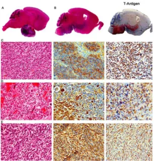
Figure 1.5. JCV-induced tumors in transgenic
animals. (A) The brain of a JCVCY (archetype) transgenic mouse with a large, extra-axial mass in the pituitary region. (B) Consecutive sections of the brain of a mouse with an intra-axial PNET centered in the brainstem and infiltrating into the basal ganglia, (H&E stain, left panel). Immunohistochemistry for T-Ag (right panel) demonstrates protein expression by the tumor but not the adjacent brain. (C) PNETS are characterized by sheaths of closely packed neoplastic cells with slightly elongated nuclei (H&E, left panel). The tumor (D) A pituitary tumor is composed of round neoplastic cells with abundant cytoplasm (H&E, left panel) and expresses cytoplasmic prolactin (middle panel) and nuclear T-Ag (right panel) by immunohistochemistry. (E) Malignant peripheral nerve sheath tumor (MPNST) is composed of fascicles of spindle-shaped cells with elongated nuclei (H&E, left panel). These tumors express S-100 (middle panel) and nuclear T-Ag (right panel) by immunohistochemistry.
Original magnification for all panels: x1000. (Luis Del Valle, Martyn K. White, and Kamel Khalili. Potential Mechanisms of the Human Polyomavirus JCV in NeuralOncogenesis. J Neuropathol Exp
1.6.3
JCV and Associated human cancer
The first indication that JCV can be involved in human cancer, come from an oligodendroglioma found in a patient with PML and Richardson reported “the disorder as a heretofore unrecognized complication of lymphatic leukemia or Hodgkin's disease, and the white matter lesions were generally discovered as incidental findings upon autopsy” (Richardson, 1961) . On the other hand, JCV has been associated with brain tumors in absence of PML lesions (Rencic et al., 1996, Del Valle et al., 2000). This association has not yet been explaned because mostly of the adult individuals are sieropositive for JC virus, with seroconversion occurring during adolescence. Also, while JCV DNA can be extracted from human tumor tissue, it is challenging to determine causality due to the ubiquitous nature of the virus (Khalili et al., 2003). Many authors found various type of brain tumors in immunocompetent individuals without PML (Boldorini et al., 1998). Del Valle and colleagues examined 85 samples of glial-tumor origin and they found that 53 to 83% of glial-tumors were positive for JCV DNA and T Ag expression (Del Valle et al., 2000). The types of human cancers associated with JCV are: gastrointestinal cancers: colorectal (Lyn et al., 2008; Laghi et al., 1999;), gastric (Murai et al., 2007), and esophageal (Del Valle et al., 2005); brain cancers: glioblastomas (Boldorini et al., 2003; Delbue et al., 2005; Del Valle et al., 2000; Pina-Oviedo et al., 2006), oligoastrocytomas (Rencic et al., 1996), oligodendrogliomas (Del Valle et al., 2002), and medullablastomas (Shiramizu et al., 2007), and lung cancer (Zheng et al., 2007). Groups have also reported that there is no association or involvement of JCV in these particolar cancers (Hayashi et al., 2001).
In another study the JCV agnoprotein has been called into the question: 11 of 16 samples of medulloblastoma resulted positive for Agno DNA sequences and agnoprotein detection by immunohistochemistry results positive in 11 of 20 samples of medulloblastoma
Association of JCV with human brain tumors
The detection of the JCV early gene in many human brain tumors have suggests that JCV may be associated with human neoplasias in the absence of immunosuppression or PML. The use of sensitive molecular allowed to detect JC viral genomic sequences and expression of T-antigen in a variety of human tumor cell types. Rencic and colleagues (Rencic et al. 1996) reported the first case of JCV T-antigen detection in an oligoastrocytoma by PCR/Southern blot hybridization, immunohistochemistry, and Western blotting. This data was further confirmed by subsequent analysis of oligodendrogliomas, in which they have revealed viral DNA in 20–75% and T-antigen expression in up to 50% of tumors (Caldarelli et al., 2000; Del Valle et al., 2001). Other studies have revealed, by RT-PCR analysis and immunohistochemistry, many tumor samples of neural origin in association with JCV tumor antigens expression: astrocytomas, anaplastic astrocytic tumors, anaplastic oligodendroglial tumor, as well as glioblastomas and ependymomas. The presence of JCV T antigen not seems to be correlated with the aggressiveness or malignancy of the glial tumor (Safak et al., 2001; Khalili et al., 2003 ).
Two separate studies of 43 medulloblastomas, nearly 77% were shown to contain viral DNA of JCV T-antigen, while 36% of the tumors analysed by immunohistochemistry showed nuclear positivity for T-antigen. (Del Valle et al., 2002a).
Medulloblastoma is the most common malignant brain tumor in children with nearly 70% appearing in children under the age of 16 and rarely occurring in individuals over 50 (Khalili et al., 2003; Molenaar and Trojanowski, 1994).
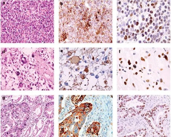
Figure 1.6. Histological and immunohistochemical evaluation of human tumors harboring JCV. Human
tumors characterized by light microscopy include medulloblastoma (panel a), glioblastoma multiforme (panel d), and adenocarcinoma of the colon (panel g). Tumors were further characterized by immunohistochemistry for their expression of cell-type-specific markers confirming the tissue of origin including the neuronal marker, synaptophysin, expressed by medulloblastomas (panel b), the glial-specific marker, GFAP (panel e), and the epithelial marker cytokeratin for adenocarcinomas (panel h). Immunohistochemical staining for JCV T-antigen shows positive staining in the nuclei of tumor cells in all three tumor types (panels c, f, and i, respectively). (panels a, d, and G: hematoxylin and eosin staining; panels b, c, e, f, h, and i: hematoxylin counterstain; All panels: original magnification 400)
(Khalili et al., Human neurotropic polyomavirus, JCV, and its role in carcinogenesis. Oncogene, 2003)
In medulloblastoma has been detected mutant genes, as the catenin of the Wnt signaling pathway. Infrequently, mutations in the p53 gene are found.
Agnoprotein, was detected in 69% of tumor samples (Safak et al., 2001); it appear involved dysregulation of the cell cycle properties due to the binding to p53 and upregulating transcription of the p21/WAF-1 (Darbinyan et al., 2002). Since, in another study, some of the medulloblastomas positive for agnoprotein were negative for T-Ag, the agnoprotein may have a role in the development of some type of medulloblatomas, indipendently by T Ag (Safak et al., 2001).
Association with non–neural human tumors
In addition to the CNS cells, JCV has also been detected in B limphocytes; a study reports that 60% of CNS limphoma samples were positive for T Ag DNA sequences and 25% of the samples showed T Ag expression (Del Valle et al., 2005), while any evidence of viral replication was detected. Hight percentage of normal tissues upper and lower human gastrointestinal tract and colon tumors, have been shown JCV DNA sequences. “Due to the potential fecal-oral transmission of JCV, has been establish that JCV DNA is present in the upper and lower human gastrointestinal tract” (Riccardiello et al., 2000, 2001). These data were confirmed by PCR/Southern blot hybridization and immunohistochemistry performed for the presence of JCV: 83% of 29 tumor samples were found positive for JCV DNA sequences (Laghi et al., 1999), “a second study detected JCV DNA in 81% of 27 samples and nuclear T-antigen by immunohistochemistry in 63% of the samples” (Enam et al., 2002). In addition, approximately 44% of the colorectal tumors also contained evidence of JCV agnoprotein. Furthemore, the disregulation of -catenin signalling in colon tumor cells, seems to be related to T Ag activity (Enam et al., 2002).
The association of JCV with human tumors and the experimental animal studies provide a strong basis to correlate the transforming ability of JCV and neoplasias.
1.6 Molecular basis of JCV T–antigen–mediated
transformation
JCV expresses three early proteins with regulatory functions: 1) the large T-Ag, 2) the small t-antigen (t-Ag) and 3) the agnoprotein encoded in the late region. Three splice variants of T-Ag have been reported (Trowbridge and Frisque, 1995). The regulatory proteins of JCV are multifunctional, in that the virus need to drive the cell cycle into the S-phase, then its proteins must be able to interact with many cellular factors and occasionally these interaction may be involved in malignant transformation.
JCV T-Ag is a nuclear phosphoprotein of 688 amino acids. It is a very versatile protein, in that it is an essential factor for viral DNA replication: T Ag works as ATP-dependent replicative DNA helicase, as DNA binding and alpha-polymerase (White and Khalili, 2005); as part of T-Ag property to drive the cells into the S-phase, its interaction with several tumor suppressor proteins can allow an aberrant stimulation of cell cycle, and this can facilitate the oncogenic transformation (Khalili et al., 2006; White et al., 2004). Some of the potential oncogenic interactions with the host cell factors are listed in this section. Results from proteins interaction studies revealed that T Ag, in a similar manner of SV40, has the capacity to bind and inactivate p53 and members of pRb family, leading to an uncontrolled proliferation. The interaction of T Ag with the oncosoppressor p53 may results in a failure of protection against DNA damage, as rather oncogenic transformations. The interaction of T-Ag with pRb activates E2F transcription factors that promote cell cycle progression (White et al., 2006).
The insulin receptor substrate 1 (IRS-1) is another factor which interact whit T Ag, allowing to development of medullosblastoma (Lassak et al., 2002), “the most prevalent solid brain tumor of childhood” (Khalili et al., 2003).
JCV T-Ag also binds to catenin (Enam et al., 2002). This interaction β-catenin/Wnt pathway is another mechanism allowing the alterations of cellular transcription of c-myc and ciclin D1 and the promotion of the cellular transformation.
Recent data suggests that the protein neurofibromin-2 or merlin (NF2) is a positive regulator of p53 and T Ag can bind this protein leading to a disruption of the control system of the cell cycle mediated by p53 (Khalili et al., 2003, White et al., 2004, Shollar et al., 2005).
The T’ proteins, obtainend by alternative splicing of the pre mRNA of T Ag, are able to interact with pRb family and this may contribute to viral replication and cellular transformation (White et al., 2005).
The other protein encoded by the early region of primate polyomaviruses is the 172 amino acid small t-Ag. The N-terminus of t-Ag is shared with the N-terminus of T-Ag but the C-terminus is a unique domain that is incorporated by alternative splicing of the early region primary transcript. t Ag function by inhibiting the cellular phosphatase PP2A, and activating kinase signaling pathways of cellular proliferation (White et al., 2004).
“Agnoprotein expression exert profound effects on the cells, including dysregulation of cell cycle progression and cell accumulation at the G2/M phase” (Darbinyan et al., 2002;).
Moreover, JCV have a mutagenic effect on cellular DNA. Genomic stability and DNA repair were found to be significantly dysregulated by JCV infection.
The complex series of activity of T-Ag expression may involve additional pathways: JCV infection and T-Ag expression can induces the expression of an anti-apoptotic protein, survivin, and this results in a . (Ambrosini et al., 1997). significant decrease in apoptosis. The authors suggeste that JCV activates this pathway in order to have enough time to replicate and complete its lytic cycle. (Pina-Oviedo et al., 2007).
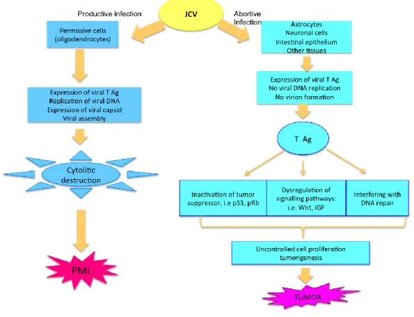
Figure 1.7: Mechanisms of JCV T-antigen-mediated cellular transformation or
demyelination (adapted from Khalili K et al., 2003)
The current data indicate that, JCV has a mutagenic effect on cellular DNA and it is able to induce instability in the cell ploydia, through the actions of T-Ag and agnoprotein.
SF2/ASF AS NEW REGULATORY PROTEIN IN JCV GENE
EXPRESSION AND REPLICATION
SF2/ASF (splicing factor 2/ alternative splicing factor) is a member of the arginine/serine–rich splicing factors family and iso ne of the key regulators of alternative splicing of many genes (Manley and Tacke, 1996). SF2/ASF was discovered and purified from HeLa cell nuclear extracts by two independent groups employing different assays (Ge et al., 1990; Krainer et al., 1990).
SF2/ASF has a modular structure consisting of two copies of N – terminal RNA recognition motifs (RRM1, RRM2), followed by a C –terminal domain rich in Arg and Ser residues, known as the Arginine–Serine-rich (RS) domain.
The RS domain interacts with the components of the core splicing apparatus to form splice site pairing, whereas the RRM1 and RRM2 domains determine the RNA–binding specificity. SF2/ASF is mainly localized to the nuclear speckles and shuttles between the cytoplasm and nucleus. Depending on splice site selection, SF2/ASF can negatively or positively regulate splicing.
Beside the roles of SF2/ASF in the regulation of gene expression through the modulation of pre – mRNA alternative splicing, it ha salso been shown a san inducer of translation initiation by suppressing the activity of 4E – BP1, an inhibitor of cap – dependent translation (Michlewski et al., 2008). SF2/ASF has been shown to be an essential host protein since SF2 – deficient mice show embrionic lethality. In addition to its role in gene expression through the regulation of splicing and translation, SF2/ASF is also up – regulated in various human tumors, including lung, kidney, colon, pancreas, small intestine, and thyroid. Up – regulation of SF2/ASF in tumor cells indicates a possible role in malignant transformation. Furthemore, over – expression of SF2/ASF is sufficient to transform NIH–3T3 and Rat1 cells, and its transforming activity is related to aberrant splicing of the tumor suppressor, BIN1, and the kinases, MNK2, and S6K1 (Karni et al., 2007).
papilloma virus type 16 late RNA control element, HPV–16 NRE, and its expression is up–regulated by viral E2 protein during the differentiation of epithelial cells infected with the virus (McPhillips et al., 2004). Retroviruses use un–spliced viral mRNA for the expression of viral regulatory and structural proteins and genomic RNA for the maintenance of the infection. The balance between the usage of un–spliced and spliced RNA by the retroviruses is extremely important, since slight differences in splicing efficiency can lead to impaired viral replication. In Rous sarcoma virus–infected cells, SF2/ASF interacts with the negative regulator of splicing elements (NRS) on full length un–spliced viral RNA and inhibits splicing. SF2/ASF has been a positive regulator of M2 ion–channel protein mRNA of influenza virus, a negative–strand RNA virus, by binding to the 3’ exon on M1 RNA (Shih et al., 1996). There is accumulating evidence that the gene expression of HIV – 1 has also been influenced by SF2/ASF. The 9 kb full–length HIV genomic transcript undergoes a series of posttranscriptional modifications that generates viral mRNAs that produce viral structural and regulatory proteins. SF2/ASF binds to exonic splicing enhancers (ESEs) downstream of the Tat-, Rev-, Env-, splice sites and promotes exon definition (Bakkour et al., 2007; Caputi et al ., 2004; Keriel et al., 2009). Therefore, SF2/ASF is thought to play a major role in the regulation of HIV-1 pre-mRNA splicing and has been proposed as a novel target for the inhibition of HIV replication. IDC16, an indole derivative that inhibits SF2/ASF activity, has been shown to be a strong inhibitor of the macrophage and T–cell – tropic strains of HIV – 1 replication (Bakkour et al., 2007).
SF2/ASF was first discovered as a cell type specific regulator of simian polyomavirus 40 (SV40) gene expression at the post -transcriptional level (Ge et al., 1990). In a recent study (Sariyer and Khalili, 2011) has been investigated the impact of SF2/ASF on human polyomavirus, JCV, gene expression and viral DNA replication in primary human fetal astrocytes, PHFA. The authors co– transfected/infected PHFA cells with the Mad–1 strains of JCV and with a plasmid ex pressing SF2/ASF in sense and antisense orientations. Viral DNA replication was analyzed at 8 days post–infection by Southern blotting. Overexpression of SF2/ASF suppressed the rate of viral replication and the expression of JCV proteins, VP1 and agnoprotein in PHFA cells. Expression of
SF2/ASF in PHFA cells also decreased the copy number of the virus during the course of JCV infection, suggested that decrease in viral gene expression by SF2/ASF had a profund impact on JCV propagation and the lytic cycle of the virus. The status of JCV transcripts during the course of viral infection was also investigated by RT-PCR. Consistent with the protein levels, over – espression of SF2/ASF in PHFA cells during the JCV infection completely inhibited viral early (T Ag) and late (VP1) mRNA production in a dose dependent manner. To investigate the possible impact of SF2/ASF on splicing of JCV transcripts, a a plasmid containing the JCV early promoter sequences ex pressing the JCV early genes was co–transfected into PHFA cells with an SF2/ASF expression plasmid. RT-PCR analyses of alternatively spliced forms of the JCV early trasnscripts revealed that SF2/ASF not only suppressed the formation of spliced gene products (T – Ag and t-Ag), but also reduced the formation of primary transcripts, suggested that SF2/ASF had a negative impact on JCV transcription. Therefore, this study shown that overespression of SF2/ASF decreased the JCV– early and late basal promoter activities in a dose dependent manner. These results revealed a novel role of SF2/ASF in trascriptional regulation. ChiP analyses from BSB8 cells overexpressing SF2/ASF assessed its ability to bind the JCV promoter; the DNA corresponding to the central region of the 98 bp tandem repeat, showed binding activity to SF2/ASF.
PURPOSE OF THE THESIS
Transcriptional suppression of JCV expression by a cellular protein, SF2/ASF, which has been mainly studied for its regulatory roles in alternative splicing, is an unexpected and novel area of JCV research. JCV gene expression is a highly regulated process, which, in permissive cells, is under tight control of the viral promoter by inducible and ubiquitously expressed cellular transcription factors. The restricted tropism of JCV to the CNS is attributed to the tissue-specific expression of the viral-early promoter and large T antigen, which is the main regulatory protein of viral replication and transcription.
Identification of SF2/ASF, which is ubiquitously expressed in body tissues, as a negative regulator of JCV tumor antigen expression may suggest a very important checkpoint for the regulation of JCV gene expression in permissive as well as nonpermissive cells.
The studies of the present thesis are based on the hypotesis that the JCV gene expression in tumor cell lines is controlled by SF2/ASF. The purpose of the thesis is to understand wheter SF2/ASF is a critical cellular protein that limits/controls the expression of JCV genes, to establish the role of SF2/ASF on integrated JCV gene expression,as well as on the maintenance of the viral transformed phenotype and also whether it may constitute a novel target for the treatment of JCV-caused diseases.
2 MATERIAL AND METHODS
2.1
Cell lines and cell culture.
BsB8 cells were derived from primitive neuroectodermal tumors that developed in transgenic mice expressing the early genome of the JC virus (Krynska et al., 1999). HJC-2 is a clonal subline of the HC-15 glioma cell line, which was established from a hamster brain tumor induced by intracerebral inoculation of neonatal hamsters with JCV (Wold et al., 1980; Raj et al. 1995; Walker et al., 1973). U-87 MG, a human malignant glioma cell line, was obtained from the American Type Culture Collection (ATCC). All cell lines and cultures were maintained at 37°C in a humidified atmosphere with 7% CO2.
2.2 Plasmid constructs
pCGT7-SF2 expression plasmid was kindly provided by Javier F. Caceres (Medical Research Council Human genetic Unit, Western general Hospital, Edinburgh EH4 2XU, Scotland). The epitope-tagged, full-length SF2/ASF expression plasmid was constructed by amplifying an SF2/ASF cDNA (Krainer et al., 1991) with specific primers and subcloning of the resulting PCR product as an XbaI-BamHI fragment into the pCGTHCFFLT7 expression vector. The
resulting vector, pCGT7-SF2 is under the control of the cytomegalovirus enhancer/ promoter (Tanaka and Herr, 1990; Cáceres et al., 1994) but also includes an NH2-terminal epitope tag, MASMTGGQQMG. This epitope tag
corresponds to the first 11 residues of the bacteriophage T7 gene 10 capsid protein and is recognized by the T7 tag monoclonal antibody (Novagen, Inc., Madison, WI). SF2/ASF domain mutations were created by using the following primers: SF2-Mut.RS (1–196)-forward 5′-ACCTTCCATCTAGATCGGGAGGTGGTG TGATTCGT-3′, SF2-Mut.RS (1–196)-reverse: 5′-ACCTTCCAGGATCCTTACCCATCA ACTTTAACCCGG-3′; SF2-RRM1 (1–100)-forward: 5′-ACCTTCCATCTAGATCGGGAG GTGGTGTGATTCGT-3′, SF2-RRM1 (1–100)-reverse: 5′-ACCTTCCAGGATCCTTA
GCCGCCGCCTCGGCCTGT -3′. SF2-Mut.RRM1 (101–248)-forward: 5′-ACCTTCCATCTAGAGGCGGGGGTG GAGGTGGCGGA-3′. SF2/ASF-RRM1 mutants were cloned into pCGT7 vector at the Xba1 and BamH1 sites and were created using the following primer pairs: SF2/ASF-RRM1(1-75) forward: 5′-ACCTTCCATCTAGATCGGGAGGTGGTGTGATTCGT- 3′, SF2/ASF-RRM1(1-75) reverse: 5′-TTCCAGGATCCTTAGTCGCGACCATACACCGCGTCTT- 3′, SF2/ ASF-RRM1 (76-100) forward: 5′-ACCTTCCATCTAGAGGCTATGATTACGATGGGTA-
3′, SF2/ASF-RRM1 (76-100) reverse:
5′-ACCTTCCAGGATCCTTAGCCGCCGCCTCGGC CTGTT-3′, SF2/ASF-RRM1 (76-110) reverse: 5′-ACC TTCCAGGATCCTTAACCTCGG GGAGCTCCGC CA-3′.
pcDNA3.1 large T antigen was amplified by using the following primers: Mad-1F (248–225), 5′-CTGGCTCGCAAAACATGTTCCCTT– 3′; Mad-1R (2575-2601), 5′-TATAACCAGCTTTACTTAACAGTTGCA-3′.
2.3 Western blot analysis
Whole cell extracts were prepared in TNN buffer (150 mM NaC, 40 mM tris-HCL, pH 7.4, 1% NP-40, 1 mM DTT, 1 mM EDTA, and protease inhibitors) from BsB8 and HJC-2 cells transfected with pCG-T7-SF2/ ASF and were separated by SDS-PAGE and transferred onto a 0.2-μm nitrocellulose membrane (BioExpress, Kaysville, UT). Blots were blocked in 10% nonfat dry milk in 1x PBST and then incubated with one of the following primary antibodies: anti-SV40 T-Ag Ab2 (1:5,000 in 2% milk/1x PBST, mouse; Calbiochem, San Diego, CA), anti-Grb2 (1:3,000 in 10% milk/1x PBST, mouse; BD Biosciences, Franklin Lakes, NJ), or anti-T7 (1:3,000 in 2% milk/1x PBST, mouse; Novagen, Gibbstown, NJ). Goat anti-mouse HRP secondary antibodies (1:5,000 in 2% milk/1x PBST) were applied, and an Amersham ECL Development kit (GE Healthcare, Fairfield, CT) was used as chemoluminescence.
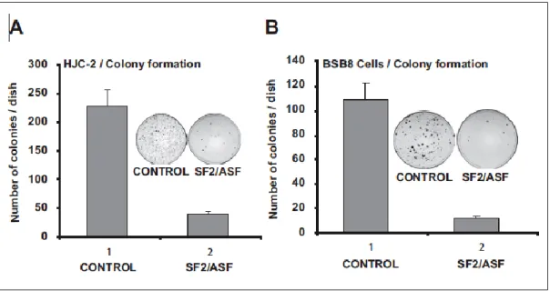
2.4 Colony formation assay
BsB8 and HJC-2 cells (5 * 105/100-mm dishes) were cotransfected either
with pCGT7 empty vector or pCGT7-SF2/ASF and pcDNA 3.1 zeo (+) plasmids (1 g each). After 24 hours, transfected and untransfected control cells were collected by trypsinization and replated in 100-mm dishes (1* 105/100-mm
dishes) in DMEM medium containing 10% FBS and G418 for selection (1 mM). Cells were maintained for 3 weeks, and the number of colonies was determined by fixing and staining the cells with 1% methylene blue. All experiments were performed in triplicate.
2.5 Soft agar growth assay
BsB8 and HCJ-2 cells (3 * 105/60-mm dishes) were either cotransfected with
pCGT7-SF2/ ASF and pcDNA 3.1 zeo (+) plasmids or infected with lenti-SF2/ASF-shRNA. After 24 hours, the cells were harvested and reseeded in 60-mm dishes (5,000 cells/dish) containing 2 mL of a 0.3% agarose suspension in DMEM plus 10% fetal bovine serum and G418 (1 mM). Plates were incubated at 37°C with 7% CO2 for 3 weeks. All experiments were done in triplicate.
2.6 MTT(3-(4,5-dimethylthiazol-2-yl)-2,5
diphenyltetrazolium bromide) assay for cell proliferation
HJC-2, BsB8, and U-87 MG cells were plated in 6-well plates (2 * 105 cells/plate)
and transfected either with pCGT7 vector alone or pCGT7- SF2/ASF expression plasmid. Seventy-two hours after transfection, cells were washed with 1x PBS and incubated with 1 mL of MTT working solution (DMEM with 0.5 mg/ mL MTT; Invitrogen, Carlsbad, CA) for 1 hour at 37°C. At the end of the incubation, the converted dye was vigorously resuspended with 1 mL acidic isopropanol (0.004 M HCL in isopropanol). The dye solution was then transferred into 1.5
mL eppendorf tubes and was centrifuged at 15,000 rpm for 5 minutes. The supernatant was transferred into new tubes, and absorbance of the converted dye was measured at a wavelength of 570 nm with background subtraction at 650 nm.
2.7 Flow cytometry analyses
HJC-2 cells were seeded at a density of 250,000 cells in 60-mm dishes and transfected with 3 £gg of pCGT7 alone or with 3 ug of pCGT7-SF2/ASF expression plasmid. Untransfected cells were used as a control. Cells were harvested at 24, 48, and 72 hours posttransfection, then fixed in 70% ethanol, and stained for 45 minutes with Guava Cell Cycle Reagent (Guava Technologies, Hayward, CA), based on standard propidium iodide (PI) chemistry for quantification for DNA in whole cells. Cell acquisition and analyses were performed by the Guava Easy cycle Mini machine and software. All experiments were performed in triplicate.
2.8 Lentiviral infection and RNA interference
Lentivirus-based U6-promoted SF2/ASF shRNA constructs were generated by cloning PCR products carrying sense and antisense oligonucleotides of SF2/ASF into the pLL3.7 vector, which also express green fluorescent protein (GFP). To knock down the expression of large T antigen, shRNA construct was generated to target the nucleotide sequences 139 to 162 of the JCV large T antigen cDNA. PCR products carrying sense and antisense oligonucleotides of large T antigen were cloned into the pLL3.7 vector. The viruses were packaged in 293T (human embryonic kidney) cells according to the procedure described previously (Song et al. 2007; Rubinson et al. 2003). HJC-2 cells were seeded in 6-well plates at 50% confluency and were incubated with 1 mL of the viral supernatants. The infected cells were then kept in regular complete medium (DMEM, 10% FBS) for
2.9 Reporter gene assays
The JCV-early promoter reporter gene construct containing the viral regulatory region has been described previously (Akan I et al., 2006). U-87 MG cells were transfected with the reporter construct in the presence or absence of expression plasmids for SF2/ASF-FL and its various deletion mutants. At 48 hours posttransfection, cells were extracted with a series of freeze/thaw cycles, and the CAT activity of the samples was determined.
3 RESULTS
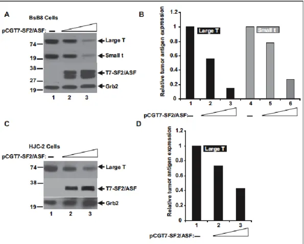
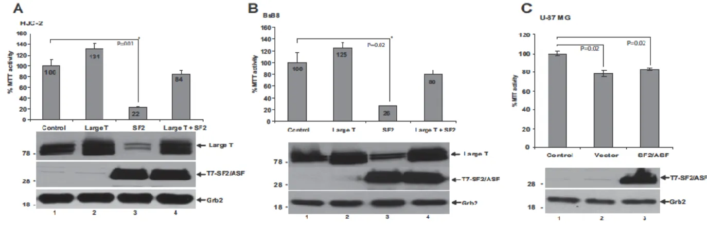
3.1 JCV tumor antigen expression is suppressed by SF2/ASF in
viral-transformed cell lines.
The impact of SF2/ASF on JCV tumor antigen expression was tested in BsB8 cells, a cell line that originated from a medulloblastoma developed in a transgenic mouse expressing the JCV-early region (Krynska et al., 1999), and in HJC-2 cells, a cell line obtained from a glioblastoma induced by intracranial injection of JCV in newborn hamsters (Wold et al., 1980; Raj et al., 1995).
Both cell lines express the JCV-early gene products, large T and small t antigens, under the control of the JCVearly promoter. To investigate the possible role of SF2/ASF in regulating the expression of large T antigen, T7-tagged SF2/ASF was introduced into BsB8 and HJC-2 cells by transient transfection (Fig. 3.1). Western blot analyses of whole cell extracts from BsB8 and HJC-2 cells revealed that SF2/ ASF suppressed the protein expression levels of large T antigen as well as small t antigen in a dose-dependent manner. These results show that SF2/ASF down-regulates JCV tumor antigen expression in the viral-transformed tumors.