1
Alma Mater Studiorum – Università di Bologna
DOTTORATO DI RICERCA IN
SCIENZE VETERINARIE
Ciclo XXIX
Settore Concorsuale di afferenza: 07/H3 Settore Scientifico disciplinare: VET/06
An integrated approach to improve the knowledge of
Ostreid herpesvirus type 1 and the comprehension of
mortality events in the Pacific oyster Crassostrea gigas
Presentata da: Dott.ssa Erika Astrid Virginie Burioli
Coordinatore Dottorato
Relatore
Prof. Arcangelo Gentile Prof. Marialetizia Fioravanti
Correlatori
Dr. Maryline Houssin Dr. Marino Prearo
3
Collaborations
The present manuscript set out the results obtained thanks to a three-year collaboration between different research institutes, located in Italy and France. In particular, the main part of molecular analyses has been conducted at the LABÉO Frank Duncombe Laboratory, situated in Caen (Normandy – France), in the Microbiology-Virology Unit, where I have been hosted for 18 months. A huge contribution was also made by the Istituto Zooprofilattico Sperimentale del Piemonte Liguria e Valle d’Aosta and especially by the Laboratorio di Ittiopatologia (Turin - Italy).
5
Acknowledgements
Ci sono essenzialmente quattro categorie di persone che vorrei ringraziare in modo particolare. Le prime sono quelle che mi hanno sempre incorragiata a seguire le mie aspirazioni e che non hanno mai cercato di reprimere il mio gusto dell’avventura, qualunque fossero stati gli ostacoli e la difficoltà, insegnandomi fin dall’inizio che la cosa più preziosa che possedevo era la libertà di scegliere ogni giorno.
Le seconde sono sono state al mio fianco quando i dubbi comparivano e se dovessi fare nomi, Katia (che capisce bene di cosa parlo) e Tommy sareste i primi della lista.
Poi ci sono le persone che mi hanno regalato la loro fiducia, così, senza alcuna garanzia. Queste persone le voglio ringraziare poiché senza di loro non avrei potuto raggiungere questo traguardo: la Professoressa Marialetizia Fioravanti (questa volta dovrebbe essere l’ultima!!!), la Dottoressa Maryline Houssin (grazie a lei la Normandia sembra un posto quasi accogliente…) e il Dottore Marino Prearo (Doctor YES).
Vorrei ringraziare anche coloro che, al contrario, non hanno reso il compito facile, ma che hanno così contribuito, a loro modo, a non farmi dimenticare mai l’obiettivo.
E poi ci sono tutti gli altri:
gli allevatori che hanno partecipato al progetto: Alessandro e Francesca, Andrea, Cristian, Eugenio, Leonardo, Samuele e Stefano e il loro equipagi;
e i colleghi e amici: Guy, Elise, Lorena, Lucie, Marzia, Paola, Suzanne, Claudio, Gabriele, Enrico, Paolo e Vasco con i quali abbiamo a volte fatto le ore piccole giusto per qualche ostrica! Un giorno mi ringrazierete, nessuno saprà aprile le ostriche meglio di voi. Ah dimenticavo: piatta o concava?
E soprattutto c’è la mia Mamie, che mi fa sempre credere che sono la persona più importante del mondo.
Il y a principalement quatre types de personnes que je voudrais remercier d’une façon particulière.
Les premières sont celles qui m’ont toujours encouragée à suivre mes aspirations, sans jamais chercher à brimer mon gout pour l’aventure, quelles que soient les obstacles et les difficultés, m’apprenant depuis le début que la chose la più précieuse que nous possédons est la liberté de choisir chaque jour.
Les secondes sont celles qui ont été là quand l’ombre du doute pointait son nez. Katia (qui comprend bien de quoi je parle) et Tommy, vous etes les premiers sur la liste.
Il y a aussi les personnes qui m’ont donné en cadeau leur confiance, comme ça, sans garantie et sans qui rien n’aurait meme commencé: le Professeur Marialetizia Fioravanti (promis, cette thèse est la dernière!!!), le Docteur Maryline Houssin (qui fait presque sembler la Normandie un endroit accueillant…) e le Docteur Marino Prearo (Doctor YES).
Les dernier groupe comprend en revanche ceux qui m’ont rendu les choses plus difficiles mais qui ont contribué, eux aussi à leur manière, à ce que je n’oublie jamais mon objectif.
Et puis il y a tous les autres:
les éleveurs et leurs équipages: Alessandro et Francesca, Andrea, Cristian, Eugenio, Leonardo, Samuele e Stefano;
et les collègues et amis: Guy, Elise, Lorena, Lucie, Marzia, Paola, Suzanne, Claudio, Gabriele, Enrico, Paolo e Vasco avec lesquels on a veillé parfois jusqu’à tard juste pour quelques huitres! Un jour vous me remercierez, personne ne saura les ouvrir mieux que vous. Ah, j’oubliais: plate ou creuse?
Et surtout il y a ma Mamie, qui me fait toujours croire que je suis la personne la plus importante du monde.
7
Table of contents
List of Figures ... 9 List of Tables ... 11 List of Abbreviations ... 13 Introduction ... 17Part I: Literature Review ... 25
Chapter 1.1: The host: the Pacific oyster Crassostrea gigas (Thunberg, 1793) ... 27
1.1.1 Taxonomy and species distribution ... 27
1.1.2 Habitat ... 29
1.1.3 Anatomy ... 30
1.1.4 Reproduction cycle and development ... 33
1.1.5 Immune defences ... 37
1.1.6 Triploidy ... 43
1.1.7 Production cycle ... 47
1.1.8 Mass mortality events affecting C. gigas ... 48
Chapter 1.2: Ostreid herpesvirus type 1 (OsHV-1) ... 53
1.2.1 Viral infections in bivalve molluscs ... 53
1.2.2 Herpesviruses in molluscs ... 54
1.2.3 Taxonomic aspects of Herpesvirales ... 55
1.2.4 Biological characteristics ... 57
1.2.5 General structure of herpesviruses ... 58
1.2.6 Genome arrangement of OsHV-1 ... 59
1.2.7 Genetic diversity of OsHV-1 ... 63
1.2.8 Replication cycle of OsHV-1 ... 64
1.2.9 OsHV-1transmission and resistance in environment ... 67
1.2.10 Genetic-based host resistance to OsHV-1 ... 68
Chapter 1.3: Diagnostic methods for OsHV-1 ... 69
1.3.1 Clinical signs ... 69
1.3.2 Histology ... 69
1.3.3 Transmission electron microscopy ... 69
1.3.4 In situ hybridisation (ISH) ... 70
8
1.3.6 Polymerase Chain Reactions (PCRs) ... 70
1.3.7 Virus isolation on cell cultures ... 71
Part II: Experimental Approaches ... 73
Chapter 2.1: What is the health status related to OsHV-1 of wild stocks of molluscs along the Italian Mediterranean coast? ... 75
2.1.1 Which is/are the species of cupped oysters present is the natural beds in Italy? ... 76
2.1.2 Determination of the health status of wild Pacific oyster stocks in Italy related to OsHV-1 and genetic polymorphism of the virus ... 87
2.1.3 Is OsHV-1 is detectable in other mollusc species in recognised infected areas? ... 104
Chapter 2.2: Sequencing of the complete genome of OsHV-1 µVAr ... 108
Chapter 2.3: Multi-site tests: a study of OsHV-1 and disease co-factors in field environment ... 137
2.3.1 Materials and Methods ... 139
2.3.2 Results ... 173
2.3.3 Discussion ... 215
2.3.4 Disease management ... 219
Chapter 2.4: Description of mortality events in farmed populations ... 223
2.4.1Description of a mortality event in farmed adult Pacific oysters Crassostrea gigas in Italy associated with the isolation of V. aestuarianus and Tenacibaculum sp. ... 223
2.4.2 Description and investigation on a mortality event in spat during June 2016 in Normandy .... 235
General conclusions and Perspectives ... 245
Bibliography ... 249
Appendix A List of samples ... 277
Appendix B Protocols ... 289
Appendix C List of sequences ... 297
9
List of figures
Figure 1 Geographical distribution of C. gigas/C. angulata p. 29
Figure 2 External shell features of C. gigas p. 30
Figure 3 General anatomy of C. gigas after the removal of right valve p. 31
Figure 4 Diagram of the digestive system of Crassostrea p. 33
Figure 5 Photomicrographs of C. gigas female and male gonads, stained using Pregnant-Gabe's trichrome p. 35
Figure 6 Developmental stages of C. gigas p. 36
Figure 7 Pediveliger larva of C. gigas p. 36
Figure 8 Cytometry analysis of SSC and FSC of haemolymph whole cells p. 38 Figure 9 Normal fertilisation events in diploid x diploid crossing p.44
Figure 10 Chemical induction of triploidy in oysters p. 45
Figure 11 Obtainment of triploid individuals using sperm from tetraploid males p. 45
Figure 12 Comparison of chromosome segregation during Meiosis I and resulting gametocytes II in diploid and triploid individuals p. 46
Figure 13 Common producing cycle of C. gigas in Europe, from seed to commercial product p. 47
Figure 14 Comparative number of C. gigas sampling in response to mortality reports during 2012 p. 50
Figure 15 Exogenous factors influencing oyster health status p. 51
Figure 16 Irido-like virion from Crassostrea angulata in TEM p. 53
Figure 17 ICTV’s classification of herpesviruses p. 56
Figure 18 Structure of a herpesvirus particle p. 59
Figure 19 Genome organisation of Herpesviridae within six classes p. 60
Figure 20 Organisation of the OsHV-1 genome p. 60
Figure 21 Layout of ORFs in the OsHV-1 genome p. 62
Figure 22 Hypothetic replication cycle of OsHV-1 p. 66
Figure 23 Participants in herpesvirus (HSV, HHV-8, EBV) entry and virus-induced cell fusion p. 66 Figure 24 Location of the sampling campaigns carried out during 2012, 2014, and 2015 p. 75
Figure 25 Alignment of the C. gigas and C. angulata alleles of the CG44 nuclear marker p. 79
Figure 26 Neighbor-Joining tree of the cupped oyster individuals, based on COI gene analysis p. 85
Figure 27 Sequence alignments of the different Italian OsHV-1 genotypes p. 90
Figure 28 Layout of Open Reading Frames in the OsHV-1 µVar genome p. 110
Figure 29 Study sites p. 139
Figure 30 “Caorle” study site p. 141
Figure 31 “Caleri” study site p. 142
Figure 32 “Giulianova” study site p. 143
Figure 33 “Varano” study site p. 144
Figure 34 “Gaeta” study site p. 144
Figure 35 “Orbetello” study site p. 145
Figure 36 “La Spezia” study site p. 146
10
Figure 38 “San Teodoro” study site p .145
Figure 39 “Baie des Veys” study site p. 148
Figure 40 “Meuvaines” study site p. 148
Figure 41 Distribution of young oyster individuals in the lantern-net p. 149
Figure 42 Floating oyster bags in the San Teodoro Lagoon p. 150
Figure 43 Different lantern-nets used during the survey p. 151
Figure 44 Technique used in the sites of Meuvaines and Baie des Veys p. 152
Figure 45 Meshed-bags placed in lantern-nets p. 153
Figure 46 Device used for the first months in France p. 153
Figure 47 Diploid individuals after one month in meshed-bag p. 154
Figure 48 A to I Trend of the shell length of C. gigas p. 174
Figure 49 Final shell length in November 2014 per ploidy and site p. 179
Figure 50 Lantern-net in open water sites p. 180
Figure 51 Lantern-net in lagoon sites p. 181
Figure 52 Tubes used in France in 2015 for the first months p. 182
Figure 53 Graphical representation of the trend of water temperature p. 183
Figure 54 Graphical representation of the trend of water salinity p. 185
Figure 55 A to D Cumulative mortality observed during 2014 and 2015 p. 187
Figure 56 A to D Mortality rates between two successive samplings in 2014 and 2015 p. 192
Figure 57 A to F Prevalence of OsHV-1 p. 201
Figure 58 A to F Mean viral load of OsHV-1 in positive samples p. 207
Figure 59 Mean load of V. aestuarianus in oyster flesh p. 214
Figure 60 Liquefactive lesion in the adductor muscle of a moribund specimen p. 227
Figure 61 Histological section of adductor muscle with filamentous bacteria p. 228
Figure 62 Tenacibaculum sp. isolated on Zobell agar p. 229
Figure 63 Trend of cumulative mortality after experimental infection with Tenacibaculum sp. p. 230
Figure 64 Yellowish area in the mantle of an oyster after experimental infection with Tenacibaculum sp. p. 230
Figure 65 Necrotic area in the adductor muscle after experimental infection with Tenacibaculum sp. p. 231
Figure 66 Evolution of mortality rate in spat in 2016 in France p. 235
Figure 67 Histological section of C. gigas spat at low magnification p. 239
Figure 68 A-B Histological section of C. gigas spat adductor muscle p. 240
Figure 69 Histological section of C. gigas spat mantle and digestive gland p. 240
Figure 70 Histological section of C. gigas spat proliferative cells at high magnification p. 241
11
List of tables
Table 1 Taxonomic classification of the Pacific oyster C. gigas p. 27
Table 2 Temperature and salinity ranges for adult C. gigas p. 29
Table 3 Polymerase Chain reaction for the detection of Ostreid herpesvirus type 1 p. 72
Table 4 Presentation of the 1278 analysed mollusc samples p. 76
Table 5 Results of the molecular analyses for oyster identification p. 81
Table 6 Frequencies of the allele “angulata” and haplotype “angulata” p.84
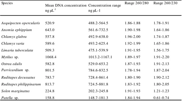
Table 7 Concentration and purity of DNA extracted from molluscs p. 106
Table 8 Description of the study sites concerned by the survey p. 140
Table 9 Description of the sampling campaigns conducted in 2014 p. 156
Table 10 Description of the sampling campaigns conducted in 2015 p. 158
Table 11 Number of sequences obtained from the different regions targeted for sequencing p. 199
13
List of abbreviations
°C degrees centigrade
µL microlitres
AbHV-1 Abalone herpesvirus type 1
AMP ANOVA ARPAT ARPAV AntiMicrobial Peptide ANalysis Of Variance
Agenzia Regionale per la Protezione Ambientale della Toscana Agenzia Regionale per la Protezione Ambientale del Veneto
ASW Artificial Sea Water
ATP AVG
Adenosine TriPhosphate Abalone Viral Ganglioneuritis
AVNV Acute Viral Necrosis Virus
Bcl-2 B-cell lymphoma 2
BI-1 Bax Inhibitor 1
BIR Baculoviral IAP Repeat
BIRP Baculovirus Inhibitor of apoptosis Repeat Protein
BLAST Basic Local Alignment Search Tool
BoHV-2 Bovine herpesvirus type 2
bp base pairs
C. angulata Portuguese oyster Crassostrea angulata
C. gigas
CCV CFU
Pacific oyster Crassostrea gigas Channel Catfish Virus
Colony Forming Unit
COI Cytochrome c oxidase subunit I
cPCR conventional Polymerase Chain Reaction
CT Cycle Threshold
d days
DENV DENgue Virus
DNA DeoxyriboNucleic Acid
dUTP deoxyUridine TriPhosphates
E Early
EBV Epstein-Barr Virus
EC European Commission
EcSOD Extracellular SuperOxide Dismutase
EFSA European Food Safety Authority
EHV-1 Equine HerpesVirus type 1
EHV-2 Equine HerpesVirus type 2
EM EURL
Electron Microscopy
European Reference Laboratory
FAO Food and Agriculture Organization of the United Nations
fg femtograms
FSC Forward SCatter
g grams
g g-force
GNV Gill Necrosis Virus
GU H&E
Genomic Unit
Haematoxylin Eosin staining
HCMV Human CytoMegaloVirus
14
HIV Haemocyte Infection Virus
HLA Human Leukocyte Antigen
HSV Herpes Simplex Virus
HVEM HerpesVirus Entry Mediator
IAP IC
Inhibitor of Apoptosis Internal Control
ICTV International Committee on Taxonomy of Viruses
IE Immediate Early
IFN InterFeroN
IHC ImmunoHistoChemistry
IPNV Infectious Pancreatic Necrosis Virus
IRL Internal Repeat Long
IRS Internal Repeat Short
ISG Interferon Stimulated Gene
ISH In Situ Hybridisation
IκB Nuclear Factor-Kappa B Inhibitor
La Late
L Litres
LPS
MALDI-TOF
LipoPolySaccharide
Matrix-Assisted Laser Desorption/Ionization Time Of Life
mg Milligrams
min Minutes
MJC MaJor Capsid protein
mL MLSA
Millilitres
Multi Locus Sequence Analysis
mm Millimetres
mtDNA Mitochondrial DNA
N Sample Size
NADPH Nicotinamide adenine dinucleotide phosphate (reduced form)
NF-κB
NGS
Nuclear Factor-Kappa B Next Generation Sequencing
NM Nuclear Marker
nm Nanometres
OIE Office International des Epizooties
ORF Open Reading Frame
OsHV-1 Ostreid herpesvirus type 1
OsHV-1-SB Ostreid herpesvirus type 1 Scapharca broughtonii strain
OVVD Oyster Velar Virus Disease
PCR Polymerase Chain Reaction
Poly(I:C) Polyinosinic: polycytidylic acid
qPCR quantitative PCR
REPAMO REseau de PAthologie des MOlluques
RER Rough Endoplasmic Reticulum
RFLP Restriction Fragment Length Polymorphism
RING Really Interesting New Gene
RNA RiboNucleic Acid
ROS Reactive Oxygen Species
rtPCR real-time PCR s SBS Second Sequencing By Synthesis SSC Side SCatter
15
SuHV-1 TEM
Suis HerpesVirus type 1
Transmission Electron Microscopy
TGN THV
Trans Golgi Network Tupaia HerpesVirus
TNF Tumor Necrosis Factor
TNFR Tumor Necrosis Factor Receptor
TRAIL Tumor-necrosis-factor Related Apoptosis Inducing Ligand
TRL Terminal Repeat Long
TRS Terminal Repeat Short
UL Long Unique region
US Short Unique region
UV UltraViolet
VZV Varicella Zoster Virus
WHO World Health Organization
17
19
Introduction
Consumption of fish, defined as finfish (vertebrates) and shellfish (invertebrates), provides energy, protein and a range of other important nutrients, with beneficial health outcomes for humans (FAO/WHO, 2010; EFSA, 2014). Moreover, eating fish is part of the cultural traditions of many people, in several populations around the world. Thanks to aquaculture activities, 73 million tons of fish are produced every year in the world, 89% of which by the Asian continent (FAO, 2014). It is interesting to highlight that 22% of this production consist in shellfish, oysters in particular, with a production of 5 million tons by year. The genus Crassostrea represents 99% of this production. Unlike other forms of aquaculture and thanks to their filter-feeding habits, bivalve farming can be considered as a sustainable animal-derived protein source.
In Europe, with 631,800 t per year, the bivalve molluscs represent 21% of the total aquaculture production. Essentially, these bivalves belong to few genera or species: mussels (Mytilus spp.) rank first with 495,000 tons produced in 2014, far forward oysters (Crassostrea spp.) with 89,000 t/year, and clams, Ruditapes philippinarum (Adams & Reeve, 1850) with 32,000 t/year (FAO, 2014). The European flat oyster, Ostrea edulis (Linnaeus, 1758) and the European carpet shell, Ruditapes decussatus (Linnaeus, 1758) represent only 3% of the oyster production and 10% of the clam production respectively. Italy ranks third among bivalve mollusc producing countries in Europe with 110.645 t/year (FAO, 2014). Though clams and mussels still account for the bulk of national production with 31.600 t/year and 79.000 t/year respectively (FAO, 2014), the Pacific cupped oyster, Crassostrea gigas (Thunberg, 1793) is becoming an increasingly important product, even if it represents today less than 1% of the total bivalve production and concerns mainly three areas, Sardinia and, to a lesser extent, Liguria, and the Po Delta. In the past, in Italy, the culture of the native flat oyster O. edulis has been practised, since antiquity to the end of the 19th century, before of being almost completely abandoned nowadays. During the late sixties, several experimental trials of cupped oyster farming were conducted along the Italian coasts, through the introduction of both C. gigas and Crassostrea angulata (Lamarck, 1819), but the real producing activities started about ten years ago. However, it is difficult to establish if some Crassostrea spp. wild populations, especially in the Northern Adriatic, were not yet present, may be introduced inadvertently long time ago by shipping from their native area in East Asia. In any case, finding optimal environmental conditions, cupped oysters successfully established and spread,
20
and today wild populations are intensively present along the Northern and Eastern Adriatic coasts. France remains the main cupped oyster producer in Europe with 76,000 t produced in 2014 (FAO, 2014).
In Europe, the farming technique varies among the regions, in order to fit to the different environmental conditions. Intertidal oyster cultivation is the most significant technique practised in France along the Atlantic and Channel coasts, but in Mediterranean Sea, where the tidal range is restrained, other techniques must be used such as gluing oysters onto ropes, in the “Etang de Thau” (France), farming them in lantern-nets or baskets, or using floating bags in the “Stagno di San Teodoro” (Italy). The seed supplying is achieved by two ways: natural collection on the wild, mainly practised in the Arcachon Bay and in Charente-Maritime (France) and hatchery production. This second source is not dependant on annual fluctuations, it allows procurement over a longer period of the year, it provides also triploid individuals, batches of homogeneous size, a low rate of individuals attached to each other, and the possibility of genetic selection. Actually, no commercial hatchery for C. gigas is present in Italy and the spat collection in open sea is not performed, compelling the producers to import spat from France. Currently, the farming cycle is about three years but may be shorter in the Mediterranean waters. The progresses accomplished in the zootechnical field had influenced positively the intensification of farming practices and a specialization of some coastal areas in oyster culture. However, the high stocking density increases host contact rates and stress, and reduces water quality making aquaculture susceptible to disease outbreaks. Moreover, the common use of bivalve stock transfers between productive areas, also situated in different countries, improves the risk of the insurgence and diffusion of infective diseases.
The rearing cycle, from spat to commercial product, takes place in an environment scarcely controllable and the oyster farming, during its history, has always been characterised by fluctuations of the production. Livestocks are subjected to natural environmental conditions that may compromise their growth and survival. In this scenario, infectious diseases have a heavy impact in marine aquaculture, with important economic consequences (Lafferty et al., 2015). For instance, a report titled “Procédures de couverture des risques conchylicoles”, delivered by the “Conseil général de l’agriculture, de l’alimentation et des espaces ruraux”, “Conseil général de l’environnement et du développement durable”, and “Inspection générale des affaires maritimes” jointly, appointed by the French Ministry for Food, Agriculture and Fisheries and Ministry for Ecology, Energy, Sustainable Development
21
and Sea, established at 172 million euros the amount of damages caused by summer mortalities of Pacific oysters only for the year 2008. Besides, contrary to what happens in finfish aquaculture, chemotherapy and vaccination are not suitable solutions in the case of mollusc disease control. In the past, the oyster industry has been periodically affected by various infectious diseases caused by parasites, viruses, or bacteria. In France, the European flat oyster production has fallen from 20,000 tons in the first seventies to 2,300 tons in 1985 (Grizel, 1985; Goulletquer and Heral, 1997) because of the insurgence of two parasitic diseases: marteiliosis in 1968, and bonamiasis in 1979, due to the protozoans Marteilia refringens (Comps, 1970a; Herrbach, 1971, Grizel et al., 1974) and Bonamia spp. (Pichot et al., 1980, Comps et al., 1980, Comps, 1983). Nearly in the same period, farmed and wild populations of Portuguese cupped oyster C. angulata were practically decimated in few years by an irido-like virus (Comps et al., 1976). This event has been responsible of the introduction of another species of cupped oyster, the Pacific oyster C. gigas, to guarantee the prosecution of farming activities. Regrettably, since April-May 2008, juvenile stages of Pacific oyster, in turn, have been affected by dramatic mortality events in all the French producing regions, with mortality rates ranging between 60% and 100% (Cochennec-Laureau et al., 2009; Renault et al., 2009). These events were associated with a new variant of the Ostreid herpesvirus type 1 (OsHV-1) termed µVar (Segarra et al., 2010) and since them, they were observed in several regions of the world, always correlated with the presence of OsHV-1 microvariants (OIE, 2014). In addition, commercial size oysters are affected by anomalous mortalities since 2012 in France, associated with Vibrio infections, and even if the disease course is less acute than in the case of young oysters, these events represent a significant threat to the oyster industry for the huge economic losses they generate.
Given the extent of these phenomena, and the increasing alarm of farmers, international cooperation is highly desirable, as expressed during the Annual Meeting of National Reference Laboratories for Molluscs Diseases in Rochefort on the 18th March 2013. Joint operations could accelerate the comprehension of these events and the finding of some solutions. A priority must be given to the study of OsHV-1. In fact, despite the heavy economic impact of virus-induced mortalities, international effective measures to prevent and control the diseaseare nowadays unavailable, in part because of the scarce information on the real diffusion of the virus and its variants, and their relative effective pathogenicity. Furthermore, some important questions emerge and, to try to answer them, a team work, made up of Italian and French Research Institutes was realised, with the final aim to give some solid
22
elements for future risk assessment activities, for the prevention of spread, and to limit economic losses that farmers have to face.
The main fields were:
- What is the real diffusion of OsHV-1 in the wild? Considering the close contact between wild and reared oyster populations and the excellent water capacities as vehicle of infectious agents, it is important to gather information on the health status of natural bivalve populations related to OsHV-1, in order to assess the potential risk of their contamination by infected farmed individuals or, on the contrary, to establish if they may act as virus reservoir.
- What is the genetic diversity of OsHV-1, especially in the wild? What are the evolutionary relationships among the genotypes?
- It has been postulated that the virulence of OsHV-1 microvariants is higher but what is the origin of this virulence if compared to the reference genotype, present before 2008? What are the putative virulence factors?
- What are the other factors that influence the OsHV-1 capacity of infection and pathogenicity? Even if a new variant was associated with the recrudescence of mortality events in young oysters in 2008, it is assumed that other factors may play an important role in the intensification of these events, such as environmental factors, synergy of the pathogenic effects during co-infections, and host factors such as physiological state (Baud et al., 2012).
In a period of expansion of oysters farming in Italy, the present work aimed to contribute to this challenge, approaching several important aspects connected with oyster health management in Italy. The information collected during the present study will aslo contribute to improving the global knowledge on oyster pathology.
The first part of the manuscript, called “Literature Review”, gathers information on the Pacific oyster C. gigas and on its pathogens, OsHV-1 in particular.
The second part discusses the results of the experimental activities and is partitioned in four chapters. The first chapter is dedicated to the improvement of knowledge of the health status of wild populations of molluscs related to OsHV-1. This concerns also investigations on genetic aspects of the viral specimens isolated in field and the identification of the species of
23
cupped oysters present in Italy, through the development of a molecular tool. The second chapter relates the sequencing of the whole genome of OsHV-1 µVar. The third chapter details the results of the multi-site tests: in order to evaluate the possible effects of different factors on the insurgence of anomalous mortality events, oyster pools, from the same batches, were placed in several sites characterised by different environmental conditions, in Italy and France. The effect of ploidy, age of oysters, and allocation date on mortality rate, prevalence and load of OsHV-1 and Vibrio aestuarianus were tested, along with the effect of environmental parameters, and presence of potentially pathogenic microorganisms. Finally, in the fourth chapter we described two mortality events occurred in farmed stocks: the first affecting adult individuals in San Teodoro (Italy) and associated with bacterial pathogens, and the second occurred in Normandy and affecting spat during June 2016.
25
PART I
27
1.1. The host: the Pacific oyster Crassostrea gigas (Thunberg, 1793)
1.1.1. Taxonomy and species distribution
The Pacific oyster C. gigas, also called Japanese oyster, is a marine bivalve mollusc, member of the Ostreidae family. Its exact classification (Marshall and Gofas, 2015) is reported in Table 1.
Table 1 Taxonomic classification of the Pacific oyster Crassostrea gigas.
To date, the genus Crassostrea includes 22 species (Bouchet and Gofas, 2012): Crassostrea aequatorialis (d'Orbigny, 1846)
Crassostrea angulata (Lamarck, 1819) Crassostrea ariakensis (Fujita, 1913)
Crassostrea belcheri (G. B. Sowerby II, 1871) Crassostrea bilineata (Röding, 1798)
Crassostrea brasiliana (Lamarck, 1819) Crassostrea columbiensis (Hanley, 1846) Crassostrea corteziensis (Hertlein, 1951)
Crassostrea cuttackensis (Newton & Smith, 1912) Crassostrea dactylena (Iredale, 1939)
Crassostrea dianbaiensis (Xia, Wu, Xiao & Yu, 2014) Crassostrea gigas (Thunberg, 1793)
28 Crassostrea ingens (Zittel, 1865)
Crassostrea mangle (Amaral & Simone, 2014) Crassostrea nippona (Seki, 1934)
Crassostrea praia (Ihering, 1907)
Crassostrea rhizophorae (Guilding, 1828) Crassostrea rivularis (Gould, 1861) Crassostrea sikamea (Amemiya, 1928) Crassostrea tulipa (Lamarck, 1819) Crassostrea virginica (Gmelin, 1791)
The species Crassostrea laperousii (Schrenk, 1861), Crassostrea posjetica (Razin, 1934) and Crassostrea talienwhanensis (Crosse, 1862) are considered as synonyms of C. gigas. In contrast, even if there is evidence of viable interspecific hybridisation between C. gigas and the Portuguese oyster C. angulata (Gaffney and Allen, 1993; Huvet et al., 2001, 2002, 2004) and after numerous controversies, the two species are nowadays considered as distinct species. However, the phenotypic characters are not sufficient to discriminate the two species. Several genetic studies, based on mitochondrial DNA (Boudry et al., 1998; O’Foighil et al., 1998) and microsatellite data (Huvet et al., 2000a, 2000b) provided evidence that the two taxa are genetically distinct although closely related (Batista et al., 2005). In particular, the estimation on nucleotide divergence (5.26%) of the cytochrome c oxidase subunit I gene (COI) sequence suggests that populations of C. gigas and C. angulata are closely related and may have diverged only one to two million years ago (O’Foighil et al., 1998). Studies involving microsatellite markers also confirmed that there are low but clear genetic differences between the two taxons. Before the intentional introduction of C. gigas from Japan to France during the early seventies, to face the mass mortality events that wiped out the Portuguese oyster from French coasts, the two populations were believed to have a well separated geographical distribution: C. angulata on the European Atlantic seaboard (that explains the common name “Portuguese oyster”) and C. gigas in Asia. However, from the recent phylogenetical studies, it seems clear that the European C. angulata populations were introduced in the XVI or XVIIth century from Taiwan, firstly in Portugal before their spread northward. Nevertheless, the relationship between the two taxa in overlapping home range locations is not clear. Actually, C. angulata and C. gigas are listed as separate but very closely related species and they still potentially conspecifics. Their global geographical distribution is represented indiscriminately on Figure 1.
29
Fig. 1 Geographical distribution of C. gigas/C. angulata
Continuous line: native area; dotted line: introduced populations (adapted from OBIS ver. 1.0)
1.1.2. Habitat
Natural Pacific oyster populations are usually established in sheltered areas along the coasts, in the intertidal zone or until a depth of about ten meters, preferably in estuarine or lagoonal environments. In fact, these ecosystems characterised by high trophic levels resulting from the mixing of inland waters, rich in nutrients, with marine waters, are suitable for filter-feeding species. As an intertidal species, it is very tolerant to various abiotic conditions. C. gigas is a euryhaline organism, able to adapt to a wide range of salinities. Both optimal and tolerance ranges for growth and spawning are very large, as reported in Table 2. The species is also extremely resistant to environmental stress from high metal exposure (Rainbow and Phillips, 1993; Boening, 1999; Funes et al., 2006). In the areas where wild beds develop, they tend to constitute reefs that provide a shelter for other marine species, even if the invasive character of C. gigas is recognised (PROGIG, 2009).
Growth Spawning Growth Spawning
3-35 (11-34) 16-35 (20-25) 10-42 (35) 10-30 (20-30)
Te mpe rature (°C) Salinity (ppt)
Table 2 Temperature and salinity ranges for adult C. gigas. Optimal ranges in parentheses (Mann et al., 1991)
30 1.1.3. Anatomy
Mollusca is one of the largest and diverse animal phylum, second only to Arthropoda, with over 46,000 described marine species, allocated in seven major classes, with a soft body as common character. In the case of bivalves, soft tissues are protected by a hard shell consisting in two valves, held together by a horny ligament. Ostreidae valves are asymmetrical. The left valve of C. gigas is larger and deeply cupped (Figure 2). It is always the left valve to be cemented to the substrate, guarantying the sessile life to this mollusc. The external shell colouring varies, on the basis of the geographical origin, from off-white, brownish, to deep purple. The inside of the shell is pearly-white with a single large muscle scar. As observed for the colour, the shape is also fashioned by the influence of both environmental and genetic factors. Along the European coasts, C. gigas and O. edulis populations can share the same habitat. However, the two species are easily distinguishable: the left valve in O. edulis is less cupped, the shape is rounder, the junction line between the two valves is not curled and the presence of a dozen lateral hinge teeth is only observed in the genus Ostrea (personal observation).
Fig. 2 External shell features of C. gigas (E. Burioli) Umbo Hinge Left valve Right valve Ventral margin Posterior margin Anterior margin
31
Molluscs are metazoan triploblastic coelomates, even if the coelom is reduced just to the cavities surrounding the gonads, the heart and the excretory organs.
The mantle, also called pallium, is an extension of the dorsal tegument that hangs down, forming two lobes around the body that constitute the mantle cavity (Figure 3). It is composed of connective tissue surrounding muscle fibers, haemolymphatic vessels, nerves, and is covered by a single layer of epithelium. The two lobes are fused in the cephalic region and form a cap that protects the mouth and the ciliated labial palps. The edges of two halves of the mantle have three folds and the medial fold shows very short tentacles with numerous sensorial organs. The mantle cavity protects the gills and the excretory, anal and genital orifices. This chamber serves for the ingress of water and nutrients and for the expulsion of the excreta and genital products. Important physiological functions are performed by the mantle, such as the production of the shell and the energy storing, in the form of glycogen that is the primary energy store in bivalves (Gabbott, 1983). The mantle epithelium includes numerous glandular cells producing mucus with protective functions and acting as a barrier against external agents.
Fig. 3 General anatomy of C. gigas after the removal of right valve (E. Burioli)
In the Ostreidae family, the foot is atrophic, even if it is present in larvae before settlement. The two valves are joined together by the ligament along the hinge line and by the massive adductor muscle ventroposteriorly (Figure 3). Its contraction controls valve opening. These mechanisms permit the regulation of the water flow. Oysters are monomyarian molluscs. Adductor muscle consists in two adjacent distinct parts. The fist, sometimes called vitreous muscle, is characterised by a translucent off-white aspect and represents two-thirds of
Hinge ligament
Mantle Labial palps
Gills
32
the total bulk of the muscle. The remainder is crescent-shaped with a pure white opaque aspect. The two parts of the adductor muscle contract at different speeds: the rapid closing of the valves is accomplished by the contraction of the translucent part while the elasticity and tonus of the white part counteract the pulling force of the ligament for long time (Marceau, 1904a, b). Consequently, the adductor muscle is mostly developed in individuals living in the intertidal zone. Finally, short repeated shell movements are observed during spawning or to help the pseudofaeces disposal.
A heart, arteries, veins, and open sinuses constitute the circulatory system that is partially closed. The sinuses, or lacunae, are spaces of varying size, within the connective tissues, without a wall. They are interposed between arteries and veins with a function similar of the capillaries in vertebrates (Galtsoff, 1964). The heart is situated in the pericardium (Figure 3), a thin-walled chamber located between the visceral mass and the adductor muscle. The three-chambered heart consists in a ventricle and two dark-coloured auricles that are covered with a tall columnar epithelium that constitutes a part of the excretory system (Franc, 1960).
The haemolymph is colourless and unlike other mollusc classes, it lacks haemocyanin (Galtsoff, 1964) but contains two main groups of circulating cells termed haemocytes. Haemocytes are not confined to the vessels: they are able to amble throughout the tissues or move to a target in response to a molecular signal.
Exchange of gases takes place essentially in the gills (Figures 3), but the mantle also contributes, even if in a lesser extent, in the respiration. Bivalves maintain a constant water flow through their gills for respiration and feeding. The gills in filter-feeding molluscs are more complex, due to their multiple functions and consist in four folds of tissue suspended from the visceral mass, two for each side.
The digestive system (Figure 4) consists of the mouth, short esophagus, stomach, crystalline style sac, digestive gland, midgut, rectum, and anus. The crystalline style is a peculiar feature of bivalves and some gastropods with a mechanical and enzymatic role in the digestion of food. The stomach is surrounded by the dark-brown digestive gland, made of an important number of tubules emptying in larger ducts leading finally to the stomach. Undigested food is discharged as feces thanks to the ciliary action of epithelium, as peristaltic movements are absent.
The nervous system of oyster is simple. It consists of a primitive system of visceral and cerebral ganglia in which ganglia pairs are connected together through commissures and with
33
the other pairs through connectives. Several nerves originate from the ganglia and extend to different parts of the body.
To date, the organs of oysters currently known to have sensory function are limited to the short tentacles that fringe the mantle and the pallial organ inside the cloaca. Tentacles contain photo- and chemoreceptors able to detect slight chemical and physical changes in their environment.
Fig. 4 Diagram of the digestive system of Crassostrea (Galtsoff, 1964)
an: anus; cl: cloaca; cr.s: crystalline style sac;dig.div: digestive gland; int: intestine; oe: esophagus; st: stomach
Excretion is carried out by the nephridia, pericardial glands, and also the mantle epithelium, that contributes to this function.
The gonad is located within a connective layer, between the digestive gland and the outer epithelium. In sexually mature oysters, many branching tubules can be observed and converge into a gonoduct. Because Pacific oysters are sequential hermaphrodite, male and female gonads cannot be observed simoultaneously in the same individual.
1.1.4. Reproduction cycle and development
Reproduction is one of the most important physiological processes in the life cycle of bivalve species. C. gigas is an alternative sequential hermaphrodite. After settlement, for several weeks, the immature gonad of juveniles remains nonfunctional and ambisexual. It
34
contains both male and female primary germ cells which will transform into mature spermatozoa or eggs during the following spring-summer. Usually, a predominance of males is observed in the 1-year old population, due to the more rapid multiplication of male germ cells that suppresses the oocyte development. However, the environmental conditions, such as warm waters, are able to influence the number of individuals that develop directly into females (Coe, 1936). During their lifetime, oysters invert sex several times. In wild populations, male are more numerous, with a 5:3 sex-ratio (Le Dantec, 1968).
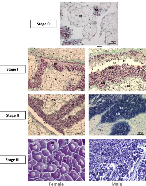
C. gigas exhibits a seasonal reproductive cycle. In Europe, after spawning, a period of quiescence is observed, in autumn. The restart of gametogenesis is observed in winter, soon or later, depending on the geographic location (Chávez-Villalba et al., 2003). In late-winter and spring, oysters tend to accumulate glycogen in their tissues that will be metabolised in lipids during the vitellogenesis. The gonadal development, determined by histological analysis, can be divided into five stages, on the basis of descriptions of Heude-Berthelin et al. (2001) (Figure 5). During the stage 0 (quiescent stage), no trace of sexuality is present and only an undifferentiated germinal epithelium is observed; the stage I (early growth stage) is characterised by small follicles and numerous spermatogonia or oogonia; in stage II (late growth stage) the follicles developed actively and a majority of primary gametes with few free spermatozoa and oocytes also are present; stage III is the maturation stage during which gametes are densely packed follicles, filling completely the follicles. At this stage, oocytes are pear-shaped and appear compressed. The vitellogenesis is completed and a distinct nucleus and nucleolus is observed. Spermatozoa are oriented with tails toward the follicle lumen. The stage IV embraces the spawning and the reabsorbing stages and phagocytosis is present. In Europe, gamete release occurs between July and August, depending on environmental factors (Enríquez-Díaz, 2009) mainly due to the geographic location. Spawning is induced by both sudden fluctuations of physic environmental parameters, temperature in particular, and chemical signals released wit gonadal fluids of other individuals, allowing the synchronisation among individuals. Egg fertilisation occurs in the water medium.
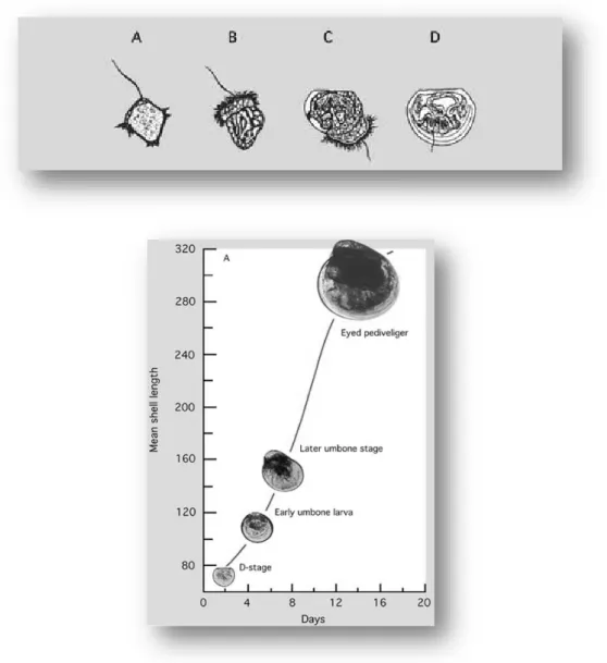
Larvae show planctonic behaviour for a temperature-depending duration. During its planctonic life, the young oysters get through different stages reported in figure 6.
35
Fig. 5 Photomicrographs of C. gigas female and male gonads, stained using Pregnant-Gabe's trichrome.
EC: ciliated epithelium; G: undifferentiated gonia; Oo: oocyte; Og: ovogonia; S: spermatid; Spc: spermatocyte; Spg: spermatogonia; Spz: spermatozoa; TG: gonadic tubule; TR: storage tissue.
(adapted from Jouaux, 2010)
Stage 0
Stage I
Stage II
Stage III
36
Fig. 6 Developmental stages of C. gigas from the early trochophore (A) to the fully shelled D-larva stage (D). The ciliated swimming feeding organ (velum) can be seen in B and early shell valve formation in C
http://www.fao.org/docrep/007/y5720e/y5720e0a.htm
At the end of the planctonic stage, a foot is present and helps the pediveliger larva (Figure 6) to crawl searching a suitable substrate to attach to. After settlement, the foot regresses until it disappears completely. At this stage oysters are termed spat.
37
1.1.5. Immune defences
In order to survive in a competitive environment, organisms must be able to protect themselves from pathogens. In these conditions, all metazoan organisms have developed complex immune defence systems. The first line of defence is represented by physicochemical barriers and mucus in particular, that covers all the epithelial layers. The microbiota composition may also contribute to the health status. The adaptive immunity, acquired in vertebrates during evolution (Hoffmann et al., 1999; Hirano et al., 2011), is not present in molluscs, whose set of ancient defence strategies is collectively known as innate immunity, characterised by its universality and rapid-acting. These strategies require firstly an afferent (or sensing) component, able to perceive infection, and secondly an efferent pathway aimed to eradicate infection, and involve both cellular and humoral components. Infection is perceived thanks to the detection of pathogen-associated molecular patterns (PAMPs) (Beutler, 2004). The phagocytosis in invertebrates, evidenced firstly in starfish by Metchnikoff (1884), is the main cell-mediated immune defence. This function is exercised by haemocytes (Lorteau et al., 1995) after their mobilisation and recruitment to the site of infection. Cellular response is coupled with humoral responses: cytokines, enzymes and other immune effectors.
In particular, the knowledge of antiviral defence mechanisms mainly derives from studies conducted on human and vertebrates where innate defences and adaptive immune system collaborate together. This is not the case of molluscs. However, they compensate the lack of adaptive response with a complex innate antiviral defence system that is greatly expanded if compared to vertebrates (Zhang et al., 2013; Venier et al., 2011).
Haemocytes
Haemocytes are present in the circulatory torrent, in vessels and sinuses, but also infiltrated throughout the tissues. They also migrate by diapedesis to the surface of epithelial layers. These cells are immunocompetent but they are also involved in different physiological processes, such as gonad resorption, excretion, digestion and transport of nutrients and shell repair (Cheng, 1981). The term haemocyte refers to three main distinct populations of circulating cells: blast-like cells, hyalinocytes, and granulocytes (Hine, 1999; Bachère et al., 2004), as shown in Figure 8 thanks to flow cytometry analysis using the Side Scatter (SSC) and Forward Scatter (FSC) parameters. Granulocytes are distinguished from other haemocytes by the possession of cytoplasmic granules and a low nucleus:cytoplasm ratio.
38
These granules may have different tinctorial properties allowing the subdivision of cells in acidophilic, basophilic, and neutrophilic (Auffret, 1989; Tiscar and Mosca, 2004). Blast-like cells and hyalinocytes are usually classified as agranular haemocytes. The first ones have a spherical or ovoid central nucleus surrounded by a thin rim of cytoplasm while hyalinocytes have a reniform irregular eccentric nucleus and an abundant cytoplasm. To date, the ontogeny of the cell line and the functionality of the different cell types have not been clearly characterised yet. However, even if the main phagocytic activity is assumed to be carried out by granulocytes, hyalinocytes are also able to phagocyte as observed with some protozoan parasites (Chagot et al., 1992; Mourton et al., 1992). During immune defence, haemocytes perform key role acting in different ways. After the perception of infections or damages due to chemicals, by the afferent component, haemocytic infiltration is observed around the lesion site. In severe infections, haemocytosis is evident in vessels and sinuses (Feng, 1988). Phagocytosis against small particulate such as viruses, bacteria or parasites, described by Cheng (1981), is carried out by haemocytes following different steps: chemotaxis, recognition/binding, engulfing and degradation. A huge respiratory burst is induced after phagocytosis, generating the release of a variety of intermediate ROS (Lambert et al., 2003) with antimicrobial action (Babior, 1984; De Lorgeril et al., 2011). When the dimension of the exogenous element exceeds the phagocytosis capacity, encapsulation has been observed in different molluscs with parasitic diseases (Smolowitz et al., 1998; Marino et al., 2005). Moreover, the bivalve haemolymph is incapable of clotting, intended as plasma gelation after vessel injury, but haemocytes display a spontaneous property of aggregation, acting as vertebrate platelets, and resulting in cell clots.
39 Toll-like receptors
As in vertebrates, viral PAMPs are recognised by pattern recognition receptors (PPRs) and this association activates complex signaling pathways that lead to the secretion of pro-inflammatory cytokines and antiviral factors. Many components of the innate immunity present in molluscs are conserved in vertebrates, even if PPRs are much more diversified in bivalves, in absence of adaptive immunity. The oyster genome counts a conspicuous number of genes related to immunity aspects such as apoptosis, cytokine activity and inflammatory response (Zhang et al., 2012). Through the use of different approaches, the transcriptome response of C. gigas to OsHV-1 infections has been investigated (Renault et al., 2011; Jouaux et al., 2013; Rosani et al., 2014; Segarra et al., 2014b; He et al., 2015). TRLs are transmembrane cell receptors, mainly present on plasma and endosomal membranes, characterised by external leucine-rich repeat domains (LRRs), binding with PAMPs, and by an internal Toll/Interleucine-1 receptor (TIR). The association PAMP/TRL activates TIR that interacts with immune adaptor proteins, such as myeloid differentiation primary response protein 88 (MyD88), leading to the activation of NF-kB. In turn, NF-kB induces the transcription of immune effectors. In mammals, various TRLs recognise different herpesvirus features such as dUTPase and viral DNA/RNAs (Cai and Zheng, 2012; Paludan et al., 2011). 83 TRLs have been reported in the oyster genome, many more than in mammals such as human and mice in which only 10 and 12 TRLs are encoded respectively (Leulier and Lemaitre, 2008). Four of these TRLs resulted up-regulated during OsHV-1 infection and may be considered specific for antiherpesviral response (He at al., 2015). The four TRLs differ from each other in domain structure and temporal expression: CgTLR-v 1, a plasma membrane TRL, is up-regulated with an expression peak 24 hours post inoculation (hpi), CgTRL-v2 is characterised by the intracytoplasmatic location of LRR and a maximum expression 12 hpi, CgTRL-v3 and CgTRL-v4 are small TRL-like proteins, expressed mainly 6 hpi, lacking LRR and transmembrane domains. Thus, both have cytoplasmic location but their exact function is unknown. Renault et al. (2011) and Segarra et al. (2014b) observed that MyD88-like adaptors, associated with TRL activation, were also up-regulated. The oyster genome has eight MyD88-like adaptors (Zhang et al., 2012), two of which were highly up-regulated. However these two MyD88-like adaptors contain no death domain, suggesting that they may not be able to transduce the signal downstream. This is confirmed by the observation of only a slight up-regulation of down-stream elements (He et al., 2015).
40 Apoptosis
Apoptosis is a physiological process of programmed cell death largely present in nature (Tittel and Steller, 2000), which allows the normal cell turn-over and plays a homeostatic role. However, it is also an important immune response to infective or toxic agents (Elmore, 2007) and it has been evidenced to be particularly developed in C. gigas, with 48 genes coding for apoptosis inhibitors, suggesting a high regulation level (Zhang et al., 2012). Two apoptotic pathways are present in molluscs (Terahara and Takahashi, 2008; Sokolava, 2009), including C. gigas, and show a high complexity level (Zhang et al., 2011): the extrinsic or death receptor pathway and the intrinsic or mitochondrial pathway (Estévez-Calvar et al., 2013). Capsases are essential components in both. In brief, an initiator capsase, in response to appropriate stimulus, activates an effector capsase, which cleaves several cellular subtrates to induce cell death. Capsase-2, and two effector capsases have been described in the genus Crassostrea (Zhan et al., 2014), in particular capsase-3 has been reported in C. gigas exposed to cadmium and noradrenaline (Sokolova et al., 2004). In vertebrates, a death transmembrane receptor, member of the TNF receptor gene (TNFRs) superfamily, activates the capsase cascade after recognition of extracellular ligands (Bridgham et al., 2003), such as TNF-α. The C. gigas genome has expanded TNF and TNFR genes (Guo et al., 2015), up-regulated during OsHV-1 infection (He et al., 2015). TNF-α homologous has been also described in other members of the family Ostreidae such as O. edulis (Martín-Gómez et al., 2014), together with members of the TNFR family in Chlamys (Li et al., 2009) and Mytilus (Philipp et al., 2012). Nevertheless, different other ligands have been described in Crassostrea (Yang and Wu, 2010; Zhang et al., 2011, 2015), TRAIL (Apo2-ligands) and the Fas-ligand in particular. It has also been shown in C. gigas that the death receptor pathway in haemocytes is also induced following cell-adhesion mediated by integrin-like molecules (Terahara et al., 2005). Extrinsic apoptosis is in turn regulated via the NF-κB pathway and all the key components of the anti-apoptotic signaling cascade (NF-κB-IκB-IAP) have been found in molluscs (Montagnani et al., 2004). The intrinsic apoptosis pathway is activated after exposure to environmental stress such as the presence of toxicants or radiation exposure, UV for instance, inducing genotoxic damages (Steinert, 1996), presence of cytokines, ROS production, viral parasitism, etc., representing a set of non-receptor-mediated stimuli. In vertebrates, these stimuli act on the permeabilisation of the mitochondria outer membrane that consequently induces the release of pro-apoptosis factors in the cytosol, mainly cytochrome c. The regulation of the apoptotic event in mitochondria is carried out through Bcl-2 (Cory and Adams, 2002) and BI-1 (Robinson et al., 2011) genes. Members of the Bcl-2 family have been described in C. gigas
41
(Zhang et al., 2011, 2015) and BI-1 in mussels (Estévez-Calvar et al., 2013). In molluscs, as in humans, the implication of the tumor suppressor p53 gene has also been demonstrated, for instance during UV-exposure (Estévez-Calvar et al., 2013). Inhibitor of apoptosis proteins (IAP), that are characterised by the presence of Baculoviral IAP Repeat (BIR) domains, are able to induce a negative control on apoptosis interacting with capsases (Deveraux and Reed, 1999) and have been largely evidenced in oyster with the presence of 48 IAP genes (Zhang et al., 2011, 2015). Some pathogens are also able to inhibit apoptosis, preventing cell death. It is the case in several viruses, in order to accomplish their complete replication cycle or during the latent phase such as in herpes simplex virus (HSV) (Perng et al., 2000).
RNA interference
The RNA interference is a defence mechanism essentially directed to viruses and is present in plants and animals. The evidence of microRNAs and up-regulation of ribonuclease genes with OsHV-1 infection in C. gigas (Xu et al., 2014; He et al., 2015) suggest that RNA interference is probably a defence pathway in molluscs (Segarra et al., 2014a), as observed during viral infections in other invertebrates like shrimps (Robalino et al., 2005). Seven genes encoding retinoic acid-inducible gene (RIG) receptors (RLRs) have been evidenced in C. gigas genome and several were up-regulated during OsHV-1 infection (He et al., 2015). RLRs are a group of PRRs able to recognise viral dsRNA and trigger an inflammatory response (Cagliani et al., 2014). An oyster homolog of the double-stranded RNA-specific adenosine deaminase (ADAR) was also up-regulated (95 times the control) during OsHV-1 infection (He et al., 2015), with a peak at 24 hpi. ADAR is an enzyme involved in mRNA editing, altering host and viral gene expression and function. For instance an up-regulation of this gene is observed during hepatitis D virus infection in humans (Jayan and Casey, 2002) leading to the inhibition of viral replication.
Interferons
To date, no interferon (IFN) homologs, a class of cytokines inducing vertebrate cell into an antiviral state (Randall and Goodbourn, 2008), has been found in invertebrates such as oysters (He et al., 2015), suggesting that the IFN pathway is a vertebrate innovation (Loker et al., 2004). However, transcriptomic analyses on C. gigas infected by OsHV-1 revealed the presence of key components of an IFN pathway (Renault et al., 2011; Rosani et al., 2014; Segarra et al., 2014b; He et al., 2015), such as IFN-stimulated gene (ISGs) and interferon regulatory factors (IFRs). The upregulation of ISGs has been evidenced miming viral
42
infection through poly(I:C) injection (Green et al., 2014). In fact, the induction of these genes leads to the successive inhibition of OsHV-1 replication in the same individuals (Green and Montagnani, 2013; Green et al., 2014). In particular, oyster viperin results to be one of the earliest upregulated genes during viral infections in C. gigas (Green et al., 2014; Rosani et al., 2014) and it seems to be able to inhibit other virus replication such as dengue virus (DENV-2) replication (Green et al., 2015).
Oxidative burst
Oxidative burst consists in the generation of reactive oxygen species (ROS) to degrade pathogens or macromolecules, such as what happens in microglial cell during herpesvirus simplex infection (Schachtele et al., 2010). Several oxidase genes are up-regulated during OsHV-1 infection (He et al., 2015), with a peak at 24 hpi: three cytochrome P450 oxidase genes, two multicopper oxidase genes, as observed by Renault et al. (2011) also, and a spermine oxidase gene. Unlike what occurs in vertebrates, the main generation of ROS in C.
gigas probably not lies in NADPH oxidases. In fact, Nox2 homologues were not found in
oyster genome, and Nox3 and Nox5 were not up-regulated by the presence of OsHV-1 (He et
al., 2015). In concert with up-regulation of pro-oxidative genes, several genes encoding
antioxidant enzymes are down-regulated during OsHV-1 infection (Corporeau et al., 2014; He
et al., 2015). This concerns a glutathione peroxidase (GPX) gene, eight glutathione
S-transferase (GSTs) genes, three catalase genes, two peroxiredoxin-like genes, and five extracellular superoxide dismutases (SODs).
An excessive ROS production can also induce irreversible host cell damages leading to cell death and tissue injury.
Other defences
The role of proteinase regulation is unclear. The action of host protease inhibitors can limit the potential damage caused by pathogens. In fact, proteases are common pathogen virulence factors, promoting the tissue invasion by microorganisms, such as Perkinsus marinus (La Peyre et al., 1996) or vibrios (Hasegawa et al., 2009). However, during OsHV-1 µVar infection, the five metalloproteinase genes are up-regulated while the five genes encoding tissue inhibitors of metalloproteinases are down-regulated (He et al., 2015). This increased production of proteinases may induce the destruction of viral or damaged host proteins.
43
The presence of active antimicrobial peptides and proteins (AMPs) has been evidenced in oysters, with common features with other Phylum AMPs (see review Bachère et al., 2015). In C. gigas, in particular, defensins (Gueguen et al., 2006; Gonzalez et al., 2007a), proline-rich peptides (Schmitt et al., 2012), molluscidin (Seo et al., 2013b), bactericidal/permeability increasing proteins (Gonzalez et al., 2007b), macrophage expressed gene 1-like protein (He et al., 2011) and other potentially antimicrobial molecules, such as lysozyme (Takahashi et al., 1986), ubiquitin (Seo et al., 2013a), and histones (Kawasaki and Iwamuro, 2008) have been characterised. To date, it is unknown if invertebrate AMPs can play a role in antiviral defence.
1.1.6. Triploidy
The majority of Metazoa is diploid. It means that somatic cells possess a karyotype with a paired set of chromosomes (2n), for instance n=10 in C. gigas. However, polyploidy, a generic term referring to specimens with more than two sets of chromosomes, occurs in nature and it is involved in evolution mechanisms (Hegarty and Hiscock, 2007). Thus, triploidy has been especially observed in plants (review Ramsey and Schemske, 1998) and it is estimated that between 30% and 50% of angiosperm species are polyploid (Grant, 1981). Even if it may also occur in animals (Mable, 2004), and mainly in insects, amphibians (Stöck et al., 2002), and fish, this character is infrequent (Orr, 1990). Burton and Husband (2000) observed that this alteration induces deep consequences on the individual physiology, which usually cause a lower fitness in natural environment. The main changes concern gene expression (see reviews by Adams and Wendel, 2005; Chen and Ni, 2006; Birchler et al., 2007) and disturbance in cell division. Moreover, unpaired polyploidy, such as triploidy (3n), is even less common in nature. Nevertheless, it has been successfully induced in several fish species (see review by Pifarrer et al., 2009). In fact, triploid specimens are usually sterile (Allen and Stanley, 1978) and this feature is very favourable for production. In this way, no energy is devoted to gonadal development, in favour of somatic growth. The induction of triploidy was also carried out on different shellfish species, such as the bivalves C. virginica, C. gigas, Chlamys varia, Mytilus spp., Pecten maxima, and T. philippinarum and also gastropods such as Haliotis discus hannai, since the early 1980s (see review by Beaumont and Fairbrother, 1991; Pifarrer et al., 2009). From an economical point of view, the use of sterile triploid oysters has three main positive implications: as a consequence of what exposed above, triploids grow faster (Nell, 2002), they enable all year-round consumption, and they are assumed to evade the physiological stress induced by reproduction. In fact, during late
44
spring and summer, the significant development of the gonad and the depletion of glycogen stores affect the organoleptic properties of cupped oysters, making them unmarketable for most consumers. Remembering that C. gigas is considered in Europe as a non-native invasive species but also an important economic activity, an additional positive aspect in favour of the use of sterile triploids is the containment of its diffusion in nature. For all these reasons, a survey conducted in 2008 showed that triploid seed produced in hatcheries represented 78.7% of the spat produced in hatcheries in France (Cultures Marines n°23, février 2009).
The induction of triploidy in the genus Crassostrea was first performed in C. virginica by Stanley et al. (1981), through the use of cytochalasin-B. The same method was then carried out for the production of triploid C. gigas. At the spawning stage, mollusc oocytes are arrested at the prophase or metaphase of Meiosis I (Colas and Dubé, 1998). The egg fecundation induces the resumption of the meiosis just after the spermatozoa entry and the extrusion of the two polar bodies (Figure 9).
45
Physical or chemical shocks, during Meiosis I or Meiosis II, are able to prevent the extrusion of a polar body, producing triploids (Figure 10). Usually, the treatment is applied during the release of the second polar body because it ensures better efficiency and larva survival (Guo et al., 1992). However, the high toxicity of cytochalasin-B to operators and animals (Pitts, 1994), the insufficient rates of survival and triploidisation in treated larvae led to the development of other methods based on the obtainment and use of tetraploid fertile males for reproduction. Diploid sperm, from tetraploid males, is used to fertilise normal haploid oocytes, generating a triploid line (Guo et al., 1996; McCombie et al., 2005) (Figure 11). To date, different methods are used to obtain tetraploid individuals, and two patents for the production of tetraploid males have been recorded.
Fig. 10 Chemical induction of triploidy in oysters. Fig. 11 Obtainment of triploid individuals using sperm from tetraploid males.
46
In shellfish, triploidy does not necessarily produce complete sterility, but rather a decrease of gonadal development. Allen and Downing (1986) reported a probable spawning in triploids produced by treating newly fertilises eggs with cytochalasin B. In particular, environmental conditions seem to influence the degree of gonadal development as reported in France during the 2003 heatwave (Normand et al., 2008). Gametogenesis has been also observed in triploids issued from tetraploid x diploid crossing and a new classification, was suggested to describe gametogenesis in triploid oysters (Jouaux et al., 2010): the specimens displaying germinal cell proliferation are called α individuals and those producing only few gametes are termed β individuals. However, to date, the effect of the different methods to obtain triploid specimens on gametogenesis capabilities has not been evaluated yet.
One of the main problems that interferes with the triploid gametogenesis is the chromosomic segregation (Figure 12). In fact, during the meiosis I, homolog chromosomes are divided randomly between the daughter cells: two in one and only one in the other (Gong et al., 2004). The result is the generation of a majority of aneuploid gametes, with an average of 15 (n=10 + 5) chromosomes (Normand, 2005).