The Pathophysiology of IgA Nephropathy
Hitoshi Suzuki,*‡Krzysztof Kiryluk,†Jan Novak,‡Zina Moldoveanu,‡Andrew B. Herr,¶ Matthew B. Renfrow,§ Robert J. Wyatt,** Francesco Scolari,††Jiri Mestecky,‡储
Ali G. Gharavi,† and Bruce A. Julian‡储
*Department of Internal Medicine, Division of Nephrology, Juntendo University Faculty of Medicine, Tokyo, Japan;
†Department of Medicine, Columbia University, College of Physicians and Surgeons, New York, New York; Departments
of‡Microbiology,§Biochemistry and Molecular Genetics, and储Medicine, University of Alabama at Birmingham,
Birmingham, Alabama;¶Department of Molecular Genetics, Biochemistry and Microbiology, University of Cincinnati
College of Medicine, Cincinnati, Ohio; **Department of Pediatrics, University of Tennessee Health Sciences Center and
the Children’s Foundation Research Center at the Le Bonheur Children’s Hospital, Memphis, Tennessee; and††Second
Division of Nephrology, Montichiari Hospital, University of Brescia, Montichiari, Italy
IgA nephropathy (IgAN) was described histologically for the first time in 1968 by Berger and Hinglais as les de´poˆts intercap-illaires d’IgA-IgG (intercapillary deposits of IgA-IgG).1Over the ensuing decades, this renal disease has been recognized as the most common primary glomerulo-nephritis and has been shown to arise from a systemic process wherein the kid-neys are damaged as innocent bystand-ers. The latter point is best illustrated by the experience with renal
transplanta-tion. IgAN frequently recurs in allo-grafts; in contrast, kidneys from donors with subclinical IgAN are clear of IgA de-posits shortly after transplantation into recipients with non-IgAN renal dis-eases.2The glomerular IgA eluted from tissue specimens from patients with IgAN is exclusively of the IgA1 sub-class, predominantly in the polymeric form, and, most importantly, glycosy-lated aberrantly. Specifically, this aber-rant IgA1 exhibits galactose deficiency
in the O-linked glycans in the hinge re-gion of the heavy chain. Blood levels of a similarly aberrantly glycosylated IgA1 are higher in patients with IgAN than in healthy controls or patients with other kidney diseases. However, as we discuss here, a high circulating load of galactose-deficient IgA1 alone does not induce the renal injury. Rather, several sequential processes or hits are necessary for the clinical expression of IgAN.
PATHOGENESIS OF IgAN
Four processes come together to induce re-nal injury that culminates in IgAN: aber-rant glycosylation of IgA1, synthesis of antibodies directed against cient IgA1, binding of the galactose-defi-cient IgA1 by the anti-glycan/glycopeptide antibodies to form immune complexes, and accumulation of these complexes in the glomerular mesangium to initiate renal injury. We recently performed a
genome-Published online ahead of print. Publication date available at www.jasn.org.
H.S. and K.K. contributed equally to this work. Correspondence: Dr. Bruce A. Julian, Division of Nephrology, Department of Medicine, THT 643, 1530 Third Avenue South, Birmingham, AL 35294. Phone: 205-934-9045; Fax: 205-934-7742; E-mail: [email protected]
ABSTRACT
Here we discuss recent advances in understanding the biochemical, immunologic, and genetic pathogenesis of IgA nephropathy, the most common primary glomer-ulonephritis. Current data indicate that at least four processes contribute to development of IgA nephropathy. Patients with IgA nephropathy often have a genetically determined increase in circulating levels of IgA1 with galactose-defi-cient O-glycans in the hinge-region (Hit 1). This glycosylation aberrancy is, how-ever, not sufficient to induce renal injury. Synthesis and binding of antibodies directed against galactose-deficient IgA1 are required for formation of immune complexes that accumulate in the glomerular mesangium (Hits 2 and 3). These immune complexes activate mesangial cells, inducing proliferation and secretion of extracellular matrix, cytokines, and chemokines, which result in renal injury (Hit 4). Recent genome-wide association studies identify five distinct susceptibility loci—in the MHC on chromosome 6p21, the complement factor H locus on chromosome 1q32, and in a cluster of genes on chromosome 22q22—that potentially influence these processes and contain candidate mediators of disease. The significant variation in prevalence of risk alleles among different populations may also explain some of the sizable geographic variation in disease prevalence. Elucidation of the pathogenesis of IgA nephropathy provides an opportunity to develop disease-specific therapies.
wide association study (GWAS) that iden-tified five susceptibility loci for IgAN and provided molecular candidates for these processes.3
Hit 1: Hereditary Increase in Galactose-Deficient Circulating IgA1
As is the case for other immunoglobu-lins, IgA1 is glycosylated. An altered pattern of its glycosylation has been recognized as a potentially pathogenic abnormality in IgAN for nearly 20 years.4The key feature is the deficiency of galactose in the hinge region of the IgA1 heavy chains. The hinge region of IgA1 extends by 13 amino acids longer than the hinge region of IgA2 and is found only in humans and higher pri-mates (Figure 1A).5It can carry up to six relatively short and simple sugar chains, called O-linked glycans, each attached by glycosidic linkage to an ox-ygen atom of a serine or threonine. These glycans are synthesized in stepwise fashion and include up to six different forms (Figure 1B). Patients with IgAN have increased circulating levels of IgA1 with abbreviated glycans composed of N-acetylgalactosamine (GalNAc), with or without sialic acid, that are devoid of a ga-lactose moiety, as in Figure 1B structures I and II. A GalNAc-specific lectin, Helix aspersa agglutinin, is frequently used in an ELISA to measure the amount of ga-lactose-deficient IgA1 (IgA1 with hinge-region O-glycan structures I and II in Figure 1B) after treatment of the IgA1 with neuraminidase to remove terminal sialic acid.6 Recent high-resolution mass spectrometry studies demonstrate significant heterogeneity in the composition and attachment sites of these glycans.7–12 Determina-tion of the precise structural abnor-mality in IgAN is complicated by the multiplicity of O-glycosylation sites and combinatorial possibilities of gly-can variants. However, examination of IgA1-producing cells shows that aber-rant O-glycosylation in IgAN is driven by combined abnormalities in expres-sion or activity of the glycosyltrans-ferases involved in the sequential post-translational modification of IgA1
(Figure 1), implicating complex regu-latory defects.13
In patients with IgAN, the predominant sites where the cells secreting
galactose-de-ficient IgA1 originate from and reside re-main uncertain. Circulatory IgA1 is pro-duced mainly in the bone marrow, whereas aberrantly glycosylated IgA1 may be syn-A
B
-Pro-Val-Pro-Ser-Thr-Pro-Pro-Thr-Pro-Ser-Pro-Ser-Thr-Pro-Pro-Thr
-Pro-Ser-Pro-Ser-Cys-Ser/Thr I Ser/Thr α2,6 α2,3 β1,3 II Ser/Thr III β1,3 α2,3 Ser/Thr IV β1,3 α2,6 Ser/Thr V β1,3 α2,6 Ser/Thr VI 255 228 230 232 236
Figure 1. Human IgA1: hinge-region amino acid sequence (A) and possible glycan variants (B). (A) IgA1 contains up to six O-glycans per hinge region: five major sites are
shown (in orange or magenta) and the sixth site is Thr233.9Novel approaches using
IgA-specific bacterial proteases and lectin binding, and, more recently, high-resolu-tion mass spectrometry with electron capture and electron transfer dissociahigh-resolu-tion, have been used to determine O-glycan heterogeneity, the sites of glycosylation, and the
microheterogeneity at the individual sites.6,7,9,45,46 The model of intact IgA1 was
generated from published crystal and solution structures of IgA1.47,48 N- and
O-glycans were modeled using the GlyProt server and related databases (http://www.
glycosciences.de), based on observed IgA1 glycoforms.9,49For clarity, the O-glycans
are shown with transparent spheres for each atom, and are colored orange for GalNAc-galactose residues and magenta for GalNAc; the illustrated O-glycan
distri-bution was taken from a study by Takahashi et al.9(B) The variants of O-glycans on
circulatory IgA1. Galactose-deficient glycans present in elevated amounts in patients
with IgAN are represented by structures I and II in magenta.6,19 Galactosylated
variants are in orange as structures III to VI. The largest O-glycan on circulatory IgA1 is a GalNAc-galactose with two sialic acids, i.e., tetrasaccharide, structure VI. IgA1 with GalNAc and sialylated GalNAc (structures I and II in magenta) is present at elevated serum levels in patients with IgAN due to the changes in expression and
activity of specific glycosyltransferases, ST6GalNAcII, and C1GalT1.50The stability of
C1GalT1 during translation is controlled by Cosmc, a foldase.51Structure I is
gener-ated by a GalNAc-transferase52; structure II, by ST6GalNAcII53; structure III, from
structure I by C1GalT154; and structures IV to VI, by sialyltransferases. Symbols:
thesized in response to a mucosal infection, and thus abnormalities in the mucosal re-sponse to common microbial or food anti-gens may be involved in production of ga-lactose-deficient IgA1.14 For example, blood levels of galactose-deficient IgA1 di-rected against mucosal pathogens are in-creased in patients with IgAN compared with those of healthy controls.14This pro-cess may include dysregulated innate im-mune responses through Toll-like recep-tors.15
Serum levels of galactose-deficient IgA1 are above the 90th percentile for healthy controls in as many as 70 to 80% of IgAN patients.6Furthermore, 40 to 50% of first-degree relatives of IgAN patients have ele-vated levels comparable to that of patients, demonstrating significant heritability of this trait.16,17The heritability of galactose-deficient IgA1 is observed in diverse racial groups and is not explained by variation in serum IgA levels, suggesting that distinct genetic mechanisms influence IgA1 glyco-sylation and production. These data also indicate that aberrant IgA1 glycosylation precedes clinically overt disease and consti-tutes an inherited risk factor for the devel-opment of IgAN. Moreover, because most persons with elevated levels of galactose-deficient IgA1 do not exhibit clinical signs of renal injury, this hereditary defect is in-sufficient to cause IgAN, implicating addi-tional pathogenic hits as described in this article.16
GWAS data have identified a major locus on chromosome 22q12.2 influenc-ing susceptibility to IgAN.3This locus is also associated with variation in serum IgA levels and has been previously asso-ciated with risk of inflammatory bowel disease,18further implicating this inter-val in the regulation of mucosal inflam-mation. Two cytokine genes within the associated region, LIF and OSM, are ex-cellent positional candidates, as both are expressed in B cells and may participate in the regulation of mucosal immunity. It is not yet known if this locus also influ-ences aberrant IgA glycosylation. Further studies, including resequencing of this locus and evaluation of its effect on se-rum levels of galactose-deficient IgA1, will clarify causal variants and their role in the synthesis of IgA1.
Hit 2: Circulating Antibodies Directed against
Galactose-Deficient IgA1
Aberrantly glycosylated IgA1 in the blood of patients with IgAN is found nearly exclusively within immune com-plexes bound to IgG or IgA1 antibodies. We have recently shown that these IgG antibodies recognize GalNAc-contain-ing epitopes on the galactose-deficient hinge region O-glycans of IgA1, defining an autoimmune component to IgAN.19 Furthermore, these IgG autoantibodies exhibit unique features in the comple-mentarity-determining region 3 (CDR3) of the variable region of their heavy chains.13Specifically, the third position in CDR3 is typically serine in patients with IgAN, a feature necessary for effi-cient binding of the IgG to galactose-de-ficient IgA1. Importantly, serum levels of IgG antibodies specific for galactose-deficient IgA1 correlated with disease severity, as assessed by the magnitude of proteinuria. It is not known whether the CDR3 serine substitution origi-nates from somatic mutations that arise during maturation of the anti-body-producing cells or from inherited germline mutations. These antibodies are also present in sera of healthy indi-viduals, albeit at lower levels. One can postulate that these antibodies are pro-duced in response to bacterial or viral cell-surface GalNAc-containing glyco-conjugates on commensal or infectious microorganisms and then cross-react with galactose-deficient O-linked gly-cans on IgA1. A predominant IgA1 au-toantibody response19directed against galactose-deficient IgA1 may explain why some patients have IgA1 as the sole Ig isotype in the glomeruli. It is to be noted, however, that the presence of IgG in the renal biopsy specimens also correlates with mesangial and endo-capillary cellularity.20
The strongest signals in the recent GWAS for IgAN were localized within the MHC complex (encoding polypep-tides that present antigens to T cells),3,21 a region highly associated with risk for many autoimmune disor-ders. Based on careful conditional anal-yses, we identified three independent
susceptibility loci within the MHC com-plex.3 The strongest genetic effect was observed for the MHC-II locus contain-ing the HLA-DQB1, DQA1, and DRB1 genes. This effect appeared to be con-veyed by a highly protective haplotype DRB1*1501-DQA1*0102-DQB1*0602. Specifically, the DQB1*0602 allele re-duced the odds of disease by over 50% per copy. This is a relatively common classical HLA allele, present in 10 to 20% of Europeans and 2 to 10% of Asians.22 The second independent genetic effect is from a region encompassing two genes encoding transporters associated with antigen processing (TAP1 and TAP2) and two genes encoding components of the immunoproteasome (PSMB8 and PSMB9). These molecules process anti-gens in the cytosol and transport them into the endoplasmic reticulum for de-livery to the cell surface in association with MHC-I molecules. It is not yet clear which of these four genes is involved in the susceptibility to IgAN. Finally, the third locus of association is on chromo-some 6p21 and encodes MHC-II mole-cules DPA1, DPB1, and DPB2, and thus also relates to the process of antigen presentation. Taken together, these new genetic findings strongly implicate adaptive immunity in the pathogenesis of IgAN, and define the genetic context required for the recognition of galac-tose-deficient IgA1 as an antigen and for generation of pathogenic anti-glycan antibodies.
Hit 3: Formation of Pathogenic IgA1-Containing Immune Complexes
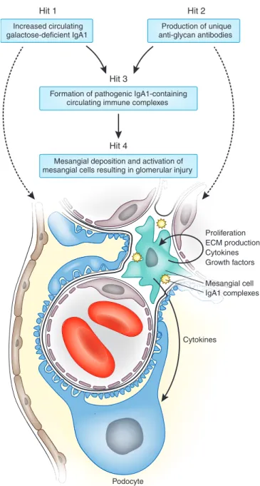
It is generally agreed that, in IgAN, the mesangial cells represent the primary target of pathogenic deposits formed by circulating immune complexes (Figure 2, solid lines) or by lanthanic deposits of aberrantly glycosylated IgA1, followed by binding of newly generated anti-glycan antibodies to form immune complexes in situ (Figure 2, broken lines).23 The presence of circulating IgA1-containing immune complexes is not unique to patients with IgAN. Such complexes can be detected in persons without apparent renal disease,
includ-ing healthy individuals and patients with Henoch-Schoenlein purpura without nephritis.19,24,25 The com-plexes in patients with Henoch-Schoenlein purpura without nephritis consist of IgA, but not IgG, and are of smaller mass than the complexes found in patients with IgAN. As these persons do not develop overt renal disease, it can be assumed that these IgA com-plexes are not nephritogenic. In con-trast, patients with Henoch-Schoenlein purpura with nephritis have larger cir-culating immune complexes contain-ing IgA and IgG.24 By analogy with other human diseases caused by im-mune complexes, it is likely that, in IgAN, the molecular proportion of an-tigens (galactose-deficient IgA1) and antibodies (IgG or IgA1) determines the size of the formed immune com-plexes and, consequently, their rate of removal from the circulation, as well as biologic activity. The pathogenic circu-lating IgA1-IgG immune complexes in patients with IgAN are relatively large (⬎800 kD) and thus may be excluded from entry into the hepatic space of Disse to reach the asialoglycoprotein receptor (ASGP-R) on hepatocytes, the normal catabolic pathway for circula-tory IgA1. As a result, these immune complexes enter the renal circulation. Due to the unique location of the mes-angium between the fenestrated endo-thelial lining of the capillaries and the glomerular basement membrane, the mesangium is prone to deposition of immune complexes. While it is not completely understood what deter-mines the entry of circulating immune complexes into the mesangium, the factors involved likely include the size of immune complexes, their amount, and local hemodynamic factors.26The biologic activity of large circulating immune complexes with galactose-deficient IgA1 increases in IgAN pa-tients during episodes of macroscopic hematuria.27 However, it is not known whether this increase in activity is due to greater production of galactose-deficient IgA1, anti-glycan antibodies, or other un-defined factors influencing the formation of these complexes and/or their
composi-Podocyte Pod Cytokines IgA1 complexes Proliferation ECM production Cytokines Growth factors Mesangial cell Increased circulating galactose-deficient IgA1 Production of unique anti-glycan antibodies Hit 2 Hit 1
Formation of pathogenic IgA1-containing circulating immune complexes
Hit 4 Hit 3
Mesangial deposition and activation of mesangial cells resulting in glomerular injury
Figure 2. Proposed pathways involved in the pathogenesis of IgAN: multi-hit mechanism. Hit 1: Production of galactose-deficient IgA1 by a subpopulation of IgA1-secreting cells. IgA1
production may be affected by the IgAN-associated locus on chromosome 22q12.2.3Hit 2:
Formation of anti-glycan antibodies with specific characteristics of the variable region of the heavy chain that recognize galactose-deficient IgA1. Hit 3: Formation of immune complexes from autoantigen (galactose-deficient IgA1) and O-glycan-specific antibodies. Hits 2 and 3
may be regulated by the three MHC loci on chromosome 6p21 associated with risk of IgAN.3
Hit 4: Deposition of pathogenic immune complexes in the mesangium, activation of mesangial cells, and induction of glomerular injury. Hits 3 and 4 may be affected by genotype at the complement factor H locus on chromosome 1q32 that regulates the alternative complement
cascade.3The first pathway assumes formation of immune complexes in the circulation and their
subsequent mesangial deposition (solid lines).13,19,55,56An alternative theory proposes that some
of the aberrantly glycosylated IgA1 molecules are in the mesangium as lanthanic deposits (left broken line) and are later bound by newly generated anti-glycan antibodies to form immune
tion.28,29MHC risk alleles may participate in this step by influencing the efficiency of antigen presentation, recognition, and processing, and subsequent activation of autoreactive B cells.
Hit 4: Mesangial Deposition of IgA1-Containing Immune Complexes, Cell Activation, and Initiation of Glomerular Injury The pathogenetic importance of im-mune complexes has been shown by in vitro studies. The glomerular injury of IgAN histologically manifests as prolifer-ation of mesangial cells and expansion of extracellular-matrix components. The detailed mechanisms of activation of mesangial cells remain to be elucidated. Nonetheless, cultured human mesangial cells provide a convenient model for evaluating the biologic activities of IgA1-containing complexes. Immune com-plexes from patients with IgAN contain-ing galactose-deficient IgA1 bind to the cells more efficiently than do uncom-plexed IgA1 or immune complexes from healthy controls. Complexes with galac-tose-deficient IgA1 induce cultured hu-man mesangial cells to proliferate, se-crete extracellular matrix components, and release humoral factors such as TNF␣, IL-6, and TGF. These factors can, in turn, alter podocyte gene expres-sion and glomerular permeability.30,31In contrast, uncomplexed galactose-defi-cient IgA1 or relatively small immune complexes (⬍800 kD) have no stimula-tory effect on cellular proliferation.
The cellular receptors on mesangial cells involved in the binding of IgA1 are not well characterized. IgA1-containing immune complexes display a high affin-ity for the extracellular-matrix compo-nents fibronectin and type IV collagen in the mesangium, and preferentially bind and activate mesangial cells. None of the well-known IgA receptors (CD89, poly-meric Ig receptor, ASGP-R) and comple-ment receptors (CR 1–3) have been con-firmed on human mesangial cells.32,33 However, transferrin receptor (CD71), which is expressed on the surface of pro-liferating human mesangial cells, can bind polymeric IgA1.34Moreover, CD71 on human mesangial cells effectively
binds immune complexes containing ga-lactose-deficient IgA1, leading to en-hanced expression of CD71.35,36 This binding creates a positive feedback loop, causing overexpression of CD71 on pro-liferating mesangial cells. However, it is not known whether CD71 is the only re-ceptor that binds IgA1-containing im-mune complexes or whether it has a di-rect pathogenic role in IgAN.
Activation of the complement system in glomeruli augments the inflammatory cascade and potentiates tissue injury in IgAN. The immune complexes with IgA1 can activate complement via the alterna-tive or lectin pathway. The pattern of gly-cosylation of IgA1 and the molecular mass of IgA1-containing immune com-plexes are also important factors in the ability of IgA1 to activate the alternative complement pathway.37Accordingly, re-nal biopsy specimens have usually de-tectable C3, while the components of the classical pathway, such as C1q, are typi-cally absent. Our recent GWAS identi-fied a major IgAN susceptibility locus within the complement factor H gene (CFH) cluster on chromosome 1q32. Products of CFH and its neighboring CFHR (CFH-related) genes participate in the modulation of the alternative path-way by binding C3a and C5a convertases. Mutations in CFH lead to uncontrolled activation of the alternative pathway and cause inherited forms of membranopro-liferative glomerulonephritis type II, a disease pathologically distinct from IgAN. However, carriers of a common deletion encompassing the neighboring CFHR1 and CFHR3 genes had an ap-proximately 30% decreased risk of devel-oping IgAN. The risk was almost 60% lower in the rare individuals who carry two copies of this deletion.3The role of CFHR1 and CFHR3 proteins in the reg-ulation of complement cascade is cur-rently under active investigation. Based on early experimental data, however, CFHR1 and CFHR3 compete with CFH for binding to C3b, the key activator of the terminal portion of the complement pathway.38 Therefore, a relative loss of CFHR1 and CFHR3 may enhance the in-hibitory action of CFH and thus convey protection against local inflammation.
These mechanistic issues have impor-tant clinical and therapeutic implications because subclinical findings consistent with IgAN are common in the general population. Necropsy series found glo-merular IgA deposits in 2% and 4% of individuals in Singapore and Germany, respectively.39,40 Even more striking, a study in Japan showed that 16% of 510 renal allografts at engraftment were af-fected, of which 19 (3.7%) had a mesan-gioproliferative nephritis.41To date, the glycosylation pattern of this lanthanic glomerular IgA has not been examined to determine if it differs from that found in IgAN patients. Such an analysis would clarify whether the IgA deposits are clin-ically silent because they have a different composition that renders them relatively inert or because there is an intrinsic hy-poresponsiveness of the kidney in which they are deposited.
POTENTIAL NEW DIAGNOSTIC AND PROGNOSTIC MARKERS In Blood
Based on the central role of galactose-deficient IgA1 in the pathogenesis, Moldoveanu et al.6 investigated the value of serum levels of this protein as a diagnostic test. By receiver operating characteristic (ROC) curve analysis, the serum level of galactose-deficient IgA1 that provided a 0.77 sensitivity had a specificity of 0.90 to distinguish IgAN patients from healthy controls, while a level with a specificity of 1.00 had a sen-sitivity of 0.44.6Other groups have repli-cated these findings.4,42Importantly, the serum level of galactose-deficient IgA1 may be significantly elevated long before the diagnosis of IgAN (Olson S. et al., un-published observation).
IgG specific for galactose-deficient IgA1 represents another potential bio-marker, as serum levels of this antibody are significantly elevated in IgAN pa-tients, and the levels correlate with pro-teinuria. ROC curve analysis indicates that when the specificity of the level of serum IgG antibody directed against ga-lactose-deficient IgA1 reached 0.95, the corresponding sensitivity was 0.88.13
Table 1. Summary of the four hits involved in the pathogenesis of IgAN Hit Pathogenic Process Putative Environmental Factors Involved Putative Genetic Factors Involved Potential Clinical Biomarkers Potential Novel Therapeutic Approaches 1 Hereditary increase in circulating galactose-deficient IgA1 Potential role of mucosal exposure to infectious or dietary antigens Strong evidence for high heritability of serum galactose-deficient IgA1 level Potential role of chromosome 22q12.2 Serum galactose-deficient IgA1 level (HAA-based ELISA) Suppression of synthesis of galactose-deficient IgA1 Enzymatic boost of galactose transfer to IgA1 hinge-region O -glycans Suppression of sialylation of galactose-deficient O -glycans 2 Circulating antibody directed against galactose-deficient IgA1 Potential role of mucosal exposure to infectious or dietary antigens Potential role of three MHC-II loci in antigen presentation and humoral response to galactose-deficient IgA1 O -glycans Serum anti-glycan antibodies (dot-blot assay) Alteration of processing and presentation of galactose-deficient IgA1 O -glycopeptides Specific B-cell depletion therapy 3 Formation of pathogenic IgA1-containing immune complexes Unknown Unknown Circulating and/or urinary immune complexes Competitive blockade of immune complex formation by non-cross-linking anti-glycan antibodies or specific glycopeptides 4 Mesangial deposition of IgA1-containing immune complexes, cell activation and initiation of glomerular injury Unknown Protective effect of common deletion in CFHR1 and CFHR3 Circulating and/or urinary complement degradation products, or novel markers of glomerular injury Suppression of the alternative complement pathway Targeted CHFR1/3 depletion Blocking mesangial cell signaling induced by nephritogenic IgA1-containing immune complexes HAA; Helix aspersa agglutinin, a lectin specific for terminal GalNAc
In Urine
Urinary proteomics holds promise for devel-opment of noninvasive tests for IgAN. A sub-set of mesangial immune complexes appar-ently enters the urinary space. Aberrantly galactosylated IgA1 within immune com-plexes has been found in the urine of patients withIgANbutnotinpatientswithnon-IgAN proteinuric glomerular diseases.43It is also possible to develop a diagnostic test without a detailed knowledge of the pathogenesis, based on analysis of the urinary peptidome. In a preliminary study, Julian et al.44found that analysis of urine samples by capillary electrophoresis, coupled online with mass spectrometry, distinguished patients with primary IgAN from patients with IgA-im-mune-complex renal disease due to cirrhosis, even if clinical proteinuria was absent. Genetic
The GWAS of sporadic IgAN identified five novel genetic variants with relatively strong protective effects against IgAN.3 While these variants are all common, their frequencies vary significantly across differ-ent contindiffer-ental populations and closely parallel the prevalence rates of IgAN. For example, African populations, which have the lowest reported prevalence of IgAN, carry the most protective alleles, while Asians, who have the highest reported prevalence, have significantly fewer protec-tive variants. The ROC analysis for a ge-netic risk score based on these five alleles is estimated in the range of 0.60 to 0.63, with the risk score explaining up to 7% of dis-ease variation among Asian and Caucasian populations. Identification of additional genetic susceptibility loci in follow-up GWAS studies of Caucasian populations will likely improve the predictive power of the genetic risk score. Additionally, a com-posite risk score based on a combined as-sessment of circulating, urinary, and ge-netic disease biomarkers holds promise to ultimately provide a tool for the noninva-sive diagnosis of IgAN.
POTENTIAL APPROACHES FOR DISEASE-SPECIFIC THERAPY The pathogenesis model (Figure 2) pro-vides an opportunity to design and test
rational therapies for IgAN (Table 1). This model predicts that the generation of nephritogenic immune complexes composed of galactose-deficient IgA1 and anti-glycan autoantibodies initiates disease. Therefore, interventions that can reduce generation of galactose-defi-cient IgA1 or anti-glycan antibodies, or block the interaction between these two components to form nephritogenic im-mune complexes, may prove effective. This result can be achieved by enhancing enzymatic activity of glycosyltransferases for synthesis of galactose-replete hinge-region O-glycans to reduce the availabil-ity of aberrantly glycosylated IgA1 for formation of nephritogenic immune complexes. Alternatively, generation of non-cross-linking, monovalent reagents (single-chain antibodies) with high af-finity for GalNAc on galactose-deficient IgA1 could theoretically prevent binding of anti-glycan antibodies. Lastly, block-ing the antibodies with a glycopeptide may be another strategy to prevent for-mation of immune complexes (Table 1). Similarly, interventions aimed at re-ducing immune complex deposition and the downstream inflammatory signals may prove beneficial. The genetic studies identify the alternative complement pathway as a prime candidate for inter-vention, and predict that targeted deple-tion of CFHR1 and/or CFHR3 would be tolerated and prove protective. More-over, blocking of specific signaling path-ways induced in mesangial cells by pathogenic IgA1-containing complexes can be theoretically accomplished by protein-kinase inhibitors, a class of drugs that is frequently used in the treatment of some types of cancer.
CONCLUSIONS
IgAN is an autoimmune renal disease arising from consequences of increased circulating levels of IgA1 with galactose-deficient hinge-region O-glycans. How-ever, this glycosylation aberrancy alone is not sufficient to induce nephritis. For the clinical manifestation of renal injury, several additional hits are required, in-cluding synthesis of circulating
antibod-ies directed against the aberrantly glyco-sylated O-linked hinge-region glycans to form immune complexes, accumulation of the complexes in the mesangium, and activation of mesangial cells. Genetic fac-tors apparently influence the expression of these hit mechanisms. Elucidation of the pathogenesis of IgAN provides an opportunity to develop a disease-specific therapy that heretofore has been missing.
DISCLOSURES
Supported in part by grants DK082753, DK078244, DK083663, DK075868, DK080301, DK077279, DK071802, and K23-DK090207 from the National Institutes of Health and a grant from the IgA Nephropathy Foundation of America.
REFERENCES
1. Berger J, Hinglais N: Les de´poˆts intercapil-laires d’IgA-IgG (Intercapillary deposits of IgA-IgG). J Urol Nephrol 74: 694 – 695, 1968 2. Silva FG, Chander P, Pirani CL, Hardy MA: Disappearance of glomerular mesangial IgA deposits after renal allograft transplantation. Transplantation 33: 241–246, 1982 3. Gharavi AG, Kiryluk K, Choi M, Li Y, Hou P,
Xie J, Sanna-Cherchi S, Men CJ, Julian BA, Wyatt RJ, Novak J, He JC, Wang H, Lv J, Zhu L, Wang W, Wang Z, Yasuno K, Gunel M, Mane S, Umlauf S, Tikhonova I, Beerman I, Savoldi S, Magistroni R, Ghiggeri GM, Bo-dria M, Lugani F, Ravani P, Ponticelli C, Al-legri L, Boscutti G, Frasca G, Amore A, Pe-ruzzi L, Coppo R, Izzi C, Viola BF, Prati E, Salvadori M, Mignani R, Gesualdo L, Berti-netto F, Mesiano P, Amoroso A, Scolari F, Chen N, Zhang H, Lifton RP: Genome-wide association study identifies susceptibility loci for IgA nephropathy. Nat Genet 43: 321– 327, 2011
4. Coppo R, Feehally J, Glassock RJ: IgA ne-phropathy at two score and one. Kidney Int 77: 181–186, 2010
5. Mestecky J, Moro I, Kerr MA, Woof JM: Mucosal immunoglobulins. In: Mucosal Im-munology, 3rd ed, edited by Mestecky J, Bienenstock J, Lamm ME, Mayer L, McGhee JR, Strober W, Amsterdam, Elsevier Aca-demic Press, 2005, pp 153–181
6. Moldoveanu Z, Wyatt RJ, Lee J, Tomana M, Julian BA, Mestecky J, Huang WQ, Anreddy S, Hall S, Hastings MC, Lau KK, Cook WJ, Novak J: Patients with IgA nephropathy have increased serum galactose-deficient IgA1 levels. Kidney Int 71: 1148 –1154, 2007 7. Renfrow MB, Cooper HJ, Tomana M, Kul-havy R, Hiki Y, Toma K, Emmett MR,
Mestecky J, Marshall AG, Novak J: Determi-nation of aberrant O-glycosylation in the IgA1 hinge region by electron capture dis-sociation Fourier transform-ion cyclotron resonance mass spectrometry. J Biol Chem 280: 19136 –19145, 2005
8. Renfrow MB, MacKay CL, Chalmers MJ, Ju-lian BA, Mestecky J, KiJu-lian M, Poulsen K, Emmett MR, Marshall AG, Novak J: Analysis of O-glycan heterogeneity in IgA1 myeloma proteins by Fourier transform ion cyclotron resonance mass spectrometry: Implications for IgA nephropathy. Anal Bioanal Chem 389: 1397–1407, 2007
9. Takahashi K, Wall SB, Suzuki H, Smith AD, Hall S, Poulsen K, Kilian M, Mobley JA, Ju-lian BA, Mestecky J, Novak J, Renfrow MB: Clustered O-glycans of IgA1: Defining mac-ro- and micmac-ro-heterogeneity by use of elec-tron capture/transfer dissociation. Mol Cell Proteomics 9: 2545–2557, 2010
10. Wada Y, Dell A, Haslam SM, Tissot B, Canis K, Azadi P, Backstrom M, Costello CE, Hans-son GC, Hiki Y, Ishihara M, Ito H, Kakehi K, Karlsson N, Hayes CE, Kato K, Kawasaki N, Khoo KH, Kobayashi K, Kolarich D, Kondo A, Lebrilla C, Nakano M, Narimatsu H, Novak J, Novotny MV, Ohno E, Packer NH, Palaima E, Renfrow MB, Tajiri M, Thomsson KA, Yagi H, Yu SY, Taniguchi N: Comparison of methods for profiling O-glycosylation: Human Pro-teome Organisation Human Disease Gly-comics/Proteome Initiative multi-institu-tional study of IgA1. Mol Cell Proteomics 9: 719 –727, 2010
11. Wada Y, Tajiri M, Ohshima S: Quantitation of saccharide compositions of O-glycans by mass spectrometry of glycopeptides and its application to rheumatoid arthritis. J Pro-teome Res 9: 1367–1373, 2010
12. Odani H, Yamamoto K, Iwayama S, Iwase H, Takasaki A, Takahashi K, Fujita Y, Sugiyama S, Hiki Y: Evaluation of the specific structures of IgA1 hinge glycopeptide in 30 IgA ne-phropathy patients by mass spectrometry. J Nephrol 23: 70 –76, 2010
13. Suzuki H, Fun R, Zhang Z, Brown R, Hall S, Julian BA, Chatham WW, Suzuki Y, Wyatt RJ, Moldoveanu Z, Lee JY, Robinson J, Tomana M, Tomino Y, Mestecky J, Novak J: Aber-rantly glycosylated IgA1 in IgA nephropathy patients is recognized by IgG antibodies with restricted heterogeneity. J Clin Invest 119: 1668 –1677, 2009
14. Smith AC, Molyneux K, Feehally J, Barratt J: O-glycosylation of serum IgA1 antibodies against mucosal and systemic antigens in IgA nephropathy. J Am Soc Nephrol 17: 3520 –3528, 2006
15. Suzuki H, Suzuki Y, Narita I, Aizawa M, Kihara M, Yamanaka T, Kanou T, Tsukagu-chi H, Novak J, Horikoshi S, Tomino Y: Toll-like receptor 9 affects severity of IgA nephropathy. J Am Soc Nephrol 19: 2384 – 2395, 2008
16. Gharavi AG, Moldoveanu Z, Wyatt RJ,
Barker CV, Woodford SY, Lifton RP, Mestecky J, Novak J, Julian BA: Aberrant IgA1 glycosylation is inherited in familial and sporadic IgA nephropathy. J Am Soc Neph-rol 19: 1008 –1014, 2008
17. Kiryluk K, Julian BA, Wyatt RJ, Scolari F, Zhang H, Novak J, Gharavi AG: Genetic studies of IgA nephropathy: Past, present, and future. Pediatr Nephrol 25: 2257–2268, 2010
18. Imielinski M, Baldassano RN, Griffiths A, Russell RK, Annese V, Dubinsky M, Kugath-asan S, Bradfield JP, Walters TD, Sleiman P, Kim CE, Muise A, Wang K, Glessner JT, Saeed S, Zhang H, Frackelton EC, Hou C, Flory JH, Otieno G, Chiavacci RM, Grund-meier R, Castro M, Latiano A, Dallapiccola B, Stempak J, Abrams DJ, Taylor K, McGovern D, Silber G, Wrobel I, Quiros A, Barrett JC, Hansoul S, Nicolae DL, Cho JH, Duerr RH, Rioux JD, Brant SR, Silverberg MS, Taylor KD, Barmuda MM, Bitton A, Dassopoulos T, Datta LW, Green T, Griffiths AM, Kistner EO, Murtha MT, Regueiro MD, Rotter JI, Schumm LP, Steinhart AH, Targan SR, Xavier RJ, Libioulle C, Sandor C, Lathrop M, Be-laiche J, Dewit O, Gut I, Heath S, Laukens D, Mni M, Rutgeerts P, Van Gossum A, Zelenika D, Franchimont D, Hugot JP, de Vos M, Vermeire S, Louis E, Cardon LR, Anderson CA, Drummond H, Nimmo E, Ahmad T, Prescott NJ, Onnie CM, Fisher SA, Marchini J, Ghori J, Bumpstead S, Gwillam R, Tremel-ling M, Delukas P, Mansfield J, Jewell D, Satsangi J, Mathew CG, Parkes M, Georges M, Daly MJ, Heyman MB, Ferry GD, Kirsch-ner B, Lee J, Essers J, Grand R, Stephens M, Levine A, Piccoli D, Van Limbergen J, Cuc-chiara S, Monos DS, Guthery SL, Denson L, Wilson DC, Grant SF, Daly M, Hakonarson H: Common variants at five new loci associ-ated with early-onset inflammatory bowel disease. Nat Genet 41: 1335–1340, 2009 19. Tomana M, Novak J, Julian BA, Matousovic
K, Konecny K, Mestecky J: Circulating im-mune complexes in IgA nephropathy consist of IgA1 with galactose-deficient hinge re-gion and antiglycan antibodies. J Clin Invest 104: 73– 81, 1999
20. Bellur SS, Troyanov S, Cook HT, Roberts IS: Immunostaining findings in IgA nephropa-thy: Correlation with histology and clinical outcome in the Oxford classification patient cohort. Nephrol Dial Transplant 26: 2533– 2536, 2011
21. Feehally J, Farrall M, Boland A, Gale DP, Gut I, Heath S, Kumar A, Peden JF, Maxwell PH, Morris DL, Padmanabhan S, Vyse TJ, Zawadzka A, Rees AJ, Lathrop M, Ratcliffe PJ: HLA has strongest association with IgA nephropathy in genome-wide analysis. J Am Soc Nephrol 21: 1791–1797, 2010 22. Solberg OD, Mack SJ, Lancaster AK, Single
RM, Tsai Y, Sanchez-Mazas A, Thomson G: Balancing selection and heterogeneity across the classical human leukocyte antigen
loci: A meta-analytic review of 497 popula-tion studies. Hum Immunol 69: 443– 464, 2008
23. Glassock RJ: The pathogenesis of IgA ne-phropathy. Curr Opin Nephrol Hypertens 20: 153–160, 2011
24. Levinsky RJ, Barratt TM: IgA immune com-plexes in Henoch-Scho¨nlein purpura. Lancet 2: 1100 –1103, 1979
25. Hall RP, Lawley TJ, Heck JA, Katz SI: IgA-containing circulating immune complexes in dermatitis herpetiformis, Henoch-Scho¨nlein purpura, systemic lupus erythematosus and other diseases. Clin Exp Immunol 40: 431– 437, 1980
26. Sterzel RB, Lovett DH, Stein HD, Kashgarian M: The mesangium and glomerulonephritis. Klin Wochenschr 60: 1077–1094, 1982 27. Novak J, Tomana M, Matousovic K, Brown
R, Hall S, Novak L, Julian BA, Wyatt RJ, Mestecky J: IgA1-containing immune com-plexes in IgA nephropathy differentially af-fect proliferation of mesangial cells. Kidney Int 67: 504 –513, 2005
28. Novak J, Moldoveanu Z, Renfrow MB, Yanagihara T, Suzuki H, Raska M, Hall S, Brown R, Huang WQ, Goepfert A, Kilian M, Poulsen K, Tomana M, Wyatt RJ, Julian BA, Mestecky J: IgA nephropathy and Henoch-Schoenlein purpura nephritis: Aberrant gly-cosylation of IgA1, formation of IgA1-con-taining immune complexes, and activation of mesangial cells. Contrib Nephrol 157: 134 –138, 2007
29. Novak J, Mestecky J: IgA Immune-complex. In: Recent Advances in IgA Nephropathy, edited by Lai KN, Hong Kong, Imperial Col-lege Press and the World Scientific Pub-lisher, 2009, pp 177–191
30. Lai KN, Leung JC, Chan LY, Saleem MA, Mathieson PW, Lai FM, Tang SC: Activation of podocytes by mesangial-derived TNF-␣: Glomerulo-podocytic communication in IgA nephropathy. Am J Physiol Renal Physiol 294: F945–F955, 2008
31. Lai KN, Leung JC, Chan LY, Saleem MA, Mathieson PW, Tam KY, Xiao J, Lai FM, Tang SC: Podocyte injury induced by me-sangial-derived cytokines in IgA nephropa-thy. Nephrol Dial Transplant 24: 62–72, 2009
32. Leung JCK, Tsang AWL, Chan DTM, Lai KN: Absence of CD89, polymeric immunoglob-ulin receptor, and asialoglycoprotein recep-tor on human mesangial cells. J Am Soc Nephrol 11: 241–249, 2000
33. Novak J, Vu HL, Novak L, Julian BA, Mestecky J, Tomana M: Interactions of hu-man mesangial cells with IgA and IgA-con-taining circulating immune complexes. Kid-ney Int 62: 465– 475, 2002
34. Moura IC, Centelles MN, Arcos-Fajardo M, Malheiros DM, Collawn JF, Cooper MD, Monteiro RC: Identification of the transferrin receptor as a novel immunoglobulin (Ig)A1 receptor and its enhanced expression on
mesangial cells in IgA nephropathy. J Exp Med 194: 417– 425, 2001
35. Moura IC, Arcos-Fajardo M, Sadaka C, Leroy V, Benhamou M, Novak J, Vrtovsnik F, Had-dad E, Chintalacharuvu KR, Monteiro RC: Glycosylation and size of IgA1 are essential for interaction with mesangial transferrin re-ceptor in IgA nephropathy. J Am Soc Neph-rol 15: 622– 634, 2004
36. Moura IC, Arcos-Fajardo M, Gdoura A, Le-roy V, Sadaka C, Mahlaoui N, Lepelletier Y, Vrtovsnik F, Haddad E, Benhamou M, Mon-teiro RC: Engagement of transferrin recep-tor by polymeric IgA1: Evidence for a posi-tive feedback loop involving increased receptor expression and mesangial cell pro-liferation in IgA nephropathy. J Am Soc Nephrol 16: 2667–2676, 2005
37. Zhang W, Lachmann PJ: Glycosylation of IgA is required for optimal activation of the alternative complement pathway by immune complexes. Immunology 81: 137–141, 1994
38. Fritsche LG, Lauer N, Hartmann A, Stippa S, Keilhauer CN, Oppermann M, Pandey MK, Kohl J, Zipfel PF, Weber BH, Skerka C: An imbalance of human complement regulatory proteins CFHR1, CFHR3 and factor H influ-ences risk for age-related macular degener-ation (AMD). Hum Mol Genet 19: 4694 – 4704, 2010
39. Waldherr R, Rambousek M, Duncker WD, Ritz E: Frequency of mesangial IgA deposits in non-selected autopsy series. Nephrol Dial Transplant 4: 943–946, 1989
40. Varis J, Rantala I, Pasternack A, Oksa H, Jantti M, Paunu ES, Pirhonen R: Immuno-globulin and complement deposition in glomeruli of 756 subjects who had commit-ted suicide or met with a violent death. J Clin Pathol 46: 607– 610, 1993
41. Suzuki K, Honda K, Tanabe K, Toma H, Nihei H, Yamaguchi Y: Incidence of latent mesan-gial IgA deposition in renal allograft donors in Japan. Kidney Int 63: 2286 –2294, 2003
42. Shimozato S, Hiki Y, Odani H, Takahashi K, Yamamoto K, Sugiyama S: Serum under-galactosylated IgA1 is increased in Japa-nese patients with IgA nephropathy. Nephrol Dial Transplant 23: 1931–1939, 2008
43. Matousovic K, Novak J, Tomana M, Kulhavy R, Julian BA, Mestecky J: IgA1-containing immune complexes in the urine of IgA ne-phropathy patients. Nephrol Dial Transplant 21: 2478 –2484, 2006
44. Julian BA, Wittke S, Novak J, Good DM, Coon JJ, Kellmann M, Zu¨rbig P, Schiffer E, Haubitz M, Moldoveanu Z, Calcatera SM, Wyatt RJ, Sykora J, Sladkova E, Hes O, Mis-chak H, McGuire BM: Electrophoretic meth-ods for analysis of urinary polypeptides in IgA-associated renal diseases. Electrophore-sis 28: 4469 – 4483, 2007
45. Gomes MM, Suzuki H, Brooks MT, Tomana M, Moldoveanu Z, Mestecky J, Julian BA, Novak J, Herr AB: Recognition of galactose-deficient O-glycans in the hinge region of IgA1 by N-acetylgalactosamine-specific snail lectins: A comparative binding study. Biochemistry 49: 5671–5682, 2010 46. Moore JS, Kulhavy R, Tomana M, Moldoveanu
Z, Suzuki H, Brown R, Hall S, Kilian M, Poulsen K, Mestecky J, Julian BA, Novak J: Reactiv-ities of N-acetylgalactosamine-specific lec-tins with human IgA1 proteins. Mol Immunol 44: 2598 –2604, 2007
47. Boehm MK, Woof JM, Kerr MA, Perkins SJ: The Fab and Fc fragments of IgA1 exhibit a different arrangement from that in IgG: A study by X-ray and neutron solution scatter-ing and homology modellscatter-ing. J Mol Biol 286: 1421–1447, 1999
48. Herr AB, Ballister ER, Bjorkman PJ: Insights into IgA-mediated immune responses from the crystal structures of human Fc␣RI and its complex with IgA1-Fc. Nature 423: 614 – 620, 2003
49. Gomes MM, Wall SB, Takahashi K, Novak J,
Renfrow MB, Herr AB: Analysis of IgA1 N-glycosylation and its contribution to Fc␣RI binding. Biochemistry 47: 11285–11299, 2008
50. Suzuki H, Moldoveanu Z, Hall S, Brown R, Vu HL, Novak L, Julian BA, Tomana M, Wyatt RJ, Edberg JE, Alarco´n GS, Kim-berly RP, Tomino Y, Mestecky J, Novak J: IgA1-secreting cell lines from patients with IgA nephropathy produce aberrantly gly-cosylated IgA1. J Clin Invest 118: 629 – 639, 2008
51. Ju T, Cummings RD: Protein glycosylation: Chaperone mutation in Tn syndrome. Na-ture 437: 1252, 2005
52. Iwasaki H, Zhang Y, Tachibana K, Gotoh M, Kikuchi N, Kwon YD, Togayachi A, Kudo T, Kubota T, Narimatsu H: Initiation of O-glycan synthesis in IgA1 hinge region is determined by a single enzyme, UDP-N-acetyl-␣-D-galac-tosamine:polypeptide N-acetylgalactosaminyl-transferase 2. J Biol Chem 278: 5613–5621, 2003
53. Raska M, Moldoveanu Z, Suzuki H, Brown R, Kulhavy R, Hall S, Vu HL, Carlsson F, Lindahl G, Tomana M, Julian BA, Wyatt RJ, Mestecky J, Novak J: Identification and characterization of CMP-NeuAc:GalNAc-IgA1 ␣2,6-sialyltransferase in IgA1-produc-ing cells. J Mol Biol 369: 69 –78, 2007 54. Ju T, Brewer K, D’Souza A, Cummings RD,
Canfield WM: Cloning and expression of human core 11,3-galactosyltransferase. J Biol Chem 277: 178 –186, 2002
55. Julian BA, Novak J: IgA nephropathy: An update. Current Opin Nephrol Hypertens 13: 171–179, 2004
56. Mestecky J, Tomana M, Moldoveanu Z, Julian BA, Suzuki H, Matousovic K, Ren-frow MB, Novak L, Wyatt RJ, Novak J: The role of aberrant glycosylation of IgA1 mol-ecules in the pathogenesis of IgA ne-phropathy. Kidney Blood Pres Res 31: 29 – 37, 2008