Case Report
Unusual Postrhinoplasty Complication: Nasal Dorsum Cyst
Pier Giorgio Giacomini,
1Davide Topazio,
1Roberta Di Mauro,
1Stelio Mocella,
2Matteo Chimenti,
3and Stefano Di Girolamo
11Department of Otorhinolaryngology, University of Rome “Tor Vergata”, Rome, Italy
2Department of Otorhinolaryngology, Bussolengo Hospital, Italy
3Department of Pathology, University of Rome “Tor Vergata”, Rome, Italy
Correspondence should be addressed to Roberta Di Mauro; [email protected]
Received 30 March 2014; Revised 5 August 2014; Accepted 21 August 2014; Published 9 September 2014 Academic Editor: Chung-Feng Hwang
Copyright © 2014 Pier Giorgio Giacomini et al. This is an open access article distributed under the Creative Commons Attribution License, which permits unrestricted use, distribution, and reproduction in any medium, provided the original work is properly cited.
Among all the possible complications of aesthetic rhinoplasty, a rare one is the development of cystic masses on the nasal dorsum: several theories suggest that cysts develop commonly by entrapment of nasal mucosa in the subcutaneous space, but they can also originate from foreign body reactions. This report deals with two cases of nasal dorsum cysts with different pathogenesis: both patients had undergone aesthetic rhinoplasty in the past (26 years ago and 14 years ago, resp.). Both cystic masses were removed via a direct open approach and nasal reconstruction was performed successfully with autologous vomer bone. The pathologic investigations showed a foreign body inclusion cyst associated with latex rubber in the first case and a sequestration of a mucosal-lined nasal bone was not removed at the time of primary rhinoplasty in the second case. A brief review of the literature focuses on the pathophysiology and treatment options for nasal dorsal cysts following aesthetic rhinoplasty.
1. Introduction
The development of postrhinoplasty nasal dorsal irregulari-ties due to bone or cartilaginous bossae is a common com-plication, whereas cysts formation is a rare event. Reviewing previously reported cases reveals that mucous cysts are by far most prevalent in the nasal dorsum. Other locations include the nasal tip, the medial canthus, and the paranasal area [1–3]. Probably, migration or incorporation of mucosal tissue (or bony or cartilaginous fragments not accurately removed during primary surgery in the subcutaneous space) commonly accounts for cyst formation [4].
With regard to nonmucosal cysts of the subcutaneous area, a fully different pathophysiology, namely, foreign body reaction, may be considered. Petrolatum jelly migrated from the packing material and foreign body reaction to alloim-plants employed for augmentation have been reported to be possible causes [5]. Other rare pathogenetic mechanisms like congenital malformations in cleft lip nose rhinoplasty or remnants of the nasolacrimal duct have been described [6,7]. Since the postsurgical cyst development could be in part prevented, particularly by considering all pathogenetic
mechanisms, this report aims at underlining the need of further insight into this rare entity.
2. Case 1
We evaluated a 62-year-old Caucasian male with an asymp-tomatic nasal radix mass and nasal airflow obstruction. Twenty-six years ago he had undergone reduction rhino-plasty with an uneventful postoperative period; however, nasal obstruction had not improved.
A nasal dorsum mass, measuring5 × 5 mm was noted three months postoperatively and slowly increased in size over the years. On physical examination, the nasal dorsum and tip were firm on palpation and a small deformity was present in the upper nasal dorsum, appearing as a2.0×2.0 cm round, soft, and painless mass located on the midline of nasal radix (Figure 1(a)). A computerized tomography (CT) scan was conducted to exclude possible bone involvement and revealed a heterogeneous, large cystic mass on the nasal radix, measuring 68 Hounsfield units (HU), adhering without erosion to the nasal bones cranial part. It contained
Volume 2014, Article ID 617424, 4 pages http://dx.doi.org/10.1155/2014/617424
2 Case Reports in Otolaryngology
(a) (b)
(c) (d)
(e)
Figure 1: (a) Frontal view (Patient #1), (b) lateral view (Patient #1), (c) preoperative CT scan (Patient #1), (d) intraoperative view of nasal cyst removal (Patient #1), and (e) pathology specimen (Patient #1).
a cystic aspect including a homogenous density foreign body (Figure 1(b)), which was first thought to be a silicone implant. In consideration of clinical and CT-features, a presurgical diagnosis of nasal radix cyst as a consequence of previous augmentation rhinoplasty was made. The mass was success-fully removed via a horizontal glabellar incision. After a skin incision, the cyst was dissected down to the nasal bone and removed in en-bloc fashion because of its close relation to the bony dorsum (Figure 1(c)).
The removed cyst contained a liquid, clear content sur-rounded by a slightly fibrous wall. Inside the cyst, a foreign body was evacuated and revealed to be a5×2 cm piece of latex rubber excised from surgical gloves and supposedly folded to fit the underlying bony defect created by hump removal
during the primary rhinoplasty procedure. Foreign material was surrounded by foreign body-type, multinucleated giant cells, and infiltration of chronic inflammatory cells inducing granulomatous reactions (Figure 1(d)).
The depressed radix residual due to the underling small bony defect was corrected by a bone autograft taken from the vomer bone exposed during concomitant closed approach septoplasty for correction of the residual nasal obstruction. The redundant skin of the radix was trimmed and accurately sutured.
The patient’s postoperative course was uneventful and no external deformity remained. Nasal airflow was not affected by the procedure and the patient was satisfied with the aes-thetic result.
(a) (b)
(c) (d)
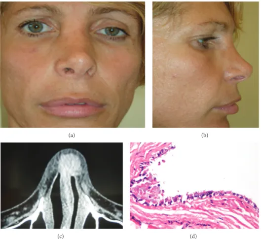
Figure 2: (a) Frontal view (Patient #2), (b) lateral view (Patient #2), (c) preoperative CT scan (Patient #2), and (d) pathology specimen (Patient #2).
3. Case 2
A 44-year-old Caucasian female with no significant medical history, who had previously undergone rhinoplasty 14 years ago, reported of a functionally satisfactory postoperative period without complications. However, three years ago, she discovered a gradually increasing3 × 3 mm mass at the nasal radix. She also complained of persistent obstruction of nasal airflow.
Physical examination revealed a spherical nasal mass (1.0 × 1.0 cm), which was on palpation that was found to be firm-elastic, mobile, painless, and covered by nor-mal skin, located on the right nasal radix (Figure 2(a)). A CT scan was performed to assess bone involvement and revealed a heterogeneous cystic mass connected to nasal bone (Figure 2(b)); the mass appeared with a poorly defined soft tissue density (90 HU) along the superior nasal dorsum. We first suspected that the cyst developed from a splinter bone of the precedent osteotomy due to the initial bone-like consistency on palpation (as reported by the patient).
The patient was admitted for removal of the mass and reconstruction of the nasal dorsum.
An open excision of the lesion was performed. The mass was found to be tightly attached to the bony dorsum and it was thus dissected down to the nasal bone and removed in
en-bloc fashion. Within the intact capsule, a mucous-lined cavity filled with a thick yellow liquid was found. Histopatho-logic investigations revealed a benign epithelial mucous cyst containing a fragment of normal cartilage and bone (Figure 2(c)).
The lesion was diagnosed as a sequestration of a mucosal-lined nasal bone not removed at the time of hump removal.
The resulting deformity of the nasal root was corrected by using autologous bony graft from the vomer. The excessive skin was removed and sutured carefully. The patient’s post-operative course was uneventful and no external deformity remained with a satisfactory final result.
4. Discussion
According to the literature, the different circumstances of the cases presented here suggest different pathways for cysts formation. The most accepted theories about the etiology of mucous cysts after aesthetic rhinoplasty include the presence of ectopic free mucosal graft implantation during surgical treatment, herniation of mucosa through intranasal incision, improper clearing of mucous epithelial remnants, or bony and cartilage parts during the operation [8,9]. Other possible causes could be intrasurgical trauma and occlusion of seba-ceous glands from scar tissue formation [10–13].
4 Case Reports in Otolaryngology Inflammatory reaction to a foreign body has also
previ-ously been described as a possible cause [5,14,15]. In our case, considering the gross and histological findings, this reaction looks like a foreign body reaction to rubber latex material, most possibly from surgical gloves inserted 26 years ago during primary surgery. This mechanism seems akin to what has been reported for silicone implants at the nasal dorsum: silicone implants have been used for rhinoplasty since 1950 and still remains widely used implant materials in selected instances such as Asian nose augmentation rhinoplasty [16–
18].
The only valid treatment of postrhinoplasty nasal dor-sal cyst is the complete resection and reconstruction. We performed a direct open approach in both cases, using a glabellar horizontal incision over the cyst. We chose the direct transcutaneous route to the lesion in order to assure the adequate exposure of the upper part of the lesion. A conventional open approach to the nasal septum was unnecessary due to the absence of concomitant dorsal or tip further deformities.
Moreover, the horizontal glabellar incision seemed well tolerated, without noticeable scar formations. A closed approach using hemitransfix incision was felt sufficient to expose the septum and to avoid any dissection and possible connection between the nasal vestibule and the dorsal area. Augmentation of the residual depressed dorsum and radix with autogenous septal bone was possible.
When treating nasal dorsal masses, one first has to exclude various nasal lesions not related to surgery: neoplas-tic lesions, benign processes, encephaloceles, gliomas, der-moids, osteomas, lipomas, granulomatous diseases (Wegener granulomatosis, sarcoidosis, and rhinoscleroma), and infec-tions (fungus, syphilis, and tuberculosis) [19].
In conclusion, according to the current literature, we would point out that postrhinoplasty nasal cysts do occur [10,12,19–21]: prevention of their formation calls for metic-ulous dissection before osteotomies and hump removal. It is advisable to carefully eliminate all bony, cartilagenous, epithelial or mucous tissues, and debris that may act as a foreign body at the nasal dorsum [19]. Proper closure of intranasal incisions, conservation of mucosal integrity, strict mucosal dissection, and separation in hump removal complete removal of adherent fragments of epithelium from cartilaginous grafts or flaps whenever used and pressure-free insertion of nasal dressings may further prevent cysts development.
Conflict of Interests
The authors declare that there is no conflict of interests regarding the publication of this paper.
References
[1] M. W. McGregor, G. B. O’Connor, and S. Saffier, “Complications of rhinoplasty. I. Skin and subcutaneous tissues,” The Journal of
the International College of Surgeons, vol. 30, no. 2, pp. 179–184,
1958.
[2] F. Riedel, C. Bersch, and K. H¨ormann, “Dorsal nasal mass formation: postrhinoplasty cyst,” HNO, vol. 55, no. 6, pp. 472– 474, 2007.
[3] C. Raine, S. L. H. Williamson, and N. R. McLean, “Mucous cyst of the alar base: a rare complication following rhinoplasty,”
British Journal of Plastic Surgery, vol. 56, no. 2, pp. 176–177, 2003.
[4] G. Rettinger, “Risks and complications in rhinoplasty,” GMS
Current Topics in Otorhinolaryngology—Head and Neck Surgery,
vol. 6, article Doc08, 2008.
[5] M. W. Pak, E. S. Chan, and C. A. van Hasselt, “Late complica-tions of nasal augmentation using silicone implants,” Journal of
Laryngology and Otology, vol. 112, no. 11, pp. 1074–1077, 1998.
[6] N. Pausch, E. Brylla, T. Schulz, J. Helmrich, A. Hemprich, and B. Frerich, “Nasenfisteln und -zysten bei Patienten mit Lippen-Kiefer-Gaumen-Spalten,” Mund-, Kiefer- und Gesichtschirurgie, vol. 10, no. 6, pp. 369–375, 2006.
[7] T. Aikawa, S. Iida, Y. Fukuda et al., “Nasolabial cyst in a patient with cleft lip and palate,” International Journal of Oral and
Maxillofacial Surgery, vol. 37, no. 9, pp. 874–876, 2008.
[8] N. C. Pausch, J. Bertolini, A. Hemprich, and T. Hierl, “Inclusion mucous cysts of the nose: a late complication after septorhino-plasty in two cleft lip patients,” Cleft Palate-Craniofacial Journal, vol. 47, no. 6, pp. 668–672, 2010.
[9] D. Y. Chang and H. R. Jin, “Foreign body inclusion cyst of the nasal radix after augmentation rhinoplasty,” Journal of Korean
Medical Science, vol. 23, no. 6, pp. 1109–1112, 2008.
[10] A. Kotzur and W. Gubisch, “Mucous cyst—a postrhinoplasty complication: outcome and prevention,” Plastic &
Reconstruc-tive Surgery, vol. 100, no. 2, pp. 520–524, 1997.
[11] M. Dini, A. Innocenti, G. L. Russo, and V. Agostini, “Postrhino-plasty mucous cyst of the nose,” Plastic and Reconstructive
Surgery, vol. 107, no. 3, pp. 885–886, 2001.
[12] E. H. Harley and J. P. Erdman, “Dorsal nasal cyst forma-tion. A rare complication of cosmetic rhinoplasty,” Archives of
Otolaryngology—Head and Neck Surgery, vol. 116, no. 1, pp. 105–
106, 1990.
[13] G. Rettinger and M. Zenkel, “Skin and soft tissue complica-tions,” Facial Plastic Surgery, vol. 13, no. 1, pp. 51–59, 1997. [14] B. A. Bassichis and J. R. Thomas, “Foreign-body inclusion cyst
presenting on the lateral nasal sidewall 1 year after rhinoplasty,”
Archives of Facial Plastic Surgery, vol. 5, no. 6, pp. 530–532, 2003.
[15] W. Lawson, S. Kessler, and H. F. Biller, “Unusual and fatal complications of rhinoplasty,” Archives of Otolaryngology, vol. 109, no. 3, pp. 164–169, 1983.
[16] C. Tham, Y.-L. Lai, C.-J. Weng, and Y.-R. Chen, “Silicone augmentation rhinoplasty in an oriental population,” Annals of
Plastic Surgery, vol. 54, no. 1, pp. 1–5, 2005.
[17] T. D. Wang, “Non-caucasian rhinoplasty,” Facial Plastic Surgery, vol. 19, no. 3, pp. 247–256, 2003.
[18] J. Yang, X. Wang, Y. Zeng, and W. Wu, “Biomechanics in augmentation rhinoplasty,” Journal of Medical Engineering and
Technology, vol. 29, no. 1, pp. 14–17, 2005.
[19] C. E. Unl¨u, G. Saylam, H. Korkmaz, E. C. Tatar, and A. Ozdek, “Nasal dorsal mucous cyst formation: a rare and preventable complication of rhinoplasty,” Kulak Burun Boˇgaz Ihtisas Dergisi, vol. 21, no. 5, pp. 294–297, 2011.
[20] T. Romo, S. S. Rizk, and G. D. Suh, “Mucous cyst formation after rhinoplasty,” Archives of Facial Plastic Surgery, vol. 1, no. 3, pp. 208–211, 1999.
[21] B. Struijs and L. J. J. M. Bauwens, “Post-rhinoplasty mucous cyst formation of the nasal dorsum,” B-ENT, vol. 6, no. 4, pp. 295– 298, 2010.
Submit your manuscripts at
http://www.hindawi.com
Stem Cells
International
Hindawi Publishing Corporationhttp://www.hindawi.com Volume 2014
Hindawi Publishing Corporation
http://www.hindawi.com Volume 2014
INFLAMMATION
Hindawi Publishing Corporation
http://www.hindawi.com Volume 2014
Behavioural
Neurology
Endocrinology
International Journal ofHindawi Publishing Corporation
http://www.hindawi.com Volume 2014 Hindawi Publishing Corporation
http://www.hindawi.com Volume 2014
Disease Markers
Hindawi Publishing Corporation
http://www.hindawi.com Volume 2014
BioMed
Research International
Oncology
Journal ofHindawi Publishing Corporation
http://www.hindawi.com Volume 2014
Hindawi Publishing Corporation
http://www.hindawi.com Volume 2014
Oxidative Medicine and Cellular Longevity
Hindawi Publishing Corporation
http://www.hindawi.com Volume 2014
PPAR Research
The Scientific
World Journal
Hindawi Publishing Corporation
http://www.hindawi.com Volume 2014
Immunology Research
Hindawi Publishing Corporation
http://www.hindawi.com Volume 2014
Journal of
Obesity
Journal ofHindawi Publishing Corporation
http://www.hindawi.com Volume 2014
Hindawi Publishing Corporation
http://www.hindawi.com Volume 2014
Computational and Mathematical Methods in Medicine
Ophthalmology
Journal ofHindawi Publishing Corporation
http://www.hindawi.com Volume 2014
Diabetes Research
Journal ofHindawi Publishing Corporation
http://www.hindawi.com Volume 2014
Hindawi Publishing Corporation
http://www.hindawi.com Volume 2014
Research and Treatment
AIDS
Hindawi Publishing Corporation
http://www.hindawi.com Volume 2014
Gastroenterology Research and Practice
Hindawi Publishing Corporation
http://www.hindawi.com Volume 2014