Sapienza University of Rome
PhD Programme in Molecular Medicine
Cycle XXXI
THE NOVEL ANTI-IL-17A MONOCLONAL ANTIBODY
SECUKINUMAB
IN THE TREATMENT OF PSORIASIS:
BIOLOGICAL EFFECTS ON ANGIOGENESIS AND
EXTRACELLULAR VESICLES
Doctoral Thesis
Presented by
Dott.ssa Silvia Carlomagno
Director of PhD Programme: Tutor:
Prof.ssa Isabella Screpanti Prof.ssa Antonella Calogero
Summary
ABSTRACT……….……….6
1. INTRODUCTION………...8
1.1 PSORIASIS……….8
1.1.1 Clinical features and classification………11
1.1.2 Comorbidities………....14
1.1.3 Pathogenesis and risk factors………...15
1.1.4 Diagnosis………...20
1.1.5 Treatments……….22
1.2 SECUKINUMAB: THE NEW ANTI-IL-17A BIOLOGIC AGENT……...27
1.3 THE ROLE OF ANGIOGENESIS IN PSORIASIS………29
1.3.1 Angiogenesis as a potential target of novel therapies………...34
1.4 THE EARLY GROWTH RESPONSE (Egr)-1 IN THE PATHOGENESIS OF PSORIASIS……….36
1.5 EXTRACELLULAR VESICLES (EVs) IN PSORIASIS………...38
1.5.1 Biogenesis and composition………...39
1.5.2 The involvement of EVs in psoriasis………..………..40
2. AIMS OF THESIS………..43
3. MATERIALS AND METHODS………...44
3.1 CELLS AND REAGENTS………...44
3.2 PREPARATION OF KERATINOCYTES CONDITIONED MEDIA……...45
3.4 RNA ISOLATION AND REAL‐TIME RT‐PCR ON CELLS AND EVs…………47
3.5 WESTERN BLOT………...49
3.6 MTS CELL PROLIFERATION ASSAY………..50
3.7 CELL MIGRATION ASSAY………...51
3.8 TUBE FORMATION ASSAY………..52
3.9 SiRNA TREATMENT ……….53
3.10 ENZYME-LINKED IMMUNOSORBENT ASSAY (ELISA)………...54
3.11 IMMUNOHISTOCHEMISTRY……….55
3.12 EVs CYTOMETRIC ANALYSIS………..56
3.13 STATISCAL ANALYSIS………...57
4. RESULTS………...58
4.1 VALIDATION OF IN VITRO PSORIATIC MODEL………..58
4.2 THE EFFECTS OF SECUKINUMAB ON IL-17A-DEPENDENT ANGIOGENESIS………61
4.3 THE ROLE OF EGR-1 AS TARGET OF THE IL-17A/SECUKINUMAB AXIS………71
4.4 IL‐17A REDUCES THE RELEASE OF EVs………..78
4.5 EVs ARE INTERNALIZED BY ACCEPTOR CELLS………..80
4.6 EVs FROM IL‐17A‐TREATED CELLS CONTAIN SPECIFIC mRNAs………..82
4.7 β‐DEFENSIN 2 mRNA IS OVER-REPRESENTED IN EVs ACCEPTOR CELLS……….85
4.8 IL‐17A‐EVs SPECIFICALLY INDUCE ENDOGENOUS β‐DEFENSIN 2 mRNA IN ACCEPTOR KERATINOCYTES……….87
5. DISCUSSION AND CONCLUSIONS………..88
6 ABSTRACT
Psoriasis is a chronic inflammatory skin disease. The role of Interleukin-17A (IL-17A) is emerging in the pathogenesis of psoriasis and considered the main driver of inflammation and dysregulated angiogenesis promoting in psoriatic keratinocytes the release of soluble mediators including the vascular endothelial factor-A (VEGF-A).
The early growth response-1 (Egr-1) transcription factor, recently demonstrated upregulated in the skin of patients with psoriasis, has been suggested to influence angiogenesis by regulating VEGF-A expression. It is reported that IL-17A increases Egr-1 expression in human keratinocytes, however little is known about the biological function of Egr-1 in psoriasis.
Notably, the successful employment of the novel therapeutic monoclonal antibody anti-IL-17A Secukinumab (Cosentyx, Novartis) is ascribable to the indirect inhibition of angiogenesis.
Beside cell-to-cell contacts and release of cytokines, hormones and second messangers, cells communicate each other through the release of extracellular vesicles (EVs) containing DNA, RNA, microRNAs and proteins. It has been reported the alteration of EVs trafficking in several diseases, but there is scare evidence of the involvement of EVs trafficking in the pathogenesis of psoriasis.
Accordingly, we investigated the molecular mechanism by which Egr-1 could affect psoriasis angiogenesis in response to IL-17A signaling and if Secukinumab reduces angiogenesis in psoriatic keratinocytes by an Egr-1-dependent mechanism. We also aimed to characterize the release, the cargo content and the capacity to transfer bioactive molecules of EVs produced by keratinocytes following IL-17A treatment compared to untreated keratinocytes.
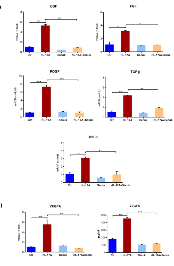
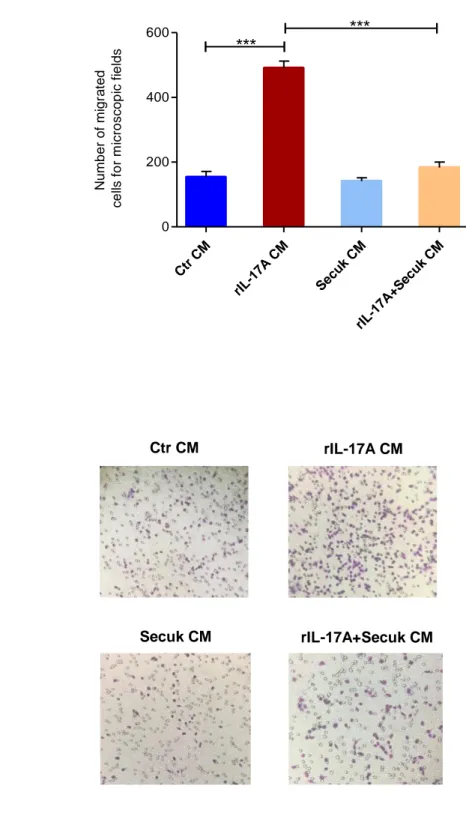
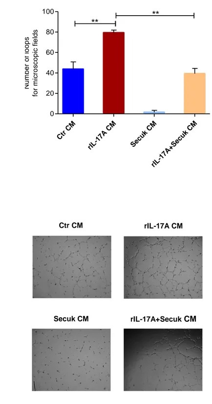
Results indicate that Secukinumab downregulates VEGFA mRNA and its soluble levels in human keratinocytes (HaCaT) treated with IL-17A. Conditioned medium from keratinocytes previously exposed to Secukinumab significantly decreases cell proliferation and migration of endothelial cells and inhibits capillary tube formation in vitro. The use of Egr-1 siRNA downregulates IL-17A-mediated VEGF-A expression. Overall, these data suggest that Secukinumab indirectly inhibits keratinocytes angiogenic property by interfering with IL-17A/Egr-1/VEGF-A axis.
As what concern the study of keratinocyte-derived EVs, results indicate that the treatment with IL-17A significantly modifies the EVs cargo and release. Vesicles from IL-17A-treated
7 cells display a specific pattern of mRNA which is abrogated by Secukinumab. Further, EVs are taken up by acceptor cells irrespective of their content but only those derived from IL-17A-treated cells enable recipient cells to express psoriasis-associated mRNA. These results imply a role of EVs in amplifying the pro-inflammatory cascade induced in keratinocytes by pro-psoriatic cytokines.
8 1. INTRODUCTION
1.1 PSORIASIS
Psoriasis is a chronic systemic inflammatory disease, with prevalent cutaneous involvement, characterized by a chronic relapsing course (Nestle FO et al., 2009). It is spread all over the world with a prevalence that varies considerably among the different populations and the various ethnic groups considered, in relation to the various geographical, environmental and genetic factors (Christophers E, 2001). Psoriasis seems to be most prevalent in Caucasian population and less frequent in Asian individuals and in black people (Gelfand JM et al., 2005; Schafer T, 2006).
In the USA, the prevalence of psoriasis has been estimated between 2.2% and 2.6%; in contrast, in Japan, the prevalence is lower and practically absent in Australians and Indians from South America. African Americans have an approximately 52% reduction in the prevalence of psoriasis compared with Caucasians (1.3% vs. 2.5%) (Saraceno R et al., 2008). In Europe the prevalence is higher in the Nordic countries than in the Mediterranean, probably due to climatic and environmental factors (Parisi R et al., 2013). The estimated prevalence of psoriasis in Southern Europe varies from 1.3% in Germany to 1.55% in Croatia and 1.58% in the UK (Saraceno R et al., 2008) (Figure 1).
9 Figure 1. Psoriasis. The prevalence of psoriasis worldwide (Greb JE et al., 2016).
In Italy the prevalence of psoriasis is about 2.9% of the population, with a notable non-homogeneous distribution among the various regions; in fact, the calculated range varies from 0.8% to 4.5% with reference to the various locations considered (Saraceno R et al., 2008). The regions that show a greater prevalence are Lazio, Abruzzo, Molise (4.5%) and Emilia Romagna (4%), while Sardinia (0.8%) is the region with the lowest prevalence, followed by Calabria, Puglia and Basilicata (1.6%) (Saraceno R et al., 2008) (Figure 2).
10 Figure 2. Psoriasis. Italian regions and percentage of people affected from psoriasis (Saraceno R et al., 2008).
The disease affects males and females equally, although it has been suggested that the onset of psoriasis is generally earlier in women than in men in some European countries (Parisi et al., 2013). Based on the age of onset, psoriasis is divided into two types (Griffiths CEM and Barker J, 2007):
type I psoriasis, present in about 75% of cases, is characterized by an early onset (before the age of 40) with a peak incidence between 16 and 20 years of age, high familiarity and a strong tendency to evolve towards a serious and/or generalized clinical form (Nevitt GJ and Hutchinson PE, 1996);
type II psoriasis, referred to as "late onset", with a peak incidence of around 50-60 years of age, a positive family history only in a limited number of cases and a less severe clinical course (Henseler T and Christophers E, 1985).
11 1.1.1 Clinical features and classification
The main clinical-pathological characteristic of psoriasis is the typical lesions consisting of chronic erythematous plaques, covered with ipercheratosic scales. Although lesions can be present throughout the body, they are most frequently located at the elbow, knee and scalp level (Nestle FO et al., 2009).
The plaque contains histopathological hallmarks that include a thickening of the epidermis (acantosis) caused by hyperproliferation of keratinocytes, hyperkeratosis and paracheratosis, or retention of nuclei in the stratum corneum; there is also a lengthening of the epidermal ridges downwards and an increase in the dilation and tortuousness of the vessels of the dermal papillae. At the level of the dermis and, to a lesser extent, the epidermis there is a rich inflammatory infiltrator, mainly consisting of T cells, myeloid and plasmacytoid dendritic cells, NK lymphocytes, macrophages and neutrophil granulocytes (Mahil et al., 2016).
The course is chronic, characterized by the alternation of acute phases and phases of remission difficult to predict. The disease can start silently for years until a stimulus of an undefined nature, sufficiently intense, determines the appearance of a psoriatic slick in the seat of injury.
Another typical, but not exclusive, feature of psoriasis is reactive isomorphism, which consists of the appearance of psoriasis patches in locations subject to mechanical stress (scarification, burns, surgical scars) (Pedace FJ et al., 1969).
From a clinical point of view, based on the form of the lesions and their localization, psoriasis can be distinguished in (Figure 3):
psoriasis vulgaris: is the commonest form of psoriasis, accounting for 90% of all cases; it is characterized by red skin lesions (plaques) covered by silvery scales and well-delineated from surrounding normal skin. Lesions appear as erythematous, a few centimeters thick and typically located on elbows and knees, but can extend and affect other skin areas (Griffiths CE et al., 2007);
guttate psoriasis: this acute form primarily affects adolescents and children; it is marked by the rapid appearance of small (less than 1 cm), punctiform and scaling lesions (papules), scattered with regularity and symmetry over the entire surface of the skin, preferring the area of the trunk and face. It is generally triggered by a bacterial infection, usually of streptococcal origin (group A β-haemolytic streptococcus), such as tonsillitis or pharyngitis, or a viral
12 infection (Martin BA et al., 1996; Griffiths CE and Barker J, 2007). Guttate psoriasis is self-limiting, resolving within 3–4 months of onset, although its long-term prognosis is unknown. One study indicated that only a third of individuals with guttate psoriasis develop classic plaque disease (Martin BA et al., 1996);
pustular psoriasis: there are three main forms;
- generalized pustular psoriasis (von Zumbusch psoriasis) is an acute form in which small pustules develop in painful inflamed skin. The patient can also have fever, chills, severe itching and diarrhea (Gooderham MJ et al., 2019);
- palmo-plantar pustulosis, consisting of yellow-brown pustules on palms and soles. About 25% of people with palmoplantar pustulosis also have chronic plaque psoriasis. Patients are predominantly women (9:1 female:male ratio) and either current or previous smokers (95%) (Kubeyinje EP and Belagavi CS, 1997);
- Hallopeau's Acrodermatitis that occurs on the fingertips, on the peri-ungueal region, while the involvement of the nail matrix determines the complete loss of the nail. It is associated with an impairment of the general state with malaise, fever and asthenia (Baran R, 1979);
erythrodermic psoriasis: the least common type of psoriasis; the whole body surface is affected by psoriasis, which can lead to hypothermia, hypoalbuminaemia, and high-output cardiac failure. Erythroderma can be caused by other diseases, including atopic dermatitis, drug eruptions, and cutaneous T-cell lymphoma. (Griffiths CE and Barker J, 2007);
inverse psoriasis: is a form of psoriasis that selectively involves the folds, recesses, and flexor surfaces such as the ears, axillae, groin folds, inframammary folds, navel, intergluteal cleft, genitals, lips, and webspaces. Inverse psoriasis causes smooth patches of red, inflamed skin that worsen with friction and sweating. Fungal infections may trigger this type of psoriasis. (Griffiths CE and Barker J, 2007);
nails psoriasis: a type of psoriasis that affect fingernails and toenails, causing pitting, abnormal nail growth and discoloration. Psoriatic nails might loosen and separate from the
13 nail bed (onycholysis) (Farber EM and Nall ML, 1974). This form is closely associated with artopathic psoriasis (Griffiths CE and Barker J, 2007);
psoriatic arthritis: is a seronegative inflammatory arthritis that occurs in the presence of psoriasis. Five types of psoriatic arthritis have been proposed distal interphalangeal joint only; asymmetrical oligoarthritis; polyarthritis; spondylitis and arthritis mutilans. Classic psoriatic arthritis consists of oligoarthritis, distal interphalangeal joint involvement, dactylitis and calcaneal enthesitis. Recently presented data indicate that its prevalence has been greatly underestimated, and may be as high as 25% in people with psoriasis. In about 10% of people with psoriatic arthritis, the arthritis appears before skin manifestations of psoriasis (Moll JMH and Wright V, 1973; Gladman DD et al., 2005; Helliwell PS and Taylor WJ, 2005).
Depending on the severity (extension on the body surface), psoriasis can be considered mild in the presence of a limited number of plaques, typically less than 3% of the skin surface is affected, moderate if 3 to 10% of the skin surface is occupied by plaques, and severe when more than 10% of the skin surface is affected (Meier M and Sheth PB, 2009).
14 Figure 3.Psoriasis. The different types of psoriasis. (A) plaque psoriasis; (B) guttate psoriasis; (C) pustular psoriasis; (D) reverse psoriasis; (E) erythrodermic psoriasis; (F) onychopsoriasis; (G) psoriatic arthritis (modified from Nestle FO et al., 2009; Boehncke WH and Schon MP, 2015).
1.1.2 Comorbidities
Several diseases have a higher incidence in psoriasis patients than the unaffected population, including Crohn's disease (Cohen AD et al., 2009), type 2 diabetes mellitus (Lee MS et al., 2014), metabolic syndrome (Arias-Santiago S et al., 2012), cardiovascular disease (Griffiths CE and Barker JN, 2007), depression and some cancers. A recent study showed that psoriasis patients are three times more likely to develop lymphomas than the unaffected population (Gelfand JM et al., 2006). An increased risk of developing skin cancer (melanoma and non-melanoma) in psoriasis patients has not been proven, but a 14-fold increased risk of developing squamocellular carcinomas has been observed in patients of phototype I-II who received 250 UV phototherapy (PUVA) sessions compared to patients who received the least number of treatments (Griffiths CE and Barker JN, 2007). Treatments with metotrexate and
15 cyclosporin in high doses can also be associated with the development of tumors ( De Oliveira Mde F et al., 2015).
The relationship between psoriasis, diabetes and cardiovascular disease is increasingly emerging Studies in psoriasis patients have found an increase in plasma levels of different inflammation markers, such as pro-inflammatory cytokines, or thrombosis regulators, suggesting that psoriatic pathology is associated with a state chronic flogosis (Fernández-Armenteros JM et al., 2019). This condition can lead to the onset of insulin resistance which, at the level of endothelial cells, leads to a reduction in the release of vasodilatory factors such as nitrogen monoxide. These events lead to the development of a state of endothelial dysfunction with increased expression of adhesion molecules, providing the basis for the development of ateroscletoric plaques and thus increasing the risk of cardiovascular pathology. Among the biomarkers identified in the plasma of patients and related to endothelial dysfunction are the soluble form of cell adhesion molecules VCAM-1 and E-selectine. To date the clinical relevance of these markers remains, however, unclear (Steyers CM and Miller FJ, 2014).
1.1.3 Pathogenesis and risk factors
The cause of psoriasis isn't yet fully understood; it is believed that clinical manifestations are determined by the combination of predisposing genetic factors and environmental triggers; among these, an important role is played by skin traumatism, infectious processes and some drugs (Kavli et al., 1985; Elder JT et al., 2009).
Its pathogenesis is therefore multifactorial and is conditioned by an abnormal immune response, probably triggered against an epidermal antigen (Lowes MA et al., 2007). Since 1970 it has been thought that the massive presence in psoriasis patients of immune system cells, especially dendritic cells and T lymphocytes, suggested a possible pathogenic role for the disregulation of the immune system in psoriasis (Nestle FO et al., 1994).
Although the literature data do not provide sufficient evidence that psoriasis is a real autoimmune disease, it shares with other immune-mediated diseases, such as Crohn's disease and diabetes mellitus, some characteristic manifestations chronic inflammation in the absence of a known antigen (Davidson A and Diamond B, 2001). For these reasons, psoriasis lesions
16 are believed to evolve from the interaction between cells and innate and adaptive immunity mediators and skin connective and epithelial tissues (Schon MP and Boechncke WH, 2005). Several pathogenic hypotheses have been formulated to explain the origin and clinical expression of psoriasis. Among the various theories, the most accredited is that psoriasis depends on an altered response of psoriasis keratinocytes to disparate inflammatory stimuli, which originate from the intervention of innate immunity cells, such as dendritic cells, polymorphucleates, mastcells, NK cells, and from adaptive immunity cells, such as CD4+ and CD8+ T-cells (Liu et al., 2007). Under normal conditions, there are, at the dermal level, a small number of T cells and CD11+ cells, while at the epidermal level Langerhans cells are present evenly in the various layers. In the psoriatic lesion, there is an increase in CD11+ dendritic cells and CD8+ T lymphocytes, which migrate at the epidermis level, respectively, at the basal layer or throughout the area. Langerhans cells continue to be present in the epidermis, but migrate at the spinosum layer level, while neutrophils accumulate at the stratum corneum level forming small aggregates (Liu et al., 2007).
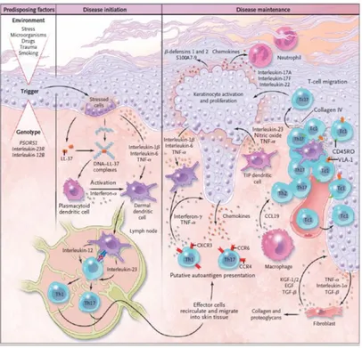
One of the pathogenic mechanisms for the development of psoriatic lesion involves, as the first event, the formation of a complex consisting of DNA fragments released by stressed or dying keratinocytes and the peptide LL-37 (Figure 4). This complex is able to activate plasmacytoid dendritic cells (pDCs) to produce IFN- α (Lande R et al., 2007).
LL-37 belongs to the family of catelicides, which relate to the class of antimicrobial peptides. These molecules are able to promote the elimination of pathogenic microorganisms and to modify the inflammatory response. In psoriasis lesions, three types of peptides are overexpressed: catelicide, β defensine and S100 proteins (Morizane S et al., 2012).
After the formation of the DNA-LL37 complex, the IFN-α produced by pDCs and the interleukin IL-1β, IL-6, the "tumor necrosis factor-α" (TNFα) produced by the keratinocytes activate the dermal dendritic cells (DDCs) that in turn produce IL23A, nitric oxide (NO) and TNFα (Zaba LC et al., 2009).
DDCs migrate to the lymph nodes, where they present the antigen to naive T cells promoting differentiation inTh17, Th1 and Th22. These cells express on their surface the receptors for the cytokines CCR4 and CCR6, which bind CCL17 and CCL20 respectively, and the "cutaneous lymphcyte-associated antigen" (CLA), a surface glycoprotein capable of binding E-selectin (Yamanaka K et al., 2008).
Through the lymphatic and blood vessels, Th17 and Th22 lymphocytes arrive at the level of the psoriatic lesion in presence of chemotactic factors CCL20 and CCL17 released by
17 keratinocytes. IL23A, produced by activated dendritic cells, induces Th17 and Th22 to release IL-17A , IL-17F, IL-22 and IFN-a. IL-17A and IL-17F stimulate keratinocytes to produce chemokines capable of recruiting neutrophils, including CCL17, CCL20, CXCL8, CXCL10, and antimicrobial peptides (LL-37 and the S100 family). (Wilson NJ et al., 2007).
CD8 T lymphocytes, which express on their Surface VLA-1 that can bind VCAM-1 expressed by endothelial cells, accumulate in the epidermis and produce IL-17 (Laggner U et al., 2011). Th1 and Th22 reach the inflammatory site and induce the production, in synergy with the Th17, of IL-22, responsible for epidermal hyperplasia, altering the differentiation of keratinocytes, with a possible contribution of IL17A (Di Meglio P et al., 2011).
Figure 4. Psoriasis. Pathogenetic model of psoriasis (Di Meglio P et al., 2011).
The greatest risk factor for psoriasis is the positive family history for the disease (Farber EM et al., 1974). It is estimated that about 30% of psoriasis patients have at least one family member with the same condition (Andressen C and Henseler T, 1982). This highlights a strong genetic component in the etiology of psoriasis.
In recent years linkage analysis studies have led to the mapping of 15 gene loci, distributed on different chromosomes, that are associated with psoriasis susceptibility, numbered 1 to 15 (Table 1). PSORS (psoriasis susceptibility) loci correspond to regions in which localize genes
18 involved in inflammatory processes or genes involved in the differentiation phases of epidermis.
Table 1. Psoriasis. PSORS loci with cytogenetic location, genomic coordinates and candidate genes (Singh et al., 2019).
The first of these loci, the PSORS1 locus, is the major genetic determinant of psoriasis which is confined to ~300 kb region in the major histocompatibility complex (MHC) (Russell TJ et al., 1972; Nair et al., 2006; Liu Y et al., 2007). The SNPs detected through genomic association studies revealed about fifteen genes (DDR1, DPCR1, MUC21, MUC22, HCG22, C6orf15, PSORS1C1, CDSN, PSORS1C2, CCHCR1, TCF19, POU5F1, PSORS1C3, HCG27, HLA-C) in the 300-kb critical region of PSORS1 are strongly associated with psoriasis (Capon et al., 2002). The association with the locus HLA is responsible for 30-50% of cases of the disease (Trembath et al., 1997, Nair RP et al., 2006). Fourteen PSORS regions (PSORS2-15) outside the MHC are reported to be associated with psoriasis. PSORS2 is associated with psoriatic patients having family history (Nair et al., 1997; Enlund et al., 1999;). PSORS3 locus represents psoriasis associated SNPs in IL2 and IL21, that takes part in T lymphocytes proliferation, Th17 differentiation, and keratinocyte proliferation (Liu et al., 2008). PSORS4 genes mainly participate in keratinization process and expressed in upper strata of the epidermis. Mutations in PSORS4 genes are reported to have high probability of triggering psoriasis vulgaris (PsV) (Capon F et al, 2001; Hüffmeier et al., 2009).
SNPs in SLC12A8 that encode a potassium/chloride transporter, is reported to be associated with psoriasis in PSORS5 locus (Hewett et al., 2002). SNPs in PSORS6 locus enhances risk of early-onset psoriasis vulgaris (Hüffmeier et al., 2009). IL23R with 19 psoriasis associated
19 SNPs, at PSORS7, has been established as one of the major susceptible gene that can trigger psoriasis (Di Meglio and Nestle, 2010). PSORS8 is reported as an overlapping susceptibility locus for both psoriasis and Crohn disease (Nair et al., 1997). The locus PSORS9, PSORS10,PSORS11 and PSORS12, are not reported to show significant association towards a distinct clinical subtype of psoriasis. As that of PSORS4, mutations in PSORS13 locus were observed to be associated with psoriasis vulgaris (Ellinghaus et al., 2010). The locus PSORS14 and PSORS15 have high association with pustular psoriasis cases (Setta-Kaffetzi et al., 2014).
There are various type of environmental factors that act on the polygenic substrate; those most involved are traumatic, infectious, pharmacological, endocrine-metabolic, alimentary and psychological (Barker J, 1991; Enamandram M and Kimball AB, 2013). Any physical trauma, such as burns and scars, can cause psoriatic lesions to appear in affected sites in predisposed persons. The phenomenon of reactive isomorphism, which implies the appearance of a psoriatic lesion on apparently healthy skin following traumatic insults, is an example of the role played by traumatic factors on the development of psoriasis. Generally it occurs within two weeks of the traumatic event at the affected site but the latency period can be even shorter (3 days) or much longer (even a year) (Weiss G et al., 2002).
Even the sunrays, which usually improve the clinical picture of most patients, in some subjects (especially those with phototype I and II) can cause an aggravation of psoriasis acting as a traumatic factor. In fact, photosensitive psoriasis is defined as the form of dermatosis in which there the appearance of new lesions following sun exposure (Rutter KJ et al., 2009). The incidence of photosensitive psoriasis varies from 14% to 24% depending on the different studies, while the phototype represents the most important factor of susceptibility (Nalluri R et al., 2010).
Infections, not only affecting the skin, but also internal organs are important elements that can trigger a latent psoriasis. The role of streptococcal infections in the pathogenesis of guttate psoriasis is known, which occurs within 15 days of disease onset (McFadden et al., 2009). More rarely, exanthematous diseases (measles and chickenpox) and HIV can be considered triggers (Naldi L et al., 2005; Patel RV and Weinberg JM, 2008).
Even some drugs can be a risk factor, that can interfere with psoriasis in various ways: aggravating a pre-existing psoriasis, causing the appearance of lesions on previously uninvolved skin areas, causing the appearance of psoriasis ex novo, favoring a resistance to treatment (Milavec-Puretić V et al., 2011 ). Among the most frequently implicated drugs are
20 antimalarials, lithium, β-blockers and nonsteroidal anti-inflammatory drugs (NSAIDs) (Basavaraj KH et al., 2010).
As far as endocrine factors are concerned, the existence of peaks of incidence at puberty and in menopause has always suggested the interference of phenomena of hormonal nature. Psoriasis may aggravate during estrogen intake and in the pre-menstrual period (Hall G and Phillips TJ, 2005).
There is numerous clinical evidence regarding the influence of metabolic disorders on the appearance or aggravation of psoriasis. In particular, dyslipidemias and obesity have considerable effects on skin disease (Naldi L et al., 2005).
Anxiety and stress are very often triggers of psoriasis that alter immune mechanisms (Raychaudhuri SP and Gross J, 2000). It is widely recognized that stress plays an important role as a trigger for psoriasis both in the first manifestation and as an aggravation of an already existing form (Heller MM et al., 2011). Stressful events can worsen psoriasis severity and even extend the period of exacerbations. The percentage of psoriatic subjects who think that stress alters the condition of their skin (stress-responders) is particularly high: from 37% to 78% (Basavaraj KH et al., 2011). Numerous studies have confirmed a higher alcohol consumption in the psoriatic population. Alcohol abuse not only acts as a triggering factor, but also as an aggravating factor in the disease, causing a greater extension of the lesions with a more evident inflammatory component (Jankovic et al., 2009).
Finally, it is estimated that in about 25% of psoriatic patients the disease is triggered by cigarette smoking, which has been particularly associated with palmar-plantar pustular psoriasis (Naldi L et al., 2005; Jin Y et al., 2009). There is a statistically significant dose-response association between the number of cigarettes smoked and the risk of disease, with a more consistent correlation for females (Setty AR et al., 2007).
1.1.4 Diagnosis
The diagnosis of psoriasis is mainly carried out on clinical observation which takes into consideration the appearance of the skin lesions, with particular attention to the color of the lesions, the size, the morphology, the distribution of the different body regions involved. The PASI index (Psoriasis Area and Severity Index) is used to monitor the pathology through the evaluation of some parameters, and combines the assessment of the severity of lesions and
21 the area affected into a single score in the range from 0 (absence of psoriasis) to 72 (severe psoriasis).
The body is divided into four sections: head (H) (10% of skin surface); arms (A) (20%); trunk (T) (30%); legs (L) (40%). For each area, the percent of area of involved skin is estimated and then transformed into a grade from 0 to 6:
0. 0% of involved area 1. < 10% of involved area 2. 10–29% of involved area 3. 30–49% of involved area 4. 50–69% of involved area 5. 70–89% of involved area 6. 90–100% of involved area.
Within each area, the severity is estimated by three clinical parameters: erythema (redness), induration (thickness) and desquamation (scaling). Severity parameters are measured on a scale of 0 to 4, where 0 indicates a complete lack of skin involvement and 4 represents the highest possible involvement. The sum of all three severity parameters is then calculated for each section of skin, multiplied by the area score for that area and multiplied by weight of respective section (0.1 for head, 0.2 for arms, 0.3 for body and 0.4 for legs) (Oji V et al., 2015).
Although this is a validated and widely used method, recommended for attributing a shared degree of psoriasis severity to both scientific and clinical applications, it has a very low sensitivity. This limit is particularly evident in the case of mild or moderate intensity diseases, with a low percentage of the body surface area (BSA) involved, and does not take into consideration the symptoms that affect the hands, nails, feet, face, or genitals . The European Medicine Agency (EMA) recommends the use of PASI in combination with the PGA index (Physician Global Assessment), which uses a 4 to 10 point scale, evaluating erythema, plaque extension and injuries involved (Robinson A et al., 2012).
Psoriasis can be considered mild, in the case in which less than 3% of the skin surface is affected, moderate, if a surface is involved from 3 to 10%, and severe, when it extends over 10% (Meier M and Sheth PB, 2009).
22 1.1.5 Treatments
In the treatment of mild and moderate psoriasis, topical drugs containing various active ingredients are often used, including corticosteroids, analogues of vitamin D and retinoids ie.
metabolites derived from vitamin A.
Glucocorticoids belong to the corticosteroid family, a group of hormones produced by the cortex of the adrenal glands. These molecules penetrate the cell by passive diffusion and bind to cytosolic glucocorticoid receptors (GC) forming a glucocorticoid-receptor complex that translocates to the nucleus and interacts with specific nuclear sequences, glucocorticoid responsive elements (GRE), regulating the expression gene. The anti-inflammatory action of glucocorticoids is caused by the activation of several genes such as IκB (NF-κB inhibitor), DUSP 1, also known as mitogen activated protein kinase (MAPK) phosphatase 1, IL-10, glucocorticoid-induced leucine zipper (GILZ) and annexin A1 (Spies CM et al., 2011). All these genes are involved in the inhibition of proinflammatory factors or in the expression of anti-inflammatory molecules (Guichard A et al., 2015). Following treatment with corticosteroids there is the appearance of local secondary effects due to the application and systemic effects due to drug absorption. The local effect is represented by cutaneous atrophy caused by the inhibition of proliferation, mitosis and protein synthesis in dermal fibroblasts causing cell cycle arrest and induction of apoptosis (Hein R et al., 1994; Amsterdam A et al., 2002). Among the systemic effects there is a decrease in the concentration of the corticotropin-releasing hormone (CRh) and of the adrenocorticotropic hormone (ACTH). Following absorption, corticosteroid levels increase in the blood leading to the appearance of clinical signs of Cushing's syndrome (Castela E et al., 2012).
Calcipotriol is an analogue of vitamin D that is used in psoriatic therapy alone or in combination with betamethasone (steroid drug). The pharmacological target is the vitamin D receptor (VDR) expressed by the keratinocytes present in the basal layer of the epidermis and, following the receptor binding, inhibits proliferation and normalizes keratinocyte differentiation (Naga Sravan Kumar Varma V et al., 2014). Several clinical studies have demonstrated an excellent efficacy and safety profile of this compound in the treatment of this pathology (Trémezaygues L and Reichrath J, 2011).
Tazarotene is a synthetic retinoid, recently introduced for the treatment of mild psoriasis; is a prodrug that is rapidly hydrolyzed in the active form that binds receptors for retinoic acid (RARs) by normalizing keratinocyte differentiation and proliferation. Although able to induce
23 a remission of the psoriatic lesion, its use is limited by the irritant action, which exerts both on healthy skin and on the plaque itself.
Among the various therapeutic options, phototherapy for psoriasis may be indicated in particular cases of the disease.
Narrowband ultraviolet B (UVB) rays are often used in psoriasis patients. The treatment involves exposure to artificial UVB rays, naturally present in sunlight. Applications are administered several times during the week and the therapy cycle can last a long time, up to twenty or thirty applications.
Another source of ultraviolet light used for the treatment of psoriasis is PUVA, which involves the association of ultraviolet radiation A (UVA) with the use of psoralens taken orally which sensitize the skin, making it more prone to treatment. The mechanism of action of the PUVA therapy is not well known, but probably determines the activation of the psolarenes, which inhibit cell reproduction. Treatment with PUVA and psolarenes is associated with an increased risk of skin cancer, especially malignant melanoma and squamous cell carcinoma, which is why PUVA therapy cannot be used in the long term.
Systemic treatment is reserved for subjects with severe or particularly extensive forms, or for those patients in whom topical or phototherapeutic treatments have proved ineffective. Systemic therapy for the treatment of psoriasis uses retinoids, methotrexate, cyclosporine A, mofetil mycophenolate and tacrolimus.
Several retinoids have been used in the treatment of psoriasis. Currently, acitretin is used, which has favorable pharmacokinetic properties, the most important of which is the shorter half-life. Acitretin has a marked effectiveness in pustular and erythrodermic psoriasis. When this drug is used on its own, the daily dosage varies from 25 to 50 mg; treatment with dosages of 10 to 20 mg is started and increased gradually (if necessary). Although complete remission is only achieved in 50% of cases, the majority of patients see the number of plaques, their extension or flaking decrease dramatically. Higher doses of Acitretin (50-75 mg/day) are more effective in less time, but are associated with more important side effects. Like all retinoids, Acitretin is also teratogenic, so it is necessary to adopt safe contraceptive measures in women of childbearing age. These must be maintained for three years after treatment has been suspended. The most easily observed side effects are dryness of the lips, nose, eyes and the thinning and fragility of the nail plates. Hyperostosis and tendon calcifications have been reported.
24
Methotrexate inhibits the enzyme dehydrofolate reductase, which is necessary for the synthesis of nucleotides and amino acids. In this way, the drug reduces DNA synthesis and inhibits mitoses, especially of rapidly proliferating cells. It is able to alter lymphocyte and neutrophil. Methotrexate should not be administered in patients with active infection, alcoholism, cirrhosis, viral hepatitis, immunological defects, renal failure and in women who do not take contraception measures.
Side effects can range from simple nausea to cancer. The recommended starting dose is 7.5 mg once a week. The use of methotrexate for the treatment of psoriasis is today less frequent than in the past.
Ciclosporin is an immunosuppressant, which inhibits the production of interleukin 2 and therefore the immune response mediated by T cells. The initial dosage is 2.5-4 mg/kg/day, divided into two doses. Given the side effects of the drug, it is necessary to monitor both blood pressure, renal function and bone marrow function. In fact, the major side effects are nephrotoxicity and hypertension, as well as hirsutism and gingival hyperplasia. Unfortunately, in addition to the side effects during treatment with Cyclosporine, the suspension usually leads to a reignition of the disease with worsening and difficulty in responding to other treatments.
Mofetil mycophenolate is able to prevent transplant rejection being an immunosuppressive agent; it has been used in the treatment of psoriasis. The recommended dose is 1 gr for 2 times a day or 500mg for 4 times a day. This drug is generally well tolerated and has a good safety profile. The main side effects are gastrointestinal disorders, anemia, leukopenia and onset of infections.
Tacrolimus has a mechanism of action very similar to that of cyclosporine and is used at a dosage of 0.1-0.15 mg/ kg/day. The most common side effects are paresthesia and diarrhea.
The new biological agents, that act on the various steps of the autoimmune-inflammatory process, offer an important alternative in the treatment of moderate to severe plaque psoriasis in adult patients who have not responded, or who have intolerance or contraindications to other systemic therapies, including cyclosporine, methotrexate and PUVA therapy (Table 2). Pro-inflammatory cytokines such as TNF-α play a central role in the pathogenesis of psoriasis. High levels of TNF-α have been found in psoriatic plaques. In plaque psoriasis, the infiltration of inflammatory cells, including T cells, leads to an increase in TNF levels in psoriatic lesions compared to levels in unaffected skin. (Gearing AJ et al., 1990). This explains why anti-TNF-α, such as Etanercept, Infliximab and Adalimumab, led to an
25 improvement in the treatment of the disease. Etanercept is a fusion protein of p75 tumor necrosis factor (TNF) receptor (p75TNF-R) with the Fc fraction of human IgG1 immunoglobulin. The protein act as a soluble receptor for TNF-α; it is a competitive inhibitor of TNF-α binding to its surface cellular receptors, p55 and p75, and therefore inhibits the biological activity of TNF. Etanercept is indicated for plaque psoriasis, psoriasis arthritis, rheumatoid arthritis and ankylosing spondylitis (Rønholt K and Iversen L, 2017). Infliximab is chimeric human-murine monoclonal antibody that binds with high affinity to both the soluble and trans-membrane forms of TNFα, inhibiting its activity. It is currently approved for the treatment of rheumatoid arthritis, Crohn's disease, ankylosing spondylitis, psoriatic arthritis and psoriasis (Laws PM and Young HS, 2012).Adalimumab is a recombinant human monoclonal antibody produced in Chinese Hamster Ovary cells. It selectively binds to TNF and neutralizes its biological function by blocking its interaction with cell membrane TNF receptors. The drug has been approved for use in psoriasis and psoriatic arthritis (Laws PM and Young HS, 2012).
Recently, a new class of biological drugs blocking both interleukins (IL)-12 and 23 was identified. This cytokines induce CD4+ naϊve lymphocytes to differentiate into T helper1 (Th1) and T helper17 (Th17) lymphocytes respectively. Ustekinumab is a fully human IgG1κ monoclonal antibody that binds the p40 protein subunit of human cytokines IL-12 and IL-23 with high affinity and specificity, inhibiting its activity and preventing binding of these cytokines with the respective receptor protein IL-12Rβ1 expressed on the surface of immune cells. Ustekinumab currently can be used in psoriasis and in psoriatic arthritis treatment (Thibodaux RJ et al., 2018). Currently there are no strong criteria to guide the choice between anti-TNF drugs available and between Etanercept, Infliximab, Adalimumab and Ustekinumab. However, the Etanercept vs Ustekinumab comparison suggests that Ustekinumab shows greater efficacy to Etanercept over a 12-week period (Sterry et al., 2004).
26 Tab.2 Psoriasis. Biologics approved for psoriasis by the United States Food and Drug Administration
27 1.2. SECUKINUMAB: THE NEW ANTI-IL-17A BIOLOGIC AGENT
IL-17A is a dimeric glycoprotein and belongs to a family of cytokines family that includes other five members, i.e. IL-17B, IL-17C,IL-17D, IL-17E and IL-17F, which are involved in inflammatory disorders and autoimmune diseases such as psoriasis and cancer (Kolls & Linden 2004). IL-17A was first described in 1993 in human peripheral blood, as an important pro-inflammatory cytokine with a critical role against extracellular microorganisms and in the pathogenesis of different autoimmune diseases (Rouvier E et al., 1993). Within the IL-17 family, IL-17A and IL-17F are central players in the adaptive immune response, particularly against bacteria and fungi while the function of IL-17B, IL-17C and IL-17D is less understood (Kolls JK and Lindén A, 2004; Iwakura Y et al., 2011). The induction and production of IL-17A during CD4+ or CD8+ T-cell differentiation is up-regulated by TGF-β, IL-6, IL-21 and IL-23 (Heidenreich et al. 2009). Specialized Th17 cell subsets of the adaptive immune response are characterized as main sources of IL-17A in vivo (Kirkham BW et al., 2014). 17A exists as either a homodimer of two 17A chains or a heterodimer with IL-17F. IL-17A and IL-17F signal through the same receptor subunit (IL-17RA and IL-17RC). However, IL-17A is approximately 10-30 times more potent than IL-17F in activating gene expression, cause to different ligand-receptor affinities (Gu C et al., 2013). IL-17A plays a critical role in the pathogenesis of a range of inflammatory diseases, including psoriasis, rheumatoid arthritis, psoriatic arthritis, autoimmune uveitis (Kirkham BW et al., 2014); it is known that this cytokine is elevated in lesions of psoriasis and in the serum of patients (Raychaudhuri SP, 2013; Chiricozzi et al., 2014). IL-17A has many functions that are relevant to psoriasis, including the direct activation of keratinocytes leading to increased production of other inflammatory mediators and the enhancement of angiogenesis (Marinoni et al., 2014). Secukinumab is a human monoclonal IgG1k antibody that has been developed to target and block the actions of IL-17A. Secukinumab was developed by Novartis and the first publication was a Phase I trial published in 2010 (Frieder J et al., 2018). In January 2015, the FDA (Food and Drug Administration) approved Secukinumab to treat adults with moderate-to-severe plaque psoriasis (Sanford M and McKeage K, 2015; Rønholt K and Iversen L, 2017). It was the first IL-17A inhibiting drug ever approved. In January 2016, the FDA approved it to treat adults ankylosing spondylitis and psoriatic arthritis (Roman et al., 2015). The recommended dose is 300 mg by subcutaneous injection (into the front of thighs, lower
28 abdomen or outer upper arms but not into areas of skins that is affected by psoriasis) at week 0, 1, 2, 3 and 4 followed by 300 mg every four weeks. The mean half-life range from 22 to 31 days. At the therapeutic concentrations used in psoriasis, Secukinumab fully neutralizes the activity of IL-17A, does not neutralize IL-17F, leaves other function of TH17 cells intact and does not directly influence the TH1 pathway. This specific mechanism of action is unique to IL-17 inhibitors and leads to the normalization of skin histolgy, including achievement of clear to almost clear skin for the majority of patients (Garnock-Jones KP, 2015).
In clinical studies, Secukinumab has demonstrated the achievement and maintenance of skin that is free or almost free of lesions (from PASI 90 to PASI 100) in 80% of patients. During the studies, 70% of patients treated with Secukinumab obtained the resolution complete (PASI 100) or almost complete (PASI 90) of cutaneous manifestations after the first year of treatment. During Phase III clinical trials, 70% or more of patients receiving Secukinumab 300 mg achieved complete (PASI 100) or near complete (PASI 90) resolution of cutaneous manifestations during the first 16 weeks of treatment (Hueber W et al., 2010; Godse K, 2017).
29 1.3 THE ROLE OF ANGIOGENESIS IN PSORIASIS
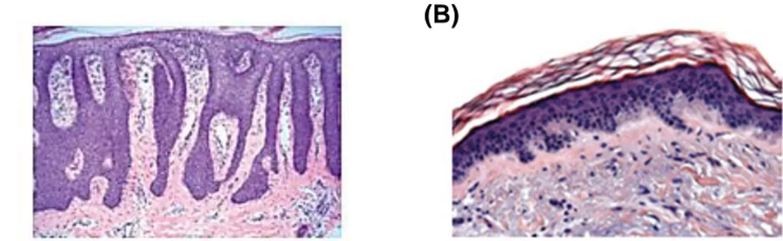
The formation of new capillaries from pre-existing blood vessels is described as angiogenesis. This process is tightly regulated by a balance between pro- and anti-angiogenic mediators. Physiological angiogenesis is induced only transiently during processes such as wound healing or pregnancy. Dysregulated angiogenesis occurs under pathological conditions such as tumor growth and chronic inflammation, as observed during psoriasis. In this condition, angiogenesis is needed for disease development and progress (Folkman 1995; Kneilling et al. 2007; Heidenreich et al. 2008). The dermis of patients with psoriasis presents peculiar histological features, including elongated, tortuous, blood capillaries, increased in diameter and number (Braverman & Sibley 1982) (Figure 5). Vascular alterations occur prematurely and precede the characteristic epidermal hyperplasia of the disease (Schon & Boehncke 2005; Griffiths & Barker 2007).
Figure 5. The role of angiogenesis in psoriasis. Psoriasis histology. (A) H&E staining of psoriasis skin. (B) Healthy skin (Heidenreich et al., 2009).
As angiogenesis is one of the key features of psoriasis, various studies have been focused on the identification of pro-angiogenic mediators in psoriatic skin. It is of interest that the remodeling of the vasculature in lesional psoriatic skin depends considerably on factors derived from the epidermis, whereas blood vascular remodeling is essential for nutrient supply of the hyperproliferative epidermis. Evidence for keratinocyte-derived pro-angiogenic signals came from a study comparing the angiogenic activity of conditioned media from keratinocytes isolated from either lesional or non-lesional skin of psoriasis patients. Conditioned media from lesional or non-lesional keratinocytes stimulated endothelial cell migration in vitro and showed strong angiogenic activity in the rat cornea micropocket assay
30 in vivo. In contrast, conditioned media from keratinocytes of healthy donors showed no pro-angiogenic response (Nickoloff et al. 1994).
Several pro-angiogenic factors are up-regulated in psoriasis development, among them vascular endothelial growth factor (VEGF), hypoxia-inducible factors (HIFs), angiopoietins (ANGs), tumor necrosis factor (TNF)-α, interleukin (IL)-8, interleukin (IL)-17, transforming growth factor (TGF)-α e TGF-β (Elder et al. 1989; Christophers 1996; Starnes et al. 2001; Creamer et al 2002; Numasaki et al. 2003; Ghoreschi et al. 2007; Heidenreich et al. 2008). Combined action of these factors stimulate endothelial cells from dermis to form new blood vessels.
VEGFs are a class of secreted polypeptides structurally similar to those of the "platelet derived growth factor" family (PDGF). The family members expressed by mammalian cells are VEGF-A, VEGF-B, VEGF-C, VEGF-D and the placental growth factor (PGF). VEGFs and its tyrosine kinase receptors VEGFR-1 and VFGR-2 (KDR) are essentially involved in vascular embryogenesis and adult neovascularization. VEGF-A, PGF and VEGF-B act mainly at the level of blood vessels, while VEGF-C and VEGF-D are essential for lymphangiogenesis. The VEGF receptors VEGFR-1 or -2 are primary expressed by vascular endothelial cells (De Vries et al. 1992; Shibuya 1995; Ferrara et al. 2003; Shibuya & Cleasson-Welsh 2006).
VEGF-A is a major regulator of physiological and pathological angiogenesis. This factor was strongly up-regulated in psoriatic skin lesions. Indeed, although this angiogenic factor can be produced by mast cells, monocytes/macrophages and infiltrating neutrophils, in psoriatic lesions the primary source of VEGF-A is keratinocytes. Physiological production of VEGFA contributes to normal proliferation, differentiation and function of the epidermis, such as epidermal barrier homeostasis (Man et al. 2006; Elias et al. 2008); in psoriasis VEGFA overexpression might contribute to keratinocytes hyperproliferation in an autocrine manner and to the epidermal changes observed in this disease (Canavese et al. 2010). Experiments based on in situ hybridization techniques and immunohistochemistry have demonstrated an increase in VEGF-A expression, both at the transcriptional and translational level, in psoriatic keratinocytes, and an increase in the expression of its receptors tyrosine kinase at endothelial cell level in dermal papillae. In psoriatic skin, VEGFR-1 and VEGF-2 receptors are detectable and functional in keratinocytes. In addition to an increased expression in the skin, psoriatic patients show a high secretion of VEGF-A in the bloodstream and these elevated plasma (serum) levels are correlated with an early onset (esordio) of the disease, with disease severity
31 and with the subsequent development of psoriatic arthritis (Creamer et al. 1996; Bhushan et al. 1999; Nielsen et al. 2002). VEGFA pathologically increased secretion by keratinocytes are triggered by a wide variety of factors, including pro-inflammatory cytokines secreted by leukocytes and the tumor growth factor TGF-α found in high concentrations in patients with psoriasis (Detmar et al. 1994). In humans, some polymorphisms of the gene encoding VEGF-A, rs2010963 and rs833061, have been associated with early development of psoriasis. The VEGFA gene is in the vicinity of the PSOR1 gene on chromosome 6p21; however, there is no linkage disequilibrium between these two genes, suggesting that they are inherited independently (Young et al. 2004, 2006).
HIFs represent heterodimeric transcription factors composed of a constitutively expressed β subunit (aryl hydrocarbon receptor nuclear translocators: ARNT, ARNT2, ARNTL) and a regulatory α subunit (HIF-1α, HIF-2α, HIF-3α) (Harris 2002; Maxwell & Ratcliffe 2002; Wenger 2002). At physiological oxygen tension, HIF-α subunits are continuously synthetized and degraded by the proteasome. For degradation, prolyl residues of HIF-α subunits are hydroxylated by prolyl hydroxylase, which are active only in the presence of normal oxygen concentrations. Under hypoxic conditions, prolyl hydroxylases are inactive; in consequence, HIF-α subunits are no longer degraded and the increasing HIF concentrations lead to nuclear translocation. Among the HIF target genes are mains regulators of angiogenesis such as VEGF, VEGFR-1, VEGF-2 and IL-8 (Elvert et al. 2003; Takeda et al. 2004; Kim et al. 2006). In psoriasis lesions, HIF-1α and -2α expression are both increased (Rosenberger et al. 2007). In epidermal keratinocytes, HIF-1α colocalizes with VEGF expression, whereas HIF-2α is expressed in the epidermis and in dermal capillaries. Epidermal hypoxia and increased HIF expression may result from the strong epidermal proliferation (Tovar-Castillo et al. 2007). Angiopoietins are a family of secreted oligomeric proteins composed of 4 proteins, ANG1, ANG2, ANG3 and ANG4. ANG1 is widespread in the adult vascular system where it is mainly produced by pericytes that surround the endothelium of the vessels and, to a lesser extent, by monocytes and neutrophils. ANG2 is generally not expressed in healthy tissues, but is released following the presence of stimuli such as thrombin and histamine (Fiedler et al. 2004). ANG1 and ANG2 bind to the receptor tyrosine kinase Tie2. This receptor is ubiquitous at the level of the vascular endothelium, where it is constitutively phosphorylated in quiescent endothelial cells suggesting an active role in vascular homeostasis (Dumont et al. 1994; Davis et al. 1996) . Tie2 is also expressed by other cell types such as monocytes, neurons and tumor cells. ANG1 is an agonist of Tie2 and, following interaction with the receptor, induces a
32 protective response, inhibiting cell apoptosis, promoting both the migration of endothelial cells and smooth muscle cells (Davis et al. 1996). Moreover, ANG1 has a clear pro-angiogenic effect and an anti-inflammatory effect on endothelial cells, reducing the expression of E-selectin, of ICAM-1 and VCAM-1 induced by VEGF-A, thus inhibiting leukocyte diapedesis (Kim et al 2000). ANG2, on the other hand, is able to antagonize the effect of ANG1 at the receptor level (Yuan et al. 2009). Following inflammatory stimuli, ANG2 is released very quickly from pericytes and inhibits the ANG1-Tie2 bond in a competitive manner, destabilizing the extracellular cell-matrix contact and activating the expression of adhesion molecules for leukocytes (Moss 2013). In psoriasis, ANG1 is expressed at the level of fibroblasts and dendritic cells, while ANG2 is expressed at the level of endothelial cells. It has been demonstrated in transgenic mouse models that the over-expression of the ANG1 / ANG2 / Tie2 system is able to increase epidermal hyperplasia, hyperkeratosis, parakeratosis and induce an increase in vascularization at the dermal level. In particular, ANG2, synthesized by endothelial cells in response to inflammatory stimuli such as TNF-α, increases the expression of ICAM-1 and VCAM-1 at endothelial cell level, facilitating leukocyte adhesion and extravasation. In this way, ANG2 could contribute to the development of psoriasis (Heidenreich et al. 2009).
Several cytokines exhibit a profound impact on angiogenesis by influencing endothelial cells proliferation, migration or survival, or by modulating the expression of pro- or anti-angiogenic factors. Among the cytokines with pro-anti-angiogenic activity, TNF, IL-8 and IL-17 are overexpressed during psoriasis.
TNF induces various pro-angiogenic factors, such as VEGF, IL-8, bFGF, in endothelial cells and exerts both pro-angiogenic and anti-angiogenic effects (Yoshida et al. 1997). Elevated levels of TNF mRNA and protein are detectable in psoriasis skin (Johansen et al. 2006). Therapies blocking the activity of TNF lead to clinical improvement of psoriasis and decreased expression of pro-angiogenic factors. The data confirm that TNF contributes to angiogenesis associated with psoriasis. It remains open whether it directly causes angiogenesis or indirectly through the induction of pro-inflammatory or angiogenic factors. IL-8 or CXCL8 belongs to the CXC family of chemokines (Baggiolini et al. 1997). IL-8 is a strong chemoattractant for neutrophils, basophils and T lymphocytes, and is involved in autoimmune, inflammatory and infectious diseases (Brat et al. 2005). Enhanced 8 and IL-8 receptor mRNA and protein is detected within the epidermis of psoriatic lesions. Immunohistochemistry localizes IL-8 protein to suprabasal keratinocytes and neutrophils
33 (Duan et al. 2001; Gillitzer & Goebeler 2001). IL-8 is a strong chemoattractant for neutrophils, basophils and T lymphocytes, and is involved in autoimmune, inflammatory and infection diseases. IL-8 can be induced by IL-1, TNF, IL-6, IFN-γ, reactive oxygen species and other mediators of cellular stress (Strieter et al. 1992). IL-8 is also a potent pro-angiogenic factor (Koch et al. 1992); it has been described to stimulate endothelial cells proliferation, migration, survival and expression of MMPs (Koch et al. 1992; Szekanecz et al. 1994; Shono et al. 1996; Li et al. 2003). Moreover, IL-8 can stimulate keratinocyte proliferation (Tuschil et al. 1992). Various cell types are capable of producing IL-8, including immune cells such as mast cells, neutrophils or T cells, keratinocytes and EC (Nickoloff et al. 1994; Biedermann et al. 2000; Gillitzer & Goebeler 2001; Karl et al. 2005).
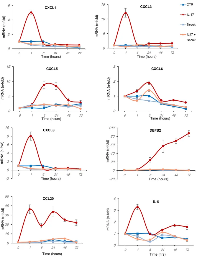
IL-17A is a pro-angiogenic factor; it can induces new vessel formation in the rat cornea micropocket assay and IL-17A overexpressing tumor cells can induce more rapid tumor growth with significantly enhanced tumor vascularization in vivo (Numasaki et al. 2003). Although it does not have a direct effect on the proliferation of endothelial vascular cells, IL-17 stimulates their migration and the production of capillary-like structure in vitro (Heidenreich et al. 2009). Moreover, IL-17A triggers the production of chemokines, growth factor and adhesion molecules by epithelial cells, fibroblasts, and EC including 6, 8, IL-1 and ICAM-IL-1, an adhesion molecule secreted by fibroblasts and keratinocytes. Thus IL-IL-17 enhances neutrophils accumulation. This interleukin is also capable of stimulating VEGF production by keratinocytes and fibroblasts. Consequently, by inhibiting the activity of IL-17A, Secukinumab inhibits angiogenesis indirectly reducing the production of VEGF by keratinocytes associated to vascular neo-formation.
34
Figure 6. The role of angiogenesis in psoriasis. Schematic representation of angiogenic signaling pathways (Heidenreich et al., 2009).
1.3.1 Angiogenesis as a potential target of novel therapies
Although angiogenesis is a central process in the evolution of psoriasis and is closely linked with the clinical manifestation, treatments aimed directly to the inhibition of this process have received little attention so far. Systemic therapies currently used in the management of psoriasis patients modulate the immune response but also have indirectly an additional anti-angiogenic effect. As an example, there is evidence that methotrexate or cyclosporine therapies, that interfere with immune activation, can indirectly inhibit/target important mediators of angiogenesis (pro-angiogenic mediators) targeting the VEGF pathway (Hirata et al. 1989; Hernandez et al 2001; Yamasaki et al. 2003). Anti-TNF-α treatments such as Infliximab, Etanercept and Adalimumab, that target a cytokine that exerts multiple effects on angiogenesis, proved to exhibit potent anti-angiogenic activities in the therapy of psoriasis (Vassalli 1992; Pandya et al. 2006). The epidermal expression of VEGF, Ang-1, An-2, MMP-9 decreases significantly during psoriasis therapy with the TNF-α antagonist Infliximab (Markham et al. 2006). The same antibody was shown to reduce the expression of VEGF and
35 its receptors during psoriasis arthritis (Cordiali-Fei et al. 2006). Similar effects occur with other TNF antagonists, Etanercept and Adalimumab, that reduces VEGF-A and thereby reduces blood supply at the site of the tissue damage and psoriatic inflammation (Wcislo-Dziadecka et al. 2015). In addition some traditional therapies are associated with a reduction in plasma VEGF-A levels and, among these, topical use of mineral tar in combination with UVB therapy.
Increasing experimental data show that directly blocking angiogenic pathways that drive cutaneous inflammation could represent a promising therapeutic strategy for psoriasis treatment. The antiangiogenic agent Neovastat may represent a new potential treatment modality in patients with moderate to severe psoriasis. Neovastat it is a VEGF antagonist and has been shown to inhibit endothelial cells proliferation in vitro and angiogenesis in vivo (Dupont et al. 2002). In a phase I/II clinical trial which involved 49 plaque psoriasis patients, was noted a statistically significant decrease in PASI score (Sauder et al. 2002). Moreover, Neovastat have an excellent tolerability. Further studies need to be performed before the drug is approved by FDA for the treatment of psoriasis. Despite the promising results obtained in pre-clinical murine models (Schonthaler et al. 2009), further studies are required before for a possible use of Bevacizumab, a humanized monoclonal antibody directed against VEGF-A and used in adjuvant therapy of colon cancer, also in psoriatic pathology (Weidemann et al. 2013). To date, only one case of a patient has been reported, subjected to Bevacizumab treatment for metastatic colon cancer, who has had a simultaneous and complete remission of his psoriasis (Akman A et al. 2009).
36 1.4 THE EARLY GROWTH RESPONSE (EGR)-1 IN THE PATHOGENESIS OF PSORIASIS
The early growth response (Egr)-1 is a zinc finger transcriptional factor (Sukhatme VP, 1990) which plays an important role in the regulation of cell growth, differentiation, cell survival and immune response (McMahon SB and Monroe JG, 1996; Qu Z et al., 1998; Sakamoto KM et al., 2004). Egr-1 is rapidly induced by a broad spectrum of extracellular signals including growth factor, cytokines and many physiologic stimuli (Thiel G et al., 2002). Egr-1 induces the expression of growth factors, growth factor receptors, extracellular matrix proteins, proteins involved in the regulation of cell growth or differentiation, and proteins involved in apoptosis, growth arrest, and stress responses (de Belle I et al., 1999; Virolle T et al., 2001; Krones-Herzig A et al., 2005; Zwang Y et al., 2011). Comparable to some other transactivators, Egr-1 associates with corepressor proteins that can modulate transcription of Egr-dependent genes. Two corepressors of Egr-1, Nab1 and Nab2, have been identified using yeast two-hybrid screening (Svaren J et al., 1996; Swirnoff AH et al., 1998). These factors bind to Egr-1 by direct protein-protein interactions, thus inhibiting the transactivating potential of Egr-1. Whereas Nab1 is constitutively expressed in most tissues and appears to be a general transcriptional regulator (Swirnoff AH et al., 1998), Nab2 may function as an important inducible regulator of gene expression (Svaren J et al., 1996).
Egr-1 was originally identified as a tumor suppressor gene in a variety of human tumor cell lines including osteosarcomas, fibrosarcomas (Huang et al., 1995), hepatocellular carcinoma, and esophageal carcinoma (Hao et al., 2002). Lung and breast carcinoma can be entirely lacking in Egr-1 expression (Huang et al., 1997; Levin et al., 1994), and deletion of the Egr-1 containing locus 5q31 has been observed in acute myelogenous leukemia (Fairman et al., 1995) and breast cancer (Ronski et al., 2005). In contrast, prostate cancer and metastatic gastric cancer can exhibit high levels of Egr-1 (Kobayashi et al., 2002; Thigpen et al., 1996). The mechanisms underlying these tissue-specific effects of Egr-1 are under active investigation.
Egr-1 also regulates inflammatory genes in many diseases such as atherosclerosis and pancreatitis (Harja E et al., 2004; Gong LB et al., 2005).
Furthermore, Egr-1 plays a crucial role in angiogenesis (Khachigian LM and Collins T, 1997; Thiel G and Cibelli G, 2002; Fahmy RG et al., 2003; Lucerna M et al., 2006). Egr-1 is critically involved in the regulation of the expression of proangiogenic genes, such as
37 VEGFA, FGF2 and IL-6 in endothelial cells or TNF-α in macrophages (Fahmy RG et al., 2003; Yan SF et al., 2000; Hao F et al., 2008; Park PH et al., 2007). Moreover, Egr-1 knockdown, or overexpression of Nab2, attenuates the proangiogenic effects of fibroblast growth factor 2 (FGF-2) and vascular endothelial growth factor (VEGF) on proliferation and differentiation of ECs (Fahmy RG et al., 2003; Lucerna M et al., 2003). Several studies have reported that Egr-1 expression in cancer cells, endothelial cells and macrophages is related to tumor progression (Lucerna M et al., 2006; Abdulkadir SA et al., 2001; Guha M et al., 2001). Collectively, these findings suggest that Egr-1 plays important roles in tumor-associated angiogenesis and tumor progression.
Interestingly, in human epidermis, it was observed that Egr-1 was decreased in both basal cell carcinoma (BCC) and squamous cell carcinoma (SCC) but was strongly increased in the skin lesions of patients with psoriasis. (Wee S et al., 2001; Fang M et al., 2017), raising the possibility that Egr-1 may contribute to differences between benign and malignant proliferation in epidermis. Emerging evidence demonstrate that IL-17A increases Egr-1 expression in human keratinocytes (Jeong et al., 2004). However, the role and the regulatory mechanism of Egr-1 in psoriasis are still unknown.
38 1.5 EXTRACELLULAR VESICLES (EVs)
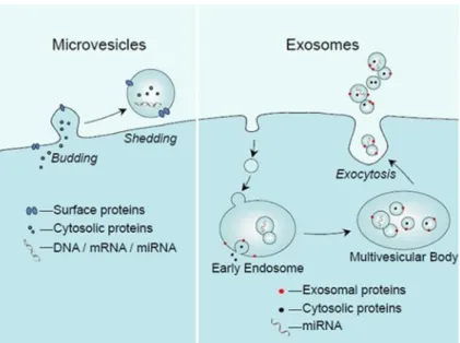
Besides cell-to-cell contacts and the release of cytokines, chemokines, growth factors, hormones and other soluble messengers, it has been recently demonstrated the ability of cells to communicate each other via the delivery of extracellular vesicles (EVs). The secretion of EVs is a universal cellular process occurring from simple organism (Archea or Gram-negative and Gram-positive bacteria) to complex multicellular organism, suggesting that this EV-mediated communication is evolutionarily conserved (Kim et al. 2013).
EVs mediate normal and pathologic intercellular communication (Thèry et al. 2009, Gyӧrgy et al. 2011). Their function depends on the cargo that they carry and the cell type from which they originate.
Many cell types are known to secrete EVs, and they include epithelial cells (van Niel et al. 2001; El Andaloussi et al. 2013), fibroblasts (Ji et al. 2008), erythrocytes (Geminard et al. 2002), platelets (Heijnen et al. 1999), mast cells (Raposo et al. 1997), tumor cells (Atay & Godwin 2014), stem cells (Zhang et al. 2014), and immune cells such as dendritic cells (DCs) (Sobo-Vujanovic et al. 2014), monocytes (Akao et al. 2011), macrophages (McDonald et al. 2014), NK cells (Lugini et al. 2012), B lymphocytes (McLellan et al. 2009), and T lymphocytes (Zhang et al. 2011).
The presence in accessible fluids (blood plasma, urine, saliva, or broncho-alveaolar lavage fluid) of exosomes containing pathology-specific biomarkers, is already exploited in clinical studies and may be useful for early detection of a disease status. Furthermore, considering the role of EVs in the intercellular communication and the potential to carry out this process at a considerable distance from the production area, the study of EVs has aroused great interest for the possible application as a transporter of iatrogenic substances to be conveyed to a specific target.
One of the major problems is the difficulty in purifying MVs and exosomes, given the low number and lack of standardized techniques and protocols (Lakhal and Wood 2011; Zhang et al.2019).
Another critical point to solve is the optimal loading of EVs with bioactive components without affecting their architecture. In fact, it has been shown that, based on the nature of the bioactive loading, the vesicles could become immunogenic, and therefore degradable.