Review Article
Aquaporin Membrane Channels in Oxidative Stress, Cell
Signaling, and Aging: Recent Advances and Research Trends
Grazia Tamma
,
1Giovanna Valenti
,
1Elena Grossini,
2Sandra Donnini,
3Angela Marino
,
4Raul A. Marinelli,
5and Giuseppe Calamita
11Department of Biosciences, Biotechnologies and Biopharmaceutics, University of Bari Aldo Moro, Bari, Italy
2Department of Translational Medicine, University of Eastern Piedmont, Novara, Italy
3Department of Life Sciences, University of Siena, Siena, Italy
4Department of Chemical, Biological, Pharmaceutical and Environmental Sciences, University of Messina, Messina, Italy
5Instituto de Fisiología Experimental, CONICET, Facultad de Ciencias Bioquímicas y Farmacéuticas, Universidad Nacional de
Rosario, Rosario, Santa Fe, Argentina
Correspondence should be addressed to Grazia Tamma; [email protected] and Giuseppe Calamita; [email protected]
Received 19 October 2017; Revised 29 January 2018; Accepted 20 February 2018; Published 27 March 2018 Academic Editor: Mark Crabtree
Copyright © 2018 Grazia Tamma et al. This is an open access article distributed under the Creative Commons Attribution License, which permits unrestricted use, distribution, and reproduction in any medium, provided the original work is properly cited. Reactive oxygen species (ROS) are produced as a result of aerobic metabolism and as by-products through numerous physiological
and biochemical processes. While ROS-dependent modifications are fundamental in transducing intracellular signals controlling
pleiotropic functions, imbalanced ROS can cause oxidative damage, eventually leading to many chronic diseases. Moreover, increased ROS and reduced nitric oxide (NO) bioavailability are main key factors in dysfunctions underlying aging, frailty,
hypertension, and atherosclerosis. Extensive investigation aims to elucidate the beneficial effects of ROS and NO, providing
novel insights into the current medical treatment of oxidative stress-related diseases of high epidemiological impact. This review focuses on emerging topics encompassing the functional involvement of aquaporin channel proteins (AQPs) and membrane transport systems, also allowing permeation of NO and hydrogen peroxide, a major ROS, in oxidative stress physiology and pathophysiology. The most recent advances regarding the modulation exerted by food phytocompounds with antioxidant action on AQPs are also reviewed.
1. Introduction
Reactive oxygen species (ROS) are unstable reactive molecules,
physiologically produced by xanthine oxidase, nicotinamide
adenine dinucleotide phosphate oxidase, lipoxygenases, and
mitochondria [1, 2]. Though oxygen is peremptory for life,
imbalances between antioxidant defense mechanisms,
over-production of ROS, or incorporation of free radicals from the
environment to living systems lead to oxidative stress. ROS
and other reactive species are implicated in a large spectrum
of biological conditions, such as mutation, tumorigenesis,
degenerative diseases, in
flammation, aging, frailty, and
devel-opment [3]. ROS exert a dual role as both deleterious and
beneficial species, the latter being of pivotal importance as
signaling molecules. At physiological levels, ROS can
improve cellular activities as they are involved in the
con-trol of the chemical balance and synaptic plasticity [4],
whereas an excess amount of ROS can damage the
endo-thelium,
leading
to
alteration
of
the
intracellular
reduction-oxidation homeostasis [5].
Among various mechanisms, the uncoupling of nitric
oxide synthase (NOS) in vascular cells has also widely been
reported to be involved in ROS generation. In that event,
NOS is turned into a peroxynitrite generator, leading to
detrimental e
ffects on vascular function, due to lipidic
perox-idation [6]. Furthermore, superoxide anions can modify
endothelial function by reducing nitric oxide (NO)
biosyn-thesis and bioavailability [7]. This issue is of particular
Volume 2018, Article ID 1501847, 14 pages https://doi.org/10.1155/2018/1501847
relevance since changes in NO release could play an
impor-tant role in endothelial function maintenance, in addition
to regulating proliferation of smooth muscle cells,
leuko-cyte adhesion, platelet aggregation, angiogenesis,
thrombo-sis, vascular tone, and hemodynamics. Hence, endothelial
dysfunction, a predictor of several cardiovascular diseases
(CVDs), is caused by imbalance between vasodilating and
vasoconstricting
agents,
including
NO,
endothelium-derived hyperpolarizing factor, prostacyclin, or
vasocon-strictive factors such as thromboxane (TXA
2) and
endothelin-1 (ET-1) [8].
NO is a gas which plays an important role in blood
pressure modulation due to its signaling action on renal,
car-diovascular, and central nervous system functions [9]. The
role of NO in vascular homeostasis also comes from the
negative regulation on coagulation and in
flammation
oper-ated by this signaling molecule.
Throughout the years, ROS and NO have been widely
considered to enter cells by freely diffusing through the cell
membrane lipid bilayer and not via specific transporters or
channels. This notion has been challenged by the discovery
of new membrane transport functions, especially those
exerted by aquaporins (AQPs), a family of membrane
chan-nel proteins widespread in nature [10, 11]. Transport of
NO and ROS by AQPs would be required for cell
homeosta-sis to play a critical role in maintaining endothelial function.
This review focuses on an emerging topic, the functional
involvement of AQPs in ROS membrane transport, with
spe-ci
fic regard to the movement of hydrogen peroxide and NO
into and out of cells, in both health and oxidative
stress-induced diseases. The emerging information and research
trends regarding the modulation exerted by food
phytocom-pounds with antioxidant action on the expression and
func-tion of AQPs are also reviewed.
2. Exogenous and Endogenous
Source of Oxidants
Reactive species (RS) derive from either endogenous or
exogenous sources. Prolonged exercise, ischemia,
inflam-mation, infection, cancer, and aging correlate with
produc-tion of free radicals. Producproduc-tion of ROS and reactive
nitrogen species (RNS) may occur through enzymatic
and nonenzymatic reactions [12, 13]. Among enzymatic
processes, NADPH oxidase (NOX), xanthine oxidase, and
peroxidases play a pivotal role in free radical generation.
For example, NOX catalyzes the production of superoxide
[14], which represents a master substrate for generation of
other RS, such as hydrogen peroxide (H
2O
2), hydroxyl
radical (OH
•), peroxynitrite (ONOO
−), and hypochlorous
acid (HOCl). The latter is synthesized in neutrophils by
myeloperoxidase, an enzyme oxidizing chloride ions when
H
2O
2is present [15, 16]. Nitric oxide (NO
•) is generated
in many tissues and results from the oxidation of
L-argi-nine to citrulline through the action of nitric oxide
synthase [17], as reported above.
Nonenzymatic reactions can also occur during oxidative
phosphorylation in mitochondria, the main RS production
site inside the cell [18]. The leakage of electrons at complex
I, complex II, or complex III associates with superoxide
production. In the mitochondrial matrix and in the cytosol,
superoxide can be converted into H
2O
2by superoxide
dis-mutase and further detoxi
fied by catalases. In addition,
ROS stimulates the generation and the release of other RS,
thereby causing a vicious circle due to increased permeability
of mitochondrial pores by ROS resulting in mitochondrial
defects leading to release of further RS [19].
Alternatively, RS also result from reaction with organic
compounds subjected to ionizing radiations. Indeed, high
doses of ionizing radiation increase the production and
release of inflammatory chemokines and RS that, in concert,
promote tissue injury [20].
Exogenous RS can originate from water and air pollution,
cigarette smoke, pesticides, dioxin, and several drugs. Once
in the body, these di
fferent compounds are metabolized,
gen-erally in the liver, generating free radicals.
3. Aquaporins, Membrane Channel Proteins of
Pleiotropic Relevance
Aquaporins (AQPs) are channel proteins widely present in
living organisms where they were initially reported to
facilitate the transport of water and certain neutral solutes
across biological membranes [21, 22]. Mammals possess
thirteen distinct AQPs (AQP0
–12) that are roughly
subdi-vided into orthodox aquaporins (AQP0, AQP1, AQP2,
AQP4, AQP5, AQP6, and AQP8) and aquaglyceroporins
(AQP3, AQP7, AQP9, and AQP10). Orthodox AQPs were
initially
described
to
conduct
only
water,
whereas
aquaglyceroporins were shown to transport water and
some small neutral solutes, particularly glycerol. These
peculiarities did not apply to AQP11 or AQP12, due to
their distinct evolutionary pathway and primary sequence
distinctions, the reason why they have been indicated as
unorthodox aquaporins [23]. The transport properties
and subcellular localization of AQP11 and AQP12 remain
unclear and a matter of debate. The functional subdivision
of AQPs has become more articulated in the light of
trans-port properties retrans-ported in recent years. Some AQPs are
also able to conduct H
2O
2and/or ammonia [24], and,
due to these biophysical properties, they are also denoted
as peroxiporins [25, 26] and ammoniaporins (or
aquaam-moniaporins) [25, 27, 28], respectively. The currently
identi
fied mammalian AQP homologues allowing passive
di
ffusion of considerable amounts of H
2O
2are AQP1,
AQP3, AQP5, AQP8, and AQP9 [29]. AQPs also facilitate
permeation of gases such as CO
2, NO, or O
2[11, 30, 31],
features that have raised a lot of interest due to the
poten-tial physiological relevance they may have in permeating
gases of biological relevance. This feature would add more
knowledge to the physiological importance of gas channels
in nature [32].
Expression and modulation of AQPs in all body
dis-tricts are the subject of intense investigation around the
world. Important roles have already been ascribed to this
family of membrane channels, in both health and disease
[21, 22, 33] (Table 1).
4. Involvement of Aquaporins in the Transport
System of Reactive Species
4.1. Aquaporin-8 as Peroxiporin Mediating Mitochondrial
H
2O
2Release in Hepatocytes. H
2O
2is a major ROS
con-stantly generated in mitochondria by the aerobic
metabo-lism. Respiratory chain-linked H
2O
2is produced by
enzymatic dismutation of superoxide radicals [34].
Com-plex I generates superoxide within the mitochondrial
matrix, whereas complex III generates superoxide in the
intermembrane space [34, 35]. Hepatic mitochondria are
not only important sources for ROS but also important
key targets for their potential damage. Under physiological
conditions, H
2O
2is the only ROS that can move out of
the mitochondria into the cytoplasm and function as a
second messenger in signal transduction pathways [35,
36]. Under oxidative stress, high ROS (H
2O
2) levels can
induce loss of mitochondrial membrane potential and
mitochondrial dysfunction with the resulting triggering of
cell death mechanisms [37, 38].
H
2O
2had been long thought to be freely diffusible across
cellular membranes, a notion that has been challenged by
both the existence of H
2O
2gradients across biological
mem-branes [39, 40] and the
finding that membrane permeability
is a rate-limiting factor in H
2O
2elimination by mammalian
cells [41]. Limited di
ffusion of H
2O
2across mitochondrial
membranes has also been suggested [42]. Hence, a
protein-facilitated di
ffusional pathway for H
2O
2across membranes
was proposed [40, 42]. H
2O
2size and chemical and
physico-chemical properties are similar to those of water [40], which
may explain H
2O
2passage through channel membrane
pro-teins such as AQPs. Accordingly, initial studies in
reconsti-tuted yeast [10] and transfected mammalian cells [43]
indicate that AQP8 and some other members of the
mamma-lian AQP family facilitate H
2O
2passage across plasma
mem-branes. Thus, AQP8 is able to function as peroxiporin.
An initial study demonstrated that AQP3 is required for
(NOX)-derived H
2O
2signaling [43]. More recent studies in
diverse nonhepatic cells have reported that plasma
mem-brane AQP8 transports NOX-generated H
2O
2that
partici-pates in intracellular signal transduction pathways [44–47].
In HeLa cells, AQP8 plays a key role in the epidermal growth
factor- (EGF-) induced entry of H
2O
2, which in turn initiates
intracellular signaling by tyrosine phosphorylation of target
proteins [44]. In B lymphocytes, AQP8-mediated H
2O
2transport has been reported to induce cell activation and
dif-ferentiation [45], whereas in leukemia cells, it has been found
to induce proliferation pathways [46, 47].
In hepatocytes, plasma membrane AQP8 is exclusively
expressed on the bile canalicular domain [48]. Therefore,
AQP8 cannot be involved in the intracellular transport
of H
2O
2generated by NADPH oxidases at sinusoidal
plasma membranes. AQP8 is also expressed in the inner
mitochondrial membranes of some cells, including
hepato-cytes [49, 50]. Experimental evidence in human hepatocyte
carcinoma HepG2 cells suggests that mitochondrial AQP8
(mtAQP8) facilitates the di
ffusional efflux of H
2O
2[51]. A
similar observation was made studying mitochondrial
AQP8b, the marine teleost orthologue of human AQP8
[52]. As reviewed below, the involvement of an
mtAQP8-mediated H
2O
2transport in normal human spermatozoa
functioning has also been suggested [53].
The knockdown of mtAQP8 expression in HepG2 cells
markedly reduces the release of mitochondrially generated
H
2O
2, and the resulting mitochondrial ROS accumulation
induces mitochondrial depolarization via the mitochondrial
permeability transition mechanism and reduced ATP levels
[51]. Interestingly, the immunological blockage of
AQP8b-mediated mitochondrial H
2O
2efflux in marine spermatozoa
Table 1: Functional relevance of mammalian aquaporins in health and disease.
Physiological functions involving aquaporins
Generation offluids
(i) Urine [150]
(ii) Cerebrospinalfluid [151]
(iii) Aqueous humor [152] (iv) Sweat [21]
(v) Saliva [21] (vi) Tears [21] (vii) Bile [153]
(viii) Gastrointestinal juices [33]
(ix) Seminalfluid [154]
Immune response and inflammation (i) Memory T-cell longevity [155]
(ii) Inflammatory response [156]
(iii) Dendritic cell maturation [157] Metabolic homeostasis and energy balance
(i) Gluconeogenesis [158] (ii) Triacylglycerol synthesis [158]
(iii) Ammonia detoxification via ureagenesis [159]
Nervous system physiology (i) Multiple functions [151] Other functions
(i) Apoptosis [160] (ii) Oxidative stress [26] (iii) Cell migration [161]
(vi) Cell volume homeostasis [162] (v) Angiogenesis [163]
Pathological states involving aquaporins (i) Cardiovascular diseases [164] (ii) Renal concentration disorders [165]
(iii) Inflammatory diseases [156]
(iv) Cholestasis [153] (v) Brain edema [151] (vi) Cataract [162]
(vii) Immune system disorders (i.e., neuromyelitis optica) [166] (viii) Malaria [167]
(ix) Obesity, diabetes, liver steatosis [158] (x) Cancer [168]
also causes ROS accumulation, mitochondrial
depolariza-tion, and decreased ATP production [52].
The oxidant-induced mitochondrial dysfunction in
HepG2 cells causes loss of viability by activating a necrotic
death pathway [51, 54]. Interestingly, mtAQP8 silencing
causes a minor loss of viability in human hepatoma HuH-7
cells but does not a
ffect viability in neither in normal rat
hepatocytes nor in the nonneoplastic human cell lines, renal
HK-2, and Chang liver cells [54]. Therefore, carcinoma cells
might be particularly susceptible to defective mtAQP8
expression. As the loss of viability in mtAQP8-knockdown
HepG2 cells is prevented by the mitochondria-targeted
oxidant MitoTempol [51], a disparity in mitochondrial
anti-oxidant defenses is likely to explain the observed differential
susceptibility among mtAQP8-knockdown cells.
Neverthe-less, it is worth mentioning that, at least for total and reduced
mitochondrial glutathione levels, there were no signi
ficant
di
fferences between HepG2, HuH-7, Chang liver cells, and
rat hepatocytes (unpublished data from Raul A. Marinelli
’s
laboratory). Further studies are needed to understand the
mechanisms that actually cause differential death in
mtAQP8-knockdown cells.
With the use of HeLa cells, the AQP8-mediated plasma
membrane H
2O
2transport has recently been reported to be
functionally modulated under stress [55]. AQP8 permeability
to H
2O
2was reversibly inhibited, thus preventing
intracellu-lar ROS accumulation during oxidative stress [55]. To the
best of our knowledge, as AQP8 expression has not been
demonstrated in HeLa cell mitochondria [44, 56], it would
be interesting to explore whether hepatocyte mtAQP8 is
under this novel regulatory mechanism of cell survival
during stress.
Another as-yet-unexplored area of research is the role
that mtAQP8-mediated H
2O
2may play in hepatocyte
physi-ology. We have recently provided evidence suggesting that
hepatocyte mtAQP8 expression can be modulated by
choles-terol via scholes-terol regulatory element-binding protein (SREBP)
transcription factors; that is, mtAQP8 is upregulated in
depleted cells and downregulated in
cholesterol-loaded cells [57]. As H
2O
2has been described to stimulate
hepatocyte cholesterogenesis via SREBPs [58], our
finding
might suggest that mtAQP8 plays a role in
SREBP-controlled cholesterol biosynthesis. For example, at low
cel-lular cholesterol levels, SREBP-dependent mtAQP8
upregu-lation could facilitate the mitochondrial H
2O
2release that
would contribute to stimulating cholesterogenesis. Further
studies are required to elucidate this issue.
4.2. AQP-Mediated H
2O
2Transport Is Critical in Sperm Cell
Motility and ROS Scavenging. The relevance of
AQP-mediated water and H
2O
2transport in human sperm cells
activity has been reported in a recent study investigating
the expression, distribution, and role of AQP3, 7, 8, and 11
in subfertile compared with normospermic subjects [53].
The investigated AQPs were found to be implicated in sperm
cell volume regulation and ROS scavenging, two functions of
critical importance in sperm counts and motility. With the
use of AQP blockers, it was suggested that chronic deficiency
in AQP-mediated H
2O
2permeability impairs ROS efflux out
of sperm cells and reduces the detoxification efficiency, with
consequent loss of sperm functionality. However, although
coordinated action of AQPs has been reported to regulate
sperm motility in the marine teleost seabream [59], further
studies are needed to con
firm the suggested
pathophysiolog-ical relevance of AQPs in human male fertility. The speci
fic
AQP homologue that, among AQP3, 7, 8, and 11, may
account for sperm cell permeability to H
2O
2remains elusive.
AQP8 features one of the highest conductances to H
2O
2among peroxiporins. However, the relevance of AQP8 as
the major H
2O
2membrane transport system in human
sperm cells remains to be proved. A recent study using
HeLa cells showed reduction of AQP-mediated water and
H
2O
2cell permeability following oxidative stress [60].
Interestingly, the diminution was prevented or reversed
when the cells were treated with antioxidant
phytochemi-cal compounds.
4.3.
AQP3
Mediates
Hydrogen
Peroxide-Dependent
Intracellular Signaling, Responses to Environmental Stress,
and Cell Migration. AQP3 is also reported to facilitate the
uptake of H
2O
2into mammalian cells [43]. Microimaging
studies using peroxy yellow 1 methyl-ester (PY1-ME), a
spe-ci
fic fluorescent probe for H
2O
2, showed AQP3-mediated
uptake of H
2O
2in HEK cells [43]. Moreover, it has been
demonstrated that T-cell migration towards chemokines is
regulated by AQP3-facilitated transport of H
2O
2that, in
turn, stimulates Rho signaling [61]. In primary keratinocytes,
H
2O
2is required to stimulate NF-
κB signaling in response to
TNF-alpha [62].
Conversely, oxidative signals seem to be important in
controlling AQP3 expression. Chrysin and resveratrol, two
antioxidant phytocompounds, have been reported to
modu-late the expression of AQP3 [63, 64]. Accordingly, severe
ultraviolet A (UVA) irradiation causes a signi
ficant reduction
in AQP3 expression secondary to increased oxidative stress
[65]. In this regard, a negative correlation between AQP3
expression and age in sun exposed skin has been described,
suggesting AQP3 as a biomarker of age-related skin
alter-ation [66].
In the colon, AQP3 is expressed in the epithelial cells
where changes in expression were found in response to
inflammation, and AQP3-depleted mice experienced
impaired recovery after chemical-induced colitis [67].
Inter-estingly, mice lacking AQP3 showed impaired healing of
super
ficial wounds in the colon. This finding elucidates the
signaling mechanism of extracellular H
2O
2in colonic
epithe-lium and suggests the implication of AQP3-mediated H
2O
2transport in innate immune responses at mucosal surfaces
[68]. AQP3-mediated H
2O
2transport has also been
described to control EGF signaling in epithelial cells [69],
playing an important role in T-cell and breast cancer cell
migration [70, 71]. However, the exact contribution of the
AQP3-mediated H
2O
2transport to these changes in cellular
function remains to be fully elucidated. Involvement of
AQP3 in trefoil peptide and EGF-mediated migration, a
vital process in in
flammatory bowel disease repair in case
of excess free radical production, has also been recently
shown [72].
4.4. AQP1-Mediated Diffusion of NO in Vasorelaxation
4.4.1. Endothelial NO Release and Oxidative Stress during
Aging. With aging, endothelial cells (ECs) undergo
consider-able remodeling processes [73, 74]. Increased endothelial
permeability, alterations in the cytoskeleton, the appearance
of
β-galactosidase staining, and the expression of several cell
cycle inhibitors [75] are also observed. Aging of ECs is
asso-ciated with an increased release of vasoconstrictors, such as
angiotensin II and endothelin, and a reduced release of
vaso-dilators, such as NO and prostacyclin [76].
Among the above factors, NO bioavailability has been
suggested to play a central role in maintaining endothelial
function [77–79]. NO is the subject of extensive studies as
one of the most relevant factors released by the endothelium,
playing an outstanding role in maintaining vascular system
function [77, 79
–81]. NO is produced by endothelial NO
syn-thase (eNOS), which transfers electrons from nicotinamide
adenine dinucleotide phosphate (NADPH) to the heme in
the amino-terminal oxygenase domain. In this way, the
sub-strate
L-arginine is oxidized to
L-citrulline and NO.
Tetrahy-drobiopterin (BH4) is an essential cofactor of eNOS exerting
a key role in the progression of NO synthesis (Figure 1). NO
formed by the vascular ECs diffuses to the adjacent cells, such
as vascular smooth muscle cells (VSMCs), platelets, and
leu-cocytes, where it exerts many of its bene
ficial actions, such as
vasodilation, antithrombotic, anti-in
flammatory, and
anti-proliferative e
ffects [82]. Endothelium-derived NO is known
to be particularly important to maintain normal vascular
tone, endothelial function, and homeostasis [83], preventing
the progression of age-related vascular disorders [80].
Decreased production of endothelium-derived NO during
aging is commonly believed to be due to decreased eNOS
activity characterizing senescent ECs [78]. In the
peroxida-tive conditions associated with aging, superoxide anion
(O
2−) can also react with NO leading to the formation of
per-oxynitrites, which, in turn, can promote protein nitration
and contribute to EC dysfunction and death [84, 85].
Fur-thermore, enhanced oxidative stress can lead to eNOS
“uncoupling” and cause endothelial dysfunction [86].
BH4 oxidation is one of the possible mechanisms of
eNOS
“uncoupling.” Intracellular BH4 levels depend on the
balance between its synthesis and degradation. In particular,
oxidative stress may lead to excessive oxidation and depletion
of BH4. As a consequence, the
flow of electrons within NOS
could be
“uncoupled” from
L-arginine oxidation and O
2−produced from the oxygenase domain [87]. Hence, eNOS
would be converted to a superoxide-producing enzyme with
reduced NO production and enhanced preexisting oxidative
stress [88, 89].
4.4.2. AQP1 and NO Flow in Vascular Senescence and
Atherosclerosis. Free diffusion (simple diffusion) through
the phospholipid bilayer composing the plasma membrane
had historically been assumed to be the only pathway
whereby NO moves into or out of cells. Thus, based on the
partition coe
fficient of NO between lipids and water [90,
91] rather than direct experimental assessment of NO di
ffu-sion across the cell membrane, NO was believed to cause
vasodilation, antithrombotic, inflammatory, and
anti-proliferative effects without need of facilitation by channels
or transporters. This assumption was not confirmed after
measurements of NO
fluxes across reconstituted
proteolipo-somes and transfected cultured cells showing that, in
addi-tion to water, the AQP1 channel could conduct NO across
plasma membranes and that the plasma membrane
repre-sents a signi
ficant barrier to NO diffusion [11]. Successively,
with the use of thoracic aortas isolated from wild-type
(Aqp
+/+) and Aqp
−/−knockout mice, it was shown that
AQP1 facilitates NO diffusion out of endothelial cells and
NO influx into vascular smooth muscle cells, and that
AQP1 conduction of NO is required for full expression of
endothelium-dependent vasorelaxation [92]. Regarding
vas-cular aging, changes in AQPs expression have been found
in animal models of kidney-clip hypertension [11]. The
traf-ficking of AQPs within cells has also been shown to change
during aging, as observed in the parotid gland [93]. The
sug-gested role of AQPs in vascular function regulation and
senescence through modulation of NO diffusion across cell
membranes opens a new avenue in understanding vascular
senescence physiology and pathophysiology. Additional
work is, however, needed since a discrepancy has been raised
by a study reporting intact NO-dependent vasorelaxation in
AQP1-depleted mice [94]. Vascular AQP1 expression was
found to undergo positive regulation with the mediation of
KLF2, the
flow-responsive transcription factor Krüppel-like
factor 2 that maintains an anticoagulant, anti-in
flammatory
endothelium with su
fficient NO bioavailability [95]. Both
in vitro and in vivo AQP1 expression was subjected to
KLF2-mediated positive regulation by atheroprotective
shear stress whereas it proved to be downregulated under
inflammatory conditions. While suggesting that endothelial
expression of AQP1 characterizes the atheroprotected,
nonin
flamed vessel wall, this finding supports the putative
continuous role of KLF2 in stabilizing the vessel wall via
cotemporal expression of eNOS and AQP1, helping to
prevent or counteract the pathogenesis of atherosclerosis.
5. Role of Vasopressin/AQP2 Axis and Oxidative
Stress in Aging
Alterations in plasma osmolality and
fluid body volume are
observed in the elderly, making old people at high risk of
developing disturbances of the water metabolism, which
can give rise to several adverse e
ffects. Aging blunts thirst
and drinking responses, making older people more
vulnera-ble to body
fluid imbalance and dehydration [96], which
can compromise cognitive function [97, 98]. Indeed,
dehy-dration is a predisposing factor for confusion in long-term
care residents [99]. Furthermore, plasma hypertonicity, a
marker of dehydration, increases the risk of ischemic stroke
in hospitalized patients [100] and may precipitate cerebral
ischemic events in susceptible elderly individuals [101].
The major hormone regulating water metabolism in the
body is vasopressin. Vasopressin is a 9-amino acid peptide
that is secreted from the posterior pituitary in response to
high plasma osmolality and hypovolemia. Vasopressin has
important roles in circulatory and water homeostasis
mediated by vasopressin receptor subtypes V1a (vascular),
V1b (pituitary), and V2 (vascular, renal). Therefore,
age-related dysfunction of the
hypothalamic-neurohypophyseal-vasopressin axis can result in multiple abnormalities in
several physiological systems that might promote a
vari-ety of morbidity such as cardiovascular and renal
dis-eases [102, 103].
At the renal level, it has been observed that aging is
accompanied by a parallel decrease in maximal urine
concen-trating ability [104]. Individuals aged 60–79 years show an
approximately 20% reduction in maximum urine osmolality,
a 50% decrease in the ability to conserve solute, and a 100%
increase in minimal urine
flow rate, when compared to
youn-ger age groups. Abnormalities in vasopressin secretion
appear to be associated with the decrease in urine
concentrat-ing ability with agconcentrat-ing: the abundance of many of the key
transport proteins responsible for urine concentrating ability
is reduced in the kidney medulla of aged rats [105]. The
reductions in water, sodium, and urea transport protein
abundances, along with their reduced response to water
restriction, contribute to the reduced ability of aged rats to
concentrate urine and conserve water [104].
The major mechanism by which vasopressin modulates
water reabsorption is by regulating the tra
fficking of the
vasopressin-sensitive water channel aquaporin-2 (AQP2) in
collecting duct principal cells. Speci
fically, binding of
vaso-pressin to the V2R increases cAMP levels, resulting in the
activation of protein kinase A (PKA). PKA-dependent
phos-phorylation of the water channel AQP2, at S256, is essential
to promote the translocation of AQP2-bearing vesicles from
an intracellular pool to the apical plasma membrane [106].
Phosphoproteomic studies have demonstrated that, besides
S256, vasopressin stimulation increases S264 and T269 but
decreases the phosphorylation of S261 [107].
Several studies performed in animal models have shown
that in aged rats, there is a large decrease in the level of
AQP2 as well as of its phosphorylated form at S256, which
can contribute to the reduced renal concentrating abilities
[105, 108]. Furthermore, AQP3 is reduced in aged rats, but
no change in the expression of AQP1 and AQP4 has been
detected in aged rats. This distinction in the regulation of
AQPs abundance may be related to the fact that only AQP2
and AQP3 expressions are under control of vasopressin.
One possible therapy to overcome the decrease in AQP2
abundance in aging might be the administration of
vasopres-sin. A recent study showed that desmopressin (dDAVP), a
selective V2R agonist, administered to 10- and
30-month-old Wag/Rij rats, decreases urine output in both rat groups
and leads to an increase in AQP2 and AQP3 abundance.
These results suggest that a decrease in AQP2 and AQP3
expression levels partially accounts for the diminution in
uri-nary concentrating ability in aging.
In general, due to an impaired ability to conserve water,
in the elderly, there is a decrease in total body water content
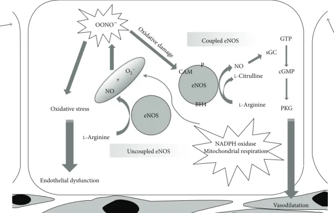
OONO− NO eNOS NADPH oxidase Mitochondrial respiration BH4 Vasodilatation eNOS CAM Oxida tive da mag e P Coupled eNOS NO sGC GTP cGMP PKG L-Citrulline L-Arginine Uncoupled eNOS Endothelial dysfunction Oxidative stress L-Arginine O2− +
Figure 1: NO release by eNOS in physiological and peroxidative conditions. While “coupled” eNOS is involved in the physiological NO
release underlying vasorelaxation, NO release by “uncoupled” eNOS is turned into OONO−(peroxynitrites) leading to an increase in
oxidative stress with consequent endothelial dysfunction. ADMA: asymmetric dimethylarginine; Akt: protein kinase B; BH4: tetrahydrobiopterin; CAM: calmodulin; Cav-1: caveolin 1; eNOS: endothelial NO synthase; cGMP: cyclic guanosine monophosphate; GTP: guanosine triphosphate; GTPCH: guanosine triphosphate cyclohydrolase I; Hsp 90: heat shock protein 90; NADPH: nicotinamide adenine dinucleotide phosphate; NO: nitric oxide; sGC: soluble guanylate cyclase; PKG: protein kinase G.
associated with a reduction of plasma volume. These changes
make the elderly much more sensitive to water overload or
dehydration resulting in abnormal movement of solutes
and, thereby, increasing the possibility of developing
hypo-or hypernatremia.
However, an increase in vasopressin levels can also be
found in aged people, where it induces water retention and
hyponatremia and stimulates calcium release from bone,
thus contributing to osteoporosis, as well as affecting the
car-diovascular system and blood pressure, thus contributing to
the development of hypertension.
For these diseases often associated with the elderly,
vaso-pressin receptor antagonists represent a promising
therapeu-tic tool. Interest in vasopressin has been renewed with the
availability of vaptans, new, potent, orally active vasopressin
receptor antagonists, initially developed for the treatment of
various forms of hyponatremia (often related to vasopressin
dysfunction) and proven to be safe in humans [109
–111].
Evaluation of the speci
fic aquaretic effect of vaptans in
aged patients treated with this antagonist might have a
profound impact in understanding the therapeutic effect
of vaptan compounds.
Interestingly, vasopressin, the levels of which are
increased after water deprivation, stimulates vascular
super-oxide production through activation of V1aR [112].
Accord-ingly, it has been shown that water deprivation increases ROS
production in the somatosensory cortex, indicating that
cere-brovascular dysfunction is related to oxidative stress [113].
5.1. Oxidative Signals and AQP2. Oxidative stress plays a key
role in modulating renal functionality, including its diluting
and concentrating ability during aging [114]. Oxidative stress
increases the risk of developing several age-related diseases
because ROS may alter cell signaling, leading to
inflamma-tion, apoptosis, and cellular senescence. During aging,
signif-icant increases in advanced glycosylation end products
(AGE) and other oxidants have been reported in kidneys
[115, 116]. Chronic inhibition of nitric oxidase synthase
reg-ulates renal water balance by reducing the expression of
AQP2 [117, 118]. Importantly, oxidative stress is often
asso-ciated with disorders linked to redox unbalance.
At a molecular level, ROS can oxidize selective amino
acids on target proteins. Oxidative dependent modifications
are being shown to be fundamental in transducing several
intracellular signals controlling pleiotropic functions such
as cell proliferation, apoptosis, autophagy, and membrane
transport. These modi
fications result from reactions between
ROS or reactive nitrogen species (RNS) and amino acid
resi-dues [26]. Oxidative modi
fications mainly occur through the
switching of the sulfur in target cysteines. However, cysteines
are not the only residues involved in oxidative modi
fications
as methionine, lysine, arginine, threonine, and proline
resi-dues can also be oxidized to reactive carbonyls [119].
Oxida-tive
sensitive
modifications include carbonylation,
nitrotyrosinylation, succinylation, S-sulfenation,
S-nitrosyla-tion, S-glutathionylaS-nitrosyla-tion, and disulfide formation.
Reversible glutathionylation results from the reaction
between glutathione and cysteine residues (PSSG) upon
exposure to RS. S-Glutathionylation is recognized as a crucial
modi
fication by which cells translate local changes of reactive
species [120].
Using a proteomic approach, Sandoval and coworkers
revealed that vasopressin stimulation is associated with
increased expression of different oxidative related proteins
such as glutathione S-transferase [121]. This observation is
likely to indicate that oxidative signaling may somehow play
a role in controlling the physiological signal transduction
cascade initiated by vasopressin. Studies from several groups
have revealed, indeed, that antioxidant compounds such as
N-acetylcysteine (NAC) rescued the reduction of AQP2
abundance observed in rats subjected to the bilateral ureteral
obstruction (BUO) [122]. Conversely, treatment with the
oxidant 4-hydroxy-2-hexenal (HHE) decreases the
abun-dance of AQP2 and activates several kinases such as
p38-MAPK and ERK [123], which have been proposed to
phos-phorylate AQP2 at S261 [124–126]. Phosphorylation at
S261 is involved in AQP2 ubiquitylation and degradation
[125, 127].
However, vasopressin is not the only factor regulating
AQP2 expression. In this respect, it has been demonstrated
that the oxidant HHE increases the expression of the
tran-scription factors NF-
κB and the enzyme NOX4 [123], both
involved in modulating the expression level of AQP2.
Specif-ically, NF-
κB decreased AQP2 mRNA and protein
abun-dance [128]. Conversely, NOX4 promotes AQP2 expression
[129]. These
findings strongly suggest that AQP2 abundance
is the result of a balanced activity between NF-
κB and NOX4.
However, how oxidative signals modulate the stability of the
target proteins remains to be clari
fied. It appears conceivable
that transient oxidative posttranslational modi
fications may
mean there is a molecular signature translating oxidative
information signaling and thus controlling the fate of
tar-get proteins. In this respect, some of the authors of this
review have recently shown that AQP2 undergoes
S-glutathionylation. It was also found that the increase in
AQP2 glutathionylation is paralleled by higher ROS
pro-duction. Conversely, low levels of ROS, measured in cells
displaying low intracellular calcium concentration,
second-ary to the expression of the calcium-sensing receptor
(CaSR), associates with reduced S-glutathionylation of
AQP2 [130]. Whether or not S-glutathionylation of
AQP2 is involved in the water imbalance observed during
aging remains to be investigated.
6. Modulatory Actions of Food Antioxidant
Phytocompounds on Aquaporins
A growing number of food phytochemicals are being found
to exert antioxidant and anti-inflammatory actions. Thanks
to their ability to interact with pivotal signaling pathways, a
number of food and herbal phytochemicals have been found
to impart health benefits modulating important cellular
func-tions such as growth, differentiation, death, and volume
homeostasis as well as redox, metabolic, and energy balance.
To date, a large number of biologically active
phytochem-icals have been identi
fied, characterized, and eventually
mod-i
fied as natural sources of novel compounds to prevent, delay,
or cure many human diseases. This is an important
achievement since secondary prevention or adjunct therapy
through dietary intervention is a cost-effective alternative
for avoiding the large burden of health care, especially that
associated with chronic illnesses.
Similarly to several other transport systems, AQPs are
also modulated by a number of food bioactive
phytocom-pounds [131, 132]. The modulatory e
ffects exerted by
benefi-cial dietary patterns, food phytochemicals, and herbal
compounds on AQPs, in both health and disease, is a fast
growing topic as their exploitation may help support current
medical treatment options to improve the prognosis of
sev-eral diseases. Flavonoid modulation of AQPs has been
reported to ameliorate forms of cerebral and retinal edemas
of different origins (AQP4) [132–135], lung injuries
(AQP1) [136], and Sjögren syndrome-associated xerostomia
(AQP5) [137], to inhibit ovarian tumor growth (AQP5) [89]
and protect against UV-induced skin damage (AQP3) [63].
Curcumin in
fluences choroid plexus AQP1 [138], ovarian
AQP3 [139], and brain AQP4 and AQP9, reducing
intracra-nial pressure in brain injury, inhibiting ovarian cancer cell
migration, and reducing brain edema, respectively [140–
142]. Modulation of AQP channel gating by curcumin has
been recently reported in a paper describing the use of HeLa
cells to investigate the effects of some antioxidant
phytocom-pounds on AQP1, AQP3, AQP8, and AQP11 [60].
Resvera-trol, a stilbene compound, was found to inhibit human
keratinocytes and ameliorate the ischemia/reperfusion injury
acting on AQP3 [64] and AQP4 [143], respectively. The
chal-cone compound phloretin also acts on the expression of
AQP9 to exert its antioxidant and anti-in
flammatory actions
[144]. Genistein and daidzein, two isoflavonoids, were found
to upregulate the expression of uterine AQP1 by increasing
the responsiveness to estrogens [145]. The monoterpenoid
carvacrol has been reported to reduce the intracerebral
hemorrhage-induced brain edema by downregulating brain
AQP4 [146]. Triterpenoids have been shown to act on the
expression of AQP1 to reduce cancer cell migration,
counter-acting metastasis, as well as to ameliorate forms of allergic
rhinitis and to downregulate kidney AQP2 to protect against
renal failures [147]. Capsaicin was found to increase the
expression level of submandibular salivary gland AQP5 to
ameliorate salivary gland hypofunction [148, 149]. While
useful information is already available, further important
achievements are expected from the ongoing studies on the
modulatory effects exerted by biologically active
phytocom-pounds on the expression and function of AQPs.
7. Conclusions and Future Perspectives
Excess of ROS within the cells and reduction of NO
bioavail-ability can largely promote cellular dysfunction, which is
linked to the development of metabolic disorders,
cardiovas-cular and renal diseases, frailty, and aging. Key roles for
AQPs as peroxiporins in the signal transduction pathways
underlying diverse cellular functions, such as di
fferentiation,
proliferation, or mobility, are suggested by the recent
evi-dence of AQP-mediated H
2O
2transport at the plasma or
mitochondrial membrane level. Dysregulation of peroxiporin
function can lead to oxidative stress and eventually cell death.
Alterations in AQP-mediated ROS and/or NO transport are
therefore assuming an increasing translational value in
phys-iology and pathophysphys-iology with promising nutraceutical
and pharmacological implications. Indeed, modulation of
the peroxiporin and/or NO channel function of AQPs at
the vascular, hepatic, testicular, or renal level may prove to
be valuable in preventing or treating cardiovascular (vascular
sti
ffness/hypertension, atherosclerosis), metabolic, and
reproductive (impaired sperm cell motility) diseases. Last
but not least, further work is warranted to investigate the
involvement of AQPs in the antioxidant and
anti-inflammatory actions exerted by food phytochemical
com-pounds in order to devise new strategies to promote health
and improve aging.
Conflicts of Interest
The authors declare they do not have any conflicts of interest.
Acknowledgments
Grazia Tamma and Giuseppe Calamita acknowledge the
financial contribution from Fondazione Cariplo and Daniel
& Nina Carasso Foundation (project LeGeReTe #1507-200
AF, #FC 2015-2440, and #FDNC Engt 00063479 supported
under the
“Thought for Food” Initiative of Agropolis
Foun-dation through the
“Investissements d’avenir” programme
with reference number ANR-10-LABX-0001-01).
References
[1] M. P. Murphy,“How mitochondria produce reactive oxygen
species,” The Biochemical Journal, vol. 417, no. 1, pp. 1–13,
2009.
[2] D. B. Zorov, M. Juhaszova, and S. J. Sollott,“Mitochondrial
reactive oxygen species (ROS) and ROS-induced ROS
release,” Physiological Reviews, vol. 94, no. 3, pp. 909–950,
2014.
[3] B. Uttara, A. Singh, P. Zamboni, and R. Mahajan,“Oxidative
stress and neurodegenerative diseases: a review of upstream
and downstream antioxidant therapeutic options,” Current
Neuropharmacology, vol. 7, no. 1, pp. 65–74, 2009.
[4] L. Zuo, T. Zhou, B. K. Pannell, A. C. Ziegler, and T. M. Best, “Biological and physiological role of reactive oxygen spe-cies—the good, the bad and the ugly,” Acta Physiologica,
vol. 214, no. 3, pp. 329–348, 2015.
[5] M. de la Paz Scribano, M. del Carmen Baez, B. Florencia et al., “Effects of atorvastatin on oxidative stress biomarkers and
mitochondrial morphofunctionality in hyper
fibrinogenemia-induced atherogenesis,” Advances in Medicine, vol. 2014, Article ID 947258, 6 pages, 2014.
[6] P. Ferroni, S. Basili, V. Paoletti, and G. Davi, “Endothelial
dysfunction and oxidative stress in arterial hypertension,”
Nutrition, Metabolism, and Cardiovascular Diseases, vol. 16, no. 3, pp. 222–233, 2006.
[7] J. Vasquez-Vivar, B. Kalyanaraman, P. Martasek et al., “Superoxide generation by endothelial nitric oxide synthase:
the influence of cofactors,” Proceedings of the National
Acad-emy of Sciences of the United States of America, vol. 95, no. 16, pp. 9220–9225, 1998.
[8] N. Panth, K. R. Paudel, and K. Parajuli,“Reactive oxygen spe-cies: a key hallmark of cardiovascular disease,” Advances in Medicine, vol. 2016, Article ID 9152732, 12 pages, 2016.
[9] V. Bauer and R. Sotnikova,“Nitric oxide—the
endothelium-derived relaxing factor and its role in endothelial functions,”
General Physiology and Biophysics, vol. 29, no. 4, pp. 319–
340, 2010.
[10] G. P. Bienert, A. L. B. Møller, K. A. Kristiansen et al.,“Specific
aquaporins facilitate the diffusion of hydrogen peroxide
across membranes,” The Journal of Biological Chemistry,
vol. 282, no. 2, pp. 1183–1192, 2007.
[11] M. Herrera, N. J. Hong, and J. L. Garvin,“Aquaporin-1
trans-ports NO across cell membranes,” Hypertension, vol. 48,
no. 1, pp. 157–164, 2006.
[12] L. A. Pham-Huy, H. He, and C. Pham-Huy,“Free radicals,
antioxidants in disease and health,” International Journal of
Biomedical Sciences, vol. 4, no. 2, pp. 89–96, 2008.
[13] I. Grattagliano, G. Calamita, T. Cocco, D. Q. Wang, and
P. Portincasa,“Pathogenic role of oxidative and nitrosative
stress in primary biliary cirrhosis,” World Journal of
Gastro-enterology, vol. 20, no. 19, pp. 5746–5759, 2014.
[14] K. Bedard, B. Lardy, and K. Krause,“NOX family NADPH
oxidases: not just in mammals,” Biochimie, vol. 89, no. 9,
pp. 1107–1112, 2007.
[15] W. Droge,“Free radicals in the physiological control of cell
function,” Physiological Reviews, vol. 82, no. 1, pp. 47–95,
2002.
[16] M. Genestra, “Oxyl radicals, redox-sensitive signalling
cas-cades and antioxidants,” Cellular Signalling, vol. 19, no. 9,
pp. 1807–1819, 2007.
[17] P. Pacher, J. S. Beckman, and L. Liaudet,“Nitric oxide and
peroxynitrite in health and disease,” Physiological Reviews,
vol. 87, no. 1, pp. 315–424, 2007.
[18] J. F. Turrens, “Mitochondrial formation of reactive oxygen
species,” The Journal of Physiology, vol. 552, no. 2, pp. 335–
344, 2003.
[19] C. Maack and M. Bohm,“Targeting mitochondrial oxidative
stress in heart failure throttling the afterburner,” Journal of
the American College of Cardiology, vol. 58, no. 1, pp. 83–
86, 2011.
[20] C. Betlazar, R. J. Middleton, R. B. Banati, and G. J. Liu,“The
impact of high and low dose ionising radiation on the central nervous system,” Redox Biology, vol. 9, pp. 144–156, 2016.
[21] P. Agre, “Aquaporin water channels,” Bioscience Reports,
vol. 24, no. 3, pp. 127–163, 2004.
[22] P. Gena, M. Pellegrini-Calace, A. Biasco, M. Svelto, and
G. Calamita, “Aquaporin membrane channels: biophysics,
classification, functions, and possible biotechnological
appli-cations,” Food biophysics, vol. 6, no. 2, pp. 241–249, 2011.
[23] K. Ishibashi, “New members of mammalian aquaporins:
AQP10–AQP12,” Handbook of Experimental Pharmacology,
vol. 190, pp. 251–262, 2009.
[24] T. P. Jahn, A. L. B. Møller, T. Zeuthen et al., “Aquaporin
homologues in plants and mammals transport ammonia,”
FEBS Letters, vol. 574, no. 1–3, pp. 31–36, 2004.
[25] A. Almasalmeh, D. Krenc, B. Wu, and E. Beitz,“Structural
determinants of the hydrogen peroxide permeability of
aqua-porins,” The FEBS Journal, vol. 281, no. 3, pp. 647–656, 2014.
[26] G. Tamma and G. Valenti,“Evaluating the oxidative stress in
renal diseases: what is the role for S-glutathionylation?,”
Anti-oxidants & Redox Signaling, vol. 25, no. 3, pp. 147–164, 2016.
[27] L. M. Holm, T. P. Jahn, A. L. B. Møller et al.,“NH3and NH4+
permeability in aquaporin-expressing Xenopus oocytes,”
Pflugers Archiv: European Journal of Physiology, vol. 450,
no. 6, pp. 415–428, 2005.
[28] S. M. Saparov, K. Liu, P. Agre, and P. Pohl,“Fast and selective
ammonia transport by aquaporin-8,” The Journal of
Biologi-cal Chemistry, vol. 282, no. 8, pp. 5296–5301, 2007.
[29] B. Wu and E. Beitz,“Aquaporins with selectivity for
uncon-ventional permeants,” Cellular and Molecular Life Sciences,
vol. 64, no. 18, pp. 2413–2421, 2007.
[30] N. L. Nakhoul, B. A. Davis, M. F. Romero, and W. F. Boron, “Effect of expressing the water channel aquaporin-1 on the
CO2permeability of Xenopus oocytes,” American Journal of
Physiology-Cell Physiology, vol. 274, no. 2, pp. C543–C548,
1998.
[31] Y. Wang, J. Cohen, W. F. Boron, K. Schulten, and
E. Tajkhorshid,“Exploring gas permeability of cellular
mem-branes and membrane channels with molecular dynamics,”
Journal of Structural Biology, vol. 157, no. 3, pp. 534–544,
2007.
[32] W. F. Boron,“Sharpey-Schafer lecture: gas channels,”
Exper-imental Physiology, vol. 95, no. 12, pp. 1107–1130, 2010.
[33] G. Calamita, C. Delporte, and R. A. Marinelli, Hepatobiliary, Salivary Glands and Pancreatic Aquaporins in Health and Disease, 2015, Place Published.
[34] A. Boveris and E. Cadenas,“Mitochondrial production of
hydrogen peroxide regulation by nitric oxide and the role of ubisemiquinone,” IUBMB Life, vol. 50, no. 4, pp. 245–250, 2000.
[35] H. Sies,“Role of metabolic H2O2generation: redox signaling
and oxidative stress,” The Journal of Biological Chemistry,
vol. 289, no. 13, pp. 8735–8741, 2014.
[36] T. Finkel,“Signal transduction by mitochondrial oxidants,”
The Journal of Biological Chemistry, vol. 287, no. 7, pp. 4434–4440, 2012.
[37] R. Solito, F. Corti, C. H. Chen et al.,“Mitochondrial aldehyde
dehydrogenase-2 activation prevents β-amyloid-induced
endothelial cell dysfunction and restores angiogenesis,”
Jour-nal of Cell Science, vol. 126, no. 9, pp. 1952–1961, 2013.
[38] F. Yin, H. Sancheti, and E. Cadenas,“Mitochondrial thiols in
the regulation of cell death pathways,” Antioxidants & Redox
Signaling, vol. 17, no. 12, pp. 1714–1727, 2012.
[39] F. Antunes and E. Cadenas,“Estimation of H2O2gradients
across biomembranes,” FEBS Letters, vol. 475, no. 2,
pp. 121–126, 2000.
[40] G. P. Bienert and F. Chaumont,“Aquaporin-facilitated
trans-membrane diffusion of hydrogen peroxide,” Biochimica et
Biophysica Acta (BBA) - General Subjects, vol. 1840, no. 5,
pp. 1596–1604, 2014.
[41] N. Makino, K. Sasaki, K. Hashida, and Y. Sakakura,“A
met-abolic model describing the H2O2elimination by mammalian
cells including H2O2permeation through cytoplasmic and
peroxisomal membranes: comparison with experimental data,” Biochimica et Biophysica Acta (BBA) - General
Sub-jects, vol. 1673, no. 3, pp. 149–159, 2004.
[42] V. G. Grivennikova, A. V. Kareyeva, and A. D. Vinogradov, “What are the sources of hydrogen peroxide production by heart mitochondria?,” Biochimica et Biophysica Acta (BBA)
-Bioenergetics, vol. 1797, no. 6-7, pp. 939–944, 2010.
[43] E. W. Miller, B. C. Dickinson, and C. J. Chang,“Aquaporin-3
intracellular signaling,” Proceedings of the National Academy of Sciences of the United States of America, vol. 107, no. 36,
pp. 15681–15686, 2010.
[44] M. Bertolotti, S. Bestetti, J. M. Garcia-Manteiga et al.,
“Tyro-sine kinase signal modulation: a matter of H2O2membrane
permeability?,” Antioxidants & Redox Signaling, vol. 19,
no. 13, pp. 1447–1451, 2013.
[45] M. Bertolotti, G. Farinelli, M. Galli, A. Aiuti, and R. Sitia,
“AQP8 transports NOX2-generated H2O2across the plasma
membrane to promote signaling in B cells,” Journal of
Leuko-cyte Biology, vol. 100, no. 5, pp. 1071–1079, 2016.
[46] F. Vieceli Dalla Sega, C. Prata, L. Zambonin et al.,
“Intracellu-lar cysteine oxidation is modulated by aquaporin-8-mediated hydrogen peroxide channeling in leukaemia cells,”
BioFac-tors, vol. 43, no. 2, pp. 232–242, 2017.
[47] F. Vieceli Dalla Sega, L. Zambonin, D. Fiorentini et al.,
“Spe-cific aquaporins facilitate Nox-produced hydrogen peroxide
transport through plasma membrane in leukaemia cells,”
Bio-chimica et Biophysica Acta (BBA) - Molecular Cell Research,
vol. 1843, no. 4, pp. 806–814, 2014.
[48] R. A. Marinelli, G. L. Lehmann, L. R. Soria, and M. J.
March-issio,“Hepatocyte aquaporins in bile formation and
cholesta-sis,” Frontiers in Bioscience, vol. 16, no. 1, p. 2642, 2011.
[49] D. Ferri, A. Mazzone, G. E. Liquori, G. Cassano, M. Svelto,
and G. Calamita, “Ontogeny, distribution, and possible
functional implications of an unusual aquaporin, AQP8,
in mouse liver,” Hepatology, vol. 38, no. 4, pp. 947–957,
2003.
[50] G. Calamita, D. Ferri, P. Gena et al.,“The inner
mitochon-drial membrane has aquaporin-8 water channels and is highly permeable to water,” The Journal of Biological
Chemis-try, vol. 280, no. 17, pp. 17149–17153, 2005.
[51] M. J. Marchissio, D. E. A. Francés, C. E. Carnovale, and R. A.
Marinelli, “Mitochondrial aquaporin-8 knockdown in
human hepatoma HepG2 cells causes ROS-induced
mito-chondrial depolarization and loss of viability,” Toxicology
and Applied Pharmacology, vol. 264, no. 2, pp. 246–254, 2012.
[52] F. Chauvigne, M. Boj, R. N. Finn, and J. Cerda,
“Mitochon-drial aquaporin-8-mediated hydrogen peroxide transport is
essential for teleost spermatozoon motility,” Scientific
Reports, vol. 5, no. 1, p. 7789, 2015.
[53] U. Laforenza, G. Pellavio, A. Marchetti, C. Omes, F. Todaro,
and G. Gastaldi,“Aquaporin-mediated water and hydrogen
peroxide transport is involved in normal human spermatozoa
functioning,” International Journal of Molecular Sciences,
vol. 18, no. 12, 2017.
[54] M. J. Marchissio, D. E. A. Francés, C. E. Carnovale, and
R. A. Marinelli, “Evidence for necrosis, but not apoptosis,
in human hepatoma cells with knockdown of mitochondrial
aquaporin-8,” Apoptosis, vol. 19, no. 5, pp. 851–859, 2014.
[55] I. Medrano-Fernandez, S. Bestetti, M. Bertolotti et al.,“Stress
regulates aquaporin-8 permeability to impact cell growth and
survival,” Antioxidants & Redox Signaling, vol. 24, no. 18,
pp. 1031–1044, 2016.
[56] R. A. Marinelli and M. J. Marchissio,“Mitochondrial
aquapo-rin-8: a functional peroxiporin?,” Antioxidants & Redox Sig-naling, vol. 19, no. 8, p. 896, 2013.
[57] M. Danielli, A. M. Capiglioni, J. Marrone, G. Calamita, and
R. A. Marinelli, “Cholesterol can modulate mitochondrial
aquaporin-8 expression in human hepatic cells,” IUBMB Life,
vol. 69, no. 5, pp. 341–346, 2017.
[58] A. M. Giudetti, F. Damiano, G. V. Gnoni, and L. Siculella, “Low level of hydrogen peroxide induces lipid synthesis in BRL-3A cells through a CAP-independent SREBP-1a
activa-tion,” The International Journal of Biochemistry & Cell
Biol-ogy, vol. 45, no. 7, pp. 1419–1426, 2013.
[59] M. Boj, F. Chauvigne, and J. Cerda,“Coordinated action of
aquaporins regulates sperm motility in a marine teleost,” Biology of Reproduction, vol. 93, no. 2, p. 40, 2015.
[60] G. Pellavio, M. Rui, L. Caliogna et al.,“Regulation of
aquapo-rin functional properties mediated by the antioxidant effects
of natural compounds,” International Journal of Molecular
Sciences, vol. 18, no. 12, 2017.
[61] M. Hara-Chikuma, S. Chikuma, Y. Sugiyama et al.,
“Chemo-kine-dependent T cell migration requires
aquaporin-3-mediated hydrogen peroxide uptake,” The Journal of
Experi-mental Medicine, vol. 209, no. 10, pp. 1743–1752, 2012.
[62] M. Hara-Chikuma, H. Satooka, S. Watanabe et al.,
“Aquapo-rin-3-mediated hydrogen peroxide transport is required for
NF-κB signalling in keratinocytes and development of
psori-asis,” Nature Communications, vol. 6, no. 1, p. 7454, 2015.
[63] N. L. Wu, J. Y. Fang, M. Chen, C. J. Wu, C. C. Huang, and
C. F. Hung,“Chrysin protects epidermal keratinocytes from
UVA- and UVB-induced damage,” Journal of Agricultural
and Food Chemistry, vol. 59, no. 15, pp. 8391–8400, 2011.
[64] Z. Wu, H. Uchi, S. Morino-Koga, W. Shi, and M. Furue,
“Res-veratrol inhibition of human keratinocyte proliferation via SIRT1/ARNT/ERK dependent downregulation of aquaporin
3,” Journal of Dermatological Science, vol. 75, no. 1, pp. 16–
23, 2014.
[65] H. Xie, F. Liu, L. Liu et al.,“Protective role of AQP3 in
UVA-induced NHSFs apoptosis via Bcl2 up-regulation,” Archives
of Dermatological Research, vol. 305, no. 5, pp. 397–406, 2013.
[66] I. Seleit, O. A. Bakry, H. S. El Rebey, G. El-Akabawy, and
G. Hamza,“Is Aquaporin-3 a determinant factor of intrinsic
and extrinsic aging? An immunohistochemical and
morpho-metric study,” Applied Immunohistochemistry & Molecular
Morphology, vol. 25, no. 1, pp. 49–57, 2017.
[67] J. R. Thiagarajah, D. Zhao, and A. S. Verkman,“Impaired
enterocyte proliferation in aquaporin-3 deficiency in mouse
models of colitis,” Gut, vol. 56, no. 11, pp. 1529–1535, 2007.
[68] J. R. Thiagarajah, J. Chang, J. A. Goettel, A. S. Verkman, and
W. I. Lencer, “Aquaporin-3 mediates hydrogen
peroxide-dependent responses to environmental stress in colonic epi-thelia,” Proceedings of the National Academy of Sciences of
the United States of America, vol. 114, no. 3, pp. 568–573,
2017.
[69] M. Hara-Chikuma, S. Watanabe, and H. Satooka,
“Involve-ment of aquaporin-3 in epidermal growth factor receptor sig-naling via hydrogen peroxide transport in cancer cells,” Biochemical and Biophysical Research Communications,
vol. 471, no. 4, pp. 603–609, 2016.
[70] P. Ricanek, L. K. Lunde, S. A. Frye et al.,“Reduced expression
of aquaporins in human intestinal mucosa in early stage
inflammatory bowel disease,” Clinical and Experimental
Gas-troenterology, vol. 8, pp. 49–67, 2015.
[71] H. Satooka and M. Hara-Chikuma,“Aquaporin-3 controls
breast cancer cell migration by regulating hydrogen peroxide
transport and its downstream cell signaling,” Molecular and
Cellular Biology, vol. 36, no. 7, pp. 1206–1218, 2016.
[72] T. Marchbank and R. J. Playford,“Trefoil factor family
permeability and aquaporin 3 levels,” The FASEB Journal, vol. 32, no. 2, pp. 1017–1024, 2018.
[73] K. Asai, R. K. Kudej, Y. T. Shen et al.,“Peripheral vascular
endothelial dysfunction and apoptosis in old monkeys,” Arte-riosclerosis, Thrombosis, and Vascular Biology, vol. 20, no. 6,
pp. 1493–1499, 2000.
[74] D. S. Celermajer, K. E. Sorensen, D. J. Spiegelhalter,
D. Georgakopoulos, J. Robinson, and J. E. Deanfield, “Aging
is associated with endothelial dysfunction in healthy men
years before the age-related decline in women,” Journal of
the American College of Cardiology, vol. 24, no. 2, pp. 471–
476, 1994.
[75] Q. Shi, K. Aida, J. L. Vandeberg, and X. L. Wang,
“Passage-dependent changes in baboon endothelial cells—relevance
to in vitro aging,” DNA and Cell Biology, vol. 23, no. 8,
pp. 502–509, 2004.
[76] M. Wang, J. Zhang, L. Q. Jiang et al.,“Proinflammatory
pro-file within the grossly normal aged human aortic wall,”
Hypertension, vol. 50, no. 1, pp. 219–227, 2007.
[77] L. J. Ignarro, G. M. Buga, K. S. Wood, R. E. Byrns, and
G. Chaudhuri, “Endothelium-derived relaxing factor
pro-duced and released from artery and vein is nitric oxide,”
Pro-ceedings of the National Academy of Sciences of the United
States of America, vol. 84, no. 24, pp. 9265–9269, 1987.
[78] F. Paneni, C. Diaz Canestro, P. Libby, T. F. Luscher, and G. G.
Camici,“The aging cardiovascular system: understanding it
at the cellular and clinical levels,” Journal of the American
College of Cardiology, vol. 69, no. 15, pp. 1952–1967, 2017.
[79] M. A. Potenza, F. Addabbo, and M. Montagnani,“Vascular
actions of insulin with implications for endothelial dysfunc-tion,” American Journal of Physiology Endocrinology and
Metabolism, vol. 297, no. 3, pp. E568–E577, 2009.
[80] L. J. Ignarro and C. Napoli,“Novel features of nitric oxide,
endothelial nitric oxide synthase, and atherosclerosis,”
Cur-rent Atherosclerosis Reports, vol. 6, no. 4, pp. 281–287, 2004.
[81] S. Taddei, A. Virdis, L. Ghiadoni et al.,“Age-related
reduc-tion of NO availability and oxidative stress in humans,”
Hypertension, vol. 38, no. 2, pp. 274–279, 2001.
[82] A. L. Sverdlov, W. P. A. Chan, N. E. K. Procter, Y. Y. Chirkov,
D. T. M. Ngo, and J. D. Horowitz,“Reciprocal regulation of
NO signaling and TXNIP expression in humans: impact of
aging and ramipril therapy,” International Journal of
Cardiol-ogy, vol. 168, no. 5, pp. 4624–4630, 2013.
[83] R. M. J. Palmer, A. G. Ferrige, and S. Moncada,“Nitric oxide
release accounts for the biological activity of
endothelium-derived relaxing factor,” Nature, vol. 327, no. 6122,
pp. 524–526, 1987.
[84] S. Donnini, M. Monti, R. Roncone et al.,“Peroxynitrite
inac-tivates human-tissue inhibitor of metalloproteinase-4,” FEBS
Letters, vol. 582, no. 7, pp. 1135–1140, 2008.
[85] M. A. Incalza, R. D'Oria, A. Natalicchio, S. Perrini, L. Laviola,
and F. Giorgino,“Oxidative stress and reactive oxygen
spe-cies in endothelial dysfunction associated with cardiovascular
and metabolic diseases,” Vascular Pharmacology, vol. 100,
pp. 1–19, 2018.
[86] M. Monti, S. Donnini, A. Giachetti, D. Mochly-Rosen, and
M. Ziche, “δPKC inhibition or varepsilonPKC activation
repairs endothelial vascular dysfunction by regulating
eNOS post-translational modification,” Journal of
Molecu-lar and CelluMolecu-lar Cardiology, vol. 48, no. 4, pp. 746–756,
2010.
[87] U. Forstermann and H. Li,“Therapeutic effect of enhancing
endothelial nitric oxide synthase (eNOS) expression and
pre-venting eNOS uncoupling,” British Journal of Pharmacology,
vol. 164, no. 2, pp. 213–223, 2011.
[88] D.-D. Chen, L.-Y. Chen, J.-B. Xie et al.,“Tetrahydrobiopterin
regulation of eNOS redox function,” Current Pharmaceutical
Design, vol. 20, no. 22, pp. 3554–3562, 2014.
[89] Y. M. Yang, A. Huang, G. Kaley, and D. Sun,“eNOS
uncou-pling and endothelial dysfunction in aged vessels,” American
Journal of Physiology Heart and Circulatory Physiology,
vol. 297, no. 5, pp. H1829–H1836, 2009.
[90] T. Malinski, Z. Taha, S. Grunfeld, S. Patton, M. Kapturczak,
and P. Tomboulian,“Diffusion of nitric oxide in the aorta
wall monitored in situ by porphyrinic microsensors,”
Bio-chemical and Biophysical Research Communications,
vol. 193, no. 3, pp. 1076–1082, 1993.
[91] M. Moller, H. Botti, C. Batthyany, H. Rubbo, R. Radi, and
A. Denicola,“Direct measurement of nitric oxide and oxygen
partitioning into liposomes and low density lipoprotein,” The
Journal of Biological Chemistry, vol. 280, no. 10, pp. 8850– 8854, 2005.
[92] M. Herrera and J. L. Garvin,“Novel role of AQP-1 in
NO-dependent vasorelaxation,” American Journal of Physiology
Renal Physiology, vol. 292, no. 5, pp. F1443–F1451, 2007.
[93] J. U. Lee, Y. W. Oh, and S. W. Kim,“Altered renal expression
of aquaporin-2 water channels in rats with experimental
two-kidney, one clip hypertension,” Journal of Korean Medical
Science, vol. 16, no. 4, pp. 462–466, 2001.
[94] V. Montiel, E. Leon Gomez, C. Bouzin et al.,“Genetic
dele-tion of aquaporin-1 results in microcardia and low blood pressure in mouse with intact nitric oxide-dependent
relaxa-tion, but enhanced prostanoids-dependent relaxarelaxa-tion,”
Pflu-gers Archiv: European Journal of Physiology, vol. 466, no. 2,
pp. 237–251, 2014.
[95] R. D. Fontijn, O. L. Volger, T. C. van der Pouw-Kraan
et al., “Expression of nitric oxide-transporting
aquaporin-1 is controlled by KLF2 and marks non-activated
endothe-lium in vivo,” PLoS One, vol. 10, no. 12, article e0145777,
2015.
[96] W. L. Kenney and P. Chiu,“Influence of age on thirst and
fluid intake,” Medicine and Science in Sports and Exercise, vol. 33, no. 9, pp. 1524–1532, 2001.
[97] J. A. Suhr, J. Hall, S. M. Patterson, and R. T. Niinisto,“The
relation of hydration status to cognitive performance in
healthy older adults,” International Journal of
Psychophysiol-ogy, vol. 53, no. 2, pp. 121–125, 2004.
[98] J. A. Suhr, S. M. Patterson, A. W. Austin, and K. L. Heffner,
“The relation of hydration status to declarative memory and working memory in older adults,” The Journal of Nutrition,
Health & Aging, vol. 14, no. 10, pp. 840–843, 2010.
[99] P. Voyer, S. Richard, L. Doucet, and P. H. Carmichael,
“Pre-disposing factors associated with delirium among demented
long-term care residents,” Clinical Nursing Research, vol. 18,
no. 2, pp. 153–171, 2009.
[100] L. Nadav, A. Y. Gur, A. D. Korczyn, and N. M. Bornstein, “Stroke in hospitalized patients: are there special risk
fac-tors?,” Cerebrovascular Diseases, vol. 13, no. 2, pp. 127–131,
2002.
[101] G. J. Rodriguez, S. M. Cordina, G. Vazquez et al.,“The
hydra-tion influence on the risk of stroke (THIRST) study,”