The Addition of Rituximab to Fludarabine Improves
Clinical Outcome in Untreated Patients with
ZAP-70-Negative Chronic Lymphocytic Leukemia
Giovanni Del Poeta,M.D.1 Maria Ilaria Del Principe,M.D.1 Maria Antonietta Irno Consalvo,Ph.D.1 Luca Maurillo,M.D.1 Francesco Buccisano,M.D.1 Adriano Venditti,M.D.1 Carla Mazzone, M.D.1 Antonio Bruno,Ph.D.1 Laura Giannı´,M.D.1 Giovanni Capelli,M.D.2 Francesco Lo Coco, M.D.1 Maria Cantonetti,M.D.1 Valter Gattei,M.D.3 Sergio Amadori,M.D.1
1Cattedra di Ematologia, Universita´ “Tor Vergata,” Roma, Italy.
2Istituto di Igiene, Istituto di Ginecologia, Univer-sita´ Cattolica “Sacro Cuore,” Roma, Italy. 3Clinical and Experimental Hematology Research Unit, Centro di Riferimento Oncologico, Aviano, Italy.
Presented in part at the 46th Annual Meeting of the American Society of Hematology, San Diego, Cal-ifornia, December 4 –7, 2004.
Supported in part by the Ministero del’Universita´ e della Ricerca Scientifica e Tecnologica, Programmi di Ricerca di Interesse Nazionale, 2003. Address for reprints: Giovanni Del Poeta, M.D., Cattedra e Divisione di Ematologia, Ospedale S. Eugenio, Universita Tor Vergata, Via Fiume Giallo, 430 MA, 0144 Roma, Italy; Fax: (011) 39 065915965; E-mail: [email protected]
Received April 28, 2005; revision received June 25, 2005; accepted July 16, 2005.
BACKGROUND.Clinical trials of monoclonal antibodies in combination with che-motherapy have reported previously unattained response rates in patients with B-cell chronic lymphocytic leukemia (B-CLL); however, the analysis of ZAP-70 protein and/or CD38 may explain better the discordant outcomes independent of treatment.
METHODS.The authors conducted a Phase II study, in which rituximab was added to fludarabine for patients with symptomatic, untreated CLL, to evaluate clinical outcomes. Sixty patients with B-CLL received 6 monthly courses of fludarabine (25 mg/m2
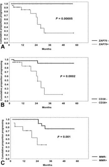
for 5 days) followed by 4 weekly doses of rituximab (375 mg/m2 ). RESULTS.On the basis of National Cancer Institute criteria, 47 of 60 patients (78%) achieved a complete remission, 9 of 60 patients (15%) achieved a partial remission, and 4 of 60 patients (7%) had no response or progressive disease. It is noteworthy that the patients experienced a long progression-free survival (PFS) from treatment (68% at 3 yrs). A significantly shorter PFS was observed in ZAP-70-positive patients (25% vs. 100% at 3 yrs; P⫽ 0.00005), in CD38-positive patients (18% vs. 91% at 3 yrs; P⫽ 0.0002), and in patients who had more minimal residual disease (36% vs. 77% at 2.5 yrs; P⫽ 0.001).
CONCLUSIONS. With the addition of rituximab to fludarabine, improved clinical outcomes were obtained, and the stratification of patients by using ZAP-70 and CD38 may help clinicians offer more aggressive and/or experimental approaches to the treatment of patients with high-risk B-CLL subtypes. Cancer 2005;104: 2743–52. © 2005 American Cancer Society.
KEYWORDS: B-cell chronic lymphocytic leukemia, treatment, fludarabine, rituximab, ZAP-70, CD38, minimal residual disease, response, outcome.
C
hronic lymphocytic leukemia (CLL) is the most common leukemia in the Western world and is characterized by the relentless accu-mulation of functionally abnormal B cells. Despite the⬎ 10-year life expectancy in early stages, patients who have progressive or ad-vanced-stage CLL die rapidly, with a median survival between 18 months and 3 years.1,2Promising results were observed whenfludara-bine was introduced for patients who were treated with alkylator agents and for patients who had symptomatic, untreated CLL.3,4
Although longer progression-free survival (PFS) was reported for the fludarabine arm in three randomized studies,5–7significant improved
survival with fludarabine therapy has not been observed to date in patients with CLL. There is an interest in developing new combina-tion therapies for CLL with agents that can be added to fludarabine to increase the complete remission (CR) rate, because disease recur-rence in patients with CLL most probably is the result of residual
© 2005 American Cancer Society DOI 10.1002/cncr.21535
tumor cells. In fact, studies using minimal residual disease (MRD) assays may be better for confirming a true CR, which is the major therapeutic objective in patients with CLL, to improve PFS and, finally, overall survival (OS). Combinations that include agents with lower toxicity than fludarabine represent a potential advantage over combination strategies with cytotoxic chemotherapy. Rituximab is a chimeric monoclonal antibody directed against the cell-surface antigen CD20, which is active in low-grade and diffuse, large-cell, non-Hodgkin lymphoma (NHL).8 –10 Rituximab
also has single-agent activity in CLL, mainly at esca-lating doses.11,12Moreover, experimental data indicate
a potential superadditive effect of combining ritux-imab with fludarabine because of diminished bcl-2 expression.13 In addition, rituximab can enhance the
sensitivity of tumor cells to chemotherapy-induced apoptosis, and one preclinical study demonstrated synergy between rituximab and fludarabine.14 Two
Phase II studies combining rituximab with fludarabine demonstrated a much higher CR rate than was ob-served previously with any other therapeutic approach that had been used previously in patients with CLL.15,16 However, the analysis of different biologic
parameters may explain better the discordant out-comes independent of the treatment observed in those studies. In fact, the evolution of risk-stratifica-tion models has advanced from clinical staging to include relevant immunologic and genetic features. Recent data from the literature indicate that unmu-tated VHgenes, CD38, ZAP-70 protein tyrosine kinase
overexpression, and MRD may predict a lower re-sponse, a shorter PFS, and a shorter OS.17–26On the
basis of the rationale of the synergistic effects of flu-darabine and rituximab, we conducted a Phase II trial investigating the safety and efficacy of a sequential application schedule in patients with previously un-treated CLL. Moreover, we evaluated the differential clinical impact of relevant biologic features, such as ZAP-70, CD38, and MRD, as determined by flow cy-tometry, on the outcomes of these homogeneously treated patients.
MATERIALS AND METHODS
This prospective Phase II study was conducted be-tween December 1997 and March 2004 in accordance with the Declaration of Helsinki. All eligible patients provided written informed consent. Patients were re-quired to have histologically and immunophenotypi-cally documented CLL, as defined by the modified National Cancer Institute (NCI) 1996 guidelines.27All
patients either required therapy for Rai Stage I–II dis-ease or had Rai Stage III–IV disdis-ease, as defined by the NCI 1996 guidelines. Eligible patients must have
re-ceived no prior therapy for CLL. An Eastern Coopera-tive Oncology Group (ECOG) performance status of 0 –3 was required. Patients were excluded if they had a positive Coombs test, active opportunistic infections, any other severe infection that was not controlled by medical therapy, or major organ dysfunction.
Treatment Plan
Patients received fludarabine (25 mg/m2)
intrave-nously daily on days 1–5 in 28-day intervals for a total of 6 cycles. Then, after a median of at least 40 days (range, 30 –155 days), patients underwent restaging (physical examination, complete blood count [CBC] with manual differential, bone marrow aspirate and biopsy) to determine their response to induction ther-apy. Patients who had stable disease or better, as defined by the NCI criteria, were treated with 4 weekly doses of rituximab (375 mg/m2) with an
administra-tion procedure identical to that described for the NHL trials (Fig. 1).28 Clinical restaging was repeated after
rituximab therapy; patients were observed for approx-imately 1 month and then completely restaged (phys-ical examination, CBC with manual differential, bone marrow aspirate and biopsy) to determine overall re-sponse according to the NCI 1996 criteria.27
Assessment and Management of Toxicity
Hematologic toxicity was graded according to the modified NCI criteria for CLL,27whereas
nonhemato-logic toxicity was graded according to the NCI
Com-FIGURE 1. Treatment scheme of fludarabine combined with rituximab. Patients received 6 monthly courses of fludarabine and 4 weekly doses of rituximab starting an average of 40 days after they completed fludarabine therapy. CR: complete remission; PR: partial remission.
mon Toxicity Criteria. Infusion toxicity was assessed according to the criteria previously described by Byrd et al.16Patients who experienced Grade 3 or 4
neutro-penia/thrombocytopenia or Grade 2 anemia were supported with growth factors (granulocyte-colony stimulating factor and erythropoietin␣) and remained without treatment until these hematologic parameters recovered within 50% of baseline values. Thereafter, these patients received the same original dose. There were no dose reductions for rituximab therapy be-cause of hematologic toxicity. Acute infusion toxicity after rituximab administration that was reversible did not require subsequent dose reduction of this agent. At the onset of fever, chills, rigors, or other infusional reactions, patients had their infusion discontinued and received an additional dose of dipheninhydra-mine (50 mg) and acetaminophen (650 mg). After the resolution of symptoms, the rituximab infusion was restarted at a rate of 50 mg per hour and then was escalated, as tolerated, to 200 mg per hour. Patients who developed an infection were observed without further CLL treatment until the infection had resolved, but no dose reductions were implemented. Nonhema-tologic toxicities, including nausea, emesis, fatigue, diarrhea, and drug-related fever and chills, required no dose reductions.
Patient Monitoring and Response Evaluation
Baseline assessment included disease history, current disease stage, B symptoms, and measurement of le-sions by physical examination and radiography (chest X-ray, computed tomography scan, ultrasound). The histologic examination of representative material (bone marrow biopsy and aspirate, immunopheno-type of peripheral blood) had to be performed at least 6 months before enrollment. Laboratory testing in-volved CBC with differential, flow-cytometric pheno-typing of lymphocytes of the peripheral blood, and clinical chemistry, including immunoglobulins (Ig), 2-microglobulin, and Coombs test.
Patients were assessed for response 4 – 6 weeks after the last rituximab infusion with a detailed clinical evaluation (physical examination with measurement of lymph nodes, liver, and spleen; CBC with differen-tial; bone marrow biopsy and aspirate). Criteria for response used the revised 1996 NCI Working Group guidelines.27A CR or partial remission (PR) had to be
maintained forⱖ 8 weeks. PFS was defined as the time from the end of treatment in the subgroup of patients who achieved a CR or PR until the last follow-up, the time at which progression occurred or further therapy for symptomatic CLL was required, or death that oc-curred from any cause. Survival was measured from
the date of initiation of treatment either until the last time the patient was seen alive or until death.
Sample Size Determination
A single-stage design according to the method of Fleming 29 was selected based on the following
as-sumptions: 1) The treatment in this study would not be considered sufficiently effective if the global re-sponse rate (CR plus PR) was ⬍ 60%. 2) The combi-nation treatment with fludarabine and rituximab would be regarded as very promising for a Phase III comparative trial if the true response rate exceeded 80%. 3) The probability of erroneously declaring the drug active for further investigation, despite a true response rate⬍ 60% (type I error), was set at 5%. 4) The probability of erroneously rejecting the therapy as not sufficiently active (⬍ 60%) if there was a promising true response rate (⬎ 90%) was 10% (type II error; power⫽ 90%).
With regard to feasibility and tolerability, a failure rate of up to 10% was considered acceptable, and the sample size for the evaluation of treatment overall efficacy was set to 60 patients. Moreover, because a major objective of the study was to detect the role of classification of patients into groups of different sizes dichotomized according to specific biologic predic-tors, such as ZAP-70, CD38, and MRD, specific post-hoc power calculations were performed for PFS on any grouping, based on the number of events and on the hazard ratio between groups at the end of the ob-served time, according to the method described by Norman and Streiner.30
Cellular Immunophenotypic Analysis
The following antibody conjugates were used: anti-CD23-phycoerythrin (PE), anti-CD5-fluorescein iso-thiocyanate (FITC), anti-CD38-PE, CD19-allophy-cocyanin (APC), CD45-FITC, CD14-PE, anti-CD95-PE, and anti-CD10-FITC (Becton Dickinson Immunocytometry Systems, San Jose, CA). Peripheral blood mononuclear cells were analyzed for surface expression of CD19/CD5/CD38 and CD19/CD5/CD23 by triple-color immunofluorescence, as described pre-viously.20
ZAP-70 protein determination also was performed by flow cytometry, as described elsewhere.23
Flow-cytometric analyses were performed on a FACS Cali-bur flow cytometer (Becton Dickinson Immunocy-tometry Systems), and CellQuest software was used to acquire and analyze data. The threshold of positivity both for CD38 and for ZAP-70 was set at ⬎ 20% of CD19-positive/CD5-positive B-cells.
MRD Flow-Cytometric Study
Between 50,000 and 300,000 total cells were analyzed in each test that was performed in bone marrow ap-proximately 1–2 months after the completion of com-bined therapy. The antibodies used were anti-CD5-APC, anti-CD20-PE, anti-CD19-peridin-chlorophyll protein, anti-CD38-PE, anti-CD20-FITC (Becton Dick-inson Immunocytometry Systems) and anti-CD79b FITC (Serotec, Oxford, United Kingdom). The 2 com-binations CD19/CD5/CD20/CD79b and CD19/CD5/ CD38/CD20 allowed to discriminate B-CLL cells from normal B cells in all samples, as described previous-ly.26The threshold for positivity was set at⬎ 5% CD19-positive/CD5-positive/CD79b-negative CLL cells.
Interphase Fluorescence in Situ Hybridization
Separate hybridizations were carried out for loci on chromosomes 11, 12, 13, and 17. For chromosomes 11 (q23), 13, and 17, commercial probes (ATM-2, Rb-1, and p53, respectively) were used (Vysis Inc., London, UK). An ␣ satellite DNA probe, CEP12, which was labeled directly with SpectrumGreen, was used to de-tect aneuploidy of chromosome 12. LSIp53 labeled with SpectrumOrange (Vysis Inc.) was used to evaluate chromosome deletion at 17p13.1. The procedure used was described previously.31
IgVH Mutation Analysis
Total RNA was extracted and reverse transcribed, as described previously.32 The resulting cyclic DNAs,
which were checked for first-strand synthesis, were amplified using a mixture of sense primers and an-nealing either to the VH1–VH6 leader sequences or to
the 5⬘ end of VH1–VH6 FR1, as reported elsewhere.33,34
These primers were used in conjunction with a mix-ture of antisense primers complementary to the germ-line JH regions. The purified, amplified products,
which were inserted into the PCR2.1-TOPO vector (In-vitrogen, Milan, Italy), were expanded in TOP10 One Shot™ competent cells (Invitrogen) and cloned. Plas-mid DNAs were isolated from overnight cultures of randomly selected colonies and were sequenced by
using an automatic DNA sequencer (Beckman
CEQ2000XL; Beckman Coulter, Hialeah, FL). Compar-isons between the obtained sequences and the se-quences of the various germline IgVHgenes were
per-formed with the IgBLAST directory (http://www. ncbi.nlm.nih.gov/igblast) using Mac Vector 7.1 se-quence-analysis software (Accelerys; Symantec, San Diego, CA). Only when the same VHDJH
rearrange-ment was identified in at least 5–10 clones was a given IgVHsequence analyzed further. Alignment of the IgVH
sequences available for each patient often revealed,
along with mutations shared by all the transcripts analyzed, several unique or partially shared muta-tions. For this reason, all mutational analyses were carried out in each IgVHtranscript separately, and the
percentage mutation assigned to a given B-CLL was the mean value of the percentage of mutation found in each transcript. VHgene sequences that deviated⬎ 2%
from the corresponding germline gene were defined as mutated.
Statistical Analysis
For the efficacy analysis, patients had to have received at least one cycle of therapy. All patients who had received at least one application of study therapy also were evaluable for toxicity. Rate comparisons between patients subgroups were performed by using the two-tailed Fisher exact test. Event-related data (survival and PFS) were estimated by using the Kaplan–Meier product-limit method. Prognostic subgroups were compared by using the log-rank test. Statistical anal-yses and Kaplan–Meier curves were performed using STATA 8.0 and Statistica威6.0 for Windows.
RESULTS
Patient Characteristics
In total, 60 patients were enrolled in the current study between December 1997 and March 2004. The pre-treatment features of these patients are summarized in Table 1. The median age was 59 years (range, 37–74 yrs). All patients met the guideline criteria for having
TABLE 1
Patient Characteristics (nⴝ 60)
Characteristic No. of patients (%)
Median age in yrs (range) 59 (37–74)
Male gender 30 (50)
Rai modified stage
Low 5 (8)
Intermediate 52 (87)
High 3 (5)
ECOG performance status
0 37 (62)
1 19 (32)
2 4 (6)
B symptoms 17 (28)
Time since first diagnosis
ⱕ 1 yr 17 (29)
2–5 yrs 29 (48)
⬎ 5 yrs 14 (23)
Bone marrow infiltration pattern
Nodular 4 (7)
Mixed 21 (35)
Diffuse 35 (58)
CLL.27 According to Rai staging criteria,1 5 patients
(8%) had low-risk (Stage 0) CLL, 52 patients (87%) had intermediate-risk (Stage I or II) CLL, and 3 patients (5%) had high-risk (Stage III or IV) CLL. In this mod-ified classification scheme, patients with intermedi-ate-risk CLL have lymphadenopathy or hepatospleno-megaly without significant cytopenias, whereas patients with high-risk CLL have anemia or thrombo-cytopenia. Much greater than one-half of patients (62%) had a good ECOG performance status of 0. The time from diagnosis ranged from 1 year to 10 years. Only 28% of patients exhibited B symptoms. Finally, the histologic patterns of bone marrow infiltration were mainly diffuse (58%) or mixed (35%).
Toxicity
All patients were evaluable for toxicity. The three most frequent side effects were infusion-related toxicity, myelosuppression, and infections. Five of 58 patients who were treated with rituximab experienced a mild, infusion-related symptom complex, which consisted of fever, chills, and rigors, during the first infusion. There were 13 opportunistic infections, including 3 dermatomal herpes zoster infections, 7 localized her-pes simplex infections, 2 episodes of Grade 3 pulmo-nary toxicity (isolated interstitial pneumonitis and Candida pneumonia), and 1 incident of fulminant B hepatitis. The reported infectious toxicity was con-fined to the first 12 months of therapy, but no signif-icant opportunistic infections were noted after this period in patients observed in CR or PR without fur-ther treatment. Hematologic toxicity (Table 2) in-cluded neutropenia (Grade 1 and/or 2 in 12 patients, Grade 3 and/or 4 in 29 patients), thrombocytopenia (Grade 1 and/or 2 in 4 patients, Grade 3 and/or 4 in 3 patients), and anemia (Grade 2 in 3 patients). In par-ticular, Grade 3 and 4 neutropenia was observed in 25 of 60 patients (41%) during fludarabine infusions and in 9 of 58 patients (16%) during rituximab treatment. Thrombocytopenia and anemia were noted only dur-ing fludarabine cycles.
Response to Treatment and Treatment Outcome
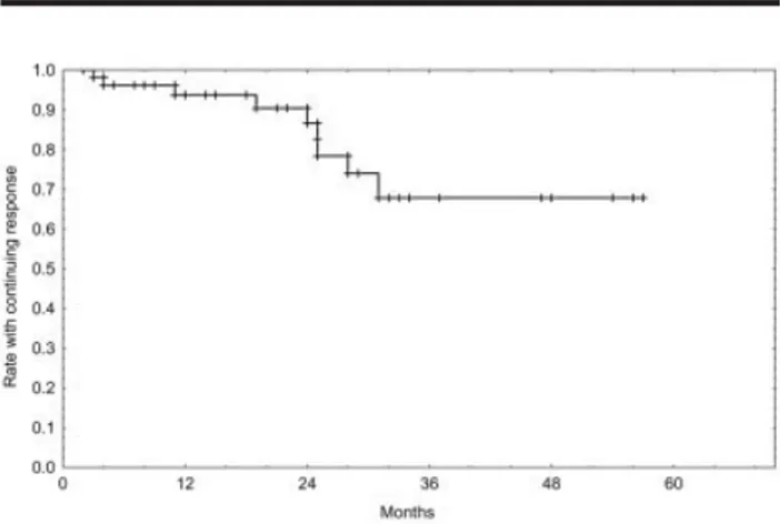
All patients enrolled on this study were evaluable for response. Response evaluation occurred at least 1 month after the completion of fludarabine therapy (induction response) and then again 2 months after the completion of all therapy (fludarabine plus ritux-imab). The response rate included an induction CR rate of 70% and an overall response rate of 92%. Of 13 patients who had a PR after induction who received rituximab, 4 patients (31%) had a conversion to a CR. The comprehensive response rate included a 78% CR rate and an overall response rate of 93% (95% confi-dence interval [95% CI], 84 –98%) (Table 3). Only 1 patient with stable disease after fludarabine therapy had a conversion to a CR after receiving rituximab therapy. Outcome data relative to PFS and OS are shown in Figures 2 and 3. After a median of 27 months of follow-up, 9 of 56 patients (16%) experienced a recurrence. The estimated 3-year PFS rate was 68% (95% CI, 46 – 83%) (Fig. 2). Among the 60 patients
TABLE 2
Hematologic Toxicity During the Treatment Period
Toxicity
No. of patients (%)
NCI Grade 1-2 NCI Grade 3-4
Anemia 3 (5) 0 (0)
Neutropenia 12 (20) 29 (48)
Thrombocytopenia 4 (7) 3 (5)
NCI: National Cancer Institute.
TABLE 3
Comprehensive Response Rate According to Baseline Patient Features (nⴝ 60) Variable No. of patients (%) OR CR PR SD PD Total group (n⫽ 60) 56(93) 47(78) 9(15) 1(2) 3(5) Low stage (n⫽ 5) 5(100) 5(100) — — — Intermediate stage (n⫽ 52) 49(94) 41(79) 8(15) — 3(6) High stage (n⫽ 3) 2(66) 1(33) 1(33) 1(33) No B-symptoms (n⫽ 43) 43(100) 42(98) 1(2) — — B-symptoms (n⫽ 17) 13(76) 5(29) 8(47) 1(8) 3(16)
OR: overall response; CR: complete response; PR: partial response; SD: stable disease; PD: progressive disease.
FIGURE 2.Kaplan–Meier analysis of progression-free survival showed the proportion of 56 patients who had a response to treatment and remained in complete or partial remission. The estimated 3-year progression-free survival rate was 68%, and 9 of 56 patients (16%) experienced a recurrence.
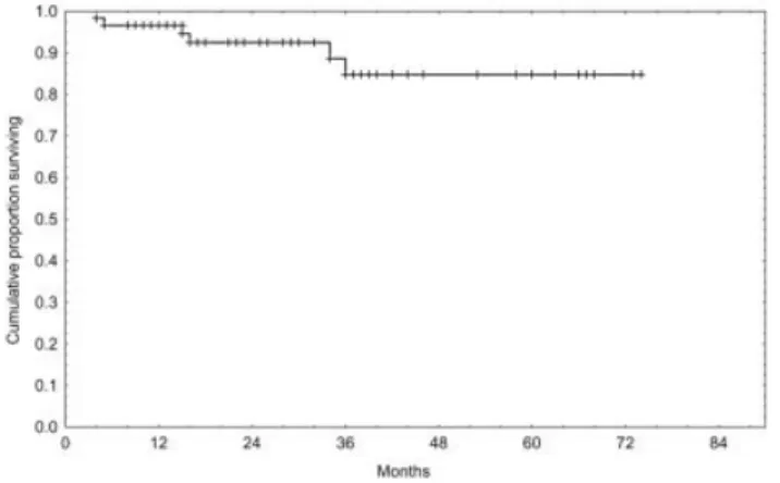
enrolled in this study, 6 patients died. One patient died in CR of fulminant B hepatitis, two other patients died of progressive CLL that was resistant to fludara-bine induction, and the other three patients died of progressive CLL after they received the prescribed protocol therapy.
Clinical and Biologic Features Predicting Response and Outcome
We examined whether age, gender, or2
-microglobu-lin level predicted a CR during fludarabine plus ritux-imab therapy. A higher CR rate was not correlated with gender, age, or2-microglobulin level (P⬎ 0.30 for all
variables). Conversely, there was a trend toward a correlation between the CR rate and the modified Rai stage, because all patients with low Rai stage CLL (n ⫽ 5 patients) achieved a CR (P ⫽ 0.05).
Twenty-eight patients (46.7%) had ZAP-70 expres-sion levels⬎ higher than 20% (range, 4–65%), and 19 of 60 patients (31.7%) presented CD38 levels ⬎ 20% (range, 1– 66%). Tests for MRD, which were performed on bone marrow by flow cytometry after the protocol therapy, were positive (⬎ 5% CD19-positive/CD5-pos-itive/CD79b-negative CLL cells) in 15 of 60 patients (25%). Forty patients were analyzed by interphase flu-orescence in situ hybridization (FISH): Twenty pa-tients (50%) had a normal karyotype, 14 papa-tients (35%) had a 13q⫺ abnormality, 4 patients (10%) had trisomy 12, 1 patient (2.5%) had 11q⫺, and 1 patient (2.5%) had 17p⫺. No significant correlation was found be-tween these cytogenetic abnormalities and ZAP-70 or CD38 expression levels. Thirty-two patients were an-alyzed for IgVHmutations: Fifteen of 16 patients with
IgVHmutations ⬍ 2% presented with ZAP-70 protein
levels⬎ 20% (P ⫽ 0.0006). Moreover, the strict
corre-lation between CD38 and IgVHmutational status was
confirmed (15 of 16 patients with IgVH mutations
⬎ 2% showed CD38 levels ⬍ 20%, P ⫽ 0.0006). With regard to clinical outcomes, all 32 patients who had ZAP-70 expression levels⬍ 20% achieved a CR or a PR (P⫽ 0.001), and a significant higher overall response rate was found among CD38-negative pa-tients (98% vs. 84%; P ⫽ 0.027) (Table 4). No signifi-cant correlation was found between cytogenetic ab-normalities and overall response. Moreover, 42 of 47 patients (93%) who achieved a CR based on NCI cri-teria were negative for MRD according to flow cytom-etry (⬍ 5% CD19-positive/CD5-positiveCD79b-nega-tive CLL cells in bone marrow; P⬍ 0.0001). Finally, all but 1 patient (15 of 16 patients) who presented with IgVHmutations⬎ 2% achieved a CR (P ⬍ 0.01).
A significantly shorter PFS was observed in ZAP-70-positive patients (25% [95% CI, 5–54%] vs. 100% at 3 yrs; P⫽ 0.00005) (Fig. 4A), in CD38-positive patients (18% [95% CI, 1–52%] vs. 91% [95% CI, 66 –98%] at 3 yrs; P⫽ 0.0002) (Fig. 4B), and in patients with higher MRD after treatment (36% [95% CI, 6 – 69%] vs. 77% [95% CI, 48 –91%] at 2.5 yrs; P ⫽ 0.001) (Fig. 4C). Moreover, unmutated patients also showed a signifi-cantly shorter PFS rate (0% vs. 50% at 2 yrs; P⫽ 0.01). Similarly, OS was significantly shorter in ZAP-70-positive patients (68% [95% CI, 40 – 85%] vs. 100% at 3 yrs; P ⫽ 0.006) and in MRD-positive patients (72% [95% CI, 42– 88%] vs. 89% [95% CI, 62–97%] at 3 yrs; P ⫽ 0.018) and showed a significant trend in CD38-positive patients (70% [95% CI, 36 – 89%] vs. 92% [95% CI, 68 –98%] at 3 yrs; P⫽ 0.059). Finally, cytogenetic abnormalities showed no significant prognostic im-pact with regard either PFS or OS.
With regard to PFS, post-hoc power was not cal-culable for ZAP-70 group comparisons, because we
FIGURE 3.Kaplan–Meier analysis of overall survival included all 60 patients with chronic lymphocytic leukemia who were treated with a sequential flu-darabine and rituximab regimen. Six patients died either from progressive disease or from opportunistic infections.
TABLE 4
Comprehensive Response Rate According to ZAP-70 and CD38 Expression Expression status No. of patients (%) OR CR PR SD PD ZAP-70 Positive (n⫽ 28)a 24(86) 16(57) 8(29) 1(4) 3(10) Negative (n⫽ 32) 32(100%) 31(97%) 1(3) — — CD38 Positive (n⫽ 19)b 16(84) 11(58) 5(26) — 3(16) Negative (n⫽ 41) 40(98) 36(88) 4(10) 1(2) —
OR: overall response; CR: complete response; PR: partial response; SD: stable disease; PD: progressive disease.
aP⫽ 0.001 (CR vs. PR vs. SD vs. PD; two-tailed Fisher exact test). bP⫽ 0.027 (CR vs. PR vs. SD vs. PD; two-tailed Fisher exact test).
found that no event (progression) took place in the group of ZAP-70-positive patients. For CD38, post-hoc power was 0.79; whereas for MRD, post-hoc power was 0.22.
DISCUSSION
In this Phase II study, we demonstrated clearly that combined immunochemotherapy with six cycles of fludarabine followed by four cycles of rituximab was safe and very effective in patients with previously un-treated B-CLL. The overall response rate was 93%, with a CR rate of 78% and a PR rate of 15%. This very high CR rate probably was achieved because almost all patients (57 of 60 patients) had CLL with low or inter-mediate Rai stage. In any event, all treated patients had progressive disease and presented with at least 1 of the following parameters: a lymphocyte doubling
time⬍ 1 year, disease progression to a more advanced Rai modified stage, or the development of systemic symptoms. Schulz et al.15 also demonstrated that
there were more CRs among patients who had Binet B CLL (43%) compared with patients who had Binet C CLL (11%) in a similar Phase II trial that was based on a regimen combining rituximab and fludarabine. A CR rate of 28% (PR rate, 49%) was obtained in patients with B-CLL who had both Rai intermediate-risk CLL (58%) and Rai high-risk CLL (42%) and were treated with a sequential scheme based on fludarabine plus rituximab.16Our sequential regimen was very effective
in the patients with intermediate Rai stage disease, as shown above (see Results), and the treatment of this progressive subset before the appearance of anemia or thrombocytopenia allowed us to obtain very good sults in terms of both response and duration of re-sponse. The long-term benefit of this therapy as it relates to PFS and OS is not yet clear, and even if it is superior to fludarabine alone remains undetermined. It is noteworthy that our patients with B-CLL who were treated sequentially with fludarabine and ritux-imab experienced a long PFS after treatment (68% were alive at 3 yrs). Recently, Cancer and Leukemia Group B (CALGB) tested this combined regimen against monotherapy with fludarabine in a retrospec-tive, Phase III clinical trial and demonstrated that, with fludarabine plus rituximab, significantly longer PFS (P⬍ 0.0001) and OS (P ⫽ 0.0006) were achieved.35
The fludarabine plus rituximab treatment regi-men is feasible and can be administered safely on an outpatient basis. Rituximab causes important and prolonged depletion of B lymphocytes. Conversely, fludarabine leads to profound depletion of T cells.36,37
The risk of opportunistic infections may increase greatly with the combination of fludarabine and ritux-imab. In the current study, toxicity was moderate, and there was a slightly increased risk of severe infections. The majority of these opportunistic infections were viral and often localized. There were only 3 patients with Grade 3 or 4 infections, 1 of which was fatal (B hepatitis). Grade 3 and/or 4 hematologic toxicity was observed almost exclusively for neutropenia (48%), compared with thrombocytopenia (5%). These per-centages of serious neutropenia were inferior to those published by Byrd et al.,16who reported that 76% of
their patients had Grade 3 or 4 neutropenia, probably because of the greater numbers of rituximab infu-sions. Nonetheless, this excess neutropenia did not predispose to an excess number of neutropenic fever episodes or life-threatening infections either in the study by Byrd et al.16or in our current study.
Moreover, there were no episodes of autoimmune hemolytic anemia, probably because the transient
B-FIGURE 4.Progression-free survival curves were based on ZAP-70, CD38, and minimal residual disease (MMR) expression. Progression-free survival was significantly shorter in patients with ZAP-70 levels⬎ 20% (ZAP-70 positive [⫹]) (A), CD38 levels ⬎ 20% (CD38⫹) (B), and (C) MRD ⬎ 5% (MMR⫹).
cell depletion caused by rituximab may avoid the de-velopment of fludarabine-associated hemolytic ane-mia. This hypothesis is corroborated by the findings of some authors who observed effective treatment of au-toimmune hemolytic anemia with rituximab.38 One
concern about the use of rituximab in patients with CLL is infusion-related toxicity. This problem arises from studies demonstrating that rituximab can cause severe infusion-related toxicity in a minority of pa-tients and that a high number of circulating B-CLL cells may predispose patients to this toxicity.39,40 In
our study, only 5 patients presented with mild infu-sion-related symptom complex, consisting of fever, chills, and rigors, during the first infusion. Probably, prior treatment with fludarabine greatly diminished the infusion toxicity observed with subsequent ritux-imab treatment, mainly because of lower leukocyte counts.
The low CR rate obtained with previous therapies in patients with CLL relates to several different mech-anisms of disrupted apoptosis.41Both fludarabine and
rituximab enhance different proapoptotic mecha-nisms, based both on bcl-2 down-modulation and caspases activation, so that this combined regimen allowed us to achieve better responses compared with single-agent fludarabine or rituximab.42
Various clinical features, including age, Rai stage, and serum2-microglobulin levels, have been
associ-ated with lower response rates and poor long-term treatment outcomes after alkylator-based and purine analog-based therapy for patients with CLL.43,44
Sev-eral standard prognostic factors, including age, gen-der, Rai stage, and 2-microglobulin levels, did not
reach a level of significance for predicting treatment outcomes either in the current study or in the ran-domized Phase II study by CALGB 9712.16
Recent studies examining molecular aberrations (i.e., p53 mutations), unfavorable cytogenetics, CD38 expression, somatic VH gene mutational status, and ZAP-70 expression, have demonstrated that all of these factors are important determinants for treat-ment outcomes in patients with CLL.45Therefore, the
analysis of unmutated VH genes, CD38 expression,
and/or ZAP-70 protein expression may be better for explaining the discordant outcomes independent of treatment that were observed in these studies. It is noteworthy that, in the current study, ZAP-70 and CD38 overexpression identified a subset of patients with a poor prognosis in terms of overall response, PFS, and OS. Thus, these two markers prospectively may identify subsets of patients who have a low like-lihood of responding to combination therapy with fludarabine and rituximab. In our experience, FISH analysis did not allow us to obtain significant different
risk subsets with regard to response and outcome, perhaps because of the small number of patients an-alyzed. Conversely, MRD assays by flow cytometry allowed us to identify patients who were at signifi-cantly greater risk of recurrence. This is important, because, based on NCI criteria, patients in CR who are MRD-positive (5 of 47 patients in the current study) may be eligible for some consolidation therapy to avoid disease recurrence. These observations may en-courage the use of cyclophosphamide combined with fludarabine and rituximab in these patient subsets with “poor-risk” B-CLL. In fact, investigators at The University of Texas M. D. Anderson Cancer Center used this triple combination and reported a CR rate of 66% in patients with previously untreated CLL.46The ultimate objective in CLL therapy is not only to achieve high CR rates but also mainly to avoid recur-rence, thus prolonging remissions and, finally, sur-vival. The introduction for stratification purposes of some biologic markers, such as ZAP-70 and CD38, into clinical practice may allow us prospectively to isolate patients who respond poorly to fludarabine plus rit-uximab and who may be potential candidates for more aggressive and/or experimental approaches.47
REFERENCES
1. Rai KR, Sawitsky A, Cronkite EP, Chanana AD, Levy RN, Pasternack BS. Clinical staging of chronic lymphocytic leu-kaemia. Blood. 1975;46:219 –234.
2. Binet JL, Leporrier M, Dighiero G, et al. A clinical staging system for chronic lymphocytic leukaemia. Cancer. 1977;40: 855– 864.
3. Keating MJ, Kantarjian H, Talpaz M, et al. Fludarabine: a new agent with major activity against chronic lymphocytic leukaemia. Blood. 1989;74:19 –25.
4. Keating MJ, Kantarjian H, O’Brien S, et al. Fludarabine: a new agent with marked cytoreductive activity in untreated chronic lymphocytic leukaemia. J Clin Oncol. 1991;9:44 – 49. 5. Johnson S, Smith AG, Loffler H, et al. Multicentre prospec-tive randomized trial of fludarabine versus cyclophospha-mide, doxorubicin, and prednisone (CAP) for treatment of advanced-stage chronic lymphocytic leukaemia. Lancet. 1996;347:1432–1438.
6. Rai KR, Peterson BL, Appelbaum FR, et al. Fludarabine com-pared with chlorambucil as primary therapy for chronic lymphocytic leukaemia. N Engl J Med. 2000;343:1750 –1757. 7. Leporrier M, Chevret S, Cazin B, et al. Randomized compar-ison of fludarabine, CAP, and CHOP in 938 previously un-treated Stage B and C chronic lymphocytic leukemia pa-tients. Blood. 2001;98:2319 –2325.
8. Maloney DG, Grillo-Lopez AJ, White CA, et al. IDEC-C2B8 (rituximab) anti-CD20 monoclonal antibody therapy in pa-tients with relapsed low-grade non-Hodgkin’s lymphoma.
Blood. 1997;90:2188 –2195.
9. Coiffier B, Haioun C, Ketterer N, et al. Rituximab for the treatment of patients with relapsing or refractory aggressive lymphoma: a multicenter Phase II study. Blood. 1998;92: 1927–1932.
10. Davis TA, White CA, Grillo-Lopez AJ, et al. Single agent monoclonal antibody efficacy in bulky non-Hodgkin’s lym-phoma: results of a Phase II trial of rituximab. J Clin Oncol. 1999;17:1851–1857.
11. O’Brien SM, Kantarjian H, Thomas DA, et al. Rituximab dose-escalation trial in chronic lymphocytic leukemia. J Clin
Oncol. 2001;19:2165–2170.
12. Byrd JC, Murphy T, Howard RS, et al. Rituximab using a thrice weekly dosing schedule in B-cell chronic lymphocytic leukemia and small lymphocytic lymphoma demonstrates clinical activity and acceptable toxicity. J Clin Oncol. 2001; 19:2153–2164.
13. Alas S, Bonavida B. Rituximab inactivates signal transducer and activation of transcription 3 (STAT3) activity in B-non Hodgkin’s lymphoma through inhibition of the interleukin 10 autocrine/paracrine loop and results in down-regulation of Bcl-2 and sensitisation to cytotoxic drugs. Cancer Res. 2001;61:5137–5144.
14. Di Gaetano N, Xiao Y, Erba E, et al. Synergism between fludarabine and rituximab revealed in a follicular lymphoma cell line resistant to the cytotoxic activity of either drug alone. Br J Haematol. 2001;114:800 – 809.
15. Schulz H, Klein SK, Rehwald U, et al. Phase II study of a combined immunochemotherapy using rituximab and flu-darabine in patients with chronic lymphocytic leukemia.
Blood. 2002;100:3115–3120.
16. Byrd JC, Peterson BL, Morrison VA, et al. Randomized Phase II study of fludarabine with concurrent versus sequential treatment with rituximab in symptomatic, untreated pa-tients with B-cell chronic lymphocytic leukemia: results from Cancer and Leukemia Group B 9712 (CALGB 9712).
Blood. 2003;101:6 –14.
17. Damle RN, Wasil T, Fais F, et al. IgV gene mutation status and CD38 expression as novel prognostic indicators in chronic lymphocytic leukemia. Blood. 1999;94:1840 –1847. 18. Hamblin TJ, Davis Z, Gardiner A, Oscier DG, Stevenson FK.
Unmutated IgV(H) genes are associated with a more aggres-sive form of chronic lymphocytic leukemia. Blood. 1999;94: 1848 –1854.
19. Hamblin TJ, Orchard JA, Davis Z, Gardiner A, Oscier DG, Stevenson FK. Immunoglobulin V genes and CD38 expres-sion in CLL. Blood. 2000;95:2455–2557.
20. Del Poeta G, Maurillo L, Venditti A, et al. Clinical signifi-cance of CD38 expression in chronic lymphocytic leukemia.
Blood. 2001;98:2633–2639.
21. Hamblin TJ, Orchard JA, Ibbotson RE, et al. CD38 expression and immunoglobulin variable region mutations are inde-pendent prognostic variables in chronic lymphocytic leuke-mia, but CD38 expression may vary during the course of disease. Blood. 2002;99:1023–1029.
22. Chen L, Widhopf G, Huynh L, et al. Expression of ZAP-70 is associated with increased B-cell receptor signaling in chronic lymphocytic leukemia. Blood. 2002;100:4609 – 4614. 23. Crespo M, Bosch F, Villamor N, et al. ZAP-70 expression as a surrogate for immunoglobulin-variable-region mutations in chronic lymphocytic leukemia. N Engl J Med. 2003;348: 1764 –1775.
24. Wiestner A, Rosenwald A, Barry TS, et al. ZAP-70 expression identifies a chronic lymphocytic leukemia subtype with un-mutated immunoglobulin genes, inferior clinical outcome, and distinct gene expression profile. Blood. 2003;101:4944 – 4951.
25. Orchard JA, Ibbotson RE, Davis Z, et al. ZAP-70 and
prog-nosis in chronic lymphocytic leukaemia. Lancet. 2004;363: 105–111.
26. Rawstron AC, Kennedy B, Evans PAS, et al. Quantitation of minimal disease levels in chronic lymphocytic leukaemia using a sensitive flow cytometric assay improves the predic-tion of outcome and can be used to optimize therapy. Blood. 2001;98:29 –35.
27. Cheson BD, Bennett JM, Grever M, et al. National Cancer Institute sponsored Working Group Guidelines for chronic lymphocytic leukemia: revised guidelines for diagnosis and treatment. Blood. 1996;29:4990 – 4997.
28. Mc Laughlin P, Grillo-Lopez AJ, Link BK, et al. Rituximab chimeric anti-CD20 monoclonal antibody therapy for re-lapsed indolent lymphoma: half of patients respond to a four-dose treatment program. J Clin Oncol. 1998;16:2825– 2833.
29. Fleming TR. One-sample multiple testing procedure for Phase II clinical trials. Biometrics. 1982;38:143–151. 30. Norman GR, Streiner DL. Biostatistics: the bare essentials.
Hamilton, Ontario, Canada: BC Dekker Inc., 1997. 31. Del Principe MI, Del Poeta G, Venditti A, et al. Clinical
significance of soluble p53 protein in B-cell chronic lym-phocytic leukemia. Haematologica. 2004;89:1468 –1475. 32. Gattei V, Degan M, Gloghini A, et al. CD30 ligand is
fre-quently expressed in human hematopoietic malignancies of myeloid and lymphoid origin. Blood. 1997;89:2048 –2059. 33. Fais F, Ghiotto F, Hashimoto S, et al. Chronic lymphocytic
leukemia B cells express restricted sets of mutated and unmutated antigen receptors. J Clin Invest. 1998;102:1515– 1525.
34. Gurrieri C, McGuire P, Zan H, et al. Chronic lymphocytic leukemia B cells undergo somatic hypermutation and intra-clonal VHDJHgene diversification. J Exp Med. 2002;196:629 – 639.
35. Byrd JC, Rai K, Peterson BL, et al. Addition of rituximab to fludarabine may prolong progression-free survival and over-all survival in patients with previously untreated chronic lymphocytic leukemia: an update retrospective comparative analysis of CALGB 9712 and CALGB 9011. Blood. 2005;105: 49 –53.
36. Morrison VA, Rai KR, Peterson BL, et al. Impact of therapy with chlorambucil, fludarabine, or fludarabine plus chlorambucil on infections in patients with chronic lym-phocytic leukemia: Intergroup Study Cancer and Leukemia Group B 9011. J Clin Oncol. 2001;19:3611–3621.
37. Byrd JC, Houdle-McGrail LL, Hospenthal DR, Howard RS, Dow N, Diehl LF. Herpes virus infections occur frequently following treatment with fludarabine: results of a prospec-tive natural history study. Br J Haematol. 1999;105:445– 447. 38. Quartier P, Brethon B, Philippet P, et al. Treatment of child-hood autoimmune haemolytic anemia with rituximab.
Lan-cet. 2001;358:1511–1513.
39. Jensen M, Winkler U, Manzke O, Diehl V, Engert A. Rapid tumor lysis in a patient with B-cell chronic lymphocytic leukemia and lymphocytosis treated with an anti-CD20 monoclonal antibody (IDEC-C2B8, rituximab). Ann
Hema-tol. 1998;77:89 –91.
40. Byrd JC, Waselenko JK, Maneatis TJ, et al. Rituximab therapy in hematologic malignancy patients with circulating blood tumor cells: association with increased infusion-related side effects and rapid blood tumor clearance. J Clin Oncol. 1999; 27:791–795.
41. Kitada S, Andersen J, Akar S, et al. Expression of apoptosis-regulating proteins in chronic lymphocytic leukemia: corre-lations with in vitro and in vivo chemoresponses. Blood. 1998;91:3379 –3389.
42. Byrd JC, Kitada S, Flinn IW, Pearson M, Shinn C, Reed J. The mechanism of tumor cell clearance by rituximab in vivo in patients with B-cell chronic lymphocytic leukemia: evidence of caspase activation and apoptosis induction. Blood. 2002; 99:1038 –1043.
43. Cheson BD, Zwiebel J. Chronic lymphocytic leukemia: stag-ing and prognostic factors. Semin Oncol. 1998;25:42–59. 44. Keating M, Lerner S, Kantarjian H, Freirech E, O’Brien S. The
serum B2-microglobulin level is more powerful than stage in
predicting response and survival in chronic lymphocytic leukemia (CLL) [abstract]. Blood. 1995;86:606a.
45. Shanafelt TD, Geyer SM, Kay NE. Prognosis at diagnosis: integrating molecular biologic insights into clinical practice for patients with CLL. Blood. 2004;103:1202–1210.
46. Wierda W, O’Brien S, Albitar M, et al. Combined fludara-bine, cyclophosphamide, and rituximab achieve a high complete remission rate as initial treatment for chronic lymphocytic leukemia [abstract]. Blood. 2001;98:771a. 47. Del Principe MI, Del Poeta G, Maurillo L, et al. Addition of
rituximab to fludarabine improves progression free survival in untreated ZAP-70 negative chronic lymphocytic leukemia (CLL) [abstract]. Blood. 2004;104:477a.