Ped Urol Case Rep 2017; 4(1):267-274 DOI: 10.14534/PUCR.2017124190
Split-appendix technique: Alternative urinary diversion for pediatric
complete incontinence
Elisa Cerchia, Francesco Molinaro, Rossella Angotti, Marina Sica, Adrian
Bianchi, Mario Messina
The Section of Pediatric Surgery, Department of Medical Sciences, Surgery and Neuroscience, University of Siena, Italy
A B S T R A C T
We report our series of selected patients with complete incontinence in whom the appendix was divided and utilized for creating two continent catheterizable stomas. All patients were treated for urinary and fecal incontinence by split appendix technique. The appendix was divided into two different parts preserving adequate perfusion and used for creating an appendicocecostomy (ACE) and an appendicovesicostomy at the same time. After a clinical and radiological follow up, our patients referred a good acceptance and an easily management of the stomas in order to stay dry for all day from urine and feces with improving of their quality of life. The combination of ACE and Mitrofanoff principle have revolutionized the management of urinary and fecal incontinence in patients who are unable to utilize their urethra to keep themselves dry.
Key Words: Split-appendix technique, appendicovesicostomy, complete incontinence, appendicocecostomy
Copyright © 2017 pediatricurologycasereports.com
Corresponding Author: Dr. Elisa Cerchia, Division of Pediatric Surgery, Dept. of Medical Sciences, Surgery and Neuroscience University of Siena. Polyclinic “Le Scotte” Viale Bracci 53100 – Siena. Email: [email protected]
Accepted for publication: 12 December 2016 Introduction
In the last decades, the introduction of intermittent catheterized stoma has revolutionized the surgery for total continent reconstruction with the purpose to preserve urinary tract function, urinary continence, elimination of fecal soiling, and preservation
of quality of life with complete self-sufficiency. The total incontinence for congenital or acquired defects represents a handicap that impairs chances of integration in society and also creates a high risk for poor self-esteem. This clinical condition is common and accounts for a large percentage of children who required more complex diversion, because treatment of one system (urinary or intestinal) could be limited management of the other. In these cases the creation of two continent catheterizable stomas by using an association of Mitrofanoff and Malone’s
PEDIATRIC UROLOGY CASE REPORTS
ISSN 2148-2969
Journal homepage: http://www.pediatricurologycasereports.com
Ped Urol Case Rep
technique is conceptually the best surgical choice [1-2]. In the last years many surgical variation to these techniques have been made for achieving the purpose of continence, taking into account the expectations and living conditions of each individual patient. We report our series utilizing an alternative surgical strategy in select clinical circumstances of complete incontinence with the aim of expanding the resources of a reconstructive surgery in accordance with the needs of the patients and their family.
Material and Methods
A retrospective review of our case series of three patients is presented, two of which were from developing countries where there is a lack of acceptance of urethral catheterization procedure. In all cases there was a depth interview to understand the needs and ability of acceptance of a permanent stoma from the patients and their family.
Case 1
A 9-year-old male patient was referred for complete incontinence in caudal regression syndrome. At the birth a loop colostomy was performed in the left upper quadrant for imperforate anus with recto-urethral fistula that was closed at the age of two months and completed by a posterior sagittal anorectoplasty. We took care of him when he was 9 years old. At physical examination, he showed a hypoplastic and curved penis with anterior hypospadias. He also had sacral and lower limb anomalies with popliteal webbing associated to equinovarus deformity of the feet, and no craniofacial or upper limb anomalies were evident. His motor and sensory development was normal. Clinically, he was continually wet and dirty for urinary and fecal incontinence, but he referred sensation of a full
bladder and to defecate. Abdominal and pelvic magnetic resonance showed a complete hypothrophic pelvic floor, a malrotated incomplete duplex kidney on right side, a bilateral hydroureteronephrosis with focal cortical thinning as the result of recurrent infections, a hypertrophic bladder detrusor and a voluminous stone in the bladder itself. Computerized tomography of the pelvis confirmed the presence of bladder stone (5x2.5 cm), the absence of sacral vertebrae from S3 to coccyx, intervertebral osteochondrosis from L4 to L5 with consequent stenosis of the channel at this level and “hourglass” appearance of the pelvic foramen. A preoperative cystography showed a bladder of low capacity with irregular wall due to hypertrophic detrusor and a left vesicoureteral reflux of III grade.
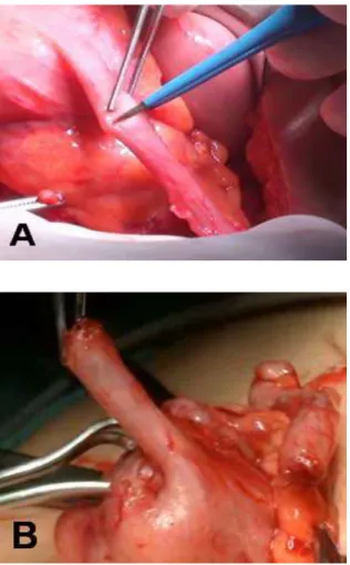
The patient was submitted to surgical reconstruction of two continent catheterizable stoma through appendix, known as split appendix technique. The skin incision was made through Mc Burney’s point, the cecum was identified and brought into the wound. The appendix was examined carefully to verify a good length and vascularization. An appendicular length of approximately 8 cm well vascularized was isolated on its mesentery. Another access was via a Pfannenstiel incision allowing to open the anterior bladder wall and to remove the great stone (4 cm in the major diameter). Then was carefully evaluated the distance between the ileo-cecal junction and the bladder wall and from this last and the abdominal wall, so it was decided to perform a Mitrofanoff appendicovesicostomy procedure (AV) in conjunction with appendicocecostomy (ACE) procedure. The appendix was divided into two different parts preserving adequate perfusion [Fig. 1 A, B].
Fig. 1 A, B. Splitting of the appendix.
The distal portion of the appendix (2/3 of the initial length) with its vascular pedicle was directed through an antirefluxing tunnel into the anterior bladder wall [Fig. 2 A, B]. The abdominal end of the conduit is brought through the abdominal wall, and a stoma was fashioned as rose-bud.
Fig. 2A, B. Performing of appendicovesicostomy. The proximal portion of the appendix (1/3 of the initial length) was kept in continuity with the cecum. A cecal myotomy was created along the anterior tenia for 2 cm and the residual appendix was tunneled with muscular and serosal layer of the cecum to provide a mechanism for fecal continence [Fig. 3 A, B].
Fig. 3 A, B. Performing of appendicocecostomy. An anastomosis of the skin, after a star-shaped incision was performed, to the apex of the spatulated appendix with creation an ACE as
rose bud. Postoperatively, a suprapubic catheter was left in situ for six weeks, initially on free drainage and after four weeks alternatively opened every 4-5 hours. A catheter 6 Ch was inserted through each conduit, that it was positioned in ACE was removed after 3 weeks and then used only for colonic irrigation once daily with 500 ml of saline solution, the other positioned in AV was removed after five weeks and the patient was trained to perform intermittent catheterization every 3 hours during the day. Fecal continence was achieved for a period of 18-20 hours, while urinary continence was obtained after suburethral endoscopic injection of dextranomer/hyaluronic acid (Deflux) and intermittent catheterization every 3 hours with an evident reduction of upper urinary tract dilatation. No stomal stenosis or leakage were present. The patient was discharged from the hospital after 2 months. After a two years of follow up by clinical and radiological controls, he referred to manage the stomas in order to stay dry for all day from urine and feces without any problems, and this allowed him to start to attend school and improve his self-esteem.
Case 2
A 16-year-old male patient was referred for complete incontinence in caudal regression syndrome. His antenatal history was unremarkable and family history revealed consanguineous parents. At the birth a loop colostomy was performed in the left upper quadrant for imperforate anus with recto-urethral fistula that was closed at the age of two years and completed by a posterior sagittal anorectoplasty. We took care of him when he was 16 years old. At physical examination, he showed sacral and lower limb anomalies with popliteal webbing associated to equinovarus
deformity of the feet with normal motor and sensory development. Clinically, he was continually wet and dirty for urinary and fecal incontinence, but he referred sensation of a full bladder and to defecate. Abdominal and pelvic magnetic resonance showed a complete hypothrophic pelvic floor with a megarectum, an agenesis of the left kidney, a right hydroureteronephrosis with focal cortical thinning as the result of recurrent infections associated to partial renal insufficiency, a hypertrophic bladder detrusor, and the absence of sacral vertebrae from S3 to coccyx. The pre-operative cystoscopy showed an urethra hardly catheterizable with dilatation of the prostatic tract and a small bladder. The patient was submitted to surgical correction by resection of megarectum, pull through of residual colon performing a neo-anus, and split appendix technique with the creation of an ACE and AV as in the previous case. An appendix of approximately 7 cm length was divided in a distal portion (2/3 of the initial length) for creating an antirefluxing tunnel into the anterior bladder wall and a proximal portion (1/3 of the initial length) kept in continuity with the cecum. Postoperatively, a suprapubic catheter was left in situ for six weeks, initially on free drainage and after four weeks alternatively opened every 4-5 hours. A catheter 8 Ch was inserted through each conduit, that it was positioned in ACE was removed after 2 weeks and then used only for colonic irrigation once daily with 400 ml of saline solution, the other positioned in AV was removed after five weeks and the patient was trained to perform intermittent catheterization every 2 hours during the day. Fecal continence was achieved for a period of 24-28 hours, while urinary continence was obtained through intermittent catheterization every 3 hours with an evident reduction of upper urinary tract
dilatation. No stomal stenosis or leakage were present. The patient was discharged from the hospital after 2 months. After one year of follow up by clinical and radiological controls, he referred a good management of the stomas with an improvement of his quality of life and renal functionality.
Case 3
A 17-year-old male patient was referred for complete incontinence in Currarino syndrome. His antenatal history showed presence of anterior meningocele treated surgically at the age of two years, sacral agenesis (S2-coccyx), fecal and urinary incontinence due to neurogenic bowel and bladder, and hypospadias. The patient underwent a surgical correction of urinary incontinence by diverticulocystoplasty with suprapubic cystostomy at an outside institution at the age of 2 years. This procedure was complicated by recurrent urinary infections and continuous loss of urine despite a regular catheterization. We took care of him when he was 17 years old. The family expressed the desire of a solution to incontinence by means of stomas easily managed to increase their independence. Abdominal and pelvic magnetic resonance showed a complete hypothrophic pelvic floor with a megarectosigmoid, and horseshoe left kidney with hydroureteronephrosis, a hypoplastic right kidney, a hypertrophic bladder detrusor, and the absence of sacral vertebrae from S2 to coccyx. The patient was submitted to surgical correction by resection of megarectosigmoid following by a termino-terminal anastomosis between the first dilated tracts of sigmoid colon with 5 cm of residual rectum allowing a reservoir for an adequate volume of feces. A very short appendix of approximately 5 cm length was divided in a distal portion (2/3 of the initial length) for
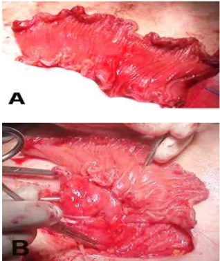
creating an antirefluxing tunnel into the posterior bladder wall and a proximal portion (1/3 of the initial length) kept in continuity with the cecum that was tubularized on a Nelaton 6 Ch for 1 cm of its distal portion with the aim to lengthen the ACE. The bladder was very small with fibrotic wall, so we decided to expand it using 16 cm of terminal ileum. The ileac loop was isolated with particular care not to destroy any of its blood supply. Normal continuity of the ileum was re-established by ileum-ileum anastomosis. The posterior wall of the bladder was dissected free from the anterior wall of the peritoneum downward toward the cul-de-sac of Douglas, and the bladder was opened in its dome. Catheters 6 Ch were placed in the ureteral orifices in order to identify them and prevent ureteral obstruction or damage from subsequent suturing. Then the segment was opened by cutting along the antimesenteric border and the ileac patch was then brought downward to the back of the bladder and anastomosed [Fig. 4 A, B].
Fig 4 A, B. Isolation of 16 cm of ileum and ileocistoplasty.
Postoperatively, two ureteral catheters and a bladder catheter was left in situ respectively for seven days and two weeks. A catheter 8 Ch was inserted through the ACE and removed after 3 weeks and then used only for colonic irrigation once daily with 400 ml of saline solution, the other positioned in AV was removed after 4 weeks and the patient was trained to perform intermittent catheterization every 3 hours during the day. Fecal continence was achieved for a period of 20-24 hours, while urinary continence was obtained through intermittent catheterization every 3 hours with an evident reduction of upper urinary tract dilatation. No stomal stenosis or leakage were present. The patient was discharged from the hospital after 1 months. After six months of follow up by clinical and radiological controls, the parents referred no infections and an easy management of the stomas.
Discussion
Urinary and fecal incontinence secondary to neurogenic bladder and bowel, respectively, are frequently encountered comorbidities in patients with myelomeningocele as well as anorectal malformation [3]. In the last decades total continent reconstruction has been increasingly performed as experience grows with various surgical techniques. Continent catheterizable channels have become an important means to achieve urinary and fecal continence. The concept of the bladder emptying through clean intermittent catheterization was introduced by Lapides in 1972 [4]. Then in 1980, Mitrofanoff described the trans-appendicular continent cystostomy as an alternative route for catheterizing the bladder when the urethra could not be used [1], including urethral strictures and injuries, bladder dysfunction associated with an intact urethral sensation (bladder or cloacal
exstrophy and epispadias, prune-belly syndrome and idiopathic dysfunctional bladder). The basic principle was the persistence of a positive pressure gradient between the conduit lumen in the antireflux tunnel and the reservoir [5]. This concept was later adopted in the management of bowel incontinence, a procedure known as the antegrade continence enema trough a catheterizable ACE and described by Malone in 1990 [6].
The appendix is the preferred conduit for a catheterizable continent stoma, in fact the main advantages of the appendix are good blood supply, satisfactory lumen and auto-lubrication. Several authors have described variations on the classical technique described by Mitrofanoff. Wedderburn [7] and Kajbafzadeh [8] reported the use of the divided appendix in children requiring simultaneous ACE and AV procedures. The split-appendix technique allows the surgeon to create two channels from a single luminal structure. An appendiceal length of approximately 9 to 15 cm was necessary for the surgeon to contemplate the appropriateness of the split-appendix technique. Additional factors involved in the decision-making process were cecal mobility and appendiceal vascular anatomy [2]. Even if the successful results of this procedure, many problems of catheterization are common. Barqawi [9] analyzed the factors associated with problematic conduits in a study of 22 ACE and AV conduits. At 4-year follow up, 5% of their AV required revision. The most common complications were stenosis in 25%, leakage in 8%, and false passage in 2%. Following revision, 97% of their Mitrofanoff channels, and 99% of the ACE conduits, were continent. In another series of 43 patients submitted to split-appendix technique, VanderBrink [10]
reported a stomal continence rate of 95% with the surgical revision rate of 19% for stomal stenosis, incontinence, difficulty catheterizing and peristomal abscess. Also in 9 of these patient, asymmetrical separation with stapled appendicocecal extension was used, mean length of the appendix for AV and ACE was 6 and 4 cm, respectively.
The stomal continence and the surgical revision in our series using a split-appendix technique is similar to other studies. Utilizing this approach, the patients in our series enjoyed the benefit of a unique surgical management of both problems. The other important factor to optimize the outcome of these patients with regards to fecal continence is to perform a technically correct operation initially in order to avoid a reoperation. In one case we found a complex situation pluri-operated that required a multiple-approach with an extended resection of megarectosigmoid and an augmented bladder as well as the split appendix made difficult by the reduced length of the appendix and the presence of a mesenterium under tension. These patients are incontinent of stool because they are not capable of having voluntary bowel movements [11]. In the most severe form, hypomotility is associated to dilation of the rectum and sigmoid colon that can lead to overflow pseudoincontinence and required a more complex bowel management program with not only daily enema but also by resection of megarectosigmoid for avoid the failure of Malone procedure.
A proper surgical selection is a fundamental component of achieving successful operative outcomes, but also it is fundamental that the family and the patient understand the need to properly use the stomas, this justifies the long-term hospitalization of our patients. We also described in our series a good follow up at two
years which allows us to exclude long-term complications and to understand what is the best attitude to allow the acceptance of permanent stomas by children.
Conclusions
The combination of ACE and Mitrofanoff principle have revolutionized the management of urinary and fecal incontinence in patients who are unable to utilize their urethra to keep themselves dry. Our results with this technique has been favorable and resulted in creation of reliable, continent catheterizable channels with a good compliance. Most of the complications are preventable by meticulous technique, and eventually multistage surgical corrections can lead to a functionally and an aesthetically superior reconstruction with a potentially excellent prognosis
Acknowledgements
The author(s) declare that they have no competing interest and financial support.
References
[1]Mitrofanoff P. Trans-appendicular
continent cystostomy in the management of the neurogenic bladder. Chir Pediatric. 1980;21(4):297-305.
[2]Cerchia E, Bulotta AL, Ruscelli M, Angotti R., Di Maggio G, Messina M. Split-appendix technique: surgical choice for complete incontinence in caudal regression syndrome. JSAS. 2013:93-97.
[3]VanderBrink BA., Levitt MA, Defoor WR,
Alam S. Creation of an
appendicovesicostomy Mitrofanoff from a preexisting appendicocecostomy utilizing the split appendix technique. J Pediatr Surg. 2014;49(4):656-9.
[4]Lapides J, Diokno AC, Silber SJ, Lowe BS. Clean, intermittent self-catheterization in
the treatment of urinary tract disease. J Urol. 1972;107(3):458-61.
[5]Hinman F Jr. Functional classification of conduits for continent diversion. J Urol 1990;144(1):27-30.
[6]Malone PS, Ransley PG, Kiely EM.
Preliminary report: the antegrade
continence enema. Lancet.
1990;336(8725):1217-8.
[7]Wedderburn A, Lee RS, Denny A,
Steinbrecher HA, Koyle MA, Malone PS. Synchronous bladder reconstruction and ACE procedure. J Urol. 2001;163(6Pt 2):2392-3.
[8]Kajbafzadeh AM, Chubak N.
Simultaneous Malone antegrade continent enema and Mitrofanoff principle using the divided appendix: report of a new technique for prevention of stoma complications. J Urol. 2001;165(6 Pt 2):2404-9.
[9]Barqawi A, de Valdenebro M, Furness PD
3rd, Koyle MA. Lessons learned from stomal complications in children with cutaneous catheterizable continent stomas. BJU Int. 2004;94(9):1344-7.
[10]VanderBrink BA, Cain MP, Kaefer M, Meldrum KK, Misseri R, Rink RC.
Split-appendix technique for simultaneous
appendicovesicostomy and
appendicocecostomy. J Ped Surg.
2011;46(1):259-262.
[11]Bischoff, Andrea, and Manuel Tovilla. A practical approach to the management of pediatric fecal incontinence. Seminars in pediatric surgery. 2010 Vol. 19. No. 2. WB Saunders, p154-159.
Access this article online http://pediatricurologycasereports.com
Quick Response Code
Pediatric Urology Case Reports is an open access journal. Articles published in this journal are licensed under the Creative Commons Attribution 4.0 International License (see http://creativecommons.org/ and http://creativecommons.org/licenses/by/4.0/).