© 2017 Surgical Neurology International | Published by Wolters Kluwer - Medknow
Editor:
Daniel Silbergeld, University of Washington Medical Center, Seattle, Washington, USA OPEN ACCESS
For entire Editorial Board visit : http://www.surgicalneurologyint.com
SNI: Neuro-Oncology
Case Report
A rare association of ganglioglioma and cavernous malformation:
Report of two cases and literature review
Biagio R. Carangelo, Giovanni Muscas
1, Clelia Miracco
2, Vitaliano F. Muzii
1Department of Neurological and Sensory Sciences, Division of Neurosurgery, Siena University Hospital, 1Department of Medicine, Surgery, and Neurosciences,
Section of Neurosurgery, University of Siena, Siena, 2Department of Medicine, Surgery, and Neurosciences, Section of Human Pathology, University of Siena, Siena,
Italy
E‑mail: Biagio R. Carangelo ‑ [email protected]; Giovanni Muscas ‑ [email protected]; Clelia Miracco ‑ [email protected]; *Vitaliano F. Muzii ‑ [email protected]
*Corresponding author
Received: 12 September 16 Accepted: 13 December 16 Published: 26 May 17
Abstract
Background: Some glial tumors have been observed in association with different
types of vascular malformations of the brain (angiogliomas). However, the association of ganglioglioma with other vascular malformations is extremely rare, with only few cases reported in the literature, one of which is referred to as “angioganglioglioma.”
Case Description: Two patients presented with acute onset of neurological symptoms,
with magnetic resonance imaging (MRI) finding of cavernoma of the left middle cerebellar penduncle, and small mass of the chiasmatic region, respectively. After microsurgical excision, histopathological examination revealed mixed ganglioglioma and cavernous malformation in both cases. Postoperative course was uneventful, and follow‑up MRI showed complete removal of the tumor with no recurrence after 4 years.
Conclusions: Angiogliomas are very uncommon tumors. In literature, we found
different interpretations of such lesions, although they should most probably be considered as distinct pathological entities. Although the association of ganglioglioma with cavernoma is extremely rare, it could be considered as a most peculiar form of angioglioma, and supports the existence of angioganglioglioma.
Key Words: Angioglioma, brain tumor, cavernoma, ganglioglioma, mixed tumor INTRODUCTION
Gangliogliomas are rare, mostly benign intraaxial tumors originating at various sites in the central nervous system (CNS). The term ganglioglioma was first introduced by Perkins in 1926, defining a neoplasm of
both astrocytic and neuronal components.[12,14]
The term “angioglioma” has been used to describe glial neoplasm with features of both glioma and vascular malformation. The cases described are very few, and such
a histological entity is only controversially accepted.[6,16]
In 2003, Kupnicka et al. first described an unusual brain tumor consisting of three components, namely, pylocytic
astrocytoma, arteriovenous malformation (AVM)‑like vascular proliferation, and gangliomatic proliferation. The term “angioganglioglioma” was proposed to define a transitional form between angioglioma and
How to cite this article: Carangelo BR, Muscas G, Miracco C, Muzii VF. A rare
association of ganglioglioma and cavernous malformation: Report of two cases and literature review. Surg Neurol Int 2017;8:94.
http://surgicalneurologyint.com/A-rare-association-of-ganglioglioma-and-cavernous-malformation:-Report-of-two-cases-and-literature-review/
This is an open access article distributed under the terms of the Creative Commons Attribution-NonCommercial-ShareAlike 3.0 License, which allows others to remix, tweak, and build upon the work non-commercially, as long as the author is credited and the new creations are licensed under the identical terms.
For reprints contact: [email protected]
Access this article online Website:
www.surgicalneurologyint.com
DOI:
10.4103/2152-7806.207115
Surgical Neurology International 2017, 8:94 http://www.surgicalneurologyint.com/content/8/1/94
ganglioglioma.[6] Several other cases of mixed tumor
and vascular malformation have been described in
the literature.[4,5,7,8‑11,17] We report two cases of mixed
ganglioglioma and cavernoma, along with a brief literature review.
CASE HISTORY
Case 1
M.C., a 58‑year‑old male presented with sudden onset of headache and postural instability owing to recurrent bleeding from a known cavernous angioma of the left middle cerebellar peduncle. Compared with previous scan, the new magnetic resonance imaging (MRI) scan showed an increased lesion with recent bleeding [Figure 1]. The lesion was completely removed via telovelo‑tonsillar approach. Postoperative course was uneventful and histopathological examination showed cavernoma‑like proliferation of vessels associated with small glioneuronal proliferation (ganglioglioma, WHO grade I) [Figure 2]. Follow‑up imaging after 4 years showed no recurrence of the lesion [Figure 1].
Case 2
F.P., a 66‑year‑old male came to our emergency department for rapidly progressing visual disturbances. Brain MRI scan showed a small mass in the chiasmatic region [Figure 3]. The tumor was completely removed by a pterional subfrontal approach. The postoperative course was uncomplicated and the histopathological examination showed cavernoma‑like vascular proliferation with diffuse glioneuronal neoplasia (ganglioglioma, grade I WHO) [Figure 4]. MRI follow‑up of the patient up to 4 years after surgery showed complete removal of the tumor with no recurrence [Figure 3].
DISCUSSION
Although uncommon, the coexistence of a benign brain tumor, mostly glioma, and a vascular malformation (namely, cavernous malformation, or AVM) has been
described and often referred to as angioglioma.[4,5,7,8‑11,17]
The existence of angioglioma as a distinguished
pathological entity is debated.[5,6,14] Historically, this
term was first used by Councilman in the 1930s to define the coexistence of glial neoplasms with abnormal vascular proliferation, vascular malformations such as AVMs and cavernomas, or their association with reactive
gliosis.[1] The name angioglioma was later used to describe
hemangioblastomas with reactive gliosis.[10] In a thorough
literature review of angiogliomas, Palma et al.[11] focused
on cavernoma‑glioma association, concluding that only the association between cavernoma and oligodendroglioma is most plausibly to be regarded as a true composite tumor. The other cases of glioma‑AVM could be classified into two groups: (a) mere coexistence of two separate lesions and (b) abundant proliferation of the vascular network of a glioma, simulating an AVM, for which the term “angiomatous glioma” seems more appropriate.
Among the authors not considering angiogliomas as
independent disease, Lombardi et al.[8] made a systematic
review of 1034 operated AVMs, finding 8 cases of suspected angioglioma. They concluded that the changes of the glial cells should be considered as the result of malformative or ischemic events in the context of the vascular malformation, rather than glial neoplasm. Moreover, even when they found atypical proliferations of glial cells with robust vascularization, the prognosis of these lesions did not differ from that of low‑grade gliomas without vascular proliferation, inducing them to consider angiogliomas just as gliomas with high vascularization.
Some authors[5,9] proposed to consider angiogliomas
as a subgroup of gliomas, whereas more recently, it has been stressed that the term angioglioma should be used for lesions lacking anatomical distinction between glial neoplasm and abnormal vascular proliferations.
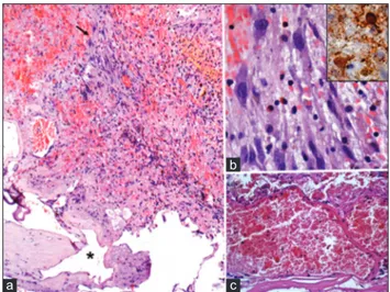
Figure 2: Case 1. (a) distinct tumor types in this picture, depicting ganglioglioma mixed with cavernous angioma at the bottom
(asterisk). The arrow indicates the tumor area enlarged in b, showing ganglion cells intermingled with small lymphocytes, erythrocytes, and glial cells. (b) inset: synaptophysin‑immunoreactive ganglion cells. (c) detail of cavernoma portion. (a‑c) haematoxylin and eosin; original magnification: (a) ×50; (b), ×400; (c), ×200. (b) inset: automated immunohistochemistry, chromogen: diaminobenzidine; original magnification: ×400
a
b
c
Figure 1: Case 1. Preoperative (a) axial FLAIR, and (b) coronal T2 MRI scans of the brain showing the tumor located in the left middle cerebellar peduncle. (c) postoperative MRI scan showing complete removal of the tumor with no recurrence 4 years after operation
Surgical Neurology International 2017, 8:94 http://www.surgicalneurologyint.com/content/8/1/94
Interestingly, Kupnicka et al.[6] described an intimate
association of AVM and ganglioglioma. The presence of neoplastic ganglion cells randomly distributed in the context of an angioglioma, led them to consider this tumor as a new distinct pathological entity, defined as angioganglioglioma, arguing a possible transitional form between angioglioma and ganglioglioma, with similarity to dysembryoplastic neuroectodermal tumors (DNT). On the other hand, ganglioglioma are rare CNS tumours defined by the presence of neoplastic ganglion and glial
cells.[5,13‑16] They are included in the WHO classification
mostly as grade I, although some examples of atypia have
been observed, originating from the glial component.[2,3,5,15]
They occur mostly in the pediatric population and in young adults, suggesting a malformative or developmental
causation with analogy to DNT.[6,16] The main histological
feature of gangliogliomas is the presence of neoplastic astrocytes or oligodentrocytes, along with atypical
neurons.[13,16] Another important histopathological aspect
of gangliogliomas, with special regard to our cases, is the absence of vascular proliferation, making these tumors be
typically described as avascular lesions.[6,14]
In both of our cases, cavernoma pattern was present, showing a complex of vascular spaces in varying sizes without intervening parenchyma, and endothelial positivity for CD34 [Figure 2b], whereas ganglion cells showed synaptophysin immunoreactivity [Figures 1b and 2a]. In case 1 [Figure 2], an abrupt separation front is appreciable between cavernous angioma and ganglioglioma, like in a “collision tumor.” However, cavernoma‑like component, which is considered peculiar of angiogliomas, and the good prognosis also associated
with angioglioma,[7,8,11,17] along with the ganglionic
component, led us to consider the hypothesis of a distinct mixed tumor with analogies to angiogangliogliomas. In the second case [Figure 4], the two components, namely, glioma and cavernoma, were closely admixed, with intermingled ganglionic cells, strongly recalling Kupnika’s description of angiogangliogliomas, although vascular component was a cavernoma instead of a AVM. Despite the different histological arrangement of the vascular and
ganglionic portions in our cases, the close proximity of ganglioglioma and cavernoma suggests the hypothesis of mixed tumors ascribable to angiogliomas, and possibly supports the existence of angiogangliogliomas. Moreover, while glial cells could simply represent a
reactive proliferation to vascular malformation,[5] this
appears much less probable for ganglionar cells shown in our cases. Instead, as proposed by some authors, the association of ganglioglioma with vascular malformation
seems to have more likely a malformative origin.[5,6]
Finally, according to Palma et al.[11] the association of
glioma with cavernoma is the one to be most reasonably considered as an independent entity.
CONCLUSION
In conclusion, we found several different interpretation
of angiogliomas proposed in the literature.[5,8,9,11] As
far as angioganglioglioma is concerned, it could be interpreted as a transition form between ganglioglioma
and angioglioma.[6] Probably, all of these conditions are
possible, although angioganglioglioma is extremely rare. Our findings may reinforce the hypothesis that the association between cavernoma and glial neoplasm is actually a distinct pathological entity, and stimulate the discussion on angioglioma and their definition.
Financial support and sponsorship
Nil.
Conflicts of interest
There are no conflicts of interest.
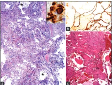
Figure 4: Case 2. (a) dilated vessels (as the ones indicated by
asterisks) of the cavernous malformation admixed with tumor cells. (a) inset: ganglion cells are identified by immunopositivity for synaptophysin. (b) cavernous vessels are evidenced by endothelial positivity for CD34. (c) in another area, the cavernous portion (bottom of picture) is quite distinct from ganglioglioma. (a), and, (c) hematoxylin and eosin; original magnification: (a) ×25; (c) ×50; (a) inset, and (b) automated immunohistochemistry, chromogen: diaminobenzidine; original magnification: A, inset, ×400; (b) ×100
a
b
c
Figure 3: Case 2. Preoperative (a) axial FLAIR, and (b) sagittal contrast enhanced MRI scans of the brain showing a suprachiasmatic inhomogeneous mass with scattered contrast enhancement. (c) four‑year follow‑up contrast‑enhanced‑T1 MRI scan showed no residual tumor or recurrence
Surgical Neurology International 2017, 8:94 http://www.surgicalneurologyint.com/content/8/1/94
REFERENCES
1. Councilman WT. The gliomatous tumors of the brain. Long Island Med J 1914;8:401-9.
2. Dash RC, Provenzale JM, McComb RD, Perry DA, Longee DC, McLendon RE. Malignant supratentorial ganglioglioma (ganglion cell-giant cell glioblastoma): A case report and review of the literature. Arch Pathol Lab Med 1999;12:342-5.
3. De Marchi R, Abu-Abed S, Munoz D, Loch MacDonald R. Malignant ganglioglioma: Case report and review of literature. J Neurooncol 2011;101:311-8.
4. Gazzeri R, De Bonis C, Carotenuto V, Catapano D, D’Angelo V, Galarza M. Association between cavernous angioma and cerebral glioma. Report of two cases and literature review of so-called angiogliomas. Neurocirugia 2011;22:562-6.
5. Hasegawa H, Bitoh S, Koshino K, Obashi J, Kobayashi Y, Kobayashi M, et al. Mixed cavernous angioma and glioma (angioglioma) in the hypothalamus – Case report. Neurol Med Chir 1995;35:238-42.
6. Kupnicka DJ, Sikorska B, Klimek A, Kordek R, Liberski PP. Angioganglioglioma: A transitional form between angioglioma and ganglioglioma? Ultrastruc Pathol 2003;27:423-32.
7. Licata C, Pasqualin A, Freschini A, Barone G, Da Pian R. Management of associated primary cerebral neoplasms and vascular malformations: 2. Intracranial arterio-venous malformations. Acta Neurochir 1986;83:38-46. 8. Lombardi D, Scheithauer BW, Piepgras D, Meyer FB, Forbes GS.
“Angioglioma” and the arteriovenous malformation-glioma association. J Neurosurg 1991;75:589-66.
9. Mathern GW, Cornford ME, Athony Verity M. Angioglioma: Definition and reappraisal. J Neuropathol Exp Neurol 1989;48:306.
10. Matyja E, Grajkowska W, Taraszewska A, Marchel A, Bojarski P, Nauman P. Advanced reactive astrogliosis associated with hemangioblastoma versus astroglial-vascular neoplasm (“angioglioma”). Folia Neuropathol 2007;45:120-5.
11. Palma L, Mastronardi L, Celli P, D’Addetta L. Cavernous angioma associated with oligo-astrocytoma-like proliferation. Report of two cases and review of the literature with a reappraisal of the term “angioglioma”. Acta Neurochir 1995;133:169-73.
12. Pant I, Suri V, Chaturvedi S, Dua R, Kanodia AK. Ganglioglioma of optic chiasm: Case report and review of the literature. Child Nerv Syst 2006;22:717-20.
13. Shuangshoti S, Kirsch E, Bannan P, Fabian VA. Ganglioglioma of the optic chiasm: Case report and review of the literature. AJNR Am J Neuroradiol 2000;21:1486-9.
14. Siddique K, Zagardo M, Guijrati M, Olivero V. Ganglioglioma presenting as a meningioma: Case report and review of the literature. Neurosurgery 2002;50:1133-5.
15. Yust-Katz S, Anderson MD, Liu D, Wu J, Yuan Y, Olar A, et al. Clinical and prognostic features of adult patients with gangliogliomas. Neuro Oncol 2014;16:409-13.
16. Zentner J, Wolf HK, Ostertun B, Hufnagel A, Campos MG, Solymosi L, et al. Gangliogliomas: Clinical, radiological, and histopathological findings in 51 patients. J Neurol Neurosurg Psychiatry 1994;57:1497-502.
17. Ziyal IM, Ece K, Bilginer B, Tezel GG, Özcan OE. A glioma with an arteriovenous malformation: An association or a different entity? Acta Neurochir 2004;146:83-6.