Vol. 120, n. 1: 71- 81, 2015
© 2015 Firenze University Press http://www.fupress.com/ijae
ITALIAN JOU R NAL OF ANATOMY AN D EM B RYOLOGY
DOI: 10.13128/IJAE-16476 Research Article - Histology and Cell Biology
Morpho-structural alterations of sub-chondral bone
tissue in patients with osteoarthritis: a scanning
electron microscopy study
Michele Attilio Rosa1, Pierfrancesco Gugliandolo1, Angelo Favaloro2, Giovanna Vermiglio2, Antonio Centofanti2, Daniele Bruschetta2, Giuseppina Rizzo2
Department of Biomedical Sciences and Morpho-functional Imaging, 1Division of Orthopaedy and 2Division of
Sport Medicine, University of Messina, Italy
Submitted August 8, 2014, accepted revised September 29, 2014
Abstract
Osteoarthritis focuses principally on the degeneration of articular cartilage as a primary cause of the disease. The pathophysiological process of osteoarthritis is characterized by alteration of chondrocytes and the increased bone formation by sub-chondral osteoblasts. Infiltration of macrophages and perivascular T and B lymphocytes is observed, and these infiltrates have been demonstrated in both early and advanced disease. The morphological and phenotypic characteristics of osteocytic cells attached to the normal and the osteoarthritic matrix dif-fer from each other, suggesting that specific signalling pathways arise or are altered between matrix and cells. On this basis, we have examined biopsies of bone obtained by normal femur and by femur of subjects affected by osteoarthritis using techniques of scanning electron microscopy in order to identify the morphostructural alterations that occur in the sub-chondral bone. Our results have shown that the bone tissue of subjects not affected by any disease of bone presents a well-organized structure, while the bone tissue obtained by patients affected by osteoarthritis shows a derangement of tissue itself possibly correlated with altered func-tion of the osteoblasts, that during the pathological process produce a less mineralized extra-cellular matrix with consequent loss of the normal bone structure. In our opinion, during the osteoarthritic process there would be a defective signalling between bone cells leading to the production of an irregular, amorphous extracellular matrix by osteoblasts, characteristic of the pathological condition.
Key words
Osteoarthritis, tissue bone, scanning electron microscopy.
Introduction
The prevailing theory on the development of osteoarthritis (OA) concentrates principally on the degeneration of articular cartilage as a primary cause of the dis-ease, suggesting that instability of the mechanical pressure on the joints leads to progressively more severe damage to the cartilage (Pessler et al., 2008) until a point where the only therapeutic option is the total replacement of the joint (Ayral et al., 2005; Hill et al., 2007).
The pathophysiological process of osteoarthritis is progressive, triggered by a change in the microenvironment of the chondrocytes and includes chondrocyte mito-sis, increased synthesis of proteoglycan and type II collagen which are the major struc-tural elements of cartilage, and increased bone formation by sub-chondral osteoblasts (Elliot et al., 1993; Nishimoto et al., 2004; McInnes et al., 2005; Baslund et al., 2005). With the increased bone formation in the sub-chondral zone, the physical properties of the bone change: it becomes more rigid with decreased elasticity, hence microfractures occur, followed by callus formation, further rigidity and additional microfractures. At the cellular level, infiltration of macrophages and perivascular T and B lymphocytes is observed, and these infiltrates have been demonstrated in both early and advanced dis-ease (Goldenberg et al., 1983; Oehler et al., 2002; Pearle et al., 2007; Benito et al., 2005).
Cartilage breaks off and the exposed subchondral bone becomes the new joint sur-face; this leads to the characteristic smooth appearance with increased radio-opacity and sclerosis in the cancellous bone beneath. The typical symptom is pain and, with the progression of osteoarthritis, the joint mobility decreases with limitation of the range of movement. The pain induced in the lower limb joints by load determines a limp of escape, so called because the patient tends to shorten the stance phase on the corresponding foot (Oehler et al., 2002). The observation that the morphologi-cal and phenotypic characteristics of osteocytic cells attached to the normal and the OA matrix differ from each other suggests that specific signalling pathways between matrix and cells arise or become altered (Scanzello et al., 2009).
In a healthy joint, processes of growth, modelling, and remodelling occur constantly during the life, but are active to different degrees in the mineralized tissues. Modelling is defined as formation or resorption at a given site: it varies bone mass but also modi-fies the shape of bone. During remodelling, processes at the cellular level allow for cell recruitment, differentiation, proliferation, and migration to surfaces (Burr, 2004; Portelli et al., 2014). Subsequently, a resorption phase follows and the cells can act more quickly or more slowly and for longer or shorter periods of time. Remodelling does not varies bone volume, therefore remodelling processes cannot account for the sclerosis observed in OA. Moreover, the process of endochondral ossification occurs throughout life. This causes progression and duplication of the tidemark, and this it is frequently observed in joints affected by OA disease. Advancement of the tidemark leads to make the calci-fied cartilage thicker and, even though the tidemark advances, remodelling at the oste-ochondral junction follows more quickly, so that the calcified cartilage may become thinner. In a healthy joint, these processes of endochondral ossification and subchon-dral remodelling are generally in balance. In an OA joint they go out of balance.
On this basis, in the present experimental study we examined biopsies of bone obtained by femur of subjects not affected by any bone disease and by femur of sub-jects suffering from OA using techniques of scanning electron microscopy in order to identify morphostructural alterations occurring in the sub-chondral zone, that may have escaped evaluation until now.
Materials and Methods
For this case study, nine male patients 60-71 years old suffering from OA and seven patients 45-70 years old who did not present OA disease but had undergone
surgery for total meniscal traumatic lesions have been selected. Pathological bone tissue biopsies were obtained by articular heads samples who had been cut because of prosthetic substitution. Healthy bone tissue biopsies were extracted through bone coring. Anaesthesia was epidural in all cases. Every patient expressed consensus and the procedures were in conformity with the guide lines from Helsinki declaration of 1975. The present study was approved by the ethical committee.
Biopsies were fixed in 2.5%glutaraldehyde in 0.1 mol/L phosphate buffer, pH 7.4, for 24 hours at room temperature. Samples were dehydrated with ethanol and amyl acetate and critical point dried through liquid CO2. The fractured surface of bone was
then placed on a stub, coated in a Plasma Science CrC Turbo Pumped (Torr Interna-tional, New Windsor, NY, USA) and was observed in a Phenom G2 scanning electron microscope (Phenom, Eindhoven, The Netherlands).
Results
For the present study we have analyzed, by scanning electron microscopy, bone tissue biopsies taken from subjects not suffering from any disease and biopsies from patients suffering from OA. In particular, we have studied the cortical area and the subcortical area of the femoral condyle.
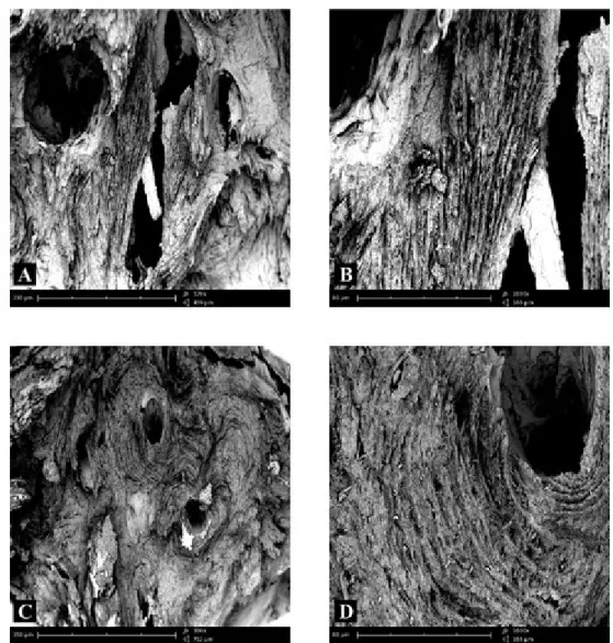
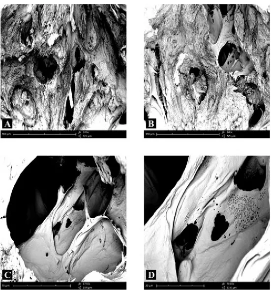
The fracture surface of femoral condyle in the cortical area of subjects not affect-ed by any bone pathology showaffect-ed numerous bone lamellae orientaffect-ed longitudinally and perfectly delineated (Fig. 1A). At high magnification, the lamellae were perfectly delineated and parallel between each other, as is the normal architecture of the bone tissue (Fig. 1B). In another microscopic field of healthy bone tissue there were numer-ous Havers’ canals (Fig. 1C) around which, at higher magnification, it was possible to observe many holes in bone (Fig. 1D).
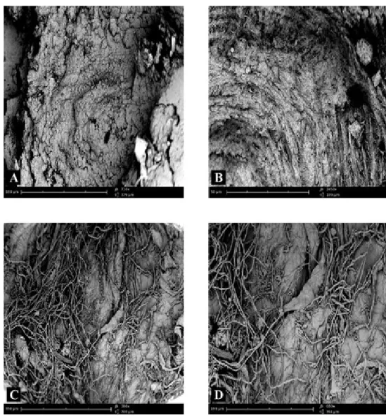
The fracture surface of femoral condyle in the subcortical area of subjects not affected by any disease of bone showed a lamellar structure more irregular than that seen in the cortical area (Fig. 2A). At high magnification, it was possible to detect bone lamellae irregularly organized and arranged more disorderly than those observed in the cortical area (Fig. 2B). Moreover, a rich fibrillar structure was evident, that appeared constituted by a dense network of collagen fibres (Fig. 2C) which pen-etrate into bone lamellae (Fig. 2D); such fibres, known as perforating or Sharpey’s fibres, are known to firmly anchor, periostium, joint capsule, ligaments and tendons to the bone tissue.
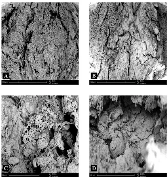
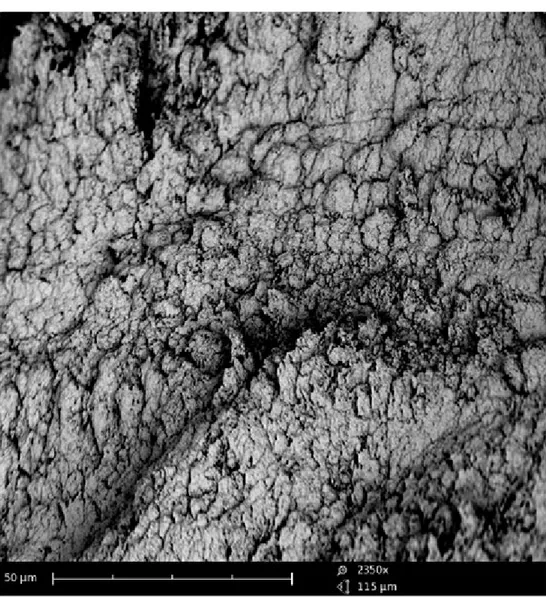
The fracture surface of femoral condyle in the cortical area of patients with OA showed a generally disorganized structure. At low magnification, the bone was quite uneven (Fig. 3A) and it was no longer possible to recognize bone lamellae regularly oriented and arranged concentrically around Havers’ canals (Fig. 3B). Higher magni-fication revealed the presence of an amorphous structure that made the bone tissue almost unrecognizable (Fig. 3C). In some microscopic fields the structure was most regularly organized for the presence of superimposed layers of bone tissue that may demonstrate new formation of strengthened and thickened tissue (Fig. 3D), indicat-ing osteosclerosis. The observation of other microscopic fields highlighted extensive neoformation of an amorphous matrix similar to roof tiles that thickened the bone tissue (Fig. 4).
The fracture surface of femoral condyle in the subcortical area of patients with OA showed a more organized structure than the cortex. The bone lamellae were relatively well organized and maintained a parallel orientation between each other (Fig. 5A); however, there were areas with diffuse osteosclerosis and areas of erosion of bone tis-sue, possibly conferring fragility (Fig. 5B). At high magnification of the erosion areas
Figure 1 – Compound panel of fracture surfaces of cortical zone of bone tissue from a biopsy of healthy
femoral condyle showing bone lamellae oriented longitudinally and perfectly delineated (A, B); the lamel-lae are perfectly delineated and parallel between them showing a normal lamellar architecture of the bone tissue is shown (B). In another microscopic field, the presence of Havers’ canals is evident (C); around these lamellae many holes are visible in bone (D).
it was possible to see areas of bone tissue arranged into sheets which showed lesions and signs of bone resorption (Fig. 5C); a further magnification of the same area revealed the presence of numerous small areas of erosion giving an almost speckled appearance to the bone, which perhaps may fuse into large and diffuse areas of ero-sion (Fig. 5D).
Figure 2 – Compound panel of fracture surfaces of sub-cortical zone of bone tissue from a biopsy of healthy
femoral condyle showing more irregular lamellar structure than figure 1 (A); at high magnification, the pres-ence of bone lamellae irregularly organized is clearly visible (B). The observation of another microscopic field allows to see a rich fibrillar structure constituted by a dense network of collagen fibres (C), penetrating into bone lamellae (D).
Discussion
Osteoarthritis is the most common joint disorder and is characterized by carti-lage loss, new bone formation at the margins of joints, changes in subchondral bone, and recurrent synovitis (Fuerst et al., 2009). Skeletal development and
homeosta-Figure 3 – Compound panel of fracture surfaces of cortical zone of bone tissue from a biopsy of femoral
condyle affected by OA, showing a generally disorganized structure (A), in which it is no longer possible to recognize bone lamellae regularly oriented and arranged concentrically around Havers’ canals (B). At higher magnification, it is easier to see the amorphous structure, that makes bone tissue almost unrecognizable (C); in other microscopic fields, the structure is more regularly organized for the presence of superimposed lay-ers of bone tissue (D).
sis depend on the specific regulation of resorption and new bone formation, which include the degradation and removal of the extracellular matrix and the synthesis and deposition of new constituents (Bord et al., 1997).
Radio-diagnostic techniques show relevant alterations in patients affected by OA such as reduced articular space in 99.5% cases, osteophytes in 98,1%,cases and sub-chondral bone sclerosis in 88,3% cases (Audrey et al., 2014).
Although numerous studies have been carried out on bone tissue affected by OA, most authors have focused on molecular and proteic characteristics of this disease
Figure 4 – Fracture surfaces on cortical zone of bone tissue from a biopsy of femoral condyle affected by
demonstrating that a key role in pathogenesis is played by integrins (Prasadam et al., 2013). Moreover, it has been demonstrated that matrix metalloproteinases are capable of degrading all components of connective tissue (Murphy and Reynolds, 1993) and are likely to be involved in bone matrix degradation during OA (Everts et al. 1992).
Figure 5 – Compound panel of fracture surfaces of sub-cortical zone of bone tissue from a biopsy of
femo-ral condyle affected by OA, showing relatively well organized bone lamellae which maintain a pafemo-rallel orien-tation among them (A); in other microscopic fields erosion areas are detectable in the bone tissue, that may make the tissue fragile (B). A higher magnification of erosion areas allows to recognize bone tissue arranged into sheets showing evident lesions and clear signs of bone resorption (C); an even higher magnification reveals the presence of numerous small areas of erosion damage giving almost a speckled appearance to the bone (D).
Other studies, carried out through optical and laser scanning confocal microscopy, have highlighted a different morphology of the osteocytes in the arthritic bone tissue as compared with normal tissue: osteocytes from arthritic tissue are round shaped due to increase in cytoplasm volume (Sanchez et al., 2008).
There are few data on morpho-structural aspects of bone tissue affected by OA. The present results have shown a general derangement of bone tissue in patients affected by OA. We observed many alterations in OA bone, such as a important struc-tural alterations of bone lamellae. Moreover, we revealed the absence of cartilage on the articular surface involved in the OA process and a specific increase in underlying bone lacunae and bone tissue, making the tissue almost unrecognizable.
These results, obtained through scanning electron microscopy, underline how bone tissue affected by OA undergoes continuous bone reshaping with consequent loss of nor-mal lamellar architecture typical of healthy bone tissue. Because modeling and remod-eling are responsible for adjusting the geometry of the joint to new conditions, many anti-activation agents may prevent the alterations in joint shape that accompany OA.
Our results showed the presence of superimposed layers of bone tissue that dem-onstrate new formation of strengthened and thickened tissue. In our opinion, these data confirm the highlighted alterations of subchondral bone occurring every time an articulation is affected by arthritic process. In fact, it has been previously demon-strated that during OA disease a higher amount of extracellular matrix proteins is produced by osteoblasts (Prasadam et al., 2013). In this respect, bone matrix serves as an organized framework offering mechanical support to and mediating biologi-cal activities of bone cells and also mediating signals that maintain bone homeostasis and remodelling (Green et al., 1995).
Bone cells, like most other matrix-associated cells, cannot survive or differentiate without adhesion to their matrix (Popov et al., 2011). Consequently, bone cell mor-phology and function depend strongly on matrix quality under conditions in which biological signals are constant (Prasadam et al., 2013).
The results of our study underline the derangement of the bone tissue affected by OA and its correlation with the altered functionality of osteoblasts that during the pathological process produce a higher mineralized extracellular matrix with conse-quent derangement of the normal bone structure.
Previous studies have shown that osteocytes in a normal extracellular matrix pre-sent a dense network of dendrites allowing important interaction among cells them-selves, whereas cells from OA matrix have a wrinkled surface with a small amount of dendrites (Pazzaglia et al., 2012). Thus, during the OA process there would be a lack of signalling between osteocytes, typical of the normal bone tissue, generating an irregular matrix and therefore the typically irregular bone tissue observed in the cur-rent study. The results of in vivo tests and of analysis of osteocyte morphology from subchondral bone of patients affected by OA and subjected to knee prosthesis surgery have shown alterations in the cytoskeleton (Bord et al., 1997).
In our opinion, the alterations detected in this research may be caused by the con-stant production of amorphous extracellular matrix produced by osteoblasts due to pathological conditions. Structural alterations that originate from this process inevi-tably affect the general macroscopic morphology of the joint affected by the patho-logical process, generating osteophytes and subchondral osteosclerosis that cause the typical symptoms of OA.
Further investigations - in progress - by light, scanning electron, and confocal laser scanning microscopy and using markers for osteoblasts and osteoclasts, could offer support to present results in order to understand the alterations occurring dur-ing bone remodelldur-ing typical of OA.
References
Audrey H.X., Abd Razak H.R., Andrew T.H. (2014) The truth behind subchondral cysts in osteoarthritis of the knee. Open Orthop. J. 8: 7-10.
Ayral X., Pickering E.H., Woodworth T.G., Mackillop N., Dougados M. (2005) Syno-vitis: a potential predictive factor of structural progression of medial tibiofemo-ral knee osteoarthritis e results of a 1 year longitudinal arthroscopic study in 422 patients. Osteoarthritis Cartilage 13: 361-367.
Baslund B., Tvede N., Danneskiold-Samsoe B., Larsson P., Panayi G., Petersen J., Petersen L.J., Beurskens F.J.M., Schuurman J., van de Winkel J.G.J., Parren P.W.H.I., Gracie J.A., Jongbloed S., Liew F.Y., McInnes I.B. (2005) Targeting inter-leukin-15 in patients with rheumatoid arthritis: a proof-of-concept study. Arthritis Rheum. 52: 2686-2692.
Benito M.J., Veale D.J., FitzGerald O., van den Berg W.B., Bresnihan B. (2005) Synovial tissue inflammation in early and late osteoarthritis. Ann. Rheum. Dis. 64: 1263-1267. Bord S., Horner A., Hembry R.M., Reynolds J.J., Compston J.E. (1997) Distribution of
matrix metalloproteinases and their inhibitor, TIMP-1, in developing human oste-ophytic bone. J. Anat. 191: 39-48.
Burr D.B. (2004) Anatomy and physiology of the mineralized tissues: Role in the pathogenesis of osteoarthrosis. Osteoarthritis Cartilage. 12: S20–S30.
Elliott M.J., Maini R.N., Feldmann M., Long-Fox A., Charles P., Katsikis P., Bren-nan F.M., Walker J., Bijl H., Ghrayeb J., Woody J.N. (1993) Treatment of rheuma-toid arthritis with chimeric monoclonal antibodies to tumor necrosis factor alpha. Arthritis Rheum. 36: 1681-1690.
Everts V., Delaissé J.M., Korper W., Neohof A., Vaes G., Beertsen W. (1992) Degra-dation of collagen in the bone resorbing compartment underlying the osteoclast involves both cysteineproteinase and matrix metalloproteinases. J. Cell. Physiol. 150: 221-231.
Fuerst M., Bertrand J., Lammers L., Dreier R., Echtermeyer F., Nitschke Y., Rutsch F., Schäfer F.K.W., Niggemeyer O., Steinhagen J., Lohmann C.H., Pap T., Rüther W. (2009) Calcification of articular cartilage in human osteoarthritis. Arthritis Rheum. 60: 2694-2703.
Goldenberg D.L., Egan M.S., Cohen A.S. (1983) Inflammatory synovitis in degenera-tive joint disease. J Rheumatol. 9: 204-209.
Green J., Schotland S., Stauber D.J., Kleeman C.R., Clemens T.L. (1995) Cell-matrix interaction in bone: type I collagen modulates signal transduction in osteoblast-like cells. Am. J. Physiol. 268: C1090-C1103.
Hill C.L., Hunter D.J., Niu J., Clancy M., Guermazi A., Genant H., Gale D., Grainger A., Conaghan P., Felson D.T. (2007) Synovitis detected on magnetic resonance imaging and its relation to pain and cartilage loss in knee osteoarthritis. Ann. Rheum. Dis. 66: 1599-1603.
McInnes I.B., Liew F.Y. (2005) Cytokine networks towards new therapies for rheuma-toid arthritis. Nat. Clin. Pract. Rheumatol. 1: 31-39.
Murphy G., Reynolds J.J. (1993) Extracellular matrix degradation. In: Royce P., Stein-mann B. (eds.) Connective Tissue and Its Heritable Disorders. New York, Wiley-Liss. Pp. 287-316.
Nishimoto N., Yoshizaki K., Miyasaka N., Yamamoto K., Kawai S., Takeuchi T., Hashimoto J., Azuma J., Kishimoto T. (2004) Treatment of rheumatoid arthritis with humanized anti-interleukin-6 receptor antibody: a multicenter, double-blind, placebo-controlled trial. Arthritis Rheum. 50: 1761-1769.
Oehler S., Neureiter D., Meyer-Scholten C., Aigner T. (2002) Subtyping of osteoarthrit-ic synoviopathy. Clin. Exp. Rheumatol. 20: 633-640.
Pazzaglia U.E., Congiu T., Franzetti E., Marchese M., Spagnuolo F., Di Mascio L., Zarattini G. ( 2012) A model of osteoblast-osteocyte kinetics in the development of secondary osteons in rabbits. J. Anat. 220: 372-383.
Pearle A.D., Scanzello C.R., George S., Mandl L.A., DiCarlo E.F., Peterson M., Sculco T.P., Crow M.K. (2007) Elevated high-sensitivity C-reactive protein levels are asso-ciated with local inflammatory findings in patients with osteoarthritis. Osteoar-thritis Cartilage 15: 516-523.
Pessler F., Chen L., Dai L., Gomez C., Diaz-Torne C., Paessler M., Scanzello C., Cakir N., Einhorn E., Schumacher H.R. (2008) A histomorphometric analysis of synovial biopsies from individuals with Gulf War Veteran’s illness and joint pain compared to normal and osteoarthritis synovium. Clin. Rheumatol. 27: 1127-1134.
Popov C., Radic T., Haasters F., Prall W.C., Aszodi A., Gullberg D., Schieker M., Docheva D. (2011) Integrins alpha2beta1 and alpha11beta1 regulate the survival of mesenchymal stem cells on collagen I. Cell Death Dis. 2: e186.
Portelli M., Matarese G., Militi A., Logiudice G., Nucera R., Lucchese A. (2014) Tem-poromandibular joint involvement in a cohort of patients with Juvenile Idiopatic Arthritis and evaluation of the effect induced by functional orthodontic appliance: clinical and radiographic investigation. Eur. J. Paediatr. Dent. 15: 63-66.
Prasadam I., Farnaghi S., Feng J.Q., Gu W., Perry S., Crawford R., XiaoY. (2013) Impact of extracellular matrix derived from osteoarthritis subchondral bone osteo-blasts on osteocytes: role of integrinβ1 and focal adhesion kinase signaling cues. Arthritis Res. Ther. 15: R150.
Sanchez C., Deberg M.A., Bellahcene A., Castronovo V., Msika P., Delcour J.P., Crie-laard J.M., Henrotin Y.E. (2008) Phenotypic characterization of osteoblasts from the sclerotic zones of osteoarthritic subchondral bone. Arthritis Rheum. 58: 442-455. Scanzello C.R., Umoh E., Pessler F., Diaz-Torne C., Miles T., DiCarlo E.,. Potter H.G.,
Mandl L., Marx R., Rodeo S., Goldring S.R., Crow M.K. (2009) Local cytokine pro-files in knee osteoarthritis: elevated synovial fluid interleukin-15 differentiates ear-ly from end-stage disease. Osteoarthritis Cartilage 17: 1040-1048.