~ 1 ~
INTRAVENOUS ALFAXALONE ANAESTHESIA IN TWO SQUAMATE SPECIES: EUBLEPHARIS MACULARIUS AND MORELIA SPILOTA
CHEYNEI
Tesi per il XXIX Ciclo del Dottorato in Scienze Veterinarie, Curriculum Scienze Cliniche Veterinarie
Dipartimento di Scienze Veterinarie, Universita’ degli Studi di Messina
Tutor: Prof. Filippo Spadola Cotutor: Prof. Zdenek Knotek Dr. Manuel Morici
Sommario
L’anestesia negli Squamati è una costante sfida della medicina e chirurgia dei rettili. Le differenze morfo-fisiologiche di questi taxa, rendono difficilmente applicabile i comuni concetti di anestesiologia veterinaria usati con successo negli altri animali da compagnia. Diversi protocolli anestetici sono stati utilizzati, sia per l’induzione che per il mantenimento, sia negli ofidi che nei sauri, ma con risultati variabili. Di fatti la maggior parte dei protocolli risultano in induzione o recuperi troppo brevi o troppo lunghi. L’obbiettivo di questa tesi dottorale è di valutare l’efficacia di un anestetico steroideo (alfaxalone), somministrato per via endovenosa in due specie di squamati usati come modello: il geco leopardo (Eublepharis macularius) e il pitone tappeto (Morelia spilota cheynei). Due metodi di somministrazione endovenosa (vena giugulare nei gechi e vena caudale nei serpenti) sono stati analizzati e descritti, usando un dosaggio di anestetico di 5 mg/kg in 20 gechi leopardo, e di 10 mg/kg in 10 pitoni tappeto. Nei gechi il tempo di induzione, il tempo di perdita del tono mandibolare, l’intervallo di anestesia chirurgica e il recupero completo sono stati rispettivamente di 27.5 ± 30.7 secondi, 1.3 ± 1.4 minuti, 12.5 ± 2.2 minuti and 18.8 ± 12.1 minuti. Nei pitoni tappeto, il tempo di induzione, la perdita di sensazione, il tempo di inserimento del tubo endotracheale, l’intervallo di anestesia chirurgica e il recupero sono stati rispettivamente di 3.1±0.8 minuti, 5.6±0.7 minuti, 6.9±0.9 minuti, 18.8±4.7 minuti, e 36.7±11.4 minuti. La somministrazione endovenosa di alfaxalone si è dimostrata un metodo efficace di induzione per una successiva anestesia gassosa in entrambi i modelli sperimentali, applicabile in sicurezza in altre specie di ofidi e sauri.
~ 2 ~
AbstractThe anaesthetic management of reptilian patient represents unique challenges. The marked morphology and physiology differences of reptiles do not allow the application of the same anaesthetic concepts used in the other domestic animal species. Many anaesthetic protocols have been used for induction to anaesthesia in snake and lizard species, generally with varying results. Most of these protocols resulted in prolonged induction time and recovery time. The aim of this doctoral thesis is to evaluate the efficacy of a neuroactive steroid anaesthetic drug (alfaxalone) administered intravenous in two squamate models: leopard geckos (Eublepharis macularius) and jungle carpet python (Morelia spilota cheynei). Two methods are described for intravenous anaesthesia with alfaxalone in 20 leopard geckos and in 10 jungle carpet python; a dose of 5 mg/kg of alfaxalone was used in leopard geckos, while a dose and 10 mg/kg of alfaxalone was used in carpet python. The venous access through the jugular vein (in geckos) and tail vein (in python) were successfully used. In the leopard geckos, the induction time, the mandibular tone lost time, interval of deep anaesthesia and the full recovery time were 27.5 ± 30.7 seconds, 1.3 ± 1.4 minutes, 12.5 ± 2.2 minutes and 18.8 ± 12.1 minutes, respectively. In the carpet python, the induction time occurred within 3.1±0.8 minutes, while the time of pain sensation loss, the mean tracheal tube insertion time, the interval of surgical state of anaesthesia, and the time of full recovery occurred within 5.6±0.7 minutes, 6.9 ± 0.9 minutes, 18.8 ± 4.7 minutes, and 36.7 ± 11.4 minutes, respectively. Intravenous administration of alfaxalone proved to be a valuable method of induction to inhalation anaesthesia in both studied squamate models.
Abstrakt
Anestezie plazů představuje v mnoha směrech náročnou výzvu. Anatomické a fyziologické odlišnosti plazů neumožňují volit stejné anestetické přístupy jako u řady domestikovaných zvířat. Pro indukci anestezie bylo dosud u hadů a ještěrů použito mnoho anestetických protokolů, avšak většinou s rozličnými výsledky. Většina těchto protokolů vedla k prodloužené době nástupu účinku (tzv. induction time) a rovněž i fázi probouzení. Cílem disertační práce bylo zhodnotit účinnost neuroaktivního steroidního anestetika (alfaxalon) podávaného intravenózně u dvou zvolených zástupců šupinatých (Squamata): gekončíka nočního (Eublepharis
macularius) a krajty diamantové (poddruh Morelia spilota cheynei). Pro intravenózní
použití alfaxalonu byly u 20 gekončíků nočních a 10 krajt diamantových zvoleny dvě metody; u gekončíků byla použita dávka 5 mg/kg, zatímco u krajt byla použita dávka 10 mg/kg. Jako žilní přístup byla zvolena jugulární žíla (u gekončíků) a ocasní žíla (u krajt). U gekončíků byl čas nástupu účinku, čas vymizení tonu čelisti, interval
~ 3 ~
hluboké anestezie a čas navrácení všech reflexů a úplného probuzení (tzv. full recovery time) 27.5 ± 30.7 sekund, respektive 1.3 ± 1.4 minut, 12.5 ± 2.2 minut a 18.8 ± 12.1 minut. U krajt došlo k nástupu účinku během 3.1 ± 0.8 minut, zatímco ke ztrátě citlivosti, možnosti zaintubování (zavedení endotracheální kanyly), celkové doby chirurgické fáze anestezie a času úplného probuzení došlo během 5.6 ± 0.7 minut, respektive 6.9 ± 0.9 minut, 18.8 ± 4.7 minut a 36.7 ± 11.4 minut. Intravenózní podávání alfaxalonu představuje u obou zvolených zástupců šupinatých vhodnou metodu pro indukci anestezie před vlastní inhalační anestezií.
~ 4 ~
INTRODUCTIONNotes on Squamata clinical morphology, physiology, anaesthesia and analgesia
The order Squamata, or the “scaled reptiles”, are the largest modern order of reptiles, comprising all lizards and snakes. Historically, the order Squamata has been divided into three suborders: Lacertilia, Serpentes, and Amphisbaenia. Squamata are the most variably sized order of reptiles, ranging from the 16 mm (0.63 in) dwarf gecko (Sphaerodactylus ariasae) to the 5.21 m green anaconda (Eunectes murinus). Since the diffusion as pet, either for conservation purpose or moreover for collection and breeding hobby, the scaled reptile are common sight at veterinary specialized practice. Obviously surgery and anaesthesia occupy a great piece of reptile veterinary work.
Reptile anaesthesia is a challenging aspect of herpetological medicine.
Running anaesthesia in reptiles has been challenging for 3 primary reasons. First, there is an enormous diversity of species across the class Reptilia, one of the most phylogenetically diverse animal classes with greater than 8000 species. Second, there is a lack of systematic research dedicated to advancing the current knowledges. Third, the reptile literature is predominant with anecdotal information, which has been widely accepted as dogma for clinicians (Mans and Sladky 2012). Reptiles, and in our case, squamates diverge from mammals or avian patients both physiologically and anatomically, thus it is impossible to extrapolate and apply on them all veterinary anaesthesia knowledges concerning other taxonomical orders.
First of almost of the Squamates are ectothermic (body temperature is dependant of the environmental temperature). Some of them thermoconform passively to their thermal environment (e.g. many nocturnal tropical geckos), but others use behavioural and physiological mechanisms to regulate their body temperature. Each species possess a preferred optimal body temperature (POBT). Moreover, every single species possess a specific preferred optimal temperature zone (POTZ), and as a result the familiarity regarding this aspect, and as well obviously systematics, taxonomic and ecology of squamates are paramount of importance for a good herpetological veterinarian. Any changes in POBT/POTZ may significantly affect their metabolism, physiology, and consequently drug efficiency and elimination. Consequently, the temperature at which one maintains a patient is one of the most important factor to success in reptilian medicine, and obviously in anaesthesia. There are well-known evidences that reptiles downregulate their body temperature in response to hypoxia and/or inadequate tissue oxygen delivery (Andrews and Pough 1985; Hicks and Bennett 2004). This is referred to as hypoxia-induced hypothermia (Hicks and Wang 1999). Hypothermia induced by hypoxia decreases metabolic rate
~ 5 ~
through the direct effect of temperature on tissue oxygen demand and through depression of the rate of aerobic metabolism (Hicks and Wang 2004). Several anaesthetic can cause apnoeic phases in scaled reptiles, and thus consequently cause hypothermia; this is the reason why is clearly essential to maintain proper temperature during any kind of anaesthetic procedures.
Understandably, squamate metabolism assumes paramount of importance during anaesthesia and analgesia (Mosley 2005). As well known, metabolism works in firm connexion with environmental and body temperature, and it can tremendously affect the response to anaesthetic, or drugs in general. The reptilian resting metabolic rate is one tenth to one third lower than the resting oxygen consumption rate of mammals of an equivalent size. Minimum and maximum oxygen consumption rates of individual reptilian species range from almost zero to values similar to those of a resting mammal (Ultsch and Jackson 1982). A decrease in an animal’s metabolic rate results in declines of drug metabolism, leading altered onset, duration of effect, time to recovery, extraction and if not, accumulation and cellular damage.
Another significant point is the cardiovascular system and blood physiology of scaled reptiles. Their cardiovascular system, it is characterized by several exceptional anatomic arrangements and physiologic functions.
The heart of a Squamate is located ventrally in the thoracic cavity, but is found caudally in elongate lizards and snakes. Heart comprises a sinus venosus, two atria, and a three-uncompleted-chambered ventricle; the cavum venosum, cavum
arteriosum, and cavum pulmonale. There is “considerable” functional separation of
oxygenated and deoxygenated blood in the squamate ventricle despite the anatomical continuity of its three chambers (White 1976). Based upon the anatomy of the squamate heart several patterns of circulation of blood returning to the heart from the systemic and pulmonary circulation have been proposed: a complete mixing, a partial mixing, and a high degree of separation between unoxygenated and oxygenated blood being distributed differentially to the two aortae (Lillywhite and Burggren 1987; Jacobson 2007). Separation of oxygenated and deoxygenated blood can permit the systemic return to the heart to be shunted to the systemic output (right-to-left-shunt) to bypass the lungs during diving (Lillywhite and Donald 1989; Jacobson 2007), either also the pulmonary return to be shunted to the lungs (left-to-right shunt) during periods of extreme ventilation. The incomplete septa allow mixing of oxygenated and deoxygenated blood, and this must to be considered during anaesthesia of a Squamate. In fact shunting play a role in inhaled anaesthetic uptake and elimination, potentially causing postponed induction, or an unexpectedly rapid recovery, it can affect systemic blood pressure, and arterial/venous oxygen concentration, impacting on anaesthetic monitoring.
An additional cardiovascular feature is the renal portal system (RPS); it is a vein plexus that bypassing the systemic circulation, reflow the blood from the caudal
~ 6 ~
quarter of the body to the kidney, and it is present in all reptilian species.
Precisely, RPS is a ring of vessels surrounding the kidney, comprising cranial portal vein and caudal portal vein. The blood flows from the caudal part through iliac veins, and it breaks into afferent renal portal vein, which transfers blood to the kidneys (O’Malley 2005; Murray 2006). The RPS is necessary to perfuse kidney during dehydration period, avoiding ischemic damage (Holz et al. 1997; Holz and Raidal 2006). The RPS makes challenging drugs administration in reptiles, and it must to be considered during drugs administration. In fact cranial quarter drug administration is advised from literature (Holz et al. 1997a, b; Kummrow et al. 2008; Mans and Sladky 2012; Innis et al. 2014). Several studies shown that RPS clinical significance is marginal, and that almost certainly RPS have a great impact just in dehydrated animals (Holz et al. 1997a, b; Giorgi et al. 2015).
Main physiological features that can cause anaesthetic inaccuracies are the blood characteristics of scaled reptiles.
The intracellular concentration of haemoglobin in squamate-RBCs is about 32% and the oxygen carrying capacity of blood is about 8 to 10 volumes %, depending on the haemoglobin concentration. Reptilian haemoglobin tends to be more oxidised than mammalian haemoglobin (Wood and Lenfant 1976) and has a lower O2 binding
capacity (Wood and Lenfant 1976), and it must to be considered during anaesthesia monitoring, or even ventilation phases. Forward talking about monitoring we will deep this concept.
The respiratory system is extremely varied from physiological point of view. Squamates have slower respiration rate, and lower rate of oxygen consumption than mammals and avian.
The lungs of squamate reptiles are freely suspended in the coelomic cavity and are not located in a closed pleural space. In reptiles, the lungs tend to be sac-like with varying degrees of partitioning. Highly aerobic species such as Varanus
exanthematicus, Varanus niloticus, and Varanus dumerili, tend to have highly
partitioned lungs with numerous septae and invaginations that increase the surface area for gas exchange. Lizards tend to have paired lungs where most snakes have a single, functional right lung (Wang et al. 1998; Jacobson 2007). Functional elements of the lung are referred as ediculi and faveoli. The tracheal rings of squamates are incomplete, and caution is needed when placing an endotracheal tube. Additionally, the trachea bifurcates proximally, so unintentional endobronchial intubation may happen. The lungs of reptiles tend to have a larger tidal volume but a smaller respiratory surface area. Squamates lack in diaphragm, and respiration is performed with axillary or chest muscles movements (Shelton et al. 1986). Moreover several squamates species possess air sacs; it must be taken in consideration when inhalation anaesthesia is performed, since gas accumulation in air sac may be more than an
~ 7 ~
option.
The control of respiration in reptiles is poorly understood. Evidence shown that respiration is controlled by oxygen, carbon dioxide concentrations in the blood as well as PH, and environmental temperatures; moreover they can tolerate hypoxic conditions (Glass and Wood 1983; Johansen et al. 1970; O’ Malley 2005). In fact, reptiles tolerate large fluctuations of blood gas tensions during the ventilation/ apnoeic cycle. Respiration is generally stimulated by CO2, but tends to be less
sensitive to CO2 in diving and fossorial species (White 1969; Glass and Johansen
1976; Shelton and Burggreen 1976). For example, respiration by the aquatic snake
Acrochordus is more sensitive to O2 than CO2 (Wood and Lenfant 1976).
All reptiles do not produce urine more concentrated than plasma. Most of the squamate are uricotelic (excrete nitrogenous waste as uric acid); uric acid is obviously produced after catabolic liver activity and it is expelled as a semisolid. In the squamates kidney urine is extremely dilute, therefore uric acid remains in solution. Urine empties into the cloacal urodeum, and then into the bladder (just in few species, e.g. Iguana iguana, not in snakes) or large intestine, where water is reabsorbed, causing uric acid precipitation; it cause an excretion of nitrogenous catabolic waste with little water dispersion.
Liver of Squamata plays an important roles in tolerance to anaerobic metabolism, hypothermia, and adaptation to the environment.
The liver of Squamata has a lower metabolic capacity compared with mammalian livers (Berner 1999), and the metabolic rate is very sensitive to changes in temperature (Penick and Paladino 1996). The lower metabolic rates of reptilian liver is the reason for prolonged effects commonly seen with drugs, and obviously it contribute to prolonged anaesthetic recoveries shown when used drugs requires hepatic metabolism (e.g. ketamine, Glenn et al. 1972; Cooper 1974; Boever and Caputo 1982; Wood et al. 1982; Bienzle and Boyd 1992; Mosley 2005; Sinner and Graf 2008)
All of these specific physiological features must to be considered when a squamate need to be anesthetized, or if an anaesthetic protocol used in mammals and avian, would be verified in squamates.
Several induction and maintenance anaesthetic procedures have been investigated (Lumb 2004).
Inhalant agents are considered not reliable and limitative in squamates species. Isoflurane (Spelman et al 1996; Mosley et al. 2003a, b, 2004; Schumacher and Yelen, 2006; Trnková 2007), and Sevoflurane have been promoted as a rapid and safe anaesthetic agent in reptiles (Rooney et al. 1999; Bertelsen et al. 2005a, b;
~ 8 ~
Hernandez-Divers et al. 2005). Even though it has been suggested that N2O may
hasten the induction phase in reptiles (Custer et al. 1980), in point of fact no researches have been published. In Squamata, the pulmonary ventilation may considerably decrease in response to hyperoxia. A research reported that an exposure to 100% O2 decreased minute ventilation in the Javan wart snake (Acrochordus javanicus, Glass and Johansen 1976). These findings suggest that exposure to high
O2 levels could be partially responsible for the hypoventilation seen during
anaesthetic induction in squamate, and in most of other reptilian species. In any case, the induction of anaesthesia with isoflurane in oxygen (O2) may be prolonged, and so
injectable agents, such as benzodiazepines, alpha- 2-agonists, cyclohexanones, propofol and neuroactive steroids are often preferred to reduce induction times (Heard 2001; O et al. 2015). Just in specific case (venomous snake) the inhalation anaesthesia (e.g. in anaesthetic box) can be chosen. On the other hand, gaseous anaesthetic agents are ideal for maintenance of anaesthesia in reptiles. Tracheal tube insertion is particularly important in anaesthetised reptiles, in order to perform a great maintenance. Besides face mask must to be avoided, when possible (e.g. small species). Adequate sedation or anaesthesia is required to produce jaw relaxation for intubation (the exception is snakes, where conscious intubation is sometimes performed). Endotracheal tubes come in a wide variety of shapes and sizes. Uncuffed tubes are usually used, to avoid damage to the delicate tracheal ring. Bradypnoea or apnoea occurs during anaesthesia in reptiles and they thus require assisted ventilation. This can be performed either by an assistant or by using a mechanical ventilator. Peak airway pressure 10–15 cm H2O and a ventilation rate of 4–8
breaths/minute are essential.
Many injectable anaesthetic drugs have been used for induction in Squamata, with varying results.
Most result in prolonged induction and recovery times, with some exceeding 24 hours. (Glenn et al. 1972; Harding 1977; Chudzinski et al. 1989; Charland et al. 1991;Stirl et al. 1996; Schumacher et al. 1997; Bennet 1998; Anderson et al. 1999; Carregaro et al. 2009).
Ketamine hydrochloride or tiletamine/zolazepam is commonly used as part of the reptile anaesthetic protocol (Cooper 1974; Harding 1977; Ogunranti 1987; Arena et al. 1988; Knotek 2004; Knotek et al. 2005; Alves Junior et al. 2012b). Its advantages include the possibility to administer intramuscularly or intravenously, and a wide safety margin. However, muscle relaxation is poor and analgesia insignificant. Recovery can be excessively prolonged (Schumacher and Yelen, 2006). Medetomidine or other alfa-2 are usually combined with ketamine and an opioid agent (Greer et al. 2001; Chittick et al. 2002; Heaton-Jones et al. 2002; Olsson and Phalen 2012).
~ 9 ~
safety margin is wide, induction is smooth, and recovery is smooth and rapid (Bennet et al. 1998; Anderson et al. 1999; Santos et al. 2008; Ziolo and Bertelsen 2009; McFadden et al. 2011; Alves Junior et al. 2012a, b). The major disadvantage is the requirement for intravenous or intraosseous access for administration of this agent. Also a continuous rate infusion or intermittent administration of boluses can be used to maintain anaesthesia (Schumacher and Yelen, 2006). As in other species, cardio-respiratory depression occurs with propofol, and apnoea too (Bennett et al. 1998). Fosfopropofol was also tested with unpredictable results (Schroeder and Johnson 2013).
Neuromuscular blocking agents provide immobilisation, but no analgesia, and IPPV will be required if they are used (Kaufman et al. 2003; Redrobe 2004; Bosso et al. 2009).
In any animal that undergo surgery analgesia have to be administered. Opioid agents, such as tramadol and buprenorphine, morphine also have the advantage of providing mild-to-moderate sedation, and may reduce the doses of other anaesthetic agents required (Machin 2001; Kummrow et al. 2008; Sladky et al. 2008; Divers et al. 2010; Mosley 2011; Mans 2014). Butorphanol is wide know that it not has analgesic propriety in some reptilian species (Trnková et al. 2008; Fleming and Roberston 2012; Williams et al. 2016). Analgesia requirements should be assessed throughout anaesthesia; voluntary movement, or elevations in heart or respiratory rate during a procedure may indicate pain (Schumacher and Yelen, 2006). As with other species, pre-emptive analgesia should be provided where possible (Čermáková et al. 2014). Multi-modal therapy is advisable for many procedures, to reduce possible side effects from one group of drugs. Commonly, a non-steroidal anti-inflammatory drug (NSAID) is used in conjunction with an opioid agents. Local anaesthetics may also be used topically (Rivera et al. 2011; Knotek et al. 2014; Spadola et al. 2015) or by local infiltration (Chiu et al. 2009) for certain cases.
Anaesthetic monitoring and peri-anaesthetic care of a Squamate patient
The heart rate and rhythm may be monitored visually, but the use of a Doppler flow device is preferred. Electrocardiogram (ECG) can be used in Squamata, with leads positioned as in other species (Germer et al. 2015). As with other species, various reflexes can be monitored to assess anaesthetic depth. Deepening of anaesthesia will reduce muscular tone. After administration of any anaesthetic drug, loss of the righting reflex is used to indicate anaesthetic induction. The palpebral reflex is also lost at this stage. The corneal reflex should be maintained during surgical anaesthesia; this reflex is lost at a deep plane of anaesthesia. The corneal and palpebral reflexes cannot be assessed in species with spectacles, including snakes and some lizards. Tail-pinch, toe-pinch and cloacal pinch reflexes should also be
~ 10 ~
monitored during anaesthesia (Schumacher and Yelen, 2006). Snakes and many lizard appear to lose muscular tone gradually, starting with the head and moving caudally. During recovery from anaesthesia, the reverse occurs, with tail tone returning first. Assessment of tail pinch reflex is, therefore, a useful measure of depth of anaesthesia in snakes. As discussed above concerning physiology, reptile haemoglobin differs from that in mammals. For this reason, pulse oximeters designed to measure relative arterial oxygen saturation (SpO2) calibrated for mammals, cannot
give exact values related to the “real” reptile’s SpO2 (Schumacher and Yelen, 2006).
End-tidal carbon dioxide concentration ETCO2 measurements are enormously useful
for monitoring reptilian respiratory performance, but just if are used paediatric one, which can work with small mL sampling.
Fasting is necessary in squamate species; a fasting of 24-48 hours can be performed. In snakes, a fasting period can be done, long. As already mentioned, during anaesthesia, reptile patient should be maintained within its species-specific POTZ. Heat pads, hot water blankets are ideal, but gloves containing warm water can be a cheaper alternative. Heat sources should usually be covered with a towel to prevent contact burns.
Fluids should be given to all anaesthetised patients. Post anaesthesia treatment can be done in special hospitalization terrarium or tank with controlled temperature and humidity, in order to favourite activation of metabolism, and thus drugs elimination (Longley 2008).
~ 11 ~
Anaesthetic administration routeThe intramuscular route of anaesthetic administration was most common in scaled reptiles. As already explained, the hind limb and tail sites must to be avoided because of the first-pass effect of the RPS.
The epaxial muscles provide a suitable injection site in most snakes, while in lizards, the muscle mass of the forelimb (triceps and biceps) are commonly preferred.
Intracoelomic administration was recently described with unpredictable absorption rate (Schroeder and Johnson 2013). Intranasal (Schnellbacher et al. 2012; Knotek and Čermáková 2014), and intracloacal (Morici et al. in press) have been recently widely investigated in chelonian species, but no references are still in squamates.
Although intravenous drug administration is not easy in scaled reptiles, the combination of technique, practice, and skilled physical restraint, enable access to the ventral coccygeal vein in even very small snakes and lizards. Caution should be used in species that perform caudal autotomy. Although a techniques for catheterization of the coccygeal vein in lizards was described (Maxwell and Jacobson 2004; Wellehan et al 2004); to author’s opinion venous catheterization for intravenous anaesthesia is not strictly necessary, and it can certainly avoid.
Even though if wide-described, intraosseous access and catheterization for anaesthetic administration (Whitake and Krum 1999; Bennett et al 1998; Heard 2001) do not give to the reptile practitioner any kind of advantages, and it is also an extremely painful procedures.
Jugular vein access to administered drug is not commonly used in Squamata. Recently a description of the procedures have been done (Cuadrado et al. 2003; Morici et al. 2016; Di Giuseppe et al. in press). In squamates, the vena jugularis
interna develops from vena cava cranialis. Precisely, the vena jugulari comuna sinistra is the second of three trunks that unite to form left cranial cava plexus. From
its origin it passes slight laterally and then up to the neck, receiving cranially the vena jugularis externa. From this point it continues as vena jugulari interna, receiving several branches and continuing cranially to the caudal cerebral vein. (O’ Donoghue 1921). Blood collection and drug administration can be performed by skilled surgeon. Actually, with more than one probability, the “jugular vein technique” did not use exactly the jugular vein, but it uses a “subclavian plexus”, formed by the connection of several veins (vena thyroidea, vena aesophagea, vena laryngea, vena trachealis, two ramus muscularis, and vena subclavia/axillaris) to the jugular vein and the cranial cava. In this moment a standardize research is in development to understand the practicability of this technique.
Lastly, an intrathecal (subdural) technique of administration of opioids agent was described by Mans (2014) in chelonians, and it provides regional analgesia for up to
~ 12 ~
48 hours. Intrathecal administration opens a great windows in future development of reptile anaesthesia.
Alfaxalone anaesthesia
Alfaxalone (3α-hydroxy-5α-pregnane-11,20-dione) is a synthetic neuroactive steroid that increases binding of the gamma-aminobutyric acid receptor to its ligand in the central nervous system, causing in complete muscular relaxation and hypnosis (Jones 2012; Chiu et al. 2016). Alfaxalone was first marketed as an anaesthetic in 1971 co-formulated with a similar, less potent, neuroactive steroid, alfadolone (3α,21-dihydroxy-5α- pregnane-11,20-dione), and dissolved in castor oil surfactant. Due to important anaphylactic reaction it was withdraw from clinical use. In 2001, a clear colourless, surfactant-free, aqueous formulation of 1% alfaxalone dissolved with 2-hydroxpropyl-β-cyclodextrin (HPCD) was released for veterinary use in Australia (Alfaxan-CD RTU, Jurox), and recently in several other countries. The most important mechanism of anaesthetic action of alfaxalone is due to positive allosteric modulation of the GABA-A receptor, a ligand-gated chloride ion (Cl−) channel receptor for the neurotransmitter GABA, which universally inhibits neuronal excitability (Harrison and Simmonds, 1984).
Alfaxalone binds to GABA-A receptors, causing movement of Cl− ions into the cell, and thus cause a hyperpolarisation of the neuron and inhibition of action potential propagation (Lambert et al., 2003). Several studies revealed that alfaxalone possess a dual mechanism of action of alfaxalone: at low concentration, alfaxalone allosterically modulates the amplitude of GABA-induced ion currents, while, at higher concentrations, alfaxalone exerts an agonist effect, similar to barbiturates (Paul and Purdy 1992; Lambert et al. 1995).
Within the central nervous system (CNS), neurones express numerous GABA-A receptor subunit isoforms (α1–α6, β1–β3, γ1–γ3, δ, ε, θ, π, ρ1–ρ3) which determine the receptor’s agonist affinity, chance of opening, conductance and other pharmacological properties (Lambert et al., 2003). The receptor subunit specificity for binding of alfaxalone has been assessed and a research has demonstrated that alfaxalone acts as a positive allosteric modulator on the α1β1γ2L receptor isoform (Maitra and Reynolds, 1998).
No studies extensive on pharmacokinetics of alfaxalone in Squamata have been performed. Merely a study was performed in red-eared slider (Trachemys scripta
elegans, Schepard et al. 2013), and demonstrated that red-eared slider had
dose-dependent and inconsistent responses to alfaxalone, and additionally the lower ambient temperature augmented the behavioural effects of this drug in chelonian. In cat it shown a nonlinear pharmacokinetic, because effect can change between
~ 13 ~
individuals. The excretion is mainly due to hepatic metabolization, and probably by kidney lungs and brain (Warne et al. 2015). Alfaxalone, due to its formulation, can be administered subcutaneously, or intramuscularly or intravenously.
Alfaxalone-HPCD induces a dose-dependent decrease in heart rate (HR), cardiac output (CO) and arterial blood pressure in cats, (Muir et al. 2009), while it cause an increasing in HR and blood pressure in green iguana at dose of 5 mg/kg intravenously (Knotek et al. 2013). A decrease in respiratory rate (RR) was shown at 5 mg/kg intravenously in green iguana (Knotek et al. 2013). Different results were shown with intramuscular administration in green iguana (Bartelsen and Sauer 2011). Intravenous administration often results (at high dosage), in a transitory apnoea, as for propofol. In any case, even if induction and recovery are smooth, tracheal tube insertion, and ventilation is suggested (Bertelsen et al. 2005a, b; Knotek et al. 2013; Olsson et al. 2013; Bertelsen et al. 2015).
Alfaxalone is currently branded for veterinarian use in dogs and cats in numerous country, but it is used as induction agent in herptile species (Knotek et al. 2011a, b; Hadzima et al. 2014; Knotek et al. 2015; Thompson et al. 2016), due a smooth induction time, a rapid recovery time, and moreover it can be injected IM (suggested dose from 10 to 30 mg/kg, Lawrence and Jackson 1983; Johnson 2005; Bertelsen and Sauer 2011; Hansen and Bertelsen 2013; Kischinovsky et al. 2013; Olsson et al. 2013; Shepard et al. 2013). The IV administration of alfaxalone in reptilian species is feasible and advised in order to decrease induction time and recovery time (Simpson 2004; Knotek et al. 2013; Morici et al. 2016). Intravenous administration of alfaxalone is advised in reptiles to decrease induction time of inhalation anaesthesia. All the recent reports shown that intravenous administration proved feasible in anaesthesia in chelonians and some species of lizards (Knotek 2014; Knotek 2016).
Eublepharius macularius and Morelia spilota cheynei as Squamata model
In the present thesis two Squamata species have been used as models to investigate the effect of alfaxalone administered intravenously. A group of leopard geckos (Eublepharis macularius) and a group of jungle carpet python (Morelia spilota
cheynei) have been enrolled for this purpose.
Leopard gecko (Eublepharis macularius) is crepuscular ground-dwelling, insectivorous lizard naturally found in the highlands of Central Asia. Unlike most geckos, leopard gecko possess movable eyelids. It has become a well-established and popular pet in captivity. As several others lizards, it has the ability to perform caudal autotomy.
Morelia spilota cheynei, or the jungle carpet python, is a semi-arboreal, nocturnal
~ 14 ~
medium-sized pythons typically measure 1.5-2.1 m in total length. It is a common trade snake’s species. Since both species are widely spread and common as reptile pets, and thus surgery is not uncommon, the development of safe chemical immobilization methods is paramount of importance for any reptile veterinarian.
Aim of the PhD thesis
The aim of this thesis was to assess muscle relaxant, hypnotic properties and cardiac effect of alfaxalone administered IV in two squamate model. Our hypothesis was that IV administration of alfaxalone in two selected species (Eublepharius macularius and Morelia spilota cheynei) would provide a suitable anaesthetic quality, short induction time, an adequate surgical state of anaesthesia and moreover a slight influence on cardiac function. Moreover we would like to compare alfaxalone action between snake and lizard and in different venous access. Additionally, the IV administration techniques would be validate.
~ 15 ~
MATERIAL AND METHODSFIRST EXPERIMENTS: INTRAVENOUS ALFAXALONE ANESTHESIA IN EUBLEPHARIS
MACULARIUS
Animals
Twenty (2 males and 18 females) of healthy adult leopard geckos (Eublepharis
macularius) were enrolled in this study. Geckos used in this study were all from a
private breeder. An informed consent was signed by the breeder, and study was performed following the directive 2010/63/EU and ethical approval. This research has received ethical and partial financial support from Faculty of Veterinary Medicine, University of Veterinary and Pharmaceutical Sciences Brno (Institucionalni vyzkum/Ta29FVL/2016). The animals ranged in age from 2 years to 4 years. The mean body weight of geckos was 0.047 ± 0.01 kg (range 0.033 - 0.065 kg). Geckos were housed in glass terrariums (90 x 45 x 45 cm) with the air temperature maintained at 30-31 °C, and air humidity on 40-50%. Substrate consisted of folded paper towel. Recycled paper box and paper towel rolls were offered to geckos in order to provide more surface area for climbing and as hiding spots. A heating pad (RH-6, ReptiTherm® Under Tank Heaters, Zoo Med, USA) was placed under each terrarium as a heat source, while an UVA/UVB light bulb (Reptisun 5.0 UVB Mini Compact Fluorescent Bulb, Zoo Med, USA) was placed inside each terrarium as light source. The geckos were kept under 12/12 light/dark regime. Feeding with crickets or cockroaches was performed three times a week with gout loaded medium size (13-14 mm approximately). Geckos were acclimatized in terrariums for at least three weeks before the procedures. The body condition of each of geckos was evaluated within the standard clinical examination method.
Intravenous administration of alfaxalone into the jugular vein
Geckos were fasted for 24 hours before the blood collection, while access to the
water was not limited. The room air temperature was kept on 26 °C. Each of the
leopard geckos was manually restrained with the head and neck extended. The left
index and left middle finger of the operator were positioned behind the head, in contact with the mandibular branch. In this manner, the head of the gecko was slightly left-rotated, exposing the area of the right jugular vein. Skin on the right
neck was disinfected with alcohol solution and the needle of insulin set (0.5 mL- 29G
insulin syringe, BD medical, France) was gently inserted rostro-caudally into the right jugular vein (Figure 1).When a blood shot was recorded in the syringe a bolus
dose of 5 mg/kg of alfaxalone (Alfaxan® 10 mg/ml; Vétoquinol, France) was
administered. The gecko was placed on an electric heating pad (Bosch PFP 1031;
~ 16 ~
Exoterra, Germany).
Monitoring
Before alfaxalone administration, the geckos were gently manually restrained and the basal heart rate frequency (HR, in T0) was recoded using a vascular Doppler probe (8
MHz, PD1v Pocket Vascular Doppler, Ultrasound Technologies, UK). The basal respiratory rate frequency (RR, in T0) was assessed by checking the gecko’s chest
wall expansions at rest. The righting reflex was evaluated by gently turning the gecko on the dorsal recumbence. The hind limb toe-pinch reflex was evaluated applying a pressure with a small haemostatic forceps (thin gauze was placed between the clamp and the skin of the gecko). For the control of mandibular muscles tone the gecko’s mouth was manually opened. HR, RR and reflexes (righting reflex, toe-pinch reflex, and mandibular muscles tone) were assessed at 2 minute intervals for 24 minutes (T2, T4, T6, T8, T10, T12, T14, T16, T18, T20, T22 and T24), after alfaxalone
administration. The time interval from the administration of alfaxalone to the loss of the righting reflex was recorded as the induction time. The time interval from the administration of alfaxalone to the loss of mandibular muscles tone was recorded as the mandibular tone lost time. The time interval from the loss of the toe-pinch reflex
to its restoration was recorded as the interval of deep anaesthesia. The time interval
from the loss of the righting reflex to its restoration was recorded as the time of full recovery. All geckos were monitored for 48 hours following the procedure.
Descriptive statistical analyses of measured indicators - minimum, maximum, mean, and standard deviation (SD), were performed by statistical software GraphPad Prism 4.03 (GraphPad Software, Inc., USA), with assessment of distribution of the data (Shapiro-Wilk test) and ANOVA followed by Bonferroni test. Differences in HR and RR values in T0 and T2, T4, T6, T8, T10, T12, T14, T16, T18, T20, T22, T24, were
~ 17 ~
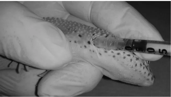
Figure 1: Intravenous (jugular vein) administration of alfaxalone in a pet leopard
geckos (Eublepharis macularius). Geckos were restrained with head and neck extended. The index and middle fingers of the surgeon were placed in contact with the mandibular branch, and the head of the gecko was slightly left-rotated, exposing the area of the right jugular vein. Subsequently, needle was inserted and when a blood shot was visible on the syringe the drug was delivered.
~ 18 ~
SECOND EXPERIMENTS: INTRAVENOUS ALFAXALONE ANESTHESIA IN MORELIA
SPILOTA CHEYNEI
Animals
Ten (2 males and 8 females) healthy sub-adult jungle carpet pythons (Morelia spilota
cheynei) were involved in this study. The mean body weight of snakes was 1.1±0.32
kg with the range from 0.72 – 1.55 kg. All the snakes used in this study came from the same captive private breed. Snakes were anaesthetized for clinical reasons, in order to perform a clinical examination and collection of pharyngeal swabs for laboratory analyses. The owner signed an informed consent, and the study was performed in compliance with directive 2010/63/EU and ethical approval. The authors did not receive any benefit by reporting the data obtained. This research has received ethical and partial financial support from Faculty of Veterinary Medicine, University of Veterinary and Pharmaceutical Sciences Brno (Institucionalni vyzkum/Ta29FVL/2016). One week before anaesthesia all snakes were housed individually in glass terrariums (Exoterra Natural Terrarium Medium/Tall, Exoterra, Hagen Inc., Canada). The air temperature was maintained at 28 °C (with maximum of 32 °C on the basking spot areas) with a multipurpose mercury vapour lamp (Exoterra Solar Glo, Hagen Inc., Canada). Snakes were kept under 10/14 light/dark regime. Air humidity within terrariums was maintained on 70% with the use of manual vaporization once a day. The snakes were kept fasted for 10 days before the anaesthesia, but clean water was offered ad libitum. After standard physical examination the blood was collected from the ventral tail vein for blood profile analyses. Packed cell volume (PCV) was measured using microhematocrit capillary tubes, total red blood cell and white blood cells counts were performed manually, using a hemocytometer with Natt and Herrick´s solution. Blood smears were prepared using a coverslip technique and differential leukocyte counts were assessed by enumeration of 200 cells in each smear. Blood chemistry was performed with the use of Abaxis VetScan Classic Analyzer; Abaxis, CA, USA. Clinically healthy snakes with the blood profile within the normal range for pythons (Centini et al. 2002; Bryant et al. 2012) were included in the investigation.
Anaesthesia and monitoring
The air temperature within the room where the anaesthesia was performed was kept on 26 °C.
The snakes were manually restrained and the basal heart rate (HR, in T0) was
recoded using a vascular Doppler probe (PD1v Pocket Vascular Doppler, Ultrasound Technologies, UK). The basal respiratory rate (RR, in T0) was assessed by checking
~ 19 ~
France) was administered intravenously into the ventral tail vein in a dose of 10 mg/mL (0.93±0.37 mL) with 1 mL syringe with 26G needle (PIC, Italy).
Snakes were placed into a fauna box (Faunarium, Exoterra, Hagen Inc., Canada) with a heating pad (Pet Mat, Australia) set up at 30 °C. HR, RR and reflexes (righting reflex, tail-pinch reflex, tracheal tube insertion) were assessed in five minute intervals (T5, T10, T15, T20, T25, T30, T35, T40, T45, T50, T55, T60) until full recovery of
snakes occurred. Righting reflex was checked with the snake being gently positioned on the dorsal recumbence. Tail-pinch reflex was evaluated applying a gentle pressure on tail extremity with haemostatic forceps. For tracheal tube insertion the snake’s mouth was gently opened manually by the surgeon (MM) and intravenous plastic catheter without needle (14, 16 or 18 gauge depending on the snake size, Artsana, Italy) was inserted into trachea. Pharyngeal swab was collected just before the tracheal tube insertion. Once the tracheal tube was inserted, snakes were ventilated with the air. Positive pressure ventilation was manually performed in those snakes that had respiratory rates lower than 1 breath per minute.
The time from alfaxalone administration to the loss of the righting reflex was recorded as the induction time. The time from alfaxalone administration to the loss of the tail-pinch-reflex was recorded as the time of pain sensation loss. The time from alfaxalone administration to the loss of mandibular tone and tracheal tube insertion was recorded as tracheal tube insertion time. The time interval from the loss of the tail-pinch reflex to its restoration was recorded as the interval of surgical state of anaesthesia. The time interval from the loss of the righting reflex to its restoration was recorded as the time of full recovery.
Descriptive statistical analyses of measured indicators - minimum, maximum, mean and standard deviation (SD) were performed by statistical software GraphPad Prism 4.03 (GraphPad Software, Inc., USA), with assessment of distribution of the data (Shapiro-Wilk test) and ANOVA followed by Bonferroni test. Differences in HR and RR values in T0 and T5, T10, T15, T20, T25, T30, T35, T40, T45, T50, T55, were compared
~ 20 ~
RESULTSFIRST EXPERIMENTS: INTRAVENOUS ALFAXALONE ANAESTHESIA IN EUBLEPHARIS MACULARIUS
The mean basal HR and RR were 90.60 ± 8.38 beats/minute and 31.4 ± 3.1 breaths/minute, respectively. The significant decrease of HR was recorded in T4 (74 ± 12.9 beats/minute) and heart rate remained significantly lower (p < 0.05) than basal HR until T24. The significant decrease of RR was recorded in T2 (26.8 ± 10.1 breaths/minute) and respiratory rate remained lower than basal RR until T24, but without any statistical significance. In four geckos, apnoea was recorded within two minutes after administration. .
Within the whole period of monitoring (T2 – T24) the mean HR was 80.6 ± 3.7 beats/minute and the mean of RR was 27.6 ± 1 breaths/minute. Results are summarized in table 1.
~ 21 ~
Table 1. Alfaxalone (5 mg/kg) intravenous induction to anaesthesia in 20 adult
leopard geckos.
Values Units Mean ± SD
Basal heart rate (HR) beats/minute 90.6 ± 8.3
HR during sedation beats/minute 80.6 ± 3.7
Basal respiratory rate (RR) breaths/minute 31.4 ± 3.1
RR during sedation breaths/minute 27.6 ± 1
Induction time seconds 27.5 ± 30.7
Mandibular tone lost time minutes 1.3 ± 1.4
Interval of deep anaesthesia minutes 12.5 ± 2.2
~ 22 ~
Figure 2. Mean (±SD) heart rate (HR) and respiratory rate (RR) of leopard geckos
(Eublepharis macularius) during anaesthesia with 5 mg kg-1 of alfaxalone administered intravenous
~ 23 ~
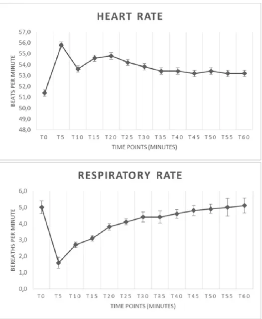
RESULTSSECOND EXPERIMENTS: INTRAVENOUS ALFAXALONE ANAESTHESIA IN MORELIA SPILOTA CHEYNEI
HR and RR values before (T0) and after the alfaxalone administration are illustrated in Figures 3. The mean basal heart rate in T0 was 51.4±3.41 beats per minutes. The mean basal respiratory rate at T0 was 5±1.25 breaths per minutes. Mean heart rate was significantly higher (p< 0.05) at T5, T15 and T20 when compared with basal HR at T0. Respiratory rate in snakes dropped in T5 and was significantly lower (p< 0.05) from T5 until T20 when compared with all other time points. In two snakes an apnoea was recorded at T5, and spontaneous breathing started again after a few minutes of assisted ventilation. Anaesthetic results are summarized in table 2. A prolonged time of full recovery was recorded just in two snakes (61.3 and 62.6 minutes, respectively).
~ 24 ~
Table 2: Induction to anaesthesia with alfaxalone (10 mg/kg) administered
intravenously to 10 sub adult jungle carpet python (Morelia spilota cheynei).
Value Induction time (minutes) Time of pain sensation loss (minutes) Tracheal tube insertion time (minutes) Interval of surgical state of anaesthesia (minutes) Time of full recovery (minutes) Minimum 1.5 4.4 5.4 14.1 28.2 Maximum 4.5 6.6 8.8 26.7 62.6 Mean 3.1 5.6 6.9 18.8 36.7 SD 0.8 0.7 0.9 4.7 11.4
~ 25 ~
Figure 3: Mean (±SD) heart rate and respiratory rate in 10 sub adult jungle carpet
pythons (Morelia spilota cheynei) during intravenous anaesthesia with alfaxalone (10 mg/kg).
~ 26 ~
DISCUSSIONLeopard geckos are one of the most common species of captive reptiles. They are the most traded species of Eublepharidae family, lacking in adhesive toe pads and, unlike other geckos, have movable eyelids. Clinical exam of leopard geckos must be gently performed, by using a proper handling, avoiding tail grasping that may cause caudal autotomy. Venipuncture in lizard species is usually performed from the tail vein. However, lizard species that perform caudal autotomy must be anesthetized before using this technique, and post procedure autotomy should be considered. For aforesaid reasons, the ventral abdominal vein is preferred in geckos (Redrobe and MacDonald 1999; Hernandez-Divers 2006). The ventral abdominal vein lies within the coelomic cavity just dorsal to the ventral midline between the umbilicus area and sternum. The lizard must be well restrained or better, it must be sedated and placed in dorsal recumbence, and the needle is inserted along the ventral midline at a shallow angle and directed cranially. There is a risk of lacerating the vessel without the ability to apply pressure post-procedure, and puncture of other visceral structures is possible (Redrobe and MacDonald 1999; Hernandez-Divers 2006). In avian medicine, the common venipuncture site of small birds, is the right jugular vein (Seymour 2005). This manual skills can be easily applied in geckos or small lizard species, in which other venipuncture sites are challenging or inaccessible for various reasons. The use of jugular venipuncture in small lizards, guaranteed safety and, fast drug delivering, reducing the time of absorption of IM or SC administration.
Preliminary results with intravenous alfaxalone administration via jugular vein in leopard geckos have been presented recently (Morici et al. 2016). During our investigation we observed that a dose of 5 mg/kg of alfaxalone administered IV in leopard geckos provided smooth, rapid induction and uneventful recovery, appropriate for intubation or for brief surgical procedures (with supplementary analgesia), while a suitable sedation was not obtain using the same dosage administered IM (personal communication).
A study investigated the IM administration of alfaxalone in leopard geckos (Henriques et al. 2015). To author’s knowledge, our report is the first that described the IV administration through jugular vein of alfaxalone in leopard geckos. When compared results from Henriques et al. (2015), our results have a rapid onset and a short recovery. In addition, Henriques et al. (2015) did not consider environment temperature. In fact, lower ambient temperature augmented the behavioural effects of alfaxalone in chelonian species (Kischinovsky et al. 2013; Shepard et al. 2013); these effects need a deep investigation also anaesthesia of lizard species. A dosage of 5 mg/kg allowed a possible safe intubation, since mandibular tone was lost within 1.3 ± 1.4 minutes, and it can be considered an excellent induction dose. The same dose used in IM did not obviously consent a good sedation level. The benefits of
~ 27 ~
alfaxalone administered intravenously in reptiles include a prompt induction time and a fast recovery; our records in leopard geckos are similar to those reported in other lizards (Knotek et al. 2013; Knotek 2016); contrariwise anaesthetic time are longer than those reported in chelonians species (Knotek 2014).
In four leopard geckos sedated IV apnoea was observed; this should be taken into consideration when using the drug at higher dosages, and one should be prepared to insert the tracheal tube, and ventilate if necessary. Certainly, an adjustment of dosage, and a combination can avoid side effect (e.g. apnoea), and provide a smooth recovery. Since our results are similar to abovementioned study on other lizard species, we can admit that the used technique and dose, are an excellent alternative to IM induction, reducing sedation onset and recovery time. Further study are needed and currently in progress, combining alfaxalone with other anaesthetic drugs. Moreover, jugular venipuncture could be easily use as administration route for drugs, or to collect blood samples.
While alfaxalone has been advised for rapid induction to anaesthesia in chameleons, iguanid lizards, agamid lizards (Knotek et al. 2013, Knotek 2016) and chelonian species (Knotek 2014), Scheelings et al. (2011) received different results in five snake species (red-bellied black snake Pseudechis porphyriacus, lowland copperhead
Austrelaps superbus, tiger snake Notechis scutatus, black-headed python Aspidites melanocephalus and eastern carpet python Morelia spilota mcdowelli). This could be
explained by different accuracy of intravenous administration in some snakes and lizards. Intravenous administration of drug in big snakes (pythons and boid snakes) is rather difficult and more challenging method than the similar method in small snakes or lizards. Nevertheless, ventral coccygeal vein is the standard site for intravenous administration of anaesthetics in snakes and this method was therefore used in the present study. Intra-cardiac administration is associated with the risk of myocardium inflammation and degeneration (McFadden et al 2011) and drug administration to palatal veins (venae palatinae) could cause haematoma (Stahl 2006). In comparison with our previous results with alfaxalone in lizards, the induction time, the time of pain sensation loss, the tracheal tube insertion time, the interval of surgical state of anaesthesia and the time of full recovery were longer in the present study with jungle carpet pythons, even with the higher dose was being used (10 mg/kg vs 5 mg/kg). It might be caused by specific anatomical and physiological differences in renal portal system in lizards and snakes.
Alfaxalone administered intravenously to ten sub-adult jungle carpet pythons (Morelia spilota cheynei) at a dose of 10 mg/kg acted rapidly. Anaesthesia was achieved in all tested snakes, and tracheal tube insertion was performed easily. Skeletal muscle relaxation, loss of the righting reflex, tail-pinch-reflex and mandibular tone were present in all pythons of this study. Within the time interval from the 5th to the 20th minute after alfaxalone administration, heart rate increased significantly while the respiratory rate decreased. In two snakes an apnoea was
~ 28 ~
present at the 5th minute after the alfaxalone administration. This is in accordance with previous experience of one author (personal communication) with alfaxalone administration to lizards in the dose of 10 mg/kg. The mean intubation time and the time of full recovery for jungle carpet pythons (Morelia spilota cheynei) in the present study were similar to the intubation time for eastern carpet pythons (Morelia
spilota macdowelli), and full recovery time for black-headed python (Aspidites melanocephalus), as reported recently by Scheelings et al. (2001).
Intravenous use of alfaxalone proved to be a suitable method of induction for inhalation anaesthesia in jungle carpet pythons. Mean heart rate increased significantly while respiratory rate decreased significantly from T5, until T20 and apnoea was recorded in two snakes at T5 after alfaxalone administration. More studies is therefore needed to be focused on optimal dosing of alfaxalone in different species of snakes.
CONCLUSION
Since our results are similar to several previously studies on other Squamata species, we can admit that alfaxalone administered at a dose of 5 mg/kg IV in adult healthy leopard geckos and at a dose of 10 mg/kg IV in healthy jungle carpet python are excellent alternatives to IM induction, reducing sedation onset, recovery time and safe tracheal tube insertion.
Alfaxalone administered at a dose of 5mg/kg into jugular plexus in leopard geckos allowed an induction time of 27.5 ± 30 seconds, a safe tracheal tube insertion in 1.3 ± 1.4 minutes, and a full recovery in 18.8 ± 12.1 minutes, while a dose of 10 mg/kg of alfaxalone administered in the tail vein in carpet python allowed a induction time of 3.1 ± 0.8 minutes, a safe tracheal tube insertion in 6.9 ± 0.9 minutes, and a full recovery time in 36.7 ± 11.4 minutes respectively. Both used dosages and administration techniques can be considered valuable and suitable in order to obtain a safe induction for inhalation anaesthesia maintenance.
Cardiac function was differently involved after alfaxalone administration. A statistical significance decrease in HR was shown in jugular administration of alfaxalone in leopard geckos, while a significantly rise in HR was noted in jungle carpet python after tail vein administration. In carpet python was also noted a statistical significant falling in RR, while pulmonary function was not altered in leopard geckos. These results reflected the dose dependant action of alfaxalone, and probably even the administration site may influenced the cardio-pulmonary function. In addition was confirm a great variability of response to alfaxalone anaesthesia in different species of Squamata; in any case further study in other species are needed.
~ 29 ~
REFERENCESAlves-Júnior JR, Bosso AC, Andrade MB, Jayme V, Werther K, Santos AL 2012a: Association of acepromazine with propofol in giant Amazon turtles Podocnemis
expansa reared in captivity. Acta Cir Bras. 27(8):552-6
Alves-Júnior JR, Bosso AC, Andrade MB, Werther K, Santos AL 2012b: Association of midazolam with ketamine in giant Amazon river turtles Podocnemis
expansa breed in captivity. Acta Cir Bras. 27(2):144-7
Anderson NL, Wack RF, Calloway L, Hetherington T, Williams JB 1999: Cardiopulmonary effects and efficacy of propofol as an anesthetic agent in brown tree snakes, Boiga irregularis. Bull Assoc Reptile Amphib Vet 9: 9-15
Andrews RM, Pough FH 1985: Metabolism of squamate reptiles: allometric and ecological relationships. Physiol Zool 58:214-231
Arena PC, Richardson KC, Cullen LK 1988: Anaesthesia in two species of large Australian skink. Vet Rec. 123(6):155-8
Bennett RA, Schumacher J, Hedjazi-Haring K, Newell SM 1998: Cardiopulmonary and anesthetic effects of propofol administered intraosseously to green iguanas. J Am Vet Med Assoc. 212(1):93-8
Berner NJ 1999: Oxygen consumption by mitochondria from an endotherm and an ectotherm. Comp Biochem Physiol B Biochem Mol Biol 124:25-31
Bertelsen MF, Buchanan R, Jensen HM, Leite CA, Abe AS, Nielsen SS, Wang T 2015: Assessing the influence of mechanical ventilation on blood gases and blood pressure in rattlesnakes. Vet Anaesth Analg. 42(4):386-93. doi: 10.1111/vaa.12221. Bertelsen MF, Mosley C, Crawshaw GJ, Dyson D, Smith DA 2005a: Inhalation anesthesia in Dumeril's monitor (Varanus dumerili) with isoflurane, sevoflurane, and nitrous oxide: effects of inspired gases on induction and recovery. J Zoo Wildl Med. 36(1):62-8
Bertelsen MF, Mosley CA, Crawshaw GJ, Dyson D, Smith DA 2005b: Minimum alveolar concentration of isoflurane in mechanically ventilated Dumeril monitors. J Am Vet Med Assoc. 226(7):1098-101
Bertelsen MF, Sauer CD 2011: Alfaxalone anaesthesia in the green iguana (Iguana
iguana). Vet Anaesth Analg. 38(5):461-6
Bienzle D, Boyd CJ 1992: Sedative effects of ketamine and midazolam in snapping turtles (Chelydra serpentina). J Zoo Wildl Med 23:201-204
~ 30 ~
Boever WJ, Caputo F 1982: Telazol (CI 744) as an anesthetic agent in reptiles. J Zoo An Med 13:59-61
Bosso AC, Santos AL, Brito FM, Alves Júnior JR, Guimarães EC 2009: The use of rocuronium in giant Amazon turtle Podocnemis expansa (Schweigger, 1812) (Testudines, Podocnemididae). Acta Cir Bras.24(4):311-5
Bryant GL, Fleming PA, Twomey L, Warren KA 2012: Factors affecting hematology and plasma biochemistry in the southwest carpet python (Morelia spilota
imbricata). J Wildl Dis. 248(2):282-94
Carregaro AB, Cruz ML, Cherubini AL, Luna SPL 2009: Influence of body temperature on rattlesnakes (Crotalus durissus) anesthetized with ketamine. Pesq. Vet. Bras. 29(12): 969-973
Centini R, Klaphake E. 2002. Hematologic values and cytology in a population of captive jungle carpet pythons, Morelia spilota cheynei. Proc ARAV 107–111.
Čermáková E, Musilová A, Barazorda Romero S, Knotková Z, Knotek Z 2014: Metody spolehlivé anestezie a analgezie plazů. Veterinární klinika 11 (6): 224-230. (in Czech)
Charland, MB 1991: Anesthesia and transmitter implantation effects of gravid garter snakes (Thamnophis sirtalis and T. elegans). Herpetol Rev 22: 46-47
Chittick EJ, Stamper MA, Beasley JF, Lewbart GA, Horne WA 2002: Medetomidine, ketamine, and sevoflurane for anesthesia of injured loggerhead sea turtles: 13 cases (1996-2000). J Am Vet Med Assoc. 221(7):1019-25
Chiu CH, Kuo YW, Hsu HT, Chu KS, Shieh C 2009: Continuous infraclavicular block for forearm amputation after being bitten by a saltwater crocodile (Crocodylus
porosus): a case report. Kaohsiung J Med Sci. 25(8):455-9 doi:
10.1016/S1607-551X(09)70542-X
Chiu KW, Robson S, Devi JL, Woodward A, Whittem T 2016: The cardiopulmonary effects and quality of anesthesia after induction with alfaxalone in 2-hydroxypropyl-β-cyclodextrin in dogs and cats: a systematic review. J Vet Pharmacol Ther doi: 10.1111/jvp.12312. [Epub ahead of print]
Chudzinski AM, Seelaender MC, Kelen EMA 1989: Standardization of anesthesia with pentobarbital in the snake Bothrops jararaca. Mem. Inst. Butantan 51: 147-152 Cooper JE 1974: Ketamine hydrochloride as an anaesthetic for East African reptiles. Vet Rec. 95(2):37-41
~ 31 ~
venipuncture techniques for blood sample collection in common chameleons (Chamaeleo chamaeleon). Vet J. 166(1):93-7
Custer RS, Bush M 1980: Physiologic and acid–base measures of gopher snakes during ketamine or halothane–nitrous oxide anesthesia. J. Am. Vet. Med. Assoc. 177: 870–874
Divers SJ, Papich M, McBride M, Stedman NL, Perpinan D, Koch TF, Hernandez SM, Barron GH, Pethel M, Budsberg SC 2010: Pharmacokinetics of meloxicam following intravenous and oral administration in green iguanas (Iguana iguana). Am J Vet Res.71:1277-1283
Fleming GJ, Robertson SA 2012: Assessments of thermal antinociceptive effects of butorphanol and human observer effect on quantitative evaluation of analgesia in green iguanas (Iguana iguana). Am J Vet Res. 73(10):1507-11
Germer CM, Tomaz JM, Carvalho AF, Bassani RA, Bassani JW 2015: Electrocardiogram, heart movement and heart rate in the awake gecko (Hemidactylus
mabouia). J Comp Physiol B. 185(1):111-8 doi: 10.1007/s00360-014-0873-5
Giorgi M, Salvadori M, De Vito V, Owen H, Demontis MP, Varoni MV 2015: Pharmacokinetic/pharmacodynamic assessments of 10 mg/kg tramadol intramuscular injection in yellow-bellied slider turtles (Trachemys scripta scripta). J Vet Pharmacol Ther. 38(5):488-96.
Glass M, Johansen K 1976: Control of breathing in Acrochordus javanicus, an aquatic snake. Physiol. Zool. 49: 328–340
Glass ML, Johansen K 1976: Control of breathing in Acrochordus javanicus, an aquatic snake. Physiol Zool 49:328-340
Glass ML, Wood SC 1983: Gas exchange and control of breathing in reptiles. Physiol Rev 63:232-260
Glenn JL, Straight R, Snyder CC 1972: Clinical use of ketamine hydrochloride as an anesthetic agent for snakes. Am. J. Vet. Res. 33: 1901-1903
Greer LL, Jenne KJ, Diggs HE 2001: Medetomidine-ketamine anesthesia in red-eared slider turtles (Trachemys scripta elegans). Contemp Top Lab Anim Sci. 40(3):9-11
Hadzima E, Mitchell AM, Knotek Z, Reavill D, Filip A, Parker S, Weston K 2014: Alfaxalone use in Xenopus laevis: comparison of IV, IM, IP and water immersion of alfaxalone with dose of 18mg/kg and 18mg/l. Proc. 21st ARAV Annual Conference, 18-24 October Orlando, pp. 60-64
~ 32 ~
Hansen LL, Bertelsen MF 2013: Assessment of the effects of intramuscular administration of alfaxalone with and without medetomidine in Horsfield's tortoises (Agrionemys horsfieldii). Vet Anaesth Analg. 40(6):e68-75 doi: 10.1111/vaa.12045 Harding KA 1977: The use of ketamine anaesthesia to milk two tropical rattlesnakes (Crotalus durissus terrificus). Vet Rec. 100(14): 289-90
Harrison NL, Simmonds MA 1984: Modulation of the GABA receptor complex by a steroid anaesthetic. Brain Research 323:287–292.
Heard DJ 2001: Reptile anesthesia. Vet Clin North Am Exot Anim Pract. 4(1):83-117
Heaton-Jones TG, Ko JC, Heaton-Jones DL 2002: Evaluation of medetomidine-ketamine anesthesia with atipamezole reversal in American alligators (Alligator
mississippiensis). J Zoo Wildl Med. 33(1):36-44
Henriques C, Almeida C, Almeida J et al. (2015) Methods of sedation in leopard gecko (Eublepharis macularius). Proceedings of 2nd International Conference on Avian herpetological and exotic mammal medicine, April, 18-23, Paris, France, p 279.
Hernandez-Divers SJ 2006: Diagnostic techniques. In: Mader DR (Ed.): Reptile medicine and surgery (2nd edn). Saunders, USA, pp. 490–532
Hernandez-Divers SM, Schumacher J, Stahl S, Hernandez-Divers SJ 2005: Comparison of isoflurane and sevoflurane anesthesia after premedication with butorphanol in the green iguana (Iguana iguana). J Zoo Wildl Med. 36(2):169-75 Hicks JW, Bennett AF 2004: Eat and run: prioritization of oxygen delivery during elevated metabolic states. Respir Physiol Neurobiol 144:215-224
Hicks JW, Wang T 1999: Hypoxic hypometabolism in the anesthetized turtle,
Trachemys scripta. Am J Physiol 277:R18-23
Hicks JW, Wang T 2004: Hypometabolism in reptiles: behavioural and physiological mechanisms that reduce aerobic demands. Respir Physiol Neurobiol 141:261-271 Holz P, Barker IK, Burger JP, Crawshaw GJ, Conlon PD 1997a: The effect of the renal portal system on pharmacokinetic parameters in the red eared slider (Trachemys scripta elegans). J Zoo Wildl Med 28:386-393.
Holz P, Barker IK, Crawshaw GJ, Crawshaw GJ, Dobson H 1997b: The anatomy and perfusion of the renal portal system in the red-eared slider (Trachemys scripta