UNIVERSITÀ DEGLI STUDI DI CATANIA
International PhD in Translational Biomedicine
XXXI cycle
DNA METHYLATION LANDSCAPE IN RETINAL
DEGENERATIVE DISEASES: IMPLICATIONS FOR PUBLIC
HEALTH AT THE CROSSROAD BETWEEN GENES AND DIET
PhD Thesis
Maugeri Andrea Giuseppe
PhD Coordinator:
Prof. Lorenzo Malatino
Tutor:
Prof. Antonella Agodi
PhD Candidate
Andrea Giuseppe Maugeri Signature
Tutor
Professor Antonella Agodi Signature
Date
Table of Contents
List of publications ... 8 Acknowledgments ... 10 Abbreviations ... 11 1 Introduction... 13 1.1 Gene-diet interaction ... 13 1.1.1 DNA methylation ... 131.1.2 Long Interspersed Nuclear Elements 1 ... 14
1.1.3 The effects of nutrients on DNA methylation ... 14
1.1.4 The effects of dietary patterns on DNA methylation ... 15
1.2 Epigenetic hallmarks in aging and age-related diseases ... 16
1.3 Age-related Macular Degeneration ... 16
1.3.1 Clinical features and epidemiology... 16
1.3.2 Major risk factors ... 18
1.3.3 Genetic risk factors ... 18
1.3.4 Nutrition and age-related macular degeneration ... 22
1.3.5 DNA methylation landscape in age-related macular degeneration ... 25
1.4 Diabetic Retinopathy ... 26
1.4.1 Clinical features and Epidemiology ... 26
1.4.2 Major risk factors ... 27
1.4.3 Genetic risk factors ... 28
1.4.4 Dietary risk factors ... 28
1.4.5 DNA methylation landscape in diabetic retinopathy ... 30
1.5 Rationale and specific aims ... 30
2 The effect of Mediterranean diet on LINE-1 methylation: a cross-sectional study in healthy women ... 36
2.1 Background ... 36
2.2 Methods ... 36
2.2.1 Study Design ... 36
2.2.2 Dietary assessment ... 37
2.2.3 Mediterranean Diet Score ... 37
2.2.4 DNA extraction and methylation analysis ... 38
2.2.5 Statistical analyses ... 38
2.3.1 Characteristics of study population ... 39
2.3.2 LINE-1 methylation ... 39
2.3.3 Mediterranean Diet Score and LINE-1 methylation ... 39
2.4 Discussion ... 40
3 Association between dietary habits and risk factors of retinal degeneration: the Kardiovize Brno 2030 Study ... 44
3.1 Background ... 44
3.2 Methods ... 45
3.2.1 Study design ... 45
3.2.2 Assessment of cardio-metabolic parameters ... 45
3.2.3 Lifestyle assessment ... 46
3.2.4 Dietary assessment ... 46
3.2.5 Principal Component Analysis... 47
3.2.6 Cluster analysis ... 47
3.2.7 Statistical analyses ... 48
3.3 Results ... 48
3.3.1 Characteristics of study population ... 48
3.3.2 Dietary patterns ... 48
3.3.3 Dietary patterns and cardio-metabolic parameters ... 49
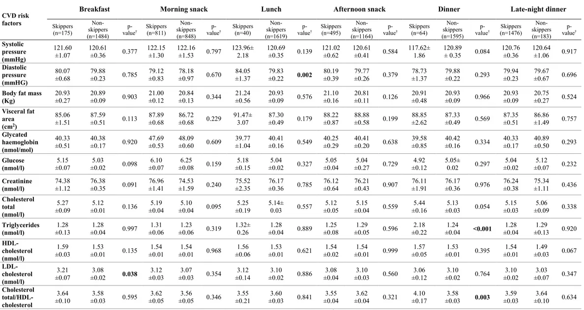
3.3.4 Eating timing, frequency and cardio-metabolic parameters ... 50
3.3.5 Skipping meals and cardio-metabolic parameters ... 50
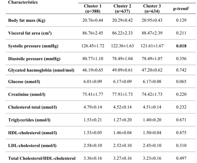
3.3.6 Clusters of eating habits and cardio-metabolic parameters ... 50
3.4 Discussion ... 51
4 Association between Complement Factor H rs1061170 polymorphism and age-related macular degeneration: a systematic review and meta-analysis ... 61
4.1 Background ... 61
4.2 Methods ... 61
4.2.1 Search strategy ... 61
4.2.2 Inclusion/Exclusion Criteria and Data Extraction ... 62
4.2.3 Quality assessment ... 62
4.2.4 Statistical analyses ... 62
4.3 Results ... 63
4.3.1 Search Findings and Studies Characteristics ... 63
4.3.3 Subgroup analysis by stage of disease and ethnicity ... 64
4.3.4 Sensitivity analysis ... 65
4.3.5 Publication Bias ... 65
4.4 Discussion ... 65
5 Association between Vascular Endothelial Growth Factor polymorphisms and age-related macular degeneration: a systematic review and meta-analysis ... 78
5.1 Background ... 78
5.2 Methods ... 78
5.2.1 Search strategy and selection criteria ... 78
5.2.2 Data extraction and quality assessment ... 79
5.2.3 Statistical analyses ... 79
5.3 Results ... 80
5.3.1 Search findings and studies characteristics ... 80
5.3.2 Meta-analysis ... 80
5.3.3 Sensitivity analysis ... 81
5.3.4 Publication bias ... 83
5.4 Discussion ... 83
6 SIRT1 regulates DNMTs functions and LINE-1 methylation in cellular models of retinal degenerative diseases: the role of resveratrol ... 90
6.1 Background ... 90
6.2 Methods ... 91
6.2.1 Cell culture and treatments ... 91
6.2.2 Determination of cell viability ... 91
6.2.3 Determination of reactive oxygen species ... 91
6.2.4 Nuclear protein extraction ... 92
6.2.5 DNMTs activity quantification ... 92
6.2.6 SIRT1 activity quantification ... 92
6.2.7 Real-time polymerase chain reaction ... 93
6.2.8 DNA extraction and LINE-1 methylation analysis ... 93
6.2.9 Statistical analyses ... 94
6.3 Results ... 94
6.3.1 Oxidative stress and inflammatory conditions affect cell viability in RPE cells ... 94
6.3.2 Oxidative stress affects DNMTs/SIRT1 functions and LINE-1 methylation in RPE cells……….94
6.3.3 Inflammatory condition affects DNMTs/SIRT1 functions and LINE-1 methylation in
RPE cells ... 95
6.3.4 Resveratrol ameliorates viability and ROS production in cells under oxidative and inflammatory conditions ... 95
6.3.5 Resveratrol restores DNMTs/SIRT1 functions and LINE-1 methylation in cells under oxidative and inflammatory conditions ... 95
6.4 Discussion ... 96
7 Characterization of SIRT1/DNMTs functions and LINE-1 methylation in age-related macular degeneration: a cross sectional study ... 102
7.1 Background ... 102
7.2 Methods ... 102
7.2.1 Study design ... 102
7.2.2 Nuclear protein extraction ... 103
7.2.3 DNMTs activity quantification ... 103
7.2.4 SIRT1 activity quantification ... 104
7.2.5 Real-time polymerase chain reaction ... 104
7.2.6 DNA extraction and LINE-1 methylation analysis ... 104
7.3 Results ... 105
7.3.1 Characteristics of study population ... 105
7.3.2 DNMTs and SIRT1 functions in AMD patients ... 105
7.3.3 LINE-1 methylation in AMD patients ... 105
7.4 Discussion ... 105
8 DNMTs functions and LINE-1 methylation in a cellular model of hyperglycaemia-induced retinopathy: the role of curcumin ... 110
8.1 Background ... 110
8.2 Methods ... 110
8.2.1 Microarray data ... 110
8.2.2 Cell culture ... 111
8.2.3 Curcumin treatment... 112
8.2.4 Determination of cell viability ... 112
8.2.5 Determination of reactive oxygen species ... 112
8.2.6 Nuclear protein extraction ... 112
8.2.7 DNMTs activity quantification ... 113
8.2.8 Real-time polymerase chain reaction ... 113
8.2.10 Statistical analyses ... 114
8.3 Results ... 115
8.3.1 Analysis of DNMTs expression using GEO datasets ... 115
8.3.2 High glucose-induced oxidative stress precedes upregulation of DNMTs functions in RPE cells ... 115
8.3.3 Effect of curcumin on viability and ROS production in RPE cells ... 116
8.3.4 Curcumin restores DNMTs functions in cells under hyperglycaemic conditions ... 116
8.3.5 LINE-1 methylation analysis ... 117
8.4 Discussion ... 117
9 Discussion and Conclusions ... 125
8
List of publications
This PhD thesis is based on the following original publications:
Barchitta M, Maugeri A. Association between Vascular Endothelial Growth Factor Polymorphisms and Age-Related Macular Degeneration: An Updated Meta-Analysis. Dis Markers. 2016; 2016:8486406.
Maugeri A, Kunzova S, Medina-Inojosa JR, Agodi A, Barchitta M, Homolka M, Kiacova N, Bauerova H, Sochor O, Lopez-Jimenez F, Vinciguerra M. Association between eating time interval and frequency with ideal cardiovascular health: Results from a random sample Czech urban population. Nutr Metab Cardiovasc Dis. 2018; pii: S0939-4753(18)30123-6.
Maugeri A, Barchitta M, Agodi A. The association between complement factor H rs1061170 polymorphism and age-related macular degeneration: a comprehensive meta-analysis stratified by stage of disease and ethnicity. Acta Ophthalmol. 2018 Oct 2. doi: 10.1111/aos.13849.
Maugeri A, Mazzone MG, Giuliano F, Vinciguerra M, Basile G, Barchitta M, Agodi A. Curcumin Modulates DNA Methyltransferase Functions in a Cellular Model of Diabetic Retinopathy. Oxidative Medicine and Cellular Longevity, vol. 2018, Article ID 5407482, 12 pages, 2018.doi.org/10.1155/2018/5407482.
Maugeri A, Barchitta M, Mazzone MG, Giuliano F, Basile G, Agodi A. Resveratrol Modulates SIRT1 and DNMT Functions and Restores LINE-1 Methylation Levels in ARPE-19 Cells under Oxidative Stress and Inflammation. Int J Mol Sci. 2018 Jul 20;ARPE-19(7). pii: E2118. doi: 10.3390/ijms19072118.
Agodi A, Maugeri A, Kunzova S, Sochor O, Bauerova H, Kiacova N, Barchitta M, Vinciguerra M. Association of Dietary Patterns with Metabolic Syndrome: Results from the Kardiovize Brno 2030 Study. Nutrients. 2018 Jul 13;10(7). pii: E898. doi: 10.3390/nu10070898.
Barchitta M, Maugeri A, Quattrocchi A, Barone G, Mazzoleni P, Catalfo A, De Guidi G, Iemmolo M, Crimi N, Agodi A. Mediterranean Diet and Particulate Matter Exposure Are Associated With LINE-1 Methylation: Results From a Cross-Sectional Study in Women. Front Genet. 2018 Oct 30;9:514. doi: 10.3389/fgene.2018.00514.
9
Maugeri A, Barchitta M, Mazzone MG, Giuliano F, Agodi A. Complement System and Age-Related Macular Degeneration: Implications of Gene-Environment Interaction for Preventive and Personalized Medicine. BioMed Res Int. 2018. https://doi.org/10.1155/2018/7532507. The publications have been adapted with the permission of the copyright owners.
10
Acknowledgments
The research was carried out at the Department of Medical and Surgical Sciences and Advanced Technologies "GF Ingrassia" of the University of Catania (Catania, Italy) in collaboration with the Department of Ophthalmology of the University of Catania (Catania, Italy), the Research and Development Department of the SIFI SpA (Catania, Italy) and the International Clinical Research Center of the St Anne’s University Hospital (Brno, Czech Republic).
The research activities were partially supported by the Department of Medical and Surgical Sciences and Advanced Technologies “GF Ingrassia”, University of Catania (Piano triennale di sviluppo delle attività di ricerca scientifica del Dipartimento - 2016-18) and by the National Program of Sustainability II (MEYS CR) (no. LQ1605). I’m also grateful to SIFI SpA for technical and financial support.
11
Abbreviations
5mC 5-Methyl-cytosines
AA Arachidonic acid
AGE Advanced glycation end-products
AHEI Alternate Healthy Eating Index
ALE Advanced lipoxidation end products
ALR2 Aldose reductase gene
AMD Age-related macular degeneration
ANCOVA Analysis of covariance
ANOVA Analysis of variance
APOE Apolipoprotein E
AREDS Age-Related Eye Disease Study
ARIC Atherosclerosis Risk in Communities
ARMS2 Age-related maculopathy susceptibility 2 ARPE-19 Human retinal pigment epithelial cells
BFM Body fat mass
BMI Body mass index
C2 Complement component 2
C3 Complement component 3
CAREDS Carotenoids in Age-Related Eye Disease Study
CFB Complement factor B
CFH Complement factor H
CFI Complement Factor I
CI Confidence interval
CNV Choroidal neovascularization
CR1 Complement receptor 1
DCCT Diabetes Control and Complications Trial
DCF 2’,7’- dichlorofluorescein
DCFDA 2’,7’ - dichlorofluorescin diacetate
DHA Docosahexaenoic acid
DM Diabetes mellitus
DME Diabetic macular edema
DMEM Dulbecco's Modified Eagle's medium
DNMT DNA methyltransferase
DR Diabetic retinopathy
EAR Estimated Average Requirements eNOS Endothelial nitric oxide synthase
EPA Eicosapentaenoic acid
FFQ Food Frequency Questionnaire
GA Geographic atrophy
GEO Gene Expression Omnibus
GOx Glucose oxidase
12
GWAS Genome Wide Association Study
HIF-2α Hypoxia-inducible factor 2α
HTRA1 High temperature requirement factor A1
HWE Hardy-Weinberg Equilibrium
IGF-1 Insulin-like growth factor 1 IL17RC Interleukin-17 receptor IQR Interquartile range
LINE Long Interspersed Nuclear Element
LPS Lipopolysaccharide
MAC Membrane attack complex
MCP Membrane cofactor protein MDS Mediterranean Diet Score MET Metabolic Equivalent of Task MTT Thiazolyl blue tetrazolium bromide
NF-kβ Nuclear factor-kβ
NPDR Non-Proliferative Diabetic retinopathy
OR Odds ratio
ORF Open reading frame
PCA Principal component analysis PDR Proliferative Diabetic retinopathy
PLEKHA1 Pleckstrin Homology Domain-containing Protein family A member 1
PUFA Polyinsatured fatty acid
RAGE Receptor for advance glycation end-products
Rco Correlation coefficient RCT Randomized controlled trial ROS Reactive oxygen species RPE Retinal pigment epithelium
SD Standard deviation
SE Standard error
SINE Short Interspersed Nuclear Element
SIRT1 Sirtuin 1
STZ Streptozotocin
TET Ten-eleven-translocation
TGF- β Transforming growth factor β
UKPDS UK Prospective Diabetes Study
VEGF Vascular endothelial growth factor
13
1 Introduction
1.1 Gene-diet interaction“Let food be thy medicine and medicine be thy food” - this quote by Hippocrates, the father of western
medicine, has inspired humans to understand how environment and foods affect individual’s health since ancient times. However, in the last decades alone, researchers and health professionals have revealed that dietary habits play a crucial role in maintaining health and in disease prevention 1. The
research, especially after the conclusion of the Human Genome Project, raised questions whether gene-diet interaction may positively or negatively influence human health. With this question in mind, researchers coined the term “Nutrigenomics”, referring to the study of dietary influence on the Genome, Transcriptome, Proteome, and Metabolome 2. Although several lines of evidence began to
show the protective effect of dietary intervention on chronic degenerative diseases by modulating molecular functions 1, further research is needed to develop novel strategies for maintaining health
and preventing diseases 3. A specific area of research that elucidates mechanisms involved in
gene-diet interaction is the Nutriepigenomics, the study of the impact of gene-diet on changes in gene expression by modulating epigenetic mechanisms 3. In 1939, Conrad H. Waddington was the first to define
Epigenomics as the causal interactions between genes and their products which bring the phenotype into being 4. This definition was later revised as the study of heritable changes in gene expression that
occur without changes in DNA sequence 5. Thus, the field of Nutriepigenomics is further revealing
the nature of diet-gene interaction, providing support for the role of nutrition in preventing diseases3.
1.1.1 DNA methylation
Epigenetic mechanisms - including DNA methylation, histone modifications, histone variants, chromatin remodelers and non-coding RNAs - regulate how and when genes are expressed without altering DNA sequence. These molecular processes characterize the epigenome which is dynamic in response to environmental signals, modifiable during normal cell differentiation and heritable in daughter cells 6. Among these mechanisms, DNA methylation is one of the most extensively studied
and best characterized. In mammals, DNA methylation is regulated by the activity of three DNA methyltransferases (DNMTs): while DNMT1 has a maintenance role, DNMT3a and 3b are de novo methylases. By contrast, the removal of methyl groups on DNA is mediated by the ten-eleven-translocation (TET) proteins 7. DNA methylation almost exclusively occurs within CpG islands –
short sequences in gene promoters and regulatory regions that typically contain about 5-10 CpG dinucleotides per 100 bp 8. In humans, approximately 60% promoters include CpG islands and ~90%
14
scattered throughout the genome 10. Some of these sequences act as retrotransposons, disrupting gene
expression and eventually leading to genomic instability. To protect from this potential deleterious effect, DNMT1 works to maintain these sequences highly methylated 11,12. DNMTs functions are
associated with several key physiological processes, including genomic imprinting, X-chromosome inactivation, regulation of gene expression, maintenance of chromosome integrity through chromatin modulation, DNA stabilization and DNA-protein interactions 13. Aberrant DNMT expression and
activity are involved in several diseases including cardiovascular diseases, obesity, type-2 diabetes and cancer 14-16.
1.1.2 Long Interspersed Nuclear Elements 1
Human genome includes almost 3 billion non-coding base pairs, of which 50% is recognized as repetitive sequences. Most of these sequences - Long Interspersed Nuclear Elements (LINEs) and Short Interspersed Nuclear Elements (SINEs) - occur discontinuously as singular scattered copies in the genome 17. LINE-1 elements - retrotransposons capable of independent and autonomous
retrotransposition via RNA intermediate - comprise approximately 17% of the genome with more than 500,000 copies. Retrotransposition of LINE-1 may lead to chromosomal instability, DNA rearrangement and alteration of ectopic gene expression in cancerous tissues 18. A full length
LINE-1 sequence is approximately 6 kb long including bidirectional, non-canonical promoter and tow open reading frames (ORF1 and ORF2) 19. After RNA polymerase II transcription, mRNA processing and
its export to the cytoplasm, ORF1 and ORF2 encode two proteins (ORF1p and ORF2p) that preferentially associate with their encoding RNA 20. The encoded ribonucleoprotein particles transfer
the RNA to the nucleus, where ORF2p triggers target-primed reverse transcription to insert a copied DNA sequence at a new site in the host cell genome 21. Although LINE-1 transcription is largely
regulated by DNA methylation of about 50 promoters, most of LINE-1 elements are truncated and cannot be transcribed 20. It follows that only one hundred LINE-1 elements are functionally capable
of retrotrasposition and only a few contribute to the vast majority of retrotransposition events 22.
LINE-1, along with Alu sequences, are the most common transposable sequences and measurement of their methylation levels has been used as a surrogate marker of global genomic DNA methylation
23. Although LINE-1 methylation is not an universally accepted marker of global methylation,
aberrant methylation of these sequences was associated with cancer, cardiovascular and neurodegenerative diseases 24-26.
1.1.3 The effects of nutrients on DNA methylation
Environmental and lifestyle factors can potentially modify DNA methylation, leading to genome reprogramming in exposed individuals and in future generations 27. Several classes of nutrients -
15
folate, polyphenols, selenium, retinoids, fatty acids, isothiocyanates and allyl compounds – modulate DNA methylation process via different mechanisms 28. Folate, a fundamental methyl donor for
cellular replication and maintenance, modulates DNA methylation, synthesis and repair. However, since folate status has been both positively and negatively associated with DNA methylation 29-35, its
effect remains to be completely elucidated. While most of the studies found that global DNA methylation levels increased with increasing folate intake 29,31-35, others observed an inverse
relationship 30. Controversial results might be attributed to unmeasured factors such as ethnicity,
genetic variants in one-carbon metabolism, lifestyles, physiological and pathological conditions, which in turn can affect DNA methylation process.
Other dietary compounds exhibit the potentiality to modulate DNA methylation. For instance, fatty acids have been suggested to affect DNA methylation process via mechanisms independent of 1-carbon nutrients. In fact, it has been demonstrated that global DNA methylation decreased with increasing intake of saturated fatty acids. Since few studies investigated the effect of n-3 and n-6 polyinsatured fatty acids (PUFAs) on DNA methylation - especially concerning eicosapentaenoic acid (EPA) 36, docosahexaenoic acid (DHA) 37 and arachidonic acid (AA) 38 - further research is
recommended to elucidate the association between dietary fat intake and DNA methylation 35.
Furthermore, several lines of evidence suggest that dietary compounds present in fruit, vegetables and spices may modulate epigenetic signatures in human cells. For instance, it has been reported that dietary phytochemicals may repair DNA damage by enhancing histone acetylation and altering DNA methylation 39.
1.1.4 The effects of dietary patterns on DNA methylation
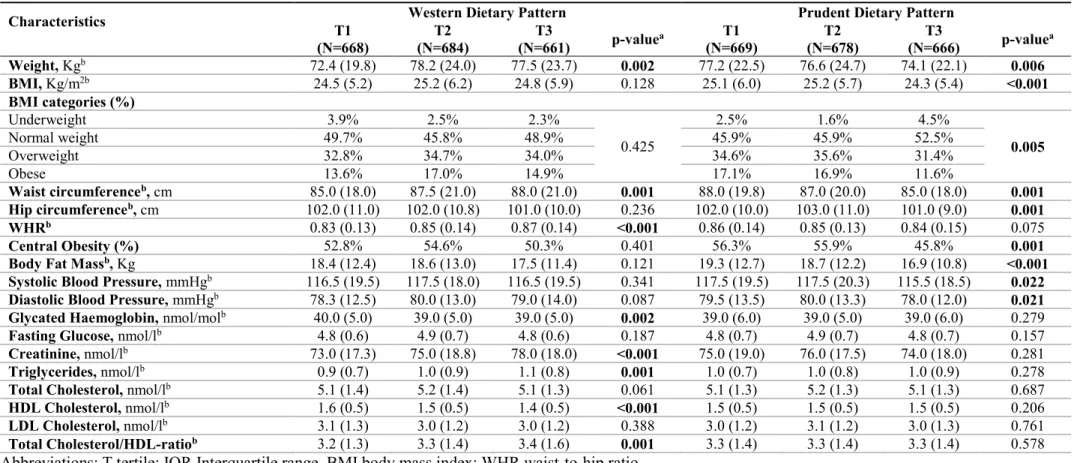
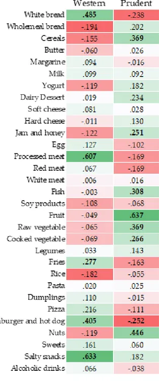
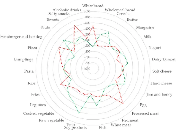
Previous studies focused on the consumption of specific foods or nutrients, but there is currently growing interest in determining how dietary patterns may affect global and local DNA methylation. A previous study - evaluating the association between diet and leukocyte DNA methylation in a cancer-free population - identified two dietary patterns: the prudent dietary pattern, characterized by a high intake of vegetables and fruits; the western dietary pattern, characterized by a high intake of energy-dense foods such as grains, meats, potato, oil, and dairy 35. While only the intake of dark green
vegetables seemed to be significantly associated with DNA methylation among specific food groups, the analysis of dietary patterns revealed a positive association between the prudent dietary pattern and global DNA methylation in a dose-response manner 35. In line with this evidence, further studies
observed that healthy women with high intake of vegetables and/or fruits had a lower risk of LINE-1 hypomethylation 32,35. The biological explanation of this relationship could be attributed to the wide
variety of nutrients and bioactive compounds provided by fruits and vegetables, including phytochemicals (phenolics, flavonoids, and carotenoids), vitamins (vitamin C, folate, and pro-vitamin
16
A), minerals (potassium, calcium, and magnesium), and fibres, which in turn modulate multiple pathways associated with epigenetic mechanisms 40,41.
1.2 Epigenetic hallmarks in aging and age-related diseases
If Genetics alone cannot explain the distinct patterns of aging nor different susceptibility to age-related diseases, for instance, between monozygotic twins 42, Epigenetics offers an explanation of
these phenomena 43. Senescence and aging are characterized by progressive loss of histones,
transcriptional changes, losses and gains in heterochromatin, global hypomethylation and local hypermethylation, and chromatin remodelling (Figure 1). These changes are heavily influenced by environmental stimuli and nutrient availability, which in turn alter intracellular metabolite concentrations 44. During aging, the overarching profile of DNA methylation changes, showing global
DNA hypomethylation and local hypermethylation that may activate specific transcriptional pathways. Although this is consistent with the abovementioned changes that occur in aging cells, it remains unclear what specific genes are directly affected by aberrant DNA methylation. While hypermethylation mainly occurs at promoter CpGs, loss of methylation occurs in repetitive regions of the genome that correlate with constitutive heterochromatin. The analysis of DNA methylation status of 26,486 autosomal CpGs in a number of human tissues revealed hypermethylation of promoter CpGs and hypomethylation of those outside during aging 45. The observed paradox -
hypermethylation of promoters versus hypomethylation of repetitive regions - is likely due to differential expression and activity of DNMTs over repeat elements and key genes affecting lifespan
46. The identification of DNA methylation markers associated with aging represents a milestone in
this area of research: in a large study, Horvath and colleagues demonstrated that DNA methylation at 353 CpGs - termed clock CpGs - accurately predict age in more than 8000 samples, including healthy human tissue and cell types including liver, kidney, immune, and brain cells and cancer samples 47,48.
Consistently, an independent study on a population aged 19-101 allowed to build a predictive model of aging using a smaller set of 71 CpGs 49. However, further advances require to understand the
underlying mechanism of these epigenetic clocks in age-related diseases. 1.3 Age-related Macular Degeneration
1.3.1 Clinical features and epidemiology
Age-related macular degeneration (AMD) is a neurodegenerative disease which leads to the progressive destruction of the neurosensory macular area, involving retinal pigment epithelium (RPE), Bruch’s membrane and choroid 50. The reasons why AMD preferentially affects the central
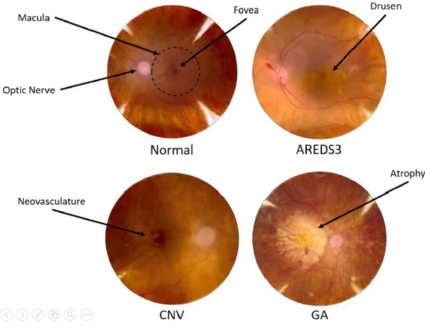
region of the retina are not well clarified. According to the severity of symptoms, the disease is classified into early, intermediate and advanced stages: while the early stage is characterized by the
17
aberrant pigmentation of the RPE and the accumulation of “drusen” - yellowish lipid-rich, protein-containing extracellular deposits accumulating between RPE and Bruch’s membrane -, the advanced stage may manifest as non-exudative (dry) or exudative (wet) AMD. The first is characterized by the geographic atrophy (GA) of RPE and thinning of the retina, which lead to the gradual loss of photoreceptors and central vision 51. The second is characterized by the growth of abnormal blood
vessels from the choroid into the normally avascular sub-RPE and sub-retinal regions (choroidal neovascularization, CNV), which negatively affects central vision 52,53. Although exudative AMD
patients represent a small proportion of total AMD cases, they account for the majority of blindness associated with AMD 54. Approximately 170 million individuals are affected by AMD worldwide,
with a prevalence that ranges from 2% to 20% among elderly people. AMD is thereby the leading cause of blindness in the developed countries and the third leading cause globally 55. Prevalence of
early AMD is positively associated with age across all ethnicities, though this is most marked in Caucasians and Hispanics and to a lesser extent in Asians and Africans. Across all age strata, Africans had the lowest prevalence of early AMD, followed by Asians. For individuals aged under 55 years, a reasonable prevalence of early AMD among Caucasians, Hispanics, Asians, and Africans is 4%, 6%, 3%, and 3%, respectively; the occurrence increases to 24%, 22%, 13%, and 11% for persons older than 75 years. With regard to advanced AMD, it has been demonstrated an exponential age-related increase in Caucasians and Asians, a moderate increase in Hispanics and a slight increase in Africans. Although prevalence of advanced AMD in individuals younger than 55 years ranges from 0% to 0.2% across all ethnicities, its occurrence increases to 6.5%, 2.4%, 1.3%, and 0.6% among persons older than 75 years in Caucasians, Asians, Hispanics, and Africans, respectively 56. As the aging population
increases, the global prevalence of AMD is anticipated to spread to 288 million by the year 2040 57,
with direct healthcare cost due to AMD that is likewise expected to increase proportionately. In the last decades, several studies - especially on Caucasians - reported consistent AMD incidence rates across different populations 56. The overall 10-year risk ranged from 11.1% to 23.7%, reflecting
partial differences in study design, follow-up time, and variation in risk factors. There was no difference in incidence rate between men and women. AMD diagnosis and staging require the exam of fundus imaging of the retina for the visualization of drusen, pigmentary changes in the RPE, neural retinal degeneration, and/or proliferative events. Diagnosis of CNV is confirmed by fluorescein angiography, which marks blood vessels between choroid and RPE 56. Clinicians classifies AMD
progression using the Age-Related Eye Disease Study (AREDS) system 51, a standard grading scale
from 1 to 4 (Figure 2):
18
Eyes include early AMD cases in which symptoms were limited to small drusen <63 μm (also referred to as “hard” drusen), a single intermediate-sized druse 63–124 μm, and/or pigmentary changes;
Eyes include those with more extensive drusen such that they have at least one large druse >124 μm (“soft” druse), multiple intermediate drusen, and/or GA not involving the central macula (AREDS3);
Eyes exhibiting GA involving the central macula and/or CNV (AREDS4). 1.3.2 Major risk factors
The main causes of AMD are characterized by the interaction of oxidative stress, aberrant RPE activity and function, apoptosis and impaired immune system activation 58,59. Smoking - leading to
oxidative stress, ischemia, hypoxia and neovascularization - is the strongest modifiable risk factor 60.
Although both current and former smoking may increase AMD risk, a protective effect has been observed for time since smoking cessation 61. In fact, subjects who had quitted smoking for more than
20 years were not at risk of advanced AMD 62,63. Several lines of evidence suggested that - together
with smoking - many other cardio-metabolic risk factors are also potential for AMD 64, including
elevated total serum cholesterol 62 higher body mass index (BMI) 65 and hypertension 66,67. The
Beaver Dam Eye Study reported that higher pulse pressure and systolic blood pressure were associated with an increased risk of early AMD lesions and exudative AMD 67. In addition, a recent
meta-analysis concluded that excessive body weight was weakly associated with increased AMD risk - especially with advanced AMD - in a dose-dependent manner, indicating that maintaining normal body weight and avoiding weight gain may confer potential protection against this disease 68.
However, it is still unclear if the observed associations may be related to an overall unhealthy lifestyle instead of a direct effect on AMD susceptibility.
To date, the only well-established protective factor against AMD is a healthy diet, characterized by high intake of omega-3, lutein, zeaxanthin, and antioxidants 69-72. Consistently, the AREDS2
formulation - a combination of zinc, b-carotene, vitamins C, and E supplements - has been shown to reduce the risk of progression to advanced AMD 73. While AREDS formulation represents the only
available treatment against non-exudative AMD, intravitreal injections of anti-vascular endothelial growth factor (VEGF) agents (i.e. ranibizumab, bevacizumab and aflibercept) may improve visual acuity in patients with exudative AMD 74-79.
1.3.3 Genetic Risk Factors
In addition to modifiable factors, genetic variants confer about 60% of the attributable risk 80, with at
19
after the discovery of genetic variants associated with AMD in the complement factor H (CFH) gene and in the 10q26 locus, which contains the age-related maculopathy susceptibility 2 (ARMS2) and high temperature requirement factor A1 (HTRA1) genes 82. For the first time, these findings pointed
out the crucial role of inflammation and oxidative stress pathways in the aetiology of AMD. Later, other genetic variants in complement genes, VEGF and apolipoprotein E (APOE) 83-86 were
associated with AMD, although they confer a smaller effect. 1.3.3.1 Complement system and Complement Factor H
The refined equilibrium between activation and inhibition of complement system is one of the most critical regulatory mechanisms to prevent self-tissue damage 87,88. In fact, on one hand, increased
complement activity may be protective against chronic low-grade inflammation and infection in early life 89, on the other hand, lack of its inhibition is associated with several diseases, such as systemic
lupus erythematosus 90, atypical haemolytic uremic syndrome 91, dense deposit disease 92, and AMD 93. Therefore, complement system activity is strictly controlled by regulatory proteins, which mainly
act by degrading complement components, increasing complement component 3 (C3) convertase decay and modulating the membrane attack complex (MAC) assembly 94-96. The first is a function of
Factor I (FI), which modulates both the classical and alternative pathways by cleaving C3 into inactive fragments 97. To prevent non-specific degradation of complement components, the
proteolytic activity of FI however requires several cofactors - such as complement receptor 1 (CR1), membrane cofactor protein (MCP), and CFH 97-101 - which accelerate C3 convertase decay 102,103.
The capacity of alternative pathway to distinguish between self and non-self is conferred by the recognition of glycosaminoglycans and sialic acid glycans (i.e. heparin-sulfate and N-Acetylneuraminic acid) on host cells 104-107. Binding of CFH to the surface of necrotic cells and to
apoptotic particles is mediated by CRP, Annexin II, DNA and histones 108-111. An additional
complement inhibitor is the decay-accelerating factor, which inhibits assembly of neo-formed C3 convertases and accelerates the decay of pre-existing convertases 96,112-116. Lastly, the regulation of
complement system may be also provided by inhibiting MAC formation via membrane bound (CD59) or fluid-phase (Vitronectin and Clusterin) inhibitors 117-122.
Several lines of evidence demonstrated that dysregulation of complement pathways - especially the alternative pathway - is involved in AMD pathogenesis. The major stressors for AMD development, such as aging, smoking, and oxidative stress, have been linked to the over-activation of the complement system (Figure 3). This has been also supported by immune-histological and proteomic studies identifying complement components as constituents of drusen 80,123-126. Increased levels of
activated complement components, which are released during complement activation, have been also observed in peripheral blood of AMD patients 127-129. Consistently, complement regulators, such as
20
Vitronectin, Clusterin and MCP, are highly expressed in drusen and RPE cells adjacent to drusen
80,130,131. Drusen are especially characterized by Amyloid beta accumulation, which in turn results in
complement activation and chronic low-grade inflammation 132. During RPE aging, it has also been
observed the accumulation of lipofuscin and bis-retinoid component, which leads to the formation of lipid peroxidation products 133, apoptosis and complement activation 134,135.
In 2005, four independent genetic association studies revealed the CFH gene on chromosome 1q32 as the first one associated with AMD risk 125,136-138, with an effect that was mainly attributed to the
rs1061170 polymorphism. This polymorphism leads to an amino acid change at position 402 of the CFH polypeptide (Y402H). Prevalence of the 402H risk variant varies across ethnicities 139, with an
increased AMD risk of 2.5 times among heterozygous individuals, and 6.0 times among homozygotes
140. This finding was confirmed by pooled analyses in both Caucasians 139 and Asians 141-143. The
rs1061170 polymorphism has been also recognized as a predictor of response to the anti-VEGF treatment: homozygotes patients were less likely to achieve a better outcome than those carrying wild type genotype, suggesting the need of more effective therapeutic strategies for this subgroup of patients 144. By contrast, the rs800292 polymorphism - a coding variant in the SCR1 domain - has
been found to be protective against AMD in both Caucasians and Asians 143. This polymorphism,
which leads to an amino acid change at position 62 of the CFH polypeptide (V62I), also conferred a better response to treatment of exudative AMD 144. To date, the role of other CFH genetic variants is
still under debate. A recent meta-analysis 145 aimed at resolving inconsistent findings about the effect
of four coding and noncoding variants (rs1410996, rs1329428, rs2274700, rs3753394). Pooled results demonstrated that these polymorphisms are significantly associated with increased AMD risk, but none of them was related to response to treatment 146.
1.3.3.2 Genetic variants in other complement components
The C3 gene is located on chromosome 19p13.3-13.2 and consists of 41 exons encoding for 1663 amino acids and 13 functional domains. The encoded protein is biologically inactive until it undergoes to conformational changes that expose binding sites to pathogenic cell surface and other complement components 147. Although emerging evidence proposed the association between C3 polymorphisms
and AMD, findings are currently controversial 148-152. The most commonly investigated is the
rs2230199 polymorphism, which leads to the R102G substitution modulating both C3 binding capacity, cofactor activity and thereby extending convertase lifetime 153. The effect of this
polymorphism on AMD risk was confirmed in Caucasians but not in Asians 154. More recently, a
meta-analysis added to the current knowledge, suggesting, on one hand, the adverse effect of rs1047286 and rs11569536, and on the other hand the protective effect of the rs2250656 155. Instead,
21
Particularly, the comparison of AMD Treatments Trials failed in demonstrating the effect of rs2230199 polymorphism on visual and anatomical outcomes, in patients treated with anti-VEGF drugs 160. However, the analysis of changes in central macular thickness after ranibizumab treatment,
showed that the minor allele of rs2250656 polymorphism was associated with improvement in retinal thickness and architecture 161.
The complement factor B (CFB) gene is located on chromosome 6p21 which includes the major histocompatibility complex class III region. Mounting evidence suggests that genetic variants in this region are associated with reduced AMD risk. In fact, previous meta-analyses confirmed the protective effect on AMD risk of the common rs641153 polymorphism - also known as R32Q - in different ethnic groups 86,162. Located 500 bp upstream from CFB gene, there is also the complement
component 2 (C2) gene, encoding for a serum glycoprotein that functions as part of the classical pathway. Two polymorphisms (i.e. rs9332739 and rs547154) have been directly associated with decreased AMD risk 86. However, these variants may be indirectly linked to AMD risk due to linkage
disequilibrium with CFB. Indeed, some common haplotypes, spanning CFB and C2 genes, are considered highly protective against AMD 163. Here again, lack of evidence exists about the effect of
CFB and C2 genetic variants on response to intravitreal anti-VEGF injections, with no significant effects on patients with exudative AMD 146,164.
Located on chromosome 4q25, the CFI gene encodes for a precursor protein in hepatocytes, macrophages, lymphocytes, endothelial cells and fibroblasts. To obtain the active protein, the precursor is cleaved into heavy and light chains, which form a heterodimeric glycoprotein. This heterodimer can prevent the assembly of convertase enzymes by cleaving C4b and C3b. The association between CFI polymorphisms and AMD was firstly reported by Fagerness et al. 165, and
further studies identified genetic variants that modulate gene expression and protein production 166-169. Although the rs10033900 polymorphism is the most investigated, evidence of an association with
AMD risk is still controversial. To date, an updated meta-analysis summarizes that carriers of rs10033900 polymorphism have a reduced risk of developing AMD in Caucasians, but not in Asians170.
1.3.3.3 Age-related maculopathy susceptibility 2 locus
The susceptibility conferred by genetic variants at chromosome 10q26 was initially proposed by linkage studies 171-173. Although it has been demonstrated a strong association between several genetic
variants in the 10q26 locus and AMD, high linkage disequilibrium between three genes (Pleckstrin Homology Domain-containing Protein family A member 1 - PLEKHA1 -, ARMS2 and HTRA1 genes) made difficult to understand the source of genetic effect at this region. Mounting evidence supports the involvement of both ARMS2 and HTRA1 in the AMD pathogenesis, whereas PLEKHA1
22
seems to be weakly associated with AMD 174-178. In the ARMS2 gene, the rs10490924 polymorphism
– which leads to an A69S change - was associated with a ≈15-fold increased risk of AMD 174-178. In
addition, a deletion-insertion polymorphism (del443ins54; in/del) in the 3-UTR of ARMS2 was associated with AMD both in Caucasian and Asians 179-181. Although molecular mechanisms
underpinning the association between ARMS2 and AMD remain to be elucidated, evidence of disorganized mitochondrial membranes and decreased number of mitochondria in RPE of AMD patients pointed out the mitochondrial dysfunction in AMD pathogenesis 182,183. In fact, ARMS2 may
affect mitochondrial function, leading to the production of reactive oxygen species, which in turn cause apoptosis and increased AMD risk 182,183.
In the HTRA1 gene, the rs11200638 polymorphism is associated with an increased risk of AMD, as confirmed by a recent meta-analysis 184. Interestingly, subgroup analyses revealed that the
polymorphism is significantly associated with CNV but not with GA, and that the effect is stronger in Caucasians than in Asians 184,185. Several lines of evidence demonstrated that the rs11200638 risk
allele is associated with higher levels of HTRA1 mRNA and protein levels. Particularly, HTRA1 may inhibit signalling of transforming growth factor β (TGF- β) proteins, which have been reported to act as negative growth regulators in the retina and RPE 186,187. In addition, HTRA1 promotes the
degradation of extracellular matrix - through enhanced expression of matrix metalloproteases - and may affect the integrity of Bruch’s membrane and RPE layer. Common haplotypes encompassing both the ARMS2 and the HTRA1 genes have also been associated with AMD risk. Among these, a common haplotype TAT tagged by rs10490924, rs11200638, and rs2293870 significantly predisposed to AMD, while the haplotype GGG significantly reduced the risk of AMD 188. Similarly,
the haplotype T-G-Wt-G tagged by rs2736911, rs10490924, in/del/Wt, and rs11200638, appeared to be protective against AMD both in Caucasian and Asians 189, while the in/del and the rs11200638
risk allele by itself were insufficient to modify HTRA1 expression levels 189.
1.3.4 Nutrition and age-related macular degeneration
A research area of increasing interest concerns the potential role of diet, antioxidant and/or mineral supplementation in preventing and delaying AMD progression. In fact, in the last decades, it has been consistently demonstrated that an adequate intake of omega-3 fatty acids, lutein, zeaxanthin, and other antioxidants, represents the only well-known protective factor against AMD onset and progression
69-72. Among these, antioxidants have been proposed to protect the macula against the oxidative stress
that leads to photoreceptor damage. A meta-analysis, including 65,250 participants from four randomized controlled trials (RCTs), evaluated the effect of antioxidants (i.e. lutein, zeaxanthin, and vitamins C and E) and/or minerals (i.e. zinc and selenium) supplementation, alone or in combination, versus placebo control subjects. Findings from this work demonstrated a no significant effect of
23
antioxidant supplementation on AMD onset 190. However, when examining evidence related to the
role of nutrition in AMD, it is worth highlighting the necessity to differentiate between disease onset and progression. In line with this need, AREDS I 191 and AREDS II 73 - two large multicentre RCTs
sponsored by the National Eye Institute - have been designed to evaluate safety and efficacy of vitamins and other nutrients supplementation for altering the natural history in patients with established disease.
The AREDS I was conducted from 1992 to 2006 to evaluate whether systemic antioxidant supplementation might ameliorate clinical aspects, natural history, and risk factors associated with AMD. AREDS I demonstrated that, in patients with at least intermediate AMD, daily long-term high-dose supplementation of 500 mg vitamin C, 400 IU vitamin E, 15 mg beta-carotene, 80 mg zinc oxide, and 2 mg cupric oxide) reduced the risk of progression to advanced AMD at 5 years from 28 to 20%
191. Next, observational studies pointed out the potential beneficial role of higher dietary intakes of
the retinal carotenoids (i.e. zeaxanthin and lutein) and omega-3 long-chain polyunsaturated fatty acids (i.e. DHA and EPA) 71,192. Lutein and zeaxanthin are xanthophyll carotenoids that have been
recognized to improve antioxidant protection, filtration of short-wavelength light, maintenance of structural integrity of cell membranes, and modulation of signal transduction pathways within the retina. DHA is one of the structural components of lipid membranes in retinal photoreceptors 193, and
its status has been found to influence the phototransduction cascade 194. DHA and EPA show also
crucial retino-protective effect by modulating gene expression 195, cellular differentiation 196, and cell
survival 196. Thus, the AREDS II rationale was based partly on these findings suggesting that retinal
carotenoids and omega-3 fatty acids influence the biological processes that have been implicated in AMD pathogenesis. Accordingly, AREDS II aimed at investigating whether the addition of 10 mg lutein and 2 mg zeaxanthin, alone or in combination with 350 mg DHA and 650 mg EPA, further reduced the risk of progression to late AMD in subjects with at least intermediate disease 197. AREDS
II demonstrated that the addition of lutein + zeaxanthin, DHA + EPA, or both components to the AREDS formulation did not further reduce the risk of progression from intermediate to late AMD, compared with the original AREDS supplement 73. Although a couple of small RCTs suggested the
potential for carotenoid supplements to enhance visual function in AMD patients 198,199, design
limitations of these studies raise the need of high-level evidence to corroborate these findings. Given the results from AREDS, diet has been proposed as a potentially modifiable factor that may influence both the development and progression of AMD. In fact, foods and nutrients may interact with each other 200. Examining dietary pattern rather than single nutrient intake may better account
for the relationships among different diet components 200. A case control study of ≈ 700 subjects
24
Index (AHEI) - significantly decreased the odds of AMD 200. This is consistent with evidence from
the Carotenoids in Age-Related Eye Disease Study (CAREDS), showing that women which reflected the healthiest smoking, physical activity and dietary habits had 71% lower odds of early AMD compared to those with unhealthy lifestyle 201. In spite of this couple of studies, there is still the need
of further research exploring the effect of healthy dietary pattern, such as Mediterranean diet, on AMD onset and progression.
1.3.4.1 Gene-Diet interaction in age-related macular degeneration
While antioxidants supplementation clearly decreases the progression from early to advanced AMD
202, evidence about the effect of their intake through the diet is still controversial, probably due to
different genetic susceptibility and/or other unmeasured effect modifiers. The Rotterdam study found a synergic interaction between CFH rs1061170 polymorphism and dietary intake of antioxidants, demonstrating that higher intake of zinc, omega-3 fatty acids, β-carotene, lutein and zeaxanthin might reduce the incidence of early AMD in subjects at higher genetic risk 203. This is in line with pooled
analysis of Blue Mountains Eye and Rotterdam cohorts, showing that dietary intake of lutein and zeaxanthin protected against the risk of early AMD, only in concurrence with at least two risk alleles of CFH rs1061170 and ARMS2 rs10490924 polymorphisms 204. Conversely, in absence of genetic
susceptibility, higher intake of lutein and zeaxanthin was associated with higher incidence of early AMD 204. The analysis of the Atherosclerosis Risk in Communities (ARIC) study added to these
controversial findings, founding that higher lutein and zeaxanthin intake was associated with lower AMD prevalence among carriers of the heterozygous CFH genotype, higher prevalence among carriers of the homozygous risk genotype, and no statistically significant effect among those with no-risk genotype 205.
In the AREDS II, increased intake of DHA and EPA was associated with decreased risk of non-exudative AMD, after adjusting for behavioural factors and genetic variants, including polymorphisms in CFH, ARMS2/HTRA1, CFB, C2, C3, and CFI genes 206. Moreover, the Blue
Mountain Eye Study found that weekly consumption of fish was associated with lower risk of advanced AMD, only among patients with the CFH homozygous risk genotype 207. Findings from a
sub-sample of the AREDS II also demonstrated a significant interaction between folate intake and the rs2230199 C3 polymorphism. In fact, the protective effect of folate intake was evident among subjects with homozygous non-risk genotype, but not in those carrying the risk allele. By contrast, no significant effect on AMD progression was observed for dietary intake of thiamin, riboflavin, niacin, and vitamins B6 and B12 208. With regard to dietary pattern, the study by Merle and colleagues
- including participants of the AREDS - was the first evaluating the interaction between genetic risk factors and overall diet 209. Particularly, they demonstrated that adherence to the Mediterranean diet
25
was associated with lower risk of progression to advanced AMD among patients with non-risk genotype, but not among those with the homozygous risk genotype 209. The significant association,
in absence of genetic susceptibility, might be explained by the protective effect of Mediterranean diet on immune and inflammatory responses.
The effect of the interaction between nutrient supplements and genetic susceptibility on the progression to advanced AMD is currently under debate. In 2008, Klein et al. demonstrated that the effect of supplementation with antioxidant and zinc on the AMD progression was higher among subjects with non-risk genotype for the CFH rs1061170 polymorphism than in high-risk subjects 210.
In addition, Seddon et al. reported that antioxidant and zinc supplementation reduced the risk of progression to the exudative AMD but not to non-exudative AMD 211. Next, Awh et al. found that
zinc supplementation reduced the progression to advanced AMD, among subjects with no risk alleles for CFH and at least one risk allele for ARMS2 212. Further, the same researchers demonstrated a
distinct effect on disease progression according to the number of risk alleles for these polymorphisms: while zinc supplementation was protective against the harmful effect of the ARMS2 risk allele, it increased the risk posed by CFH allele 212. These findings are supported by current knowledge about
physiologic implication of zinc binding to CFH, which might counteract the ability to inactivate C3 convertase 213-215. This, together with functional consequences of CFH rs1061170 polymorphism,
might cause the detrimental effect associated with concurrence of CFH risk genotypes and zinc supplementation 216.
1.3.5 DNA methylation landscape in age-related macular degeneration
Given the abovementioned evidence, a typical gene-environment interaction has been proposed in the context of retinal degenerative disorders 217. In fact, it seems reasonable that environmental effects
may be under genetic control, as well as environmental risk factors may trigger the disease in genetically susceptible subjects 82. Retinal cells show altered gene expression in response to
environmental factors (i.e. nutrient intake, light, and oxidative stress) and internal cellular signals (i.e. reactive oxygen species - ROS), calcium concentration, and DNA damage) 218. Epigenetic
mechanisms may represent the events modulating the interaction between genetic factors, life experiences and environmental exposures 5. Among these, DNA methylation and histone
modifications are strictly related to gene expression 219,220 and genome stability 18. The key
pathological features of AMD are associated with several risk factors (e.g. smoking, low omega-3 diet, excessive retinal iron levels and ageing) which induce high oxidative stress 221. In a recent
genome-wide methylation study in post-mortem RPE and choroid from AMD patients, the hypermethylation of the promoter regions of two glutathione S transferase isoforms (GSTM1 and GSTM5) was identified 222. The epigenetic downregulation of these detoxification enzymes may
26
increase the susceptibility to oxidative stress in AMD 41. The cellular redox state of the retina and
aging might also involve the activation of sirtuin 1 (SIRT1). SIRT1 upregulates the expression of hypoxia-inducible factor 2α (HIF-2α), VEGF and erythropoietin, activating both hypoxia and angiogenesis mechanisms 223.
Inflammation is another hallmark of AMD that may be triggered by changes in histone acetylation and methylation status that involve the production of inflammatory cytokines and auto-inflammatory T cells 224. A DNA methylation study, using peripheral blood mononuclear cells from one pair of
monozygotic twins and two pairs of dizygotic twins with discordant AMD phenotypes, was performed 225. Notably, the promoter region of interleukin-17 receptor (IL17RC) was hypomethylated
in the twins with AMD, enhancing IL17RC expression and, hence, increasing the chronic inflammatory response in the macula 225.
Interestingly, it has been suggested that hypomethylation of the promoter of the clusterin gene – one of the major component of drusen - is an epigenetic signature of AMD, which leads to the upregulation of clusterin gene expression in cultured RPE cells derived from AMD patients compared to age-matched healthy donors 226.
1.4 Diabetic Retinopathy
1.4.1 Clinical features and Epidemiology
Diabetic retinopathy (DR) is a specific microvascular complication of diabetes mellitus (DM) which results in the damage of small blood vessels and neurons of the retina. The earliest stages include narrowing of the retinal arteries followed by the reduction of retinal blood flow, dysfunction of the neurons of the inner retina, changes in the function of the outer retina, which in turn are associated with changes in visual function and dysfunction of the blood-retinal barrier 227. In the advanced stages,
the basement membrane of the retinal blood vessels clots and capillaries degenerate, leading to reduced blood flow and progressive ischemia. These events cause the formation of microscopic aneurysms - balloon-like structures jutting out from the capillary walls - which in turn recruit inflammatory initiating the degeneration of the retinal neurons and glial cells. DR is one of the leading cause of vision loss in middle-aged economically active people, accounting for 4.8% of the number of cases of blindness (37 million) worldwide 228. The prevalence of DR ranges from 10 to
50% dependent of population, type and duration of DM 229. It is worth underlying that, with the
increasing number of people with diabetes, the number of DR - which includes severe non-proliferative DR, non-proliferative DR (PDR) and diabetic macular edema (DME) - has been estimated to rise to 191 million by 2030 230. An European multicenter study reported that DR prevalence among
T1DM patients ranges from 25% to 60% 231. Notably, 50% of type I DM patients with no DR had
27
develop PDR by 5 years 232. The burden of DR appears to be lower in type II DM patients, with
prevalence that ranges from 25% in United Kingdom to 40% in Italy 233-236. In United Kingdom, the
5-year cumulative DR incidence in type II DM patients was 4%, rising to 16.4% after 10-years follow-up 237. In the last decades, there have been made major advances in preventing DR development and
progression. The increased awareness of DR risk factors and the access to community screening programs led to a decline in the prevalence and incidence of DR in the developed countries 238.
Moreover, RCTs showed that early treatment can reduce the risk of severe visual loss by 57% 239.
1.4.2 Major risk factors
The risk factors of DR can be broadly classified into modifiable (i.e. hyperglycaemia, hypertension, hyperlipidaemia and obesity) and non-modifiable factors (i.e. duration of diabetes, puberty, pregnancy and genetic susceptibility). The Diabetes Control and Complications Trial (DCCT) showed that glycaemic control (i.e. HbA1c value less than 7%) could reduce the risk of DR development and progression in type I and type II DM patients, respectively 233. Notably, it has been
proposed that the protective effect of immediate intensive treatment of hyperglycaemia continues regardless of glycaemia in the later course of diabetes 240. The long lasting effect of glycaemic control
- also known as metabolic memory - could counteract hyperglycaemia-induced pathological processes, such as enhanced oxidative stress and glycation of cellular proteins and lipids 241. The
Action in Diabetes and Vascular Disease trial proposed a HbA1c threshold for micro-vascular events of 6.5%: above this threshold, every 1% increase in HbA1c level was associated with a 40% higher risk of a micro-vascular complications 242. Although there is no evidence of achieving additional
benefit for microvascular events by reducing HbA1c level below these thresholds, it is not even demonstrated a harmful effect.
Despite several observational studies failed in demonstrating that blood pressure is a risk factor for DR incidence and progression 243,244, several RCTs indicated the benefits of blood pressure control
as a major modifiable factor for DR management. Among these, the UK Prospective Diabetes Study (UKPDS) was the first to point out the importance of tight blood pressure control in reducing DR
233,240. After 9 years of follow-up, tight blood pressure control reduced the risk of DR progression by
34%, while for every 10mmHg increase in systolic blood pressure the risk of early DR and PDR increased by 10% and 15%, respectively 245,246.
Findings on the effect of lipid on the development and progression of DR are currently controversial
247-249. While DCCT showed that the severity of DR increased with increasing triglycerides and
decreasing high-density lipoprotein (HDL) levels 250, the Multi-Ethnic Study of Atherosclerosis 251
and the Chennai Urban Rural Epidemiology Study Eye Study 249 showed no association between total
28
with DR and the LDL appeared to be related to DME 249. These findings were confirmed by the
Sankara Nethralaya Diabetic Retinopathy Epidemiology and Molecular Genetic Study, which demonstrated that high serum LDL and high cholesterol ratio were associated with DME 248.
With regard to obesity, recent studies found a positive correlation between BMI, waist to hip ratio (WHR) and the risk of DR 252-254. Consistently, two European prospective studies independently
demonstrated the positive association of BMI and WHR with severity of DR 252,255.
Overall, although evidence on the risk of DR related to hypertension, hyperlipidaemia and obesity is still inconclusive or controversial, it is crucial for people with diabetes to maintain an optimal cardio-metabolic status to help prevent both development and progression of DR and other diabetes-related complications.
1.4.3 Genetic Risk Factors
More recently, several twin studies 256, family studies 257-263, candidate gene studies 264-268, linkage
studies 259,268 and small-scale Genome Wide Association Study (GWAS) attempted to identify genes
in the development of DR. For instance, concordant twins with type II DM seemed to have same severity of DR severity compared to twins with type I DM 256.
Moreover, familial aggregation studies found that siblings or relatives of diabetic patients with DR showed a three-fold increased risk of developing DR than those with no DR 258-261. Among genetic
factors associated with DR onset, it is worth mentioning chromosome 1p 259, chromosomes 3 and 9 269, aldose reductase gene (ALR2) 270,271, receptor for advance glycation end-products (RAGE) gene 272, TGF-β1 gene 273, VEGF gene 274, endothelial nitric oxide synthase (eNOS) gene 274, vitamin D
receptor and insulin-like growth factor 1 (IGF-1) gene 275. However, it is not possible to draw any
conclusion since these findings are weak, inconsistent and lacking of standardization across different populations. The same limitations should be considered when interpreting results from linkage analysis and GWAS approaches. For instance, although genetic variants in chromosomes 1,3 and 12 were related to DR in Pima Indians and Mexican Americans 259,269, none of these regions reached
statistical significance. Similarly, a GWAS found five novel chromosomal regions (i.e. chromosome 1p, 10p, 10q, 13q and 5q) associated with DR 276, but none of the regions reached genome-wide
statistical significance. 1.4.4 Dietary risk factors
Even if diet is a well-established factor involved in the development of DM, its role in DR pathogenesis should be better clarified. A post hoc analysis of an RCT demonstrated that adherence to the Mediterranean diet is associated with a decreased risk of DR compared to a low-fat control diet
29
DM 278, obesity and cardiovascular risk factors in people with DM 279. The Mediterranean diet is
characterized by the intake of fruit, vegetables, whole grains, plant proteins, fish, and low-fat dairy products. In line with previous evidence, high fruit, vegetable and oily fish intake have been found to confer protective effects against the onset of DR 280-282. The protective effect of fruit and vegetables
may depend on their antioxidant content, vitamins C and E, carotenoids or polyphenols. However, evidence about the effect of vitamins C, E and carotenoid intake is not entirely consistent. Although several cross-sectional and prospective studies supported the beneficial effect of vitamin C intake against DR 281,282, others failed in demonstrating this relationship 283,284. Overall, vitamin E seemed
to be not associated with DR, but rather it appeared to increase the risk among subjects not treated with insulin 284, those taking oral hypoglycaemic agents 283, and those with poor glucose control 283.
With regard to carotenoids, while lutein intake was not associated with DR 285, its serum concentration
was lower in diabetic patients with NPDR compared to those without DR 286. Consistently, a
cross-sectional study demonstrated the beneficial effect of combined lutein/zeaxanthin and lycopene plasma concentration against DR risk 287. Fish may exert its protective effects through its omega-3 content
and not to vitamin D, as neither intake nor supplements were associated with DR. Several lines of evidence supported PUFA intake as beneficial in helping prevent DR 280,288. Particularly,
supplementation with n3-PUFA decreased the number of retinal acellular capillaries associated with diabetes and inflammatory markers in animal models of diabetes 289,290.
In spite of controversial findings, several RCTs demonstrated that some polyphenols may inhibit the onset of retinopathy 291, while antioxidant supplementation reduced retinal oxidative stress and
slowed DR progression in patients with NPDR 292,293. However, preclinical and clinical studies are
warranted to better evaluate potential strategies against DR based on dietary intervention and/or supplementation.
1.4.4.1 Biological mechanisms of dietary risk factors
While poor glycaemic control is one of the main causes of DR, the intake of healthy foods and/or nutrients appears to be protective. Several studies confirmed that hyperglycaemia, along with the DM duration, is one of the strongest risk factors of DR 294,295. Hyperglycaemia-induced pathways that
influence the development of DR include non-enzymatic protein glycation, activation of protein kinase C, activation of the hexosamine pathway, production of reactive oxygen species (ROS), and induction of HIF 294,295. Particularly, non-enzymatic protein glycation accelerates the accumulation
of advanced glycation end-products (AGEs), which are not only implicated in the loss of the retinal capillary pericytes, but also in inflammation, oxidative stress and activation of VEGF 294,295. VEGF
play an important role in DR, since it is the key driver of neovascularization in the proliferative diabetic retinopathy 296. The abovementioned pathways seem to be correlated: while the activation of