Transcriptional regulation by Inositol Polyphosphates
Thesis by
Wanda Anselmo
Submitted to
Universita’ della Calabria
In partial fulfillment of the requirements
for the degree of
Doctor in Biologia Molecolare ed Attivita’ dei Farmaci in Oncologia
Submitted December 10, 2014
Defended December 19, 2014
2
Table of Contents
Introduction to Inositol ... 3
Inositol nuclear functions ... 7
IP7 (5-diphosphoinositolpentakisphosphate) ... 10
IP7 – the new role ... 11
Results ... 14
Discussion ... 27
Materials and Methods ... 30
Cell Culture, Transfections, and Drug Treatments ... 30
Western Blotting ... 30
Dual Luciferase Assay ... 31
Flow Cytometry Assay ... 31
3
Introduction to Inositol
The discovery of inositol dates back more than a century and half and since this initial finding there have been numerous major discoveries leading towards our understanding of inositol polyphosphate signaling pathways. The German chemist Josef Scherer first isolated hexahydroxycyclohexane (1) from muscle tissue and designated this new molecule inos (Greek for muscle). Additional early discoveries include (the the identification in 1850 of high levels of phosphorous in phytate, by Theodor Harting in 1914, the determination of phytate's structure as myo-inositol-1,2,3,4,5,6-hexakisdihydrogen phosphate (2-3). Myo-inositol is the most abundant inositol found in nature and is synthesized by both prokaryote and eukaryote cells and is the primary basic isomer to which all phosphorylation occurs. Through the phosphorylation of its six hydroxyl groups in the inositol ring (4), myo-inositol produces many signaling molecules (inositols) which act as secondary messengers in the cytoplasm and nucleus. These messenger molecules (phosphorylated inositols) transmit signals originating from the receptors on the cell’s surface to target molecules inside the cell.
Inositol hexaphosphate (IP6) is the most abundant inositol in eukaryotes (5) with a concentration in mammalian cells in the range of 10-60. IP6 is derived from the hydrolysis of variable hydroxyl groups on PIP2 (Phosphatidylinositol 4,5-bisphosphate) by PLC1 of the Phospholipase C (PLC) family, generating the IP3, 1,4,5 and DAG. The former, a well-known IP3 Ca2+ regulator, is a necessary precursor for the evolution of inositol species with increased phosphorylation, while the latter is instrumental in the activation of protein kinase C (6).
4
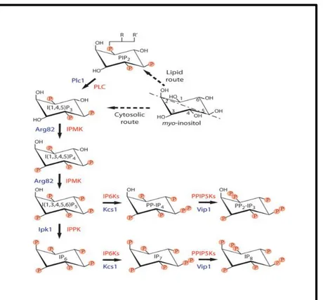
Fig. 1. IPK family members (including IPMK and IP6K mediate the sequential, combinatorial phosphorylation of IP3 to generate a novel class of poorly defined second messengers—“higher” inositol polyphosphates. Additionally, we recently characterized IPMK as a lipid inositide kinase possessing a novel “PI3K” activity. Inositol pyrophosphates such as IP7 and IP8 are generated by IP6Ks and represent high energy inositol molecules capable of pyrophosphorylating proteins.
In the last fifteen years, many studies have focused on the mechanism and metabolism of inositols in the yeast Saccharomyces cerevisiae (7). This is largely due to its characteristic of having a highly reproducible, controlled cultivation method, and the knowledge of its entire genome (8). Four inositol kinases have been indentified in eukaryotic system (budding yeast) and another two in plants and metazoans. The four enzymes discovered in yeast are: Ipk2, Ipk1, Kcs1 and Vip1. In single-celled organism (yeast), the synthesis of inositol phosphate pathways starts with the conversion of inositol 1,4,5- trisphosphate [I(1,4,5)P3 or IP3] to inositol 1,4,5,6-tetrakisphosphate [I(1,4,5,6)P4 or IP4] and then to inositol 1,3,4,5,6-pentakisphosphate [I(1,3,4,5,6)P5 or IP5) through the first enzyme Ipk2. IP5 is transformed to inositol 1,2,3,4,5,6-hexakiphosphate [I(1,2,3,4,5,6)P6 or IP6) through another kinase Ipk1. The last two kinases Kcs1 and Vip1, act as an inositol hexakiphosphate (IP6K) and inositol heptakisphosphate kinases (PP-IP5K)
5
respectively, and generate the last member of the inositol phosphate family (0): the inositol pyrophosphates [PP-IP4, PP-IP5 (IP7), PP2-IP3 and PP2-IP4 (IP8)]. Kcs1 and Vip1 collectively work together to generate IP8 from IP6.
Fig 2: Inositol phosphate and pyrophosphate pathways. Activation of phosphoinositide-specific phospholipase C (PLC) triggers the conversion of phosphatidylinositol 4,5-bisphosphate, PI(4,5)P2, to the second messengers I(1,4,5)P3 and 1,2-diacylglycerol (not shown). Metabolism of I (1,4,5)P3 occurs to generate numerous inositol phosphate and inositol pyrophosphate chemical codes. There are several evolutionarily conserved inositol phosphate kinases that contribute to the production of these regulatory molecules: in mammals there are 6 distinct kinase activities, whereas in the budding yeast there are 4 gene products. The commonly used gene name abbreviations are listed. Inositol phosphatases are omitted for clarity except for the black arrow linking I(1,3,4,5)P4 and I(1,3,4)P3, which is encoded by a 5-phosphatase INPP5.
Two other IP kinases are identified in higher eukaryotics species (mammals): IPMK and IP5K. The first one is an Ipk2 homolog and makes IP5 though IP4’s phosphorylation. The second one instead is an Ipk1 homolog to produce IP5 and IP6 (10). However in higher eukaryotes an alternative way to produce IP6 has been identified. Availed with the presence of IP3K and I(1,3,4)P3 5/6-kinases(alias ITPK1) (11-12), IP6 may be produced by phosphorylating I(1,3,4)P3 to I(1,3,4,6)P4. In this pathway I(1,4,5)P3 is phosphorylated by IP3 3-kinase to I(1,3,4,5)P4. The latter subsequently is dephosphorylated by 5-Ptase to I(1,3,4)P3 and afterwards catalyzed by IP3
6
5/6-kinase in the preferred human Ipk2/IPMK substrate I(1,3,4,6)P4. I(1,3,4,6)P4 is sequentially phosphorylated by Ipk2 and Ipk1 to make IP5 and finally IP6.
Recently studies conducted by Leymann and co-workers (13) have demonstrated that the production of IP4, IP5 and IP6 is not dependent on Iptk activity, but is dependent on Ipk2/IPMK.
Frederick JP et all. reported in their study, with mice deficient for Ipk2, that the synthesis of
inositol pentakisphosphate, hexakisphosphate and pyrophosphate species is mostly disrupted, and that Ipk2-deficient mice die around embryonic day 9.5 with multiple morphological defects, including abnormal folding of the neural tube.
Subcellular localization studies showed that Ipk1 and Ipk2 have nuclear compartment localization, whereas Kcs1 and mammalian homologs IP6K1 (14-15) are distributed in both nuclear and cytoplasmic compartments. The only exception is Vip1 where its localization is purely cytoplasmic (16). These nuclear presences suggest a crucial nuclear functions of IPs and PP-IPs.
7
Inositol nuclear functions
IP6, second to IP3, is the most studied inositol phosphate in the last 10 years. The variety of studies performed on IP6 show its diverse set of cellular functions. Recent studies demonstrated a pools of nuclear PIP2 which resulted from the nuclear-cytoplasmic shuttle of PLC isoforms. Furthermore, these studies show that PIPs are involved in nuclear function. This includes pre-mRNA splicing, nuclear calcium, chromatin structure, cell cycle, gene expression and messenger RNA export (17-19).
Genetic studies on budding yeast detected three distinct nuclear processes where IP and PP-IP were induced:
Transcriptional regulation/chromatin remodeling Efficient mRNA export from the nucleus
Telomere length maintenanceFig 3: Nuclear role within which inositols are involved. PIP 2 IP 3 IP 4 IP 5 IP 6 PP-IP4 Gene expression Chromatin Remodellin mRNA Export RNA editing
Kcs
Telomere Lenght NHEJ Kcs1 IPMK Ipk18
Studies about the role of IPs as nuclear-signaling molecules show a series of discoveries in the phosphate-responsive signaling pathway (PHO). The PHO pathway is important for chromatin remodeling and it carefully coordinates cellular responses to phosphate starvation. Recent reports identified IP7 as a negative regulator of Pho80-Pho85 CDK complex in a Pho81-dependent manner. Pho-85 is a cyclin-dependent kinase and it is associated with cyclin Pho-80. In the presence of high Pi levels, the complex Ph80-Pho85 is active and makes the phosphorylation of transcription factor Pho4. Phosphorylated Pho4 is exported to the cytoplasm and the gene targets remain turned off. Otherwise, in absence of Pi or in Pi starvation, Pho81, that is a CDK1 inhibitor, prevents Pho80-Pho85 phosphorylation and exportation of Pho4. Pho4 remains in the nucleus and binds activation sequences in the PHO5 promoter. Furthermore, Pho4 recruits the chromatin-remodeling complexes INO80 and SWI/SNF, which help displace four positioned nucleosomes from the PHO5 promoter facilitating PHO5 transcription. Therefore, during Pi starvation, yeast cells increase IP7 levels. The IP7 binds to the Pho80-Pho85 complex and its link is sufficient enough to inactivate the Pho80-Pho85 complex, the Pho4’s phosphorylation and consequently transcriptional activation and mRNA synthesis (19).
It was demonstrated that IPs plays other important roles after transcription and processing mRNA, specifically during assembly into a messenger ribonucleoprotein (mRNP) particle (20). The mRNA is generated at the end of mRNA maturation, and it is competent for mRNA export through the NPC (Nuclear Pore Complex). To allow the mRNA export, NPC requires a series of essential nuclear transport factors and IP6 (21). Additionally, Dbp5 (DEAD-box protein) requires for activation two cofactors: Gle1 and IP6. Gle1 (export factor localized at the cytoplasmic face of the NPC) alone is not able to stimulate the RNA-dependent ATPase activity of Dbp5 but needs to be tied to IP6 (22-23-24).
IPs family is also involved in two other nuclear processes. The first is the adenosine deaminases process. ADAR (adenosine deaminases acting on RNA) or ADAT (transfer RNA) is a family of enzymes that mediate the site-specific catalytic deamination of adenosine to produce inosine in RNA. Deamination of adenosine happens inside the nucleus and is seen in the crystal structure of human ADAR2 that this enzyme requires presence of IP6 ).
Phospholipase C pathway is implicated on the second process, DNA repair, through IP6 which plays a role as a positive regulator of NHEJ in mammalian cells. The components that are required
9
for NHEJ include the Ku70/80 heterodimer that binds to DNA ends, and a DNA-dependent protein kinase catalytic subunit (DNA-PKcs), that shares significant homology to the PI3-kinase family. Ku recruits DNA-PK to the DNA and the break in the DNA is repaired by a DNA ligase IV. An in vitro assay revealed that IP6 could activate the DNA-PKcs/Ku holoenzyme. The IP6 interaction appeared to be mediated through its direct binding Ku. Cells with partially depleted IPs had correlatively reduced mobility of Ku.
An additional role for inositides-regulated DNA metabolism has been proposed in the modulation of telomere length in yeast. It was shown that plc1, ipk2, and kcs1 mutants have longer telomeres when compared to wild-type cells, implicating PP-IP4 as a negative regulator of telomere length.
10
IP7 (5-diphosphoinositolpentakisphosphate)
IP7 and IP8 are defined “high energy” molecules because contain highly energetic pyrophosphate bonds. Inositol pyrophosphates are made by the IP6Ks, and recently discovered VIP1. In mammalian cells exists three IP6Ks isoforms: IP6K1, IP6K2 and IP6K3 and they are able to phosphorylate IP6 to IP7 and IP5 to PP-P4 utilizing ATP as a phosphate donor. In the first case when IP6 was the precursor, IP6Ks attached to the phosphate group at position 5 of the inositol ring and formed 5-PP-IP5/IP7 and afterwards phosphorylated to 5-PPP-IP5/IP8. Indeed, when IP5 is the substrate, IP6Ks makes a phosphorylation at position 1 and/or position 3, generating 1(3)-PP-IP4. The other class of enzyme, VIP1/PP-IP5K, not only makes a conversion of IP6 to IP7 but also catalyzes the IP7 phosphorylation to make IP8, but they are unable to use IP5 as a substrate. IP7 plays a role in the regulation of endocytosis, chemotaxis, apoptosis and telomere elongation ADD BIBLIO. Studies conducted by Snyder and co-workers showed the innovative IP7 protein pyrophosphorylation. This novel modification is characterized by the IP7’s ability to make a donation/transfer of its beta-phosphate group to protein target trough a non-enzymatic and temperature dependent reaction. However, even though it is a kinase-independent reaction, the phosphorylation mediated by IP7 requires a priming event through a canonical kinase-ATP phosphorylation on a serine residue. Therefore, it defines a new mechanism called pyro-phosphorylation, in which a phosphate group is added onto a pre-existing phosphoserine previously phosphorylated by CK2.
11
IP7 – the new role
The Proto-assay chip reported the identification of almost 259 IP7’s target proteins. The proteins shown in the list (Table 1), for the majority, belong to nuclear compartments. For the role that IP7 in the nucleus environment, we decided to investigate and characterize the interaction between the inositol pyrophosphate and the components of the Polycomb Repressive Complex 2, Enhancer of Zeste 2 (EZH2) and Embryonic Ectoderm Development (EED).
PRC2 complex is composed of five subunits: EZH2, EED, SUZ12, Rbp48 and AEBP2. EZH2 is the catalytic subunit of the complex. Its methyltransferase activity plays an important role in the trimethylation of Lysine 27 of histone 3 (26-27). Tri-methylation of H3K27 is associated with chromatin condensation and transcriptional repression of genes involved in development differentiation. The PRC2 complex, in conjunction with its positively acting Trithorax Group proteins (TrxG), can maintain heritable transcription patterns of the homeotic (Hox) genes during development and differentiation (28-29-30)
Following the idea that IP7 could have a role in the nucleus environment, we investigated the interactions between the inositol pyrophosphate and the enzyme of the chromatin repressive complex, as well as which ways the phosphorylation, that IP7 mediated on its target, could change the activity and function of them. The first approach we performed was to test the interaction between IP6K1 and EZH2.
We created two versions of IP6K1 both generated with a Myc tag: Wilde type and Kinase death. Kinase death (containing a mutation on the catalytic site), and transfected with EZH2 HA tagged. Lysates were precipitated with Myc beads. Contrary to our expectations, we didn’t see the IP6K1WT-EZH2 interaction. Instead, with IP6K1WT present, EZH2 disappeared. In our next step, we attempted to determine if this lack of interaction was directly related to EZH2 or was an IP6K1 repressor effect. For this reason, in the next experiment we performed a co-transfection on Human Embryonic Kidney 293 T-cells. For each of the IP6K1 Myc tag versions (WT and KD), we transfected with both of the components of the PRC2 complex (EED-HA tag and SUZ12-HA tag) first with one then the other.
In all cases, we assisted to a down-regulation of protein expression, when they were co-transfected with IP6K1-WT version. This led us to theorize a new repressor role for IP6K1. At this point, we
12
investigated whether or not the repressor role played by IP6K1-WT, was directed toward the exogenous plasmid. The next experiments we performed were aimed at discovering the value of the EZH2 endogenous protein levels when co-transfected with IP6K1-WT and KD versions. The results did not show any modulation in EZH2 endogenous expression protein which confirmed that the EZH2 exogenous version could potentially be an IP6K1-WT phosphorylation target. Phosphorylation of EZH2-HA could function as a signal for its degradation (31). To examine this possibility, we compared the half-lives of the exogenous and the endogenous EZH2. To this end, we transfected HA-tagged EZH2 into HEK 293 T-cells and treated cells with the protein synthesis inhibitor, cycloheximide, (Chx) at different times. Western blot analysis showed that exogenous EZH2 exhibit a half-life of almost 6h, in contrast endogenous EZH2 showed a longer half-life, almost 22h. The shorter half-life of exogenous EZH2 (HA-EZH2) may be due to reduced protein stability or the actions of inositol pyrophosphatase. Studies conducted by Hofmannand and Falquet, demonstrated the decisions about which protein is to be degraded at a specific time is made by the ubiquitination machinery, often in response to a prior event such as phosphorylation (32). Referring to this model, using a proteasome inhibitor (MG132) we examined the protein levels of EZH2-HAtag in treated and untreated cells. Although EZH2-HA was not detected in the untreated cells, likely due to its quick degradation, it is clearly detected in presence of the proteasome inhibitor MG132. Subsequently, we investigated about how IP6K1 recognizes and recruits its target and how it acts as a repressor. To investigate if the IP6K1 repressor effect is PRC2 complex dependent, we performed co-transfection experiments with over expression of the other two genes: GFP and Luciferase. In both cases we assisted and showed the down-regulation of both the GFP and Luciferase proteins levels. At this point is still unknown what the mechanism behind this down-regulation is. We hypothesized that IP6K1 can have a suppressor role during the DNA transcription process. However, it still unknown how, and at which level of this process, IP6K1 is implicated. In order to determine how IP6K1 acts during the transcription process, we chose to use the GAL4/UAS system. In the GAL4 system, the promoter drives expression of the transcriptional activator GAL4, which in turn activates the target gene (IP6K1). The GAL4 protein activates transcription of only those genes bearing GAL4 binding sites Upstream Activation Sequence (UAS). The GAL4 gene is placed near the promoter/enhancer driving ectopic expression, and UAS is fused to the target gene (Luciferase). The promoter/enhancer directs expression of GAL4, and GAL4 in turn directs transcription of the UAS–target gene.
13
The data show that, the luciferase expression is down regulated when it is in co-transfection with IP6K1-WT as expected. Experiments performed with IP6K1p-BIND (WT and KD version) co-transfected with a Luciferase gene, while it is placed under the control of a different promoter, showed no effect in regulation of the expression. However, we strikingly observed down regulation of Luciferase expression when it is co-transfected with IP6K1-KD.
During this study we identified a correlation between inositol pyrophosphates and the DNA transcription process. It is still inconclusive whether IP7 is responsible for the repressor effect on the gene transcription. However, we are continuing to make progress towards establishing a novel function of inositol pyrophosphates.
14
Results
In order to discovery and characterization of IP7 substrate I planned to begin this study utilizing a semisynthetic epitope approach. This approach employ the use of IP7S, novel inositol molecule analogue, in which the pyrophosphate moiety is replaced with a thiol-containing phosphate capable of labeling substrates in a fashion that permits the exploitation of a semisyntethic epitope by the thiophosphate ester antibody. Through a proto-array chip (Fig 4) we were able to identify two of PRC2 complex complements, EZH2 and EED, of 259 potential substrates of IP7.
Fig 4. IP7βS-mediated phosphorylation of proteins on a proto-array chip. IP7βS reactions, followed by alkylation and thiophosphate antibody detection occur on the nitrocellulose coated slide. Imaging was performed via a GenePix array scanner after application of a fluorescent secondary (Alexa Fluor 647 Goat Anti-Rabbit IgG). 259 unique proteins were identified as substrates of IP7 meeting significance criteria including duplicate and intensity analysis. (Representative of 4 arrays performed). Table 1. Subset of nuclear, chromatin relevant IP7 targets identified via IP7βS-mediated phosphorylation of proteins on a proto-array chip. Also shown are representative in vitro assay confirmation of the array results in which tagged-overexpressed proteins are immunoprecipitated from mammalian cells (293T) and subjected to IP7 phosphorylation reactions. Equal amounts of protein were present under both reaction condition (untreated/phosphorylated as verified by anti-myc tag blots (not shown). Bolded are Polycomb Group proteins involved in histone methylation.
15
To further biochemically confirm IP7’s distinct pyrophosphorylation of EZH2 we developed a novel, sensitive assay utilizing the NanoPro platform. The assay is based on an isoelectric focusing protocol in which proteins are first separated and the cross linked in a capillary-based system amenable to antibody-based-HRP detection (Fig 5A). As such, the system’s greatest utility is in its ability to characterize, separate, and resolve proteins are first according to their various phosphorylation states using a single, standard antibody. Using anti-HA tag antibody and probing 293T, HA-EZH2-expressing cell lysate, the NanoPro platform identifies two, overlapping peaks for EZH2 at respective pls of 5.70+-0.01 and 5.61+-0.01. Further reacting the lysate in vitro by the addition of recombinant CK2 and ATP shifts the peak distribution, increasing the 5.70+-0.01 peak curve area. However, addition of IP7 to the lysates results in a dramatic acidic shift to EZH2’s pl profile resulting in a peak at 4.87, suggesting we can in fact monitor IP7-mediated phosphorylation via this method (Fig 5B). Indeed, under longer image integration times (not shown), we can detect a small 4.87 peak in untreated lysates (data not shown), potentially corresponding to endogenously pyrophosphorylated EZH2.
16
Fig. 5. A) Capillary-based immunoassay platform utilizing isoelectric focusing to resolve differing phosphorylation states of a protein. B) Isoelectric focusing of 293T lysates overexpressing HA-tagged EZH2: untreated (pink) lysates contain 2 overlapping pI peaks; addition of recombinant CK2 and ATP enhances phosphorylation and shifts peak distribution (blue); addition of IP7 results in a further acidic shift (green).
Previous results shown EZH2 as a probably IP7 substrate. In order to show the putative interaction between EZH2 and IP6K1, Myc- pulldown experiments were performed in Human Embryonic Kidney 293T cells using Myc-tagged IP6K1 Wild Type, Myc-tagged IP6K1 Kinase Death versions and HA-tagged EZH2.
Fig. 6: IP6K1 interacts with EZH2. Coimmunoprecipitation assay of EZH2 and IP6K1. Lysates from 293T cells transfected with the indicated plasmids were immunoprecipitated with antibodies against Myc and HA. Whole-cell lysate (lysate) was also subjected to Western blot analysis to examine the expression of EZH2 and IP6K1.
Data shows (Fig. 6) association between exogenous EZH2, (HA)-tagged EZH2, and IP6K1 KD version (Fig. 6 third well) suggests that it interact in vivo with IP6K1 enzyme, but this association is reduced in cells co-transfected with HA-EZH2 and IP6K1 WT version suggesting that the inositol pyrophosphate could modulate the interaction (second well).
EZH2-HA + + + Anti-HA Anti-Myc IP6K1-Myc WT - + -- - + Anti-HA Anti-Myc IP6K1-Myc KD
17
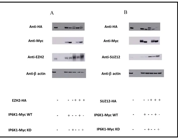
We evaluate with the same approach whether all others two PRC2 components, EED and SUZ-12, interact with IP6K1 enzyme in the same way. As a shown in Fig 7 EED-Ha tagged and SUZ12-HA tagged where co-transfected with both active and inactive catalytically forms of IP6K1 and again we assisted to a down-regulation of expression of PRC2 complex components protein when are co-transfected with IP6K1-WT version and precipitation with catalytically inactive IP6K1 (IP6K1 KD).
Fig 7: IP6K1 interacts with other both PRC2 components. Examination of the interaction between endogenous HA-EED and HA-SUZ12 (respectively panels A and B) and Myc-IP6K1 by immunoprecipitation and Western blotting with antibodies against either HA and Myc in HEK 293 cells.
Anti-Myc Anti-Myc IP6K1-Myc WT IP6K1-Myc KD + + + + + + -- - + - -- -+ + Anti-HA Anti-Myc Anti-Myc Anti-HA EED-HA IP6K1-Myc WT IP6K1-Myc KD Anti-HA Anti-Myc Anti-HA Anti-Myc SUZ12-HA IP6K1-Myc WT IP6K1-Myc KD + + + + + + - - + - + -- - + + + + - + -- - + + + + - + -- - +
18
To confirm whether EZH2 lack is a tag-dependent event or is an interaction consequence, cells HEK 293T were transfected with Flag-EZH2 and either GST or GST-IP6K1 WT (CMV-GST R11) version. Cells were lysate and subjected to SDS-PAGE. Western blotting was then performed with anti-GST and anti-Flag antibodies. As shown in Figure 8 A, EZH2 flag version when co-transfected with IP6K1 Wild Type is is enable to be detected. In addition, co-transfection experiments were performed using HA-EZH2 and either GST or GST-IP6K1 WT version. The detection of anti-GST and anti-HA with the respective antibodies was successful to demonstrate the EZH2 deficiency when is co-transfected with IP6K1 WT version.
Fig 8: Interaction between IP6K1 and EZH2 is independent by IP6K1 and EZH2 tag. Coimmunoprecipitation assay of EZH2 and IP6K1. Lysates from 293T cells transfected with the indicated plasmids were immunoprecipitated with antibodies against Flag, HA and GST. Whole-cell lysate (lysate) was also subjected to Western blot analysis to examine the expression of EZH2 tagged and IP6K1 tagged.
These results show the potential interaction between IP6K1 and PRC2 complex suggesting that the PRC2 complex subunits, EZH2, EED and SUZ12 could be, likely, IP7’s substrates. Surprising the EZH2 down regulation in co-transfection with IP6K1 active version. These results led us to suggest that EZH2’s phosphorylation IP7-mediated, could mediate the ubiquitination process and
EZH2-Flag + + CMV-GST CMV-GST R11 -Anti-Flag Anti-GST -actin EZH2-HA Anti-HA A B Anti-GST -actin CMV-GST CMV-GST R11 + + + + + -- +
19
consequently EZH2 degradation by the proteasome. Susan C. Wu et al. demonstrated how EZH2’s phosphorylation CDK1-mediated could regulates its stability and promote its degradation. The above studies and observations drive us to consider the ubiquitination process as a possible mechanism through EZH2 could be degraded.
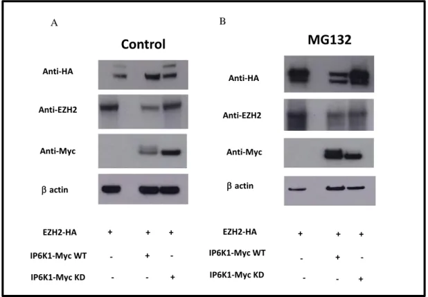
We overexpressed in HEK 293T cells, exogenous EZH2, (HA)-tagged EZH2, and exogenous IP6K1, (Myc)-tagged IP6K1, WT and KD version for 48h. Prior to harvesting, the cells were treated with or without the proteasome inhibitor, MG132 (Fig 9). Although (HA)-tagged EZH2 was not detected in the untreated cells, likely due to its quick degradation, but is clearly evident (HA)-tagged EZH2 presence when cells were treated with MG132 (Fig 9 (Panel B).
Fig. 9: Phosphorylation EZH2 is ubiquitinated and degraded via the proteasome pathway: HEK 293cells were co-trasfected with EZH2-Ha tagged and both IP6K1-Myc tagged isoforms. 4 h prior to harvesting, cells were treated with or without 25 mM MG132. Lysates were prepared under denaturing conditions and subjected Western blot analysis was performed using to using FLAG the indicated antibodies.
IP6K1-Myc KD Anti-HA Anti-Myc Anti-EZH2 actin EZH2-HA IP6K1-Myc WT +
-Control
Anti-HAMG132
A B Anti-EZH2 Anti-Myc actin EZH2-HA IP6K1-Myc KD IP6K1-Myc WT -+ + + - + -+ + + - + -- +20
On the basis of the above-mentioned results, we aimed to examine if the enzyme IP6K1 could also interact with endogenous EZH2 and if this interaction could produce the EZH2’s degradation. To evaluate our hypothesis we evaluate the EZH2 endogenous levels in cells transfected with or without HA-tagged EZH2, in presence of IP6K1 WT version and KD version. Data shown that in cells co-transfected with HA-tagged EZH2 and Myc-tagged IP6K1 WT the levels of endogenous EZH2 are similar to cells that have not been transfected with (HA)-tagged EZH2 despite levels of (HA)-tagged EZH2 are significantly reduced.
Fig. 10: Endogenous levels of PRC2 complex components: (A) HEK 293cells were co-transfected with EZH2-Ha tagged; SUZ12-Ha tagged (B); and both IP6K1-Myc tagged isoforms. Western blot analysis of HEK 293 cell extracts using the anti-HA, anti-Myc and anti-EZH2 total antibodies was performed. Data shows that endogenous EZH2 levels are not changed in presence of IP6K1 WT version, suggesting a specific IP6K1 interaction, in vivo, with EZH2 tagged version. - - - + + + - + - - + - - - + - - + A B Anti-HA Anti-HA Anti-Myc Anti-Myc
Anti- actin Anti- actin Anti-EZH2 Anti-SUZ12 EZH2-HA - - - + + + IP6K1-Myc WT IP6K1-Myc KD IP6K1-Myc WT IP6K1-Myc KD SUZ12-HA - + - - + - - - + - - + - - - + + + - + - - + - - - + - - +
21
Tai-Lung Cha et al. in a recent work reported one potential Akt phosphorylation site, serine 21, in the EZH2 protein. Through a combination of phosphorylation site prediction programs they identified other two possible EZH2 phosphorylation site: Thr-345 and Thr-487. Previous studies demonstrated that EZH2 phosphorylation on these sites appear to be less stable than total EZH2. In view of all that we tried to figured out if a possible IP7 pyrophosphorylation reaction could make an EZH2 instability form. For this reason we performed comparison stability experiment between exogenous and endogenous EZH2. We approached at this study through use of protein synthesis inhibitor, cycloheximide (Chx). To this end, we co-transfected (HA)-tagged EZH2 and (Myc)-tagged IP6K1 active and inactive forms in into HEK 293, to evaluate the exogenous levels of EZH2 (data not shown) and at the same time we made a single transfection with (Myc)-tagged IP6K1 WT and KD to measure the EZH2 endogenous levels (2 set).
Two sets of treatment time were performed, cells co-transfected with exogenous EZH2 were treated for 1-2-4 and 6h while endogenous set were treated for longer time 16-18-20-22 hr. Western blot analysis using the HA-antibodies for first set, and EZH2-antibodies for second set, have allowed the detection of differences half-life for exogenous and endogenous EZH2 (~ 6hs for exogenous EZH2) (more than 20hs for endogenous EZH2). The shorter half-life of (HA)-tagged EZH2 suggests a reduced protein stability cause probably by the pyrophosphorylation reaction.
All experiments showed a selective modulation of PRC2 complex protein expression under IP6K1 WT control, probably due by producing IP7. The inositol, in turn, is responsible of pyrophosphorylation activity that makes an instable form its targets and subsequently drive them to the degradation process. Despite all this plethora of experiments we investigated if the IP6K1 WT repressor effect shows a PRC2 complex down-regulation expression or if a general effect that it exercises, likely, is interfering during the different step that lead the protein formation.
22
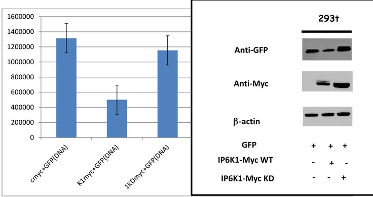
To prove the general effect exercised by IP6K1 WT co-transfection experiments were performed. HEK 293t cells were transfected first day with empty vector Myc-tagged (as a control), IP6K1 WT and IP6K1 KD Myc. 24h after the first transfection cells were transfected again with GFP plasmid and Luc-plasmid.
Fig 11. IP6K1 suppress the GFP express ion showing a general down regulation effect. Representative flow cytometry histograms for the transfection in HEK 293T cells showing difference in GFP expression levels 48 h post IP6K1 WT and KD Myc tag transfection and 24h post GFP transfection (A). Representative western blot of GFP, Myc and β-actin from HEK 293T cells. (B)
The extent to which the IP6K1 WT yield an effective down regulation of GFP and Luciferase was established by subjecting a sample of the whole cell lysates to SDS-PAGE and there solved gel to immunoblotting. Immunoblotting of beta-actin provides a measure of equivalent loading, antibodies against GFP and Luciferase showed also this case the ability of IP6K1 active form to modulate the amount of protein expression.
0 200000 400000 600000 800000 1000000 1200000 1400000 1600000 293t GFP Anti-GFP Anti-Myc IP6K1-Myc WT -actin IP6K1-Myc KD + + + - + - - - +
23
Fig 12. IP6K1 WT down regulated the transcriptional level of Luciferase: (A) HEK 293T cells were transiently co-transfected with luciferase reporter genes and IP6K1 WT and KD Myc tagged in 24-well plates, respectively;, CMV-Luciferase acted as a target gene. HEK29T cells were transiently co-transfected with cMyc-CMV Luc, IP6K1 WT Myc-CMV Luc and IP6K1KD Myc Myc-CMV Luc, using Lipofectamine 2000 in 24-well plates. 48 h later, collecting cells and using the dual-luciferase reporter assay system measured luciferase activities. The ordinate indicated relative luciferase activity. (B) Relative western blotting experiment using anti-Luciferase, anti Myc and b-actin antibodies.
Moreover results from Flow cytometry assay, confirmed that GFP fluorescence amount is enhanced, in samples co-transfected with empty vector and IP6K1-KD Myc tagged. On the contrary amount of GFP fluorescence are decreased in cells overexpressing IP6K1-WT. Same results are obtained when we performed confirmation experiments through Luciferase assay (Fig 12). 0 500000000 1000000000 1500000000 2000000000 2500000000 293t CMV-LUCIFERASE Anti-Luciferase Anti-Myc IP6K1-Myc WT -actin IP6K1-Myc KD + + + - + - - - +
24
Fig: 13. IP6K1 WT pBIND and IP6K1 KD pBIND can regulated the transcriptional level of UAS-Luciferase: HEK 293T cells were transiently co-transfected with luciferase reporter genes above UAS vector and IP6K1 WT and KD pBIND in 24-well plates, The experimental group cells were co-transfected with pBIND C vector-UAS Luc, IP6K1 pBIND-UAS Luc and IP6K1 KD-UAS using Lipofectamine 2000 in 24-well plates. 48 h later, cells were harvested and luciferase activities were measured using Dual-Luciferase Reporter Assay.
Subsequently we performed experiments using the bipartite system GAL4-UAS (add biblio PPT). This system is based on two different kind of plasmid, one activator (p-BIND), and other one is the effector (UAS). In the activator plasmid, containing GAL4 DNA-binding domain, our gene of interest IP6K1, is placed under the control of a specific promoter, while in the effector plasmid the gene of interest, in our case the Luciferase, is fused to sequence of the DNA-binding motif of GAL4 (Upstream Activating Sequences, UAS). HEK 293T cells were transfected with empty vector (as a control) and IP6K1 p-BIND (WT and KD). After 24h cells were transfected with UAS-Luciferase, and the day after we achieved a Luciferase assay. Data showed, as our expectation, the down regulation of luciferase signal under IP6K1 WT but surprisingly we assisted also to a down regulation of Luciferase under IP6K1 KD action. This result pointed out that the repressor effect is independent from its catalytic activity and that to act as a repressor, is necessary the nuclear localization but mostly important is the direct interaction with the DNA (Fig 15).
0 200000 400000 600000 800000 1000000 1200000 1400000 1600000
25
Fig 14. IP6K1 WT pBIND and IP6K1 KD pBIND do not effect the GFP expression: Representative flow cytometry histograms for the transfection in HEK 293T cells showing in all three conditions, a costant expression of GFP levels 24 h post GFP transfection. ) Relative western blotting experiment using anti-GFP, anti Myc and b-actin antibodies.
Still unknown is in which cellular compartment and in which way IP6K1 acts as repressor during this process. Could IP6K1 get it the nucleus compartment and through phosphorylation probably mediated by IP7 create a transcriptional repressive state?
To try to answer to this question, last experiment we performed, was a comparison between GFP level protein.
Co-transfections experiments DNA-DNA and DNA-RNA are compared. HEK 293 cells were transfected with IP6K1 WT and KD at 48h. 24h later cells are transfected again, one set were transfected with GFP DNA plasmid and the second set is transfected with GFP-RNA. Day after cells were collect and expression proteins levels were analyze through SDS page. Data shown the levels of the GFP protein in cells transfected GFP DNA (plasmid) were modulated in presence of IP6K1 WT. Protein’s level of GFP-RNA instead are unchanged. This result suggests an involvement of IP6K1 WT during the transcription process and supposedly its presence inside the nucleus. 0 500000 1000000 1500000 2000000 2500000 293t GFP Anti-GFP Anti-Gal4 IP6K1-pBIND WT -actin IP6K1-pBIND KD + - + - - - + + +
26
Fig 15. Comparison of GFP protein levels: HEK 293 T cells were co-transfected with cMyc, IP6K1 WT Myc tag and IP6K1 KD Myc Tag for 48h. After 24h the first transfection cells are again transfected with GFP DNA (plasmid) and GFP (RNA). 293t 293t Anti-GFP Anti-Myc -actin GFP-DNA + + + IP6K1-Myc WT - + - - - + IP6K1-Myc KD GFP-RNA + + + IP6K1-Myc WT - + - - - + IP6K1-Myc KD
27
Discussion
The inositol phosphates are known as second messenger classes and represent one of the most important signaling families. After two decades of research, the consensus is that inositol pyrophosphates are key regulators of cellular metabolism. In addition, recent studies showed that the inositol phosphates are implicated in regulation of mRNA transport from the nucleus, the stimulation of DNA-dependent protein kinase activity and influencing vesicular dynamics. In light of these studies, it is not surprising to find as a potential target of diphosphoinositol pentakisphosphate (IP7) the catalytic subunit of Polycomb Repressive Complex 2 (PRC2 complex), Enhancer of Zeste 2 (EZH2) ,which is responsible for a mark of transcriptionally silent chromatin, involved in the epigenetics modification. EZH2’s role is to maintain more condensed the chromatin configuration through the trimethylation of Lysine 27 of Histone 3 (H3K27me3). The information regarding the nuclear process where IP7 is involved in, lead us to investigate first the connection between IP7 and EZH2, as well as which consequences result from this interaction in the epigenetics system. EZH2, catalytic subunit of (PRC2), pledged in the epigenetic modification as a trimethylation of Lysine 27 of Histone 3 (H3K27me3).
We then investigated on the interaction between IP7 and EZH2 and a following EZH2’s phosphorylation, and how this post transcriptional modification can change the activity of EZH2. Because of IP7’s unable detection, we were forced to use, during the experimental work, Inositol Hexakisphosphate Kinase 1(IP6K1) the enzyme responsible of its conversion. We performed experiments aimed to evaluate the interaction between IP7 and EZH2. Data shown a completely lack on, EZH2 expression, when it is co-transfected with the active form of IP6K1. Despite it, is still unknown how IP7 can makes EZH2 disappear, we are interrogate if maybe its pyrophosphorylation reaction can affect EZH2 stability and its catalytic activity.
One report demonstrated that the Serine 21 of EZH2 can be phosphorylated by AKT, and suggested that the phospho-mimic S21D inhibited PRC2mediated H3K27 trimethylation and gene silencing (33). Furthermore, Kaneko S. et al, described during their study on, the correlation between EZH2 phosphorylation and cell-cycle, that the phosphorylation mediated by Cyclin D1 (CDK1) on two different Threonine sites of EZH2 (T345 and T487) does not product alterations of its catalytic
28
activity and the trimethylation of H3K27 (H3K27me3), were relatively constant throughout the cell cycle. (34)
On the basis of the above studies, we performed experiment aimed to evaluate the alteration of EZH2 activity when co-transfected with IP6K1. We performed experiments aimed to understand how posttranslational modification of EZH2 could affected its association with its target. Our finding was, that in HEK 293-T cells transfected with IP6K1 Wilde Type (IP6K1-WT) and KD, EZH2 endogenous levels were not modulate and we did not reveal any difference in the trimethylation of H3K27 (H3K27me3). Intrigued to discover how IP6K1 acts in different way against exogenous and endogenous protein, experiments with use of inhibitor of protein biosynthesis (Chx) were performed. Data showed differences between exogenous and endogenous half-life of proteins, approximately 6h for the EZH2 HA tagged (exogenous) and 22h for the EZH2 endogenous. This extremely differences made us wonder about: when, where and against who IP6K1 plays a repressor role. The steps through which IP6K1 could be involved during the protein disappearing are: DNA transcription, RNA production, RNA exportation and protein biosynthesis. First step to understand the mechanism of action of IP6K1, was to analyze the nature of its repressor role. Is it a general role or it is just exercised against its target? To answer at this question, we performed different experiments aimed to evaluate, first, the interaction between IP7 and the other two PRC2 complex component, Embryonic ectoderm development (EED) and SUZ-12 and second with GFP (plasmid) and Luciferase (plasmid). Data shown a completely lack on, SUZ-12, expression and down regulation of EED, GFP and Luciferase expressions, when they are singularly co-transfected with the active form of IP6K1.
At that point, we had provided evidence about the IP6K1 general repressor effect and not just selective against its target.
Another step we had to make for the comprehension of IP6K1 repressor role is where it acts. Is it acts as a repressor during the DNA transcription process? Is it acts as a repressor inhibiting the RNA exportation? Is it acts mediating proteins degradation?
To determine which role plays IP6K1 during this process we used the GAL4/UAS system. This system, originally developed in yeast, is composite by a promoter (or enhancer) that directs expression of the yeast transcriptional activator GAL4 in a particular pattern, and GAL4 in turn
29
directs transcription of the GAL4-responsive (UAS) target gene in an identical pattern (35-36). The CheckMate Mammalian Two-Hybrid System adapted this system to mammalian cells (37-38). This system is compound by two vectors, the first one is the pBIND Vector, which contain the yeast GAL4 DNA-binding domain, upstream of a multiple cloning region (in our experiment we used this vector to generate the IP6K1 pBIND). The second vector is the UAS vector (in our system the Luciferase plasmid is located under UAS GAL4 binding site).
Through this system we were able to realize a direct interaction between IP6K1 and DNA. We observed, as our expectation, a down regulation of Luciferase fluorescence when it was co-transfected with IP6K1 WT pBIND. Strikingly we detected also a Luciferase down regulation when it was co-transfected with IP6K1Kinease Death (IP6K1-KD) pBIND.
This results suggests, that IP6K1 needs to effect is repressor role a direct interaction to the DNA. The ability of this system is to generate a direct interaction between GAL4 and UAS site, so in our case between IP6K1 and DNA. Through this system we were able to figure out that IP6K1 can produce a repressor effect when it interacts with the DNA. Interesting role, likely, is played by IP7. Our deduction so, is that IP7 generated by IP6K1 can force and drive IP6K1 toward the nucleus, and so to the DNA. IP6K1 after the probably IP7 binding, can exercise by its own or by phosphorylation of a transcriptional factor, a repressor role. It still unknown the mechanism through this happens. Our finding, so is the IP6K1 could acts as a DNA transcription repressor, hypothesis validates by the result we obtained from the double transfection experiments we performed using IP6K1WT and KD (plasmid) and in vitro synthesized GFP mRNA (generated by T cells). The result we achieved by this results is that the GFP protein level after RNA transfection was not modulated compared to the GFP protein level generated from GFP DNA transfection. Ones again these results suggests the probably implication of IP6K1 during the DNA transcription progress.
30
Materials and Methods
Cell Culture, Transfections, and Drug Treatments
HEK293T, HeLa, U2OS, and NIH3T3 cells were cultured in Dulbecco’s modified Eagle’s medium (DMEM) supplemented with10%fetalbovineserumandpenicillin/streptomycin. PC3 cells were cultured in DMEM/F12 (1:1) supplemented with 10% fetal bovine serum and penicillin/streptomycin. Transfections were performed using Lipofectamine 2000 (Invitrogen catalog number 11668-019). Cycloheximide (Sigma catalog number C4859) was used at a final concentration of 100 mg/ml.
Western Blotting
HEK 293T cells were washed twice with ice-cold PBS and scraped into ice-cold lysis buffer (50 mmol/L Tris–HCl [pH 8.0], 1% Triton X-100, 150 mmol NaCl) with protease (Roche Diagnostic GmbH, Mannheim, Germany) and phosphatase inhibitors cocktail (SIGMA Chemical Co., St. Louis, MO). Lysates were incubated at 4 C with gentle rotation for 1 h. Samples were then centrifuged at 15,000g for 10 min at 4 C, and supernatants were recovered. Protein concentration was determined using the DC Protein assay kit (Bio-Rad, Hercules, CA) using BSA as standard. Then, total protein extracts equivalent to 50 mg protein/lane were resolved by SDS–PAGE under reducing condition and electrophoretically transferred to PVDF membranes (Millipore SpA, Milan, Italy). Transfer efficiency on the membranes were routinely monitored by 0.1% Red Ponceau S (SIGMA Chemical Co.). Membranes were probed with the following antibodies: FLAG M2 mouse monoclonal (Sigma catalog number F3165), HA mouse monoclonal (ROCHE 12CA5), c-Myc Mouse Monoclonal Antibody (Invitrogen R950-25), GAPDH rabbit monoclonal (Cell Signaling 14C10), GST Rabbit mAb (Cell Signaling 91C1), b-actin Rabbit mAb (Cell Signaling 13E5), EZH2 XP Rabbit mAb (Cell Signaling D2C9), EED Rabbit polyclonal (ab96801), Suz-12
Rabbit polyclonal (ab77605), GFP Rabbit XP(R) (Cell Signaling D5.1),Firefly Luciferase Goat pAb (abcam ab635).
31
Dual Luciferase Assay
Four mg of IP6K1 WT and KD Myc tagged, 4 mg of IP6K1 WT and KD pBIND and 4 mg of CMV-Luciferase Firefly and 4 mg UAS-luciferase firefly, were transfected into HEK 293A cells using Lipofectamine. After being cultured for 48 hours, cells were lysed by the addition of 100 µl of passive lysis buffer. Twenty microliters of lysate were used for determining dual luciferase activities.
Flow Cytometry Assay
Cell transformation was quantitatively determined using a Bio Rad Flow Cytometry (Bio-Rad, USA) equipped with a xenon-ion excitation lamp (488nm). Aliquot of transformed cells were collected after 48h of transformation, centrifuged, and then washed twice by saline solution. The cells were subsequently resuspended in buffered saline solution and analyzed in a FACS caliber flow cytometer. All analysis were performed at a low flow rate (20 ml min-1) with pressure setting of 0.7 bar. To avoid errors, sample were diluted with sterile seawater. Intrinsic structural parameters forward scatter and side scatter with the green fluorescence from the GFP (515 to 545nm emission filter) measured in the FL1 channel as an extrinsic parameter by analyzing data with cell process software were used to evaluate transformation efficiency.
32
Bibliography
1: Inositol: history of an effective therapy for Polycystic Ovary Sindrome- Bizzarri M, Calrlomagno G.
2: Theodor HartigUeber das Klebermehl. BotanischeZeitung 1855.
3: ANDERSON RJ. A Contribution to the chemistry of phytin: Composition of barium phytate and phytic acid. ii. A study of the properties of phytic acid and its decomposition products. Eighth paper on phytin. J BiolChem 1914; 17: 171-190.
4: Inositol pyrophosphates: between signalling and metabolism. Miranda S. C. WILSON, Thomas M. LIVERMORE and Adolfo SAIARDI. Biochemical Journal (2013) 452, 369-379 doi: 10.1042/BJ20130118.
5: Purified inositol hexakisphosphate kinase is an ATP synthase: Diphosphoinositol pentakisphosphate as a high-energy phosphate donor (phosphotransferase/pyrophosphate). SUSAN M. VOGLMAIER, MICHAEL E. BEMBENEK, ADAM I. KAPLIN, GYORGY DORMAN, JOHN D. OLSZEWSKI, GLENN D. PRESTWICH and SOLOMON H. SNYDER Proc. Natl. Acad. Sci. USA Vol. 93, pp. 4305-4310, April 1996 Biochemistry.
6: Roles of inositol phosphates and inositol pyrophosphates in development, cell signaling and nuclear processes MARCO M.TSUI, JOHN D. YORK Adv Enzyme Regul. 2010;50(1):324-37. doi: 10.1016.
7: Extraction and analysis of soluble inositol polyphosphates from yeast. Cristina Azevedo and Adolfo Saiardi Nat Protoc. 2006;1(5):2416-22.
8: Life with 6000 genes. Goffeau, A. et al. Life with 6000 genes. Science 274, 546 563–567 (1996). 9: Structural analysis and detection of biological pyrophosphates reveal that the family of VIP/diphosphoinostolpentakisphosphate kinase 1/3-kinase. LIN H, FRIDY PC, RIBEIRO AA, CHOI JH, BARMA DK, VOGEL G, FALCK JR, SHEARS SB, YORK JD, MAYR GW. J Biol Chem. 2009 Jan 16;284(3):1863-72. doi: 10.1074
33
10: Frederick JP, Mattiske D, Wofford JA, Megosh LC, Drake LY, Chiou ST, et al. An essential role for an inositol polyphosphatemultikinase, Ipk2, in mouse embryogenesis and second messenger production. Proc. Natl. Acad. Sci. USA 2005;102:8454–9.
11: Characterization of a cDNA encoding Arabidopsis thaliana inositol 1,3,4-trisphosphate 5/6-kinase. Wilson MP, Majerus PW. BiochemBiophys Res Commun 1997;232:678–81.
12: Majerus PW. Inositol phosphate biochemistry. AnnuRevBiochem 1992;61:225–50.
13: The absence of expression of the three isoenzymes of the inositol 1,4,5-trisphosphate 3-kinase does not prevent the formation of inositol pentakisphosphate and hexakisphosphate in mouse embryonic fibroblast. Leymann A, Pouillon V, Bostan A, Schurmans S, Erneux C, Pesesse X. Cell Signal 2007, 19:1497-1504.
14: Inositol pyrophosphates are required for DNA hyperrecombination in protein kinase c1 mutant yeast H.R. Luo, A. Saiardi, H. Yu, E. Nagata, K. Ye, S.H. Snyder, Biochemistry 41 (2002) 2509-2515
15: Identification and characterization of a novel inositol hexakisphosphate kinase. A. Saiardi, E. Nagata, H. R. Luo, A. M. Snowman, S.H. Snyder, J. Biol. Chem. 276 (2001) 39179-39185. 16: Cloning and characterization of two human VIP1-like inositol hexakisphosphate and diphosphoinositolpentakisphosphate kinase. Peter C. Fridy, James C. Otto, D.EricDollins, and John D. York. J Biol Chem. 2007 Oct 19;282(42):30754-62.
17: Phospholipid signalling in the nucleus. Een DAG uit het leven van de inositide signalering in de nucleus. C.S. D’Santos, J.H. Clarke, N. Divecha, Biochim. Biophys. Acta 1436 (1998) 201-232.
18: Nuclear diacylglycerol, the cell cycle, the enzymes and a red herring (or how we came to love phosphatidylcholine). N. Divecha, H. Banfic, J. Treagus, L. Vann, R. Irvine, C. D’Santos, Biochem. Soc. Trans. 25 (1997) 571-575.
19: A role for nuclear inositol 1,4,5-trisphospate kinase in transcriptional control. Odom AR, Stahlberg A, Wente SR, York JD. Science 2000, 287:2026-2029.
34
20: Integration of the multiple controls regulating the expression of the arginase gene CAR1 of Saccharomyces cerevisiae in response to different nitrogen signals: role of Gln3p, Argp-Mcm1p, and Ume6p. Dubois E, Messenguy F. Mol Gen Genet 1997, 253;568-580.
21: Role of Plc1pin regulation of Mcm1p-dependent genes. Guzinska K, Varghese R, Vancura A. FEMS Microbiol Lett 2009, 295:245-250.
22: Inositol hexakisphosphate and Gle1 activate the DEAD-box protein Dbp5 for nuclear mRNA export. Alcázar-Román AR1, Tran EJ, Guo S, Wente SR. Nat Cell Biol. 2006 Jul;8(7):711-6. Epub 2006 Jun 18.
23: Gle1 is a multifunctional DEAD-box protein regulator that modulates Ded1 in translation initiation. Bolger TA1, Wente SR. J Biol Chem. 2011 Nov 18;286(46):39750-9. doi: 10.1074/jbc.M111.299321. Epub 2011 Sep 23.
24: The DEAD-box protein Dbp5 controls mRNA export by triggering specific RNA:protein remodeling events. Tran EJ1, Zhou Y, Corbett AH, Wente SR. Mol Cell. 2007 Dec 14;28(5):850-9.
25: A phospholipase C-dependent inositol polyphosphate kinase pathway required for efficient messenger RNA export. York JD, Odom AR, Murphy R, Ives EB, Wente SR. Science 1999, 285:96-100.
26: Histone methyltransferase activity associated with a human multiprotein complex containing the Enhancer of Zeste protein. Kuzmichev A, Nishioka K, Erdjument-Bromage H, Tempst P, Reinberg D. Genes Dev 2002;16:2893-905.
27: Histone methyltransferase activity of a Drosophila Polycomb group repressor complex. Muller J, Hart CM, Francis NJ, Vargas ML, Sengupta A, Wild B, et al. Cell 2002;111:197-208.
28: Genome-wide mapping of Polycomb target genes unravels their roles in cell fate transitions. Bracken AP, Dietrich N, Pasini D, Hansen KH, Helin K. Genes Dev 2006;20:1123-36.
29: Role of histone H3 lysine 27 methylation in Polycomb-group silencing. Cao R1, Wang L, Wang H, Xia L, Erdjument-Bromage H, Tempst P, Jones RS, Zhang Y. Science 2002;298:1039-43.
35
30: Silencing of human polycomb target genes is associated with methylation of histone H3 Lys 27. Kirmizis A1, Bartley SM, Kuzmichev A, Margueron R, Reinberg D, Green R, Farnham PJ.. Genes Dev 2004;18:1592-605.
31: Wnt-1 signal induces phosphorylation and degradation of c-Myb protein via TAK1, HIPK2, and NLK. Chie Kanei-Ishii, Jun Ninomiya-Tsuji, Jun Tanikawa, Teruaki Nomura, Tohru Ishitani, Satoshi Kishida, Kenji Kokura, Toshihiro Kurahashi, Emi Ichikawa-Iwata, Yongsok Kim, Kunihiro Matsumoto, and Shunsuke Ishii. Genes Dev. 2004 Apr 1;18(7):816-29.
32: A ubiquitin-interacting motif conserved in components of the proteasomal and lysosomal protein degradation systems. Kay Hofmannand Laurent Falquet. Trends Biochem Sci. 2001 Jun;26(6):347-50.
33: Akt-Mediated Phosphorylation of EZH2 Suppresses Methylation of Lysine 27 in Histone H3 Tai-Lung Cha, Binhua P. Zhou, Weiya Xia, Yadi Wu, Cheng-Chieh Yang, Chun-Te Chen, Bo Ping, Arie P. Otte, Mien-Chie Hung Science 14 October 2005: Vol. 310 no. 5746 pp. 306-310 DOI: 10.1126/science.1118947.
34: Phosphorylation of the PRC2 component Ezh2 is cell cycle-regulated and up-regulates its binding to ncRNA. Syuzo Kaneko,1,2,4 Gang Li,1,2,4 Jinsook Son,2 Chong-Feng Xu,3 Raphael Margueron,5 Thomas A.` Neubert,3 and Danny Reinberg1. Genes Dev. 2010 Dec 1;24(23):2615-20. doi: 10.1101/gad.1983810.
35: A novel genetic system to detect protein-protein interactions. Fields, S. and Song, O. (1989) Nature340,245–6.
36: The two-hybrid system: A method to identify and clone genes for proteins that interact with a protein of interest. Chien, C. et al. (1991). Proc. Natl. Acad. Sci. USA88, 9578–82.
37: Intracellular leucine zipper interactions suggest c-Myc heterooligomerization. Dang, C.V. et al. (1991). Mol. Cell. Biol.11, 954–62.
36
38: Karyoplasmic interaction selection strategy: A general strategy to detect protein-protein interactions in mammalian cells. Fearon, E.R. et al. (1992). Proc. Natl. Acad. Sci. USA89, 7958– 62.