Contents lists available atScienceDirect
Journal of Dentistry
journal homepage:www.elsevier.com/locate/jdent
E
ffect of benzalkonium chloride on dentin bond strength and endogenous
enzymatic activity
Allegra Comba
a, Tatjana Maravic
a, Lucrezia Valente
a, Margherita Girlando
a, Sandra R Cunha
b,
Vittorio Checchi
a, Stefano Salgarello
c, Franklin R Tay
d, Nicola Scotti
e, Lorenzo Breschi
a,⁎,
Annalisa Mazzoni
aaDepartment of Biomedical and Neuromotor Sciences, DIBINEM, University of Bologna - Alma Mater Studiorum, Via San Vitale 59, 40125 Bologna, Italy bDepartment of Restorative Dentistry, School of Dentistry, University of São Paulo, Av. Prof. Lineu Prestes 2227, 05508-000 São Paulo, Brazil
cDipartimento di Specialità Medico-Chirurgiche, Scienze Radiologiche e Sanità Pubblica - Università degli Studi di Brescia, P.le Spedali Civili 1, 25123 Brescia, Italy dThe Dental College of Georgia, Augusta University, 1430 John Wesley Gilbert Drive, Augusta, GA, USA
eDepartment of Surgical Sciences, University of Turin, Dental School Lingotto, Via Nizza 230, 10100 Torino, Italy
A R T I C L E I N F O Keywords: Benzalkonium chloride Universal adhesives MMPs In situ zymography Zymographic assay Microtensile bond strength
A B S T R A C T
Objective: This in vitro study evaluated at baseline (T0) and over time (T12 months), the effect of a multi-mode universal adhesive compared with two experimental formulations blended with different concentrations of benzalkonium chloride (BAC), on bond strength and endogenous enzymatic activity.
Methods and materials: Specimens were assigned to the following groups according to the adhesive protocol: G1) All-Bond Universal (ABU) self-etch (SE); G2) ABU + 0.5% BAC SE; G3) ABU + 1% methacrylate BAC SE; G4) ABU etch-and-rinse (E&R); G5) ABU + 0.5% BAC E&R; G6) ABU + 1% methacrylate BAC E&R. Gelatin zymography was performed on dentin powder obtained from eight human third molars. Endogenous enzymatic activity within the hybrid layer was examined using in situ zymography after 24 h (T0) or 1-year storage in artificial saliva (T12). Forty intact molars were prepared for microtensile bond strength test at T0 and T12. Results were statistically analyzed with three-way ANOVA (α = 0.05).
Results: Gelatin zymography assay and in situ zymography quantification analyses indicated that all the BAC-containing formulations decreased matrix metalloproteinase expression. However, in situ zymography showed a general trend of enzymatic activity increase after aging. Microtensile bond-strength testing showed decrease in bond strength over time in all the tested groups; performances of the 1% methacrylate BAC experimental groups were worse than the control.
Conclusions: BAC-containing adhesives reduce endogenous enzymatic activity both immediately and over time. However, independently from the adhesive employed, increase in the gelatinolytic activity over time and decrease in bond strength was found (especially in the BAC + 1% methacrylate groups), probably due to im-paired polymerization properties.
Clinical significance: Adhesives containing protease inhibitors are practical and efficient tools in clinical practice for enhancement of the longevity of dental restorations. However, extensive investigation of the me-chanical and adhesive properties of the material is necessary prior to their clinical use.
1. Introduction
Adhesive systems introduced a revolution in contemporary re-storative dentistry by enabling previously inconceivable treatment plans. Currently available adhesive systems allow clinicians to bond to
tooth structure without the need for a retentive cavity design because they provide immediate bond strength to the dental substrate [1].
Regardless of advancements in adhesive dentistry over the past decade, the stability of resin-dentin bonds over time remains an issue that has not been adequately addressed [1]. Adhesion to dentin is a type
https://doi.org/10.1016/j.jdent.2019.04.008
Received 27 January 2019; Received in revised form 11 April 2019; Accepted 13 April 2019
⁎Corresponding author.
E-mail addresses:[email protected](A. Comba),[email protected](T. Maravic),[email protected](L. Valente), [email protected](M. Girlando),[email protected](S.R. Cunha),[email protected](V. Checchi),
[email protected](S. Salgarello),[email protected](F.R. Tay),[email protected](N. Scotti),[email protected](L. Breschi), [email protected](A. Mazzoni).
0300-5712/ © 2019 Elsevier Ltd. All rights reserved.
of in situ tissue engineering that creates a hybrid layer (HL) in dentin by infiltration of the demineralized collagen fibril network with adhesive resin monomers [2–4]. Although this interdiffusion layer is responsible for the retention of resin restorations, it is the most vulnerable com-ponent of the resin-dentin bond [5]. In vitro and in vivo studies showed that the degradation of resin-dentin bonds over time is caused by the hydrolytic breakdown of the resin and denuded dentinal collagenfibrils at the base of the HL [6,7]. Host-derived proteinases are responsible for the deterioration of the HL with aging [8–11].
Among the endogenous enzymes, matrix metalloproteinases (MMPs) and cysteine cathepsins are involved in the hydrolytic de-gradation of dentin collagen, with a 36–70% decrease in bond strength after 1 year of storage in artificial saliva [7,12]. Accordingly, increasing the resistance of collagen fibrils against enzymatic deterioration and inactivating of the activities of proteinases are fundamental approaches to improve the longevity of resin-dentin bonds [11,13].
Several natural and synthetic MMPs inhibitors (chlorhexidine, ga-lardin, quaternary ammonium methacrylates, various collagen cross-linking agents) have been employed experimentally as part of the bonding procedures on dentin with the intention of enhancing HL longevity [10,14–20]. Benzalkonium chloride (BAC), an antimicrobial agent containing a quaternary ammonium group, has demonstrated inhibitory potential against endogenous proteases [18]. A commercially available antimicrobial phosphoric acid etchant containing 1.0 wt% BAC (Etch-37 w/BAC, Bisco Inc, Schaumburg, IL, USA) is commercially available and does not immediate bond strength [21]. The etchant possesses anti-proteolytic properties. However, rinsing of the BAC-containing etchant in the etch-and-rinse bonding technique limits the amount that of BAC available in the hybrid layer and hence, its anti-proteolytic potential [22]. A more effective delivery system may entail incorporation of BAC into the primer and/or adhesive formulation to obtain deeper infiltration of the agent into dentin without lengthening the clinical procedures.
The objective of the present in vitro study was to evaluate at baseline (T0) and over time (T12 months), the effect of a commercially available multi-mode universal adhesive compared with two experimental for-mulations of this adhesive blended with different concentrations of BAC (0.5% BAC or 1% methacrylate BAC) on endogenous enzymatic activity and bond strength. The null hypotheses tested were that incorporation of BAC in an adhesive formulation: 1) has no effect on the activity of endogenous dentin MMPs immediately or over time; 2) has no effect on immediate bond strength and bond strength after aging for 12 months. 2. Materials and methods
2.1. Gelatin zymography of dentin powder extracts
Zymography was performed in accordance with a previous pub-lication [23]. Briefly, mineralized dentin powder was obtained from eight human third molars by triturating dentin frozen in liquid nitrogen using a Retsch mill (MM400, Retsch GmbH, Haan, Germany). Aliquots of mineralized dentin powder (100 mg) were treated as follows:
•
L1: dentin remained mineralized (DP, control);•
L2: demineralized with 10 wt% phosphoric acid for 10 min at 4 °C to simulate thefirst step of the etch-and-rinse (E&R) approach (DDP);•
L3: All-Bond Universal adhesive (ABU) applied on DP for 30 min to simulate the self-etch (SE) approach;•
L4: ABU + 0.5% BAC applied on DP for 30 min to simulate the SE approach;•
L5: ABU + 1% methacrylate BAC applied on DP for 30 min to si-mulate the SE approach;•
L6: DDP (as for L2) treated with ABU for 30 min to simulate the E&R approach;•
L7: DDP treated with ABU + 0.5% BAC for 30 min to simulate the E &R approach;•
L8: DDP treated with ABU + 1% methacrylate BAC for 30 min to simulate the E&R approach.All treatments involving adhesive resins were performed with light protection at 4 °C under constant agitation. After treatment, the speci-mens were rinsed several times with acetone and centrifuged at 4 °C (20,800 G) to remove the adhesive resins. After the aforementioned treatments, the dentin powder aliquots were suspended in extraction buffer (50 mM Tris–HCl pH 6, containing 5 mM CaCl2, 100 mM NaCl, 0.1% Triton X-100, 0.1% nonionic detergent P-40, 0.1 mM ZnCl2, 0.02% NaN3) for 24 h at 4 °C. They were further sonicated for 10 min (ca. ≈30 pulses) and centrifuged two times for 20 min at 4 °C (20,800 G) to collect the supernatant and remove powder remnants. The supernatant was concentrated using Vivaspin concentrators (10,000 kDa cut off; Vivaspin Sartorius Stedim Biotech, Goettingen, Germany) in several rounds of centrifugation of 20 min each at 25 °C (15,000 G). Bradford assay was used to determine the total protein concentration in the dentin protein extracts. The dentin protein aliquots were diluted in Laemmli sample buffer in a 4:1 ratio and subjected to electrophoresis under non-reducing conditions in 10% sodium dodecyl sulfate-polyacrylamide gel electrophoresis (SDS-PAGE) containing 1 mg/m Lfluorescein-labeled gelatin. Pre-stained low-range molecular weight SDS-PAGE standards (Bio-Rad, Hercules, CA) were used as molecular-weight markers. After electrophoresis, the gels were washed for 1 h in 2% Triton X-100 and incubated in zymography activation buffer (50 mmol/L Tris–HCl, 5 mmol/L CaCl2, pH 7.4) for 48 h. Proteolytic activity was evaluated using a long-wave ultraviolet light scanner (Chemi-Doc Universal Hood, Bio-Rad, Hercules, CA, USA). Gelatinase activities in the samples were analyzed in duplicate by ge-latin zymography. Densitometric evaluation of bands obtained from zymography was performed using the ImageJ software (National Institutes of Health, Bethesda, MD, USA).
2.2. In-situ zymography of resin-dentin interfaces
One millimeter-thick slabs of middle/deep dentin were obtained from three extracted human third molars using a low-speed saw (Micromet, Remet; Bologna, Italy) with water-cooling. Two slabs were obtained from each tooth and then further divided into 4 pieces, so that testing of all groups was performed using the same dentinal substrate. A standardized smear layer was created on each dentin surface using 600-grit silicon carbide paper. The dentin specimens were further treated as follows:
•
Group 1 (G1): ABU was directly applied on untreated dentin ac-cording to the manufacturer’s instructions for the SE mode.•
Group 2 (G2): ABU + 0.5% BAC was directly applied on untreated dentin in the same manner as ABU.•
Group 3 (G3): ABU + 1% methacrylate BAC was directly applied on untreated dentin in the same manner as ABU for the SE mode.•
Group 4 (G4): dentin surfaces were etched with 35% phosphoric-acid gel (3 M ESPE, St. Paul, MN, USA) for 15 s and rinsed with water. ABU was generously applied according to the manufacturer’s instructions in the E&R mode.•
Group 5 (G5): dentin surfaces were etched as in G4. ABU + 0.5% BAC was generously applied in the same manner as ABU.•
Group 6 (G6): dentin surfaces were etched as in G4. ABU + 1% methacrylate BAC was generously applied in the same manner as ABU.Each bonded specimen was cured for 20 s using a LED light-curing unit (Demi Plus, Kerr Corp., Middleton, USA). The adhesive-dentin interfaces were then exposed by cutting the bonded specimens vertically into 1-mm-thick sticks using a slow-speed saw with water cooling. The sticks were glued to glass slides, ground down and po-lished to obtain specimens of approximately 50μm thick, using a series
of wet silicon carbide papers. Self-quenched fluorescein-conjugated gelatin was used as the MMP substrate (E-12055, Molecular Probes, Eugene, OR, USA) for in situ zymography at the baseline (T0) and after 12 months (T12) of storage in artificial saliva at 37 °C as previously described [24]. Briefly, the fluorescent gelatin mixture was placed on top of each slab and covered with a coverslip. The slides were incubated in a humidified chamber at 37 °C overnight. During incubation, the assemblies were prevented from direct contact with water and were protected from exposure to light. After incubation, the microscopic slides were examined using a confocal laser scanning microscope (ex-citation wavelength 488 nm; emission wavelength 530 nm; Model A1-R; Nikon, Tokyo, Japan). For each specimen, a series of images were made to visualize hydrolysis of the quenchedfluorescein-conjugated gelatin substrate as an indicator of endogenous gelatinolytic activity. Enzy-matic activity was quantified as the integrated density of the fluores-cence signals using ImageJ software. Because the data were not nor-mally distributed even after nonlinear transformation, they were analyzed using the Mann-Whitney U-test (α = 0.05).
2.3. Microtensile bond strength (μTBS)
Ninety freshly extracted non-carious sound human third molars were collected after the patients’ informed consents were obtained under a protocol approved by the institutional review board of the University of Bologna, Italy. Tooth crowns were sectioned with the Micromet diamond saw with water irrigation to expose middle/deep coronal dentin. A standardized smear layer was created on the dentin with wet 600-grit silicon carbide paper. The teeth were randomly as-signed to the same six groups (N = 15) described for in situ zymography and identical bonding procedures were performed. Resin composite buildups (4-mm thick) were produced with 2 incremental layers of Filtek Z-250 (3 M ESPE) on each bonded specimen.
The specimens were sectioned to produce approximately 1-mm-thick beams for testing with the microtensile non-trimming technique. The dimension of each beam (ca. 0.9 mm × 0.9 mm × 8 mm) was re-corded using a pair of digital calipers to the nearest 0.01 mm. The bonded area was calculated for conversion of microtensile strength values (N) to units of stress (MPa). Beams were tested after 24 h (T0) and storage in artificial saliva at 37 °C for 12 months (T12). The arti-ficial saliva consisted of CaCl2 (0.7 mmoles/L), MgCl26H2O (0.2 mmoles/L), KH2PO4 (4.0 mmoles/L), KCl (30 mmoles/L), NaN3 (0.3 mmoles/L) in HEPES buffer [29]. After storage, a simplified uni-versal testing machine (Bisco Inc.) was used to stress the bonded beams to failure at a crosshead speed of 1 mm/min. Each specimen was ob-served under a stereomicroscope (Stemi 2000-C; Carl Zeiss Jena GmbH, Göttingen, Germany) at 50× magnification to determine the failure
mode. Failure modes were classified as adhesive (A), cohesive in resin composite (CC) or cohesive in dentin (CD). Premature failures that occurred during sectioning were not included in the statistical analysis because there were less than 3% debonded beams and they were si-milarly distributed among the groups.
Analysis was performed using the tooth as the statistical unit. Bond strength data from each tooth were averaged to obtain the mean bond strength for that tooth. The acquired data were evaluated for com-pliance with the normality (Shapiro-Wilk test) and equality of variance (modified Levine test) assumptions required for parametric statistical analysis. Because those assumptions were not violated, the data were analyzed with a three-factor analysis of variance, to examine the effects of “adhesive system”, “aging time” and “bonding strategy” on μTBS. Post-hoc pairwise comparisons were conducted using the Tukey method. For all analyses, statistical significance was pre-set at α = 0.05.
3. Results
3.1. Gelatin zymography
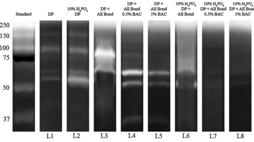
Zymography results are shown inFigs. 1 and 2. Zymographic assay revealed low expression of pro-form of MMP-9 (92 kDa) and active form of MMP-2 (66 kDa) in the mineralized dentin powder. In contrast, protease expressions in the demineralized powder were more pro-nounced. When employed in the SE mode, ABU increased the activity of MMP-9 pro-form and MMP-2 active form, while BAC-containing ad-hesive formulations showed reduced expression of MMP-2, and almost complete inhibition of the bands corresponding to MMP-9. All-Bond Universal adhesive applied in the E&R mode increased the activity of MMP-2 and inhibited MMP-9, while all the BAC-containing formula-tions tested in the E&R mode decreased MMP-2 expression and com-pletely inhibited MMP-9.
3.2. In situ zymography
Qualitative and quantitative in situ zymography results are shown in Figs.3,4,5,6,7. There was a decrease in enzymatic activity at T0 in all the experimental groups compared to controls. All the groups showed a general trend of increase in enzymatic activity after aging, except for the ABU SE + 1% methacrylate BAC group, which showed further de-crease. Overall, the experimental groups bonded with ABU + 1% me-thacrylate BAC showed the lowest level offluorescence over time, re-gardless of the bonding strategy employed (p < 0.05).
Fig. 1. Zymographic analysis of proteins extracted from dentin powder.Std: Standards (Std); L1: miner-alized dentin (DP) showing the presence of activity of pro-form of 9 (92 kDa) and active form of MMP-2 (66 kDa); L2: demineralized dentin powder (DDP) showing an increase of pro-form of MMP-9 (92 kDa), active form of MMP-2 (66 kDa);L3: DP after incuba-tion with All-Bond Universal (ABU) showing activity of active form MMP-9 (86 kDa) and MMP-2 (66 kDa);L4: DP after incubation with ABU + 0.5% BAC showing a reduction in the activity of MMP-9 active form and an increase in MMP-2 activity;L5: DP after incubation with ABU + 1% methacrylate BAC showing inhibition of MMP-9 and a decrease in the activity of MMP-2 ;L6: DDP after incubation with ABU showing MMP-2 ac-tivity and inhibition of MMP-9 ; L7: DDP after in-cubation with ABU + 0.5% BAC showing inhibition of MMP-9 and a reduction in MMP-2 activity;L8: DDP treated with ABU + 1% methacrylate BAC showing an enzymatic activity similar to that of L7.
3.3. Microtensile bond strength
Bond strength values (means and standard deviations, in MPa) at T0 and T12 months are reported inTable 1. Statistical analysis showed that only the variables “adhesive system” and “aging time” significantly affected the results, but not the variable “bonding strategy”. Post-hoc analysis indicated that ABU + 1% methacrylate BAC performed sig-nificantly worse than ABU or ABU + 0.5% BAC (p < 0.05). In adid-tion, all groups showed a significant decrease in microtensile bond strength values over time (p < 0.05).
4. Discussion
The present study investigated BAC blended within a commercially available multi-mode adhesive in terms of anti-enzymatic activity and bond strength preservation. The use of two adhesive formulations containing BAC decreased the activity of MMPs both in the dentin protein extracts and in the HL. This warrants rejection of thefirst null hypothesis that“incorporation of BAC in an adhesive formulation has no effect on the activity of endogenous dentin MMPs immediately or
over time”. Furthermore, the two formulations of BAC-containing ad-hesives influenced bond strength differently, both immediately and after aging. Even though aging in artificial saliva significantly reduced adhesion values, irrespective of whether BAC was incorporated in the adhesive formulation or the application mode (ER or SE), 1% BAC methacrylate formulation showed reduced bond strength values both immediately and over time. Hence, the second null hypothesis that “incorporation of BAC in an adhesive formulation has no effect on immediate bond strength and bond strength after aging for 12 months” has to be rejected.
Regardless of the bonding system or bonding strategy, adhesive resin monomers do not completely coat the completely or partially demineralized collagenfibrils, producing HLs that are susceptible to enzymatic hydrolytic degradation over time [25]. In addition to the enzymatic degradation of the collagen, resin-based restorations are subjected to polymer degradation by enzymes present in the saliva that results in exposure of more collagen. The unprotected collagenfibrils at the base of the hybrid layer are attacked by endogenous enzymes that are bound to thefibrils, causing the loss of the anchoring function of the HL and bond strength degradation over time [26]. These events occur Fig. 2. Zymographic assay quantification showing a generally lower enzymatic activity in BAC-containing groups.
Fig. 3. Resin-bonded radicular dentin interfaces prepared with ABU SE (a,d), ABU + 0.5% BAC SE (b,e) and ABU + 1% methacrylate BAC SE (c,f) at T0, incubated with quenched fluorescein-labeled gelatin. (a) Image acquired in green channel, showing fluorescence (identifying intense endogenous enzymatic activity) in dentinal tubules and within the HL created with ABU SE; (b) Image acquired in green channel, showingfluorescence in dentinal tubules and within the HL created with ABU + 0.5% BAC SE; (c) Image acquired in green channel, showingfluorescence in dentinal tubules and within the HL created with ABU + 1% methacrylate BAC SE; (d) Image of the HL created by the application of ABU SE obtained by merging differential interference contrast image (showing the optical density of the resin-dentin interface) and image acquired in green channel; (e) Image of the HL created by the application of ABU + 0.5% BAC SE obtained by merging differential interference contrast image and image acquired in green channel; (f) Image of the HL created by the application of ABU + 1% methacrylate BAC SE obtained by merging differential interference contrast image and image acquired in green channel. D = Dentin; HL = Hybrid Layer; R = Resin Composite (For interpretation of the references to colour in thisfigure legend, the reader is referred to the web version of this article).
even in the absence of bacteria and may be attenuated by proteolytic enzyme inhibitors [9].
Benzalkonium chloride has been reported to inhibit MMP activities within the hybrid layer, when applied on acid-etched dentin [27–29]. The quaternary ammonium salt yielded mixed results when it is in-corporated into different components of the adhesive system. A com-mercially available etchant containing 1% BAC (Bisco) was initially produced by capitalizing on the antimicrobial properties of BAC. Al-though this antimicrobial etchant was shown to possess anti-col-lagenolytic potential, dentin bonded with the use of the BAC-containing etchant showed deterioration of the HLs after 6 months [22,30], 12 months [27–29] and 18 months of aging [31]. This may be attributed to the unstable electrostatic interaction between BAC and demineralized
dentin and that close to 50% of the BAC is washed off along with the etchant [18]. Because BAC can be dissolved in ethanol and acetone, and its activity is rather consistent in different pH, it was anticipated that BAC may be incorporated into a large variety of adhesive blends without forfeiting its anti-enzymatic properties [29].
Non-covalently bound molecules, such as non-polymerizable MMP inhibitors, may leach out of the HL. Instability of these non-poly-merizable MMP inhibitors compromises long-term anti-proteolytic benefits and results only in delaying, and not preventing the degrada-tion of adhesive interfaces [32,33]. Other strategies have been proposed to circumvent this deleterious issue [28,34,35]. For example, in-corporation of compounds that copolymerize with resin monomers in the adhesive blends is promising because the agent becomes Fig. 4. Resin-bonded radicular dentin interfaces prepared with ABU E&R (a,d), ABU + 0.5% BAC E&R (b,e) and ABU + 1% methacrylate BAC E&R (c,f) at T0, incubated with quenchedfluorescein-labeled gelatin. (a) Image acquired in green channel, showing fluorescence in dentinal tubules and within the HL created with ABU E&R; (b) Image acquired in green channel, showingfluorescence in dentinal tubules and within the HL created with ABU + 0.5% BAC E&R; (c) Image acquired in green channel, showingfluorescence in dentinal tubules and within the HL created with ABU + 1% methacrylate BAC E&R; (d) Image of the HL created by the application of ABU E&R obtained by merging differential interference contrast image and image acquired in green channel; (e) Image of the HL created by the application of ABU + 0.5% BAC E&R obtained by merging differential interference contrast image and image acquired in green channel; (f) Image of the HL created by the application of ABU + 1% methacrylate BAC E&R obtained by merging differential interference contrast image and image acquired in green channel. D = Dentin; HL = Hybrid Layer; R = Resin Composite (For interpretation of the references to colour in thisfigure legend, the reader is referred to the web version of this article).
Fig. 5. Resin-bonded radicular dentin interfaces prepared with ABU SE (a,d), ABU + 0.5% BAC SE (b,e) and ABU + 1% methacrylate BAC SE (c,f) at T12 months, incubated with quenchedfluorescein-labeled gelatin. (a) Image acquired in green channel, showing fluorescence in dentinal tubules and within the HL created with ABU SE; (b) Image acquired in green channel, showingfluorescence in dentinal tubules and within the HL created with ABU + 0.5% BAC SE; (c) Image acquired in green channel, showingfluorescence in dentinal tubules and within the HL created with ABU + 1% methacrylate BAC SE; (d) Image of the HL created by the application of ABU SE obtained by merging differential interference contrast image and image acquired in green channel; (e) Image of the HL created by the application of ABU + 0.5% BAC SE obtained by merging differential interference contrast image and image acquired in green channel; (f) Image of the HL created by the application of ABU + 1% methacrylate BAC SE obtained by merging differential interference contrast image and image acquired in green channel. D = Dentin; HL = Hybrid Layer; R = Resin Composite (For interpretation of the references to colour in thisfigure legend, the reader is referred to the web version of this article).
immobilized within the polymer matrix. In the present study, BAC was blended with a commercially available multi-mode adhesive in dif-ferent concentrations and formulations (0.5% BAC or 1% BAC metha-crylate).
The results of both zymographic analyses demonstrated stronger inhibition of enzymatic activities in the BAC-containing adhesives, when compared with the BAC-free control adhesive. Protease inhibition was the most efficient in experimental groups containing 1% BAC methacrylate, at the baseline, as well as over time. Several other studies showed the potential of BAC to inhibit endogenous dentinal enzymes [18,28,31] or to prevent collagen degradation [18,22,29]. However, they are not comparable to the present study because of the adoption of different methodologies, and aqueous BAC solution was used in those studies. The present study was the first to examine the effect of in-corporation of BAC in an adhesive system on endogenous enzymatic activity. Investigations of bond strength and material properties are required before such an experimental adhesive may be recommended for clinical use.
Incorporation of 0.24–2 wt% BAC into two different E&R adhesives (Optibond Solo Plus, Kerr Corp. and All-Bond 3, Bisco) resulted in
preservation of their bond strengths to dentin after 18 months of aging. Moreover, in adhesive formulations blended with lower BAC con-centrations, increase in bond strength was noted [31]. Adper Single Bond blended with 0.5% and 1% of BAC showed no significant decline in bond strength after 6 months [22]. Likewise, Adper Single Bond blended with 0.5% BAC showed no significant decline in bond strength after 12 months of aging [29]. When a certain concentration of a new component is incorporated into an adhesive, especially in a form of a liquid, there is a risk of jeopardizing the balance between the compo-nents of the system. This may have a detrimental effect on the degree of conversion, elastic modulus, tensile stresses and other mechanical properties [36,37]. It is possible that the higher concentration of BAC utilized in the present study adversely affected the polymerization quality and mechanical properties of the resin-dentin interface, both in the E&R and SE mode. The 1% methacrylate BAC showed lower bond strength even at baseline compared to the other experimental groups. Contrary to the results of the present study, Sabatini et al. investigated the use of ABU adhesive system blended with 0.5–2% BAC or metha-crylate BAC in the E&R mode [28], The authors reported that bond strength was preserved in all the experimental groups after 6 and 12 Fig. 6. Resin-bonded radicular dentin interfaces prepared with ABU E&R (a,d), ABU + 0.5% BAC E&R (b,e) and ABU + 1% methacrylate BAC E&R (c,f) at T12, incubated with quenchedfluorescein-labeled gelatin. (a) Image acquired in green channel, showing fluorescence in dentinal tubules and within the HL created with ABU E&R; (b) Image acquired in green channel, showingfluorescence in dentinal tubules and within the HL created with ABU + 0.5% BAC E&R; (c) Image acquired in green channel, showingfluorescence in dentinal tubules and within the HL created with ABU + 1% methacrylate BAC E&R; (d) Image of the HL created by the application of ABU E&R obtained by merging differential interference contrast image and image acquired in green channel; (e) Image of the HL created by the application of ABU + 0.5% BAC E&R obtained by merging differential interference contrast image and image acquired in green channel; (f) Image of the HL created by the application of ABU + 1% methacrylate BAC E&R obtained by merging differential interference contrast image and image acquired in green channel. D = Dentin; HL = Hybrid Layer; R = Resin Composite (For interpretation of the references to colour in thisfigure legend, the reader is referred to the web version of this article).
Fig. 7. In situ zymography quantification at T0 and T12 months showing reduced enzymatic activity in BAC-containing groups, especially when AllBond Universal is associated with 1% methacrylate BAC. However, the activity increased with storage in artificial saliva for 12 months.
months of aging. A possible reason for these discrepancies may be due to the manner in which BAC was incorporated into the adhesive, or the difference in light-curing intensities utilized by the two studies.
Incorporation of methacrylate BAC into ABU in the present study yielded the worst results in terms of bond strength after aging. However, the same experimental group demonstrated the lowest level of enzymatic activity compared to other groups, especially after aging for 12 months. One can only rationalize that the abhorrent bond strength results are attributed to the poor degree of conversion of BAC methacrylate in the presence of other methacrylate resin monomers in the ABU adhesive. This could have resulted in the leaching of un-polymerized BAC methacrylate resin monomer from the resin-dentin interface. Apart from potentially killing cells in the vital dental pulp, the resin-dentin interface becomes porous after leaching of the un-polymerized resin monomer. This, in turn, facilitates ingress of water and salivary enzymes into the HL and subsequent hydrolysis of the other polymerized resin components. Although leaching of the BAC methacrylate monomer could have resulted in better MMP inhibition, the overall result showed no contribution to the longevity of resin-dentin bonds.
5. Conclusions
Although the BAC-containing adhesive formulations showed in-hibitory potential on dentinal endogenous enzymes at the baseline, this property did not seem to be long-lasting. The MMP inhibition potential of the experimental adhesive formulation was not reflected by the bond strength results. Overall, the experiment shows that adhesives blended with MMP inhibitors should be extensively tested before translation to clinical use.
Conflict of interest statement
We wish to confirm that there are no known conflicts of interest associated with this publication and there has been no significant fi-nancial support for this work that could have influenced its outcome. References
[1] M.G. Rasines Alcaraz, A. Veitz-Keenan, P. Sahrmann, P.R. Schmidlin, D. Davis, Z. Iheozor-Ejiofor, Direct composite resinfillings versus amalgam fillings for per-manent or adult posterior teeth, Cochrane Database Syst. Rev. 31 (2014) CD005620, ,https://doi.org/10.1002/14651858.CD005620.pub2.
[2] T. Maravic, A. Mazzoni, A. Comba, N. Scotti, V. Checchi, L. Breschi, How stable is dentin as a substrate for bonding? Curr. Oral Heal. Rep. 4 (2017) 248–257,https:// doi.org/10.1007/s40496-017-0149-8.
[3] Y. Liu, L. Tjäderhane, L. Breschi, A. Mazzoni, N. Li, J. Mao, D.H. Pashley, F.R. Tay, Limitations in bonding to dentin and experimental strategies to prevent bond de-gradation, J. Dent. Res. 90 (2011) 953–968,https://doi.org/10.1177/ 0022034510391799.
[4] L. Breschi, T. Maravic, S.R. Cunha, A. Comba, M. Cadenaro, L. Tjäderhane, D.H. Pashley, F.R. Tay, A. Mazzoni, Dentin bonding systems: from dentin collagen structure to bond preservation and clinical applications, Dent. Mater. 34 (2018) 78–96,https://doi.org/10.1016/j.dental.2017.11.005.
[5] N. Nakabayashi, K. Kojima, E. Masuhara, The promotion of adhesion by the in-filtration of monomers into tooth substrates, J. Biomed. Mater. Res. 16 (1982) 265–273.
[6] S.R. Armstrong, M.A. Vargas, I. Chung, D.H. Pashley, J.A. Campbell, J.E. Laffoon, F. Qian, Resin-dentin interfacial ultrastructure and microtensile dentin bond
strength afterfive-year water storage, Oper. Dent. 29 (2004) 705–712. [7] M. Hashimoto, H. Ohno, M. Kaga, K. Endo, H. Sano, H. Oguchi, In vivo degradation
of resin-dentin bonds in humans over 1 to 3 years, J. Dent. Res. 79 (2000) 1385–1391,https://doi.org/10.1177/00220345000790060601.
[8] J. De Munck, K. Van Landuyt, M. Peumans, A. Poitevin, P. Lambrechts, M. Braem, B. Van Meerbeek, A critical review of the durability of adhesion to tooth tissue: methods and results, J. Dent. Res. 84 (2005) 118–132,https://doi.org/10.1177/ 154405910508400204.
[9] D. Pashley, F. Tay, C. Yiu, M. Hashimoto, L. Breschi, R. Carvalho, Collagen de-gradation by host-derived enzymes during aging, J. Dent. Res. 83 (2004) 216–221. [10] A. Mazzoni, V. Angeloni, F.M. Apolonio, N. Scotti, L. Tjäderhane, A.
Tezvergil-Mutluay, R. Di Lenarda, F.R. Tay, D.H. Pashley, L. Breschi, Effect of carbodiimide (EDC) on the bond stability of etch-and-rinse adhesive systems, Dent. Mater. 29 (2013) 1040–1047,https://doi.org/10.1016/j.dental.2013.07.010.
[11] L. Tjäderhane, F.D. Nascimento, L. Breschi, A. Mazzoni, I.L.S. Tersariol, S. Geraldeli, A. Tezvergil-Mutluay, M.R. Carrilho, R.M. Carvalho, F.R. Tay, D.H. Pashley, Optimizing dentin bond durability: control of collagen degradation by matrix me-talloproteinases and cysteine cathepsins, Dent. Mater. 29 (2013) 116–135,https:// doi.org/10.1016/j.freeradbiomed.2008.10.025.The.
[12] M.R.O. Carrilho, S. Geraldeli, F. Tay, M. de Goes, R. Carvalho, L. Tjäderhane, In vivo preservation of hybrid layer by chlorhexidine, J. Dent. Res. 86 (2007) 529–533.
[13] Y. Nishitani, M. Yoshiyama, B. Wadgaonkar, L. Breschi, F. Mannello, A. Mazzoni, R. Carvalho, L. Tjäderhane, F. Tay, D. Pashley, Activation of gelatinolytic/col-lagenolytic activity in dentin by self etching adhesives, Eur. J. Oral Sci. 114 (2006) 160–166.
[14] L. Breschi, F. Cammelli, E. Visintini, A. Mazzoni, M. Carrilho, M. Cadenaro, S. Foulger, F.R. Tay, Influence of chlorhexidine concentration on the durability of etch-and-rinse dentin bonds: a 12-month in vitro study, J. Adhes. Dent. 11 (2009) 191–198.
[15] L. Breschi, P. Martin, A. Mazzoni, F. Nato, M. Carrilho, L. Tjäderhane, E. Visintini, M. Cadenaro, F.R. Tay, E.D.S. Dorigo, D.H. Pashley, Use of a specific MMP-inhibitor (galardin) for preservation of hybrid layer, Dent. Mater. 26 (2010) 571–578,
https://doi.org/10.1016/j.dental.2010.02.007.
[16] A. Mazzoni, V. Angeloni, N. Sartori, S. Duarte, T. Maravic, L. Tjäderhane, D.H. Pashley, F.R. Tay, L. Breschi, Substantivity of carbodiimide inhibition on dentinal enzyme activity over time, J. Dent. Res. 96 (2017) 902–908,https://doi. org/10.1177/0022034517708312.
[17] A. Cova, L. Breschi, F. Nato, A. Ruggeri, M. Carrilho, L. Tjaderhane, C. Prati, R. Di Lenarda, F. Tay, D. Pashley, A. Mazzoni, Effect of UVA-activated riboflavin on dentin bonding, J. Dent. Res. 90 (2011) 1439–1445,https://doi.org/10.1177/ 0022034511423397.
[18] A. Tezvergil-Mutluay, M.M. Mutluay, L.S. Gu, K. Zhang, K.A. Agee, R.M. Carvalho, A. Manso, M. Carrilho, F.R. Tay, L. Breschi, B.I. Suh, D.H. Pashley, The anti-MMP activity of benzalkonium chloride, J. Dent. 39 (2011) 57–64,https://doi.org/10. 1016/j.jdent.2010.10.003.
[19] A. Tezvergil-Mutluay, M.M. Mutluay, K.A. Agee, R. Seseogullari-Dirihan, T. Hoshika, M. Cadenaro, L. Breschi, P. Vallittu, F.R. Tay, D.H. Pashley, Carbodiimide cross-linking inactivates soluble and matrix-bound MMPs, in vitro, J. Dent. Res. 91 (2012) 192–196,https://doi.org/10.1177/0022034511427705. [20] L. Breschi, Chlorhexidine application to stabilize the adhesive interface: why and
how? J. Adhes. Dent. 15 (2013) 492,https://doi.org/10.3290/j.jad.a32067. [21] J. Kanca III, One Step bond strength to enamel and dentin, Am. J. Dent. 10 (1997). [22] C. Sabatini, J. Kim, P.O. Alias, In vitro evaluation of benzalkonium chloride in the preservation of adhesive interfaces, Oper. Dent. 39 (2014) 283–290,https://doi. org/10.2341/13-131-LR.
[23] A. Mazzoni, P. Scaffa, M. Carrilho, L. Tjäderhane, R. Di Lenarda, A. Polimeni, A. Tezvergil-Mutluay, F.R. Tay, D.H. Pashley, L. Breschi, Effects of etch-and-rinse and self-etch adhesives on dentin MMP-2 and MMP-9, J. Dent. Res. 92 (2013) 82–86,https://doi.org/10.1177/0022034512467034.
[24] A. Mazzoni, F.M. Apolonio, V.P.A. Saboia, S. Santi, V. Angeloni, V. Checchi, R. Curci, R. Di Lenarda, F.R. Tay, D.H. Pashley, L. Breschi, Carbodiimide in-activation of MMPs and effect on dentin bonding, J. Dent. Res. 93 (2014) 263–268,
https://doi.org/10.1177/0022034513516465.
[25] A. Mazzoni, F. Nascimento, M. Carrilho, I. Tersariol, V. Papa, L. Tjaderhane, R. Di Lenarda, F. Tay, D. Pashley, L. Breschi, MMP activity in the hybrid layer detected with in situ zymography, J. Dent. Res. 91 (2012) 467–472,https://doi.org/10. 1177/0022034512439210.
[26] M.R.O. Carrilho, R.M. Carvalho, M.F. de Goes, V. di Hipólito, S. Geraldeli, F.R. Tay, D.H. Pashley, L. Tjäderhane, Chlorhexidine preserves dentin bond in vitro, J. Dent. Res. 86 (2007) 90–94,https://doi.org/10.1177/154405910708600115. Table 1
Microtensile bond strengths of different groups immediately after bonding (T0) and after 12 months of aging (T12) in artificial saliva.
Mode SE ER
Adhesive ABU ABU+ 0.5%BAC ABU+ 1%BAC ABU ABU+ 0.5%BAC ABU+ 1%BAC
T0 42.2aA± 16.8 40.5aA± 13.1 40.8aA± 12.4 44.1aA± 13.9 39.7aA± 9.4 36.2bA± 8.7
T12 35.8aB± 17.5 30.2aB± 12.5 21.8bB± 11.6 31.6aB± 14.8 29.0aB± 7.8 17.4bB± 10.5
Data are presented as means ± standard deviations, in MPa. Different lower-case letters indicate significant differences (p < 0.05) within the same row, different upper-case letters indicate significant differences (p < 0.05) within the same column.
[27] N. Tekçe, S. Tuncer, M. Demirci, S. Balci, Do matrix metalloproteinase inhibitors improve the bond durability of universal dental adhesives? Scanning 38 (2016) 535–544,https://doi.org/10.1002/sca.21293.
[28] C. Sabatini, D.H. Pashley, Aging of adhesive interfaces treated with benzalkonium chloride and benzalkonium methacrylate, Eur. J. Oral Sci. 123 (2015) 102–107,
https://doi.org/10.1111/eos.12168.
[29] C. Sabatini, P.A. Ortiz, D.H. Pashley, Preservation of resin-dentin interfaces treated with benzalkonium chloride adhesive blends, Eur. J. Oral Sci. 123 (2015) 108–115,
https://doi.org/10.1111/eos.12176.
[30] M. El Gezawi, R. Haridy, E. Abo Elazm, F. Al-Harbi, M. Zouch, D. Kaisarly, Microtensile bond strength, 4-point bending and nanoleakage of resdentin in-terfaces: effects of two matrix metalloproteinase inhibitors, J. Mech. Behav. Biomed. Mater. 78 (2018) 206–213,https://doi.org/10.1016/J.JMBBM.2017.11. 024.
[31] C. Sabatini, S.K. Patel, Matrix metalloproteinase inhibitory properties of benzalk-onium chloride stabilizes adhesive interfaces, Eur. J. Oral Sci. 121 (2013) 610–616,
https://doi.org/10.1111/eos.12089.
[32] H. Sano, T. Takatsu, B. Ciucchi, J. Honer, W. Matthews, D. Pashley, Nanoleakage:
leakage within the hybrid layer, Oper. Dent. 20 (1995) 18–25.
[33] F.R. Tay, N.M. King, K. Chan, D.H. Pashley, How can nanoleakage occur in self-etching adhesive systems that demineralize and infiltrate simultaneously? J. Adhes. Dent. 4 (2002) 255–269.
[34] D. Pashley, F. Tay, S. Imazato, How to increase the durability of resin-dentin bonds, Compend. Contin. Educ. Dent. 32 (2011) 60–64.
[35] J.M. Antonucci, D.N. Zeiger, K. Tang, S. Lin-Gibson, B.O. Fowler, N.J. Lin, Synthesis and characterization of dimethacrylates containing quaternary ammonium func-tionalities for dental applications, Dent. Mater. 28 (2012) 219–228,https://doi. org/10.1016/j.dental.2011.10.004.
[36] M. Cadenaro, D.H. Pashley, G. Marchesi, M. Carrilho, F. Antoniolli, A. Mazzoni, F.R. Tay, R. Di Lenarda, L. Breschi, Influence of chlorhexidine on the degree of conversion and E-modulus of experimental adhesive blends, Dent. Mater. 25 (2009) 1269–1274,https://doi.org/10.1016/j.dental.2009.05.008.
[37] M. Cadenaro, T. Maravic, A. Comba, A. Mazzoni, L. Fanfoni, T. Hilton, J. Ferracane, L. Breschi, The role of polymerization in adhesive dentistry, Dent. Mater. 35 (2018) e1–e22,https://doi.org/10.1016/j.dental.2018.11.012.