REVIEW
The restorative role of annexin A1 at the
blood–brain barrier
Simon McArthur
1, Rodrigo Azevedo Loiola
3, Elisa Maggioli
3, Mariella Errede
2, Daniela Virgintino
2and Egle Solito
3*Abstract
Annexin A1 is a potent anti-inflammatory molecule that has been extensively studied in the peripheral immune
system, but has not as yet been exploited as a therapeutic target/agent. In the last decade, we have undertaken the
study of this molecule in the central nervous system (CNS), focusing particularly on the primary interface between the
peripheral body and CNS: the blood–brain barrier. In this review, we provide an overview of the role of this molecule
in the brain, with a particular emphasis on its functions in the endothelium of the blood–brain barrier, and the
protec-tive actions the molecule may exert in neuroinflammatory, neurovascular and metabolic disease. We focus on the
possible new therapeutic avenues opened up by an increased understanding of the role of annexin A1 in the CNS
vasculature, and its potential for repairing blood–brain barrier damage in disease and aging.
Keywords: Blood–brain barrier, Annexin A1, Inflammation, Metabolism, Multiple sclerosis, Stroke
© 2016 The Author(s). This article is distributed under the terms of the Creative Commons Attribution 4.0 International License (http://creativecommons.org/licenses/by/4.0/), which permits unrestricted use, distribution, and reproduction in any medium, provided you give appropriate credit to the original author(s) and the source, provide a link to the Creative Commons license, and indicate if changes were made. The Creative Commons Public Domain Dedication waiver (http://creativecommons.org/ publicdomain/zero/1.0/) applies to the data made available in this article, unless otherwise stated.
Blood–brain barrier structure
The blood–brain barrier (BBB) is the major regulator of
communication between the peripheral circulation and
the brain, acting to protect the central nervous system
(CNS) from the damaging consequences of peripheral
challenges to homeostasis, such as occur during
inflam-mation and metabolic disease. Given the tight confines
of the skull, oedema-induced elevated tissue pressure
is highly damaging to neuronal function [
1
], hence one
of the most important functions of the BBB is to limit
immune cell extravasation, and to protect brain tissue
from the development of localised inflammation.
Simi-larly, the neural environment is highly metabolically
active, needing a significant proportion of the body’s
energy supply [
2
], and is thus highly vulnerable to
meta-bolic toxins. As a defence against these, another
criti-cal feature of the BBB is the presence of a network of
highly efficient efflux transporters in cerebral endothelial
cells, acting to extrude metabolic waste products and to
prevent potentially toxic molecules from entering the
brain [
3
].
The BBB is not an isolated single anatomical structure,
but is part of the so called neurovascular unit (NVU), a
morpho-functional unit formed by multiple integrated
ele-ments of the vessel wall (endothelial cells and pericytes),
encircling perivascular astroglia, microglia cells and
inter-vening neuronal terminals [
4
] (Fig.
1
). Central to the
bar-rier function of the NVU are the endothelial cells. These
cells are markedly different to other endothelia within the
body in that they display interendothelial tight junctions
(TJs) organised in an extensive array of junctional strands;
a network of close transmembrane protein–protein links
that, together with adherens junctions and junctional
adhesion molecules, essentially prevent small molecules
and invading cells from passing across the vessel wall via
a paracellular route [
5
–
7
]. A number of different proteins
are involved in forming TJs in the brain vasculature, but
two of the most important are the molecules occludin and
claudin-5 which form homodimeric bridges linking
neigh-bouring cells [
8
]. These molecules in turn complex with a
series of intracellular elements, including the proteins zona
occludens-1, -2 and -3 (ZO-1, -2 and -3), which couple to
the actin cytoskeleton and help give junctions rigidity [
9
].
Open Access
*Correspondence: [email protected]
3 William Harvey Research Institute, School of Medicine and Dentistry, Queen Mary University, London, UK
Alongside the paracellular pathway leukocytes
migra-tion occurs also by transcellular pathways which occurs
in a dynamic interaction between leukocytes protrusions
at specific site of the endothelium of the BBB [
10
].
The critical function of the endothelium is the
selec-tive regulation of molecular uptake into the brain
paren-chyma. Given the strength of inter-endothelial TJs,
small molecule entry into the brain is essentially
neg-ligible under normal conditions. The brain does,
how-ever, require both a significant energy input [
2
], and the
removal of neuronal metabolic waste products to the
cir-culation for elimination through the kidneys. As such, an
array of small molecule transport proteins are expressed
on the surface of endothelial cells, including
transport-ers for glucose, amino acids, nucleosides and many other
cations and anions [
7
,
11
]. In addition to these
transport-ers, receptor-mediated uptake systems exist for the larger
biomolecules such as lipoproteins, peptides and protein
hormones, permitting the selective entry of molecules
like insulin and transferrin into the brain.
Complement-ing these uptake systems, the BBB expresses a range of
highly effective ATP-binding cassette (ABC) family efflux
transporter systems, most notably P-glycoprotein, breast
cancer resistance protein (BCRP), multidrug
resistance-associated protein (MRP-1 and MRP-2), which together
serve to limit exposure of the CNS to potentially
neuro-toxic molecules [
12
], and which unfortunately are also a
major barrier to the therapeutic treatment of brain
dis-eases with pharmacological agents [
13
].
The endothelial cells lie on a complex basal lamina (the
equivalent of the basement membrane in peripheral
ves-sels without the lamina reticularis) [
14
], which serves not
only to provide physical support to the endothelia, but
also includes pericytes and is a further barrier between
the circulation and the brain parenchyma [
4
]. The basal
lamina is actually a juxtaposed pair of molecular layers
indistinguishable anatomically at the level of
microves-sels, but originating from endothelial cells and from
perivascular astrocytes (parenchymal layer). The two
layers are similarly composed of members of four major
glycoprotein families: laminins, collagen IV isoforms,
nidogens and heparin sulphate proteoglycans, including
VE-Cadherin
Zona occludens-1, -2, -3
Occludin
Catenins
Claudin-5
F-actin
JAMs
Microglia
Astrocyte end-foot
Pericyte
Endothelial basal
lamina
Endothelium
Lumen
Glia limitans
Fig. 1 Schematic depiction of the principal molecular and cellular components of the neurovascular unit that regulate inter-endothelial
perlecan and agrin [
15
]. They can be distinguished
how-ever, by their complement of laminin subtypes [
16
], with
the endothelial basement layer containing laminin-411
and -511, whilst the parenchymal one contains
laminin-111 and -211 [
17
].
The basal lamina is not simply a passive substrate but
is actively involved in communication across the BBB
and in the transfer of nutritional support into the brain
parenchyma [
18
,
19
]. The contribution this molecular
component of the NVU plays in maintaining BBB
integ-rity has not been fully clarified, but enzymes such as the
matrix metalloproteinases that disrupt its structure have
been implicated in inappropriate immune cell or parasite
entry into the brain [
20
,
21
] and in oedema and
haemor-rhage during cerebrovascular incidents [
22
–
24
].
Pericytes embedded in the basal lamina communicate
with microvessel endothelial cells performing
impor-tant regulatory functions controlling vessel diameter and
cerebral blood flow [
25
,
26
], and contributing to BBB
integrity. Mice lacking brain pericytes are embryonically
lethal, but notably have developing BBBs characterised
by abnormal distribution of TJ molecules and enhanced
vascular permeability [
27
], indicating an important role
for these cells in either the development, differentiation
or maintenance of BBB function [
28
,
29
]. This activity is
confirmed by studies of murine models of reduced
cer-ebral pericyte number, with a strong negative correlation
existing between brain vessel pericyte coverage and
vas-cular permeability [
30
], emphasising the importance of
these cells, even if the fine details of how they contribute
to BBB integrity remain unclear.
Astrocytes, present on the parenchymal side of the
vascular basal lamina, are major components of the
NVU, with individual astrocytes sending out numerous
processes and endfeet that under pericyte-derived
guid-ance cues, surround and ensheath the vessel wall [
30
].
The astrocyte processes provide considerably more than
structural support however, as they not only produce
the molecular components of the parenchymal basal
lamina limitans [
31
], but also actively promote TJ
for-mation between endothelial cells [
32
,
33
]. Additionally,
astrocytes of the BBB have critical roles in brain–blood
transport; they actively regulate water uptake through
the major cerebral water channel, aquaporin-4,
local-ized on the plasma membrane of endfeet in contact with
the vessel wall [
34
]. They are also intimately involved in
nutrient uptake [
35
] and play an important role in the
removal of neuronal metabolic waste products [
36
,
37
].
Whilst less studied than other components of the
neuro-vascular unit, there is some evidence of a role for
micro-glia in the regulation of BBB integrity, particularly under
inflammatory conditions [
38
]. Evidence in vitro indicates
that inflammatory activation of microglia can disrupt
endothelial TJs through release of reactive oxygen species
and cytokines [
39
,
40
], but whether this occurs in vivo,
and the extent to which microglia influence BBB function
under normal conditions remains to be investigated.
Together with signalling input from parenchymal
neurons [
41
], the neurovascular unit generates a highly
efficient barrier to free communication between the
cir-culation and the brain, whilst permitting the selective
uptake of requisite nutrients and water, and enabling
the removal of waste products of neuronal metabolism.
Whilst this system is indeed highly effective under
nor-mal homeostatic challenges, it can be significantly
per-turbed following the onset of disease and inflammation.
The blood–brain barrier in inflammation
The BBB is not static, but actively changes and responds
to inflammatory challenge, whether originating in the
periphery or the brain parenchyma. Numerous
inflam-matory factors have been shown to enhance BBB
perme-ability, as have recently been reviewed in detail [
42
–
44
].
Changes to the BBB reflect both alterations in the
per-meability barrier to small molecules and, with particular
relevance to neurodegenerative conditions such as
multi-ple sclerosis (MS) and Alzheimer’s disease (AD), a loss of
the normal restrictions on entry of immune cells into the
brain parenchyma through changes in the expression of
leukocyte adhesion molecules [
45
,
46
]. The mechanisms
underlying these changes are complex, and commonly
involve interacting circuits and feedback loops centred
on the actions of vasoactive mediators and
pro-inflam-matory cytokines upon endothelial cells and perivascular
astrocytes [
33
]. For example, bradykinin not only acts via
B
2receptors on endothelial cells to open TJs [
47
], but also
to induce astrocytic release of interleukin-6, which itself
can further enhance endothelial cell permeability [
48
].
On first examination, enhanced BBB permeability upon
exposure to inflammatory stimulation would appear to
be maladaptive, but an explanation may lie in
consider-ation of the role the BBB plays in the induction of
sick-ness behaviours. These behaviours, commonly associated
with inflammatory disease, include deficits in memory
and attention, lethargy and anhedonia, and are thought
to provide an adaptive advantage, conserving
meta-bolic energy for the fight against infection/damage [
49
].
Changes in BBB integrity and consequent access of
circu-lating mediators to the CNS parenchyma are thought to
be one of the major communication pathways underlying
the induction of these behaviours [
50
], acting in concert
with direct vagal information. Although these behaviours
are advantageous in the short term, helping to promote
recovery, extended and/or inappropriately severe
sick-ness behaviour can be a major source of morbidity in
chronic inflammatory conditions [
51
,
52
].
Evidence for a link between disease-associated
enhanced BBB permeability and cognitive impairment
has been steadily accruing, both in age-related cognitive
decline [
51
,
53
], and in pathologies as diverse as stroke
[
52
,
54
], vascular dementia [
55
,
56
], AD [
57
–
59
], diabetes
mellitus [
60
] and obesity [
61
]. Significantly, many of these
conditions are associated with peripheral inflammatory
activity to a greater or lesser extent; hence developing
an understanding of the factors regulating BBB
perme-ability may offer the opportunity to modify the negative
cognitive aspects of many inflammatory and neurological
conditions.
Annexin A1, peripheral inflammation and the BBB
For discussion of the general response of the BBB to
peripheral and neuroinflammatory challenge we defer
to recent comprehensive reports [
1
,
42
,
62
–
64
]; in this
paper we will discuss the specific role of one particular
mediator known for its peripheral anti-inflammatory/
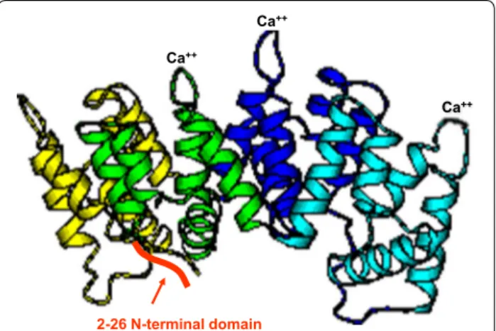
pro-resolution actions, annexin A1 (ANXA1-Fig.
2
), with
a particular focus on opportunities where it may be used
therapeutically to restore damaged BBB function.
The role of ANXA1 as a resolving/protecting
mol-ecule in the periphery is well known, as this molmol-ecule is
a secondary mediator of the anti-inflammatory effects
of glucocorticoid hormones [
66
], preventing leukocyte
migration into inflamed tissue [
67
]. Its role in the CNS
has been much less comprehensively studied, however.
ANXA1 belongs to the annexin superfamily of proteins
(ANXA1–A12) that are near-ubiquitously expressed in
eukaryotes from mould to mammals (yeasts being the
only major exception) [
68
]. ANXA1 is a
calcium-depend-ent phospholipid binding protein with a phospholipase
A
2inhibitory activity. Structurally ANXA1 presents four
repeats, three of which contain a Ca
2+-binding domain
(highly conserved among the 13 mammalian annexins)
and an N-terminal domain with multiple different
phos-phorylation sites that regulate its function [
69
,
70
], and
which confer its specific anti-inflammatory activity [
71
].
From an evolutionary point of view the structure of the
ANXA1 gene supports the hypothesis that it arose by
double duplication of an ancestral single domain gene
[
72
]. Interestingly another member of the family ANXA2
has been recently showed to be involved in miR155
regu-lation of BBB function [
73
].
Mechanistically, the auto/paracrine actions of ANXA1
are transduced by its binding to the G protein-coupled
receptor formyl peptide receptor 2 (FPR2, also known as
the lipoxin A
4receptor) [
74
], which we have shown to be
expressed on brain microvascular endothelial cells [
75
],
and by interaction with membrane phospholipids [
76
].
Numerous intracellular signalling pathways can be
acti-vated downstream of ANXA1 binding to FPR2;
activa-tion of p38 mitogen-activated protein (MAP) kinase [
77
],
activation of extracellular signal-regulated protein kinase
(ERK1/2) and mobilisation of intracellular Ca
2+[
78
], and
modification of the actin cytoskeleton through activation
of small guanosine triphosphate hydrolase (GTPases) [
79
,
80
] have all been reported.
The role of ANXA1 in the CNS has been debated for
several years [
81
], with its central functions only
hav-ing been clarified in the last decade. We and others have
shown ANXA1 to regulate microglial efferocytosis
(non-inflammatory removal of dead cells), and phagocytosis
[
82
–
84
], supporting a development and
anti-inflamma-tory role, respectively, in the brain. More recently
how-ever, we demonstrated an essential homeostatic function
of ANXA1, maintaining endothelial TJs in the BBB
[
80
], and repairing the permeabilising effect of systemic
lipopolysaccharides (LPS) on the BBB [
64
]. We propose
a dual role for ANXA1 in the CNS vasculature, serving
as a homeostatic regulator in normal conditions by
pro-moting BBB integrity, and importantly, acting to prevent
and limit the impact of pathological peripheral challenge
upon the brain.
Annexin A1 and the regulation of BBB integrity
across the lifespan
ANXA1 is a critical component of the normal BBB. It is
expressed by the brain microvascular endothelial cells in
close proximity to the plasma membrane and at points of
cell–cell contact where it co-localizes with cortex actin
microfilaments [
75
]. Deletion of the ANXA1 gene in
null mice is associated with disorganization of the actin
cytoskeleton, reduction of stress fibre formation, cell
shape changes and a loss of polarity that concludes in the
disruption of occludin and VE-Cadherin, findings which
indicate that ANXA1 participates in the regulation of
Ca++
Ca++
Ca++
2-26 N-terminal domain
Fig. 2 Crystal structure of ANXA1, showing four core Ca2+-binding domains, and the N-terminal sequence conferring specificity (2–26 N-terminal domain). Adapted from [65]
BBB permeability through its association with the actin
cytoskeleton [
75
]. In normal health conditions, ANXA1
thus plays a major protective role in the brain through
the promotion of BBB integrity. There are two stages
dur-ing life, however, when BBB function is less than optimal,
prenatal development and old age, and there is evidence
that changes to ANXA1 expression play a role in both.
The BBB was long considered immature and not fully
functional during development, but there is now
consid-erable evidence that this is not completely true, and that
many barrier functions are effective from the earliest
stages of brain ontogeny [
85
–
87
]. While this review does
not focus on the development of the BBB, it is interesting
to report that we have evidence that ANXA1 is expressed
by microglia-like cells and BBB endothelial cells during
human foetal development, further supporting a role
for the protein in prenatal brain development (Fig.
3
).
ANXA1 mainly localizes to endothelial cell cytoplasm
and plasma membranes (Fig.
3
b) and to a lesser extent to
the endothelial nucleus (Fig.
3
d). It has been suggested
that in vitro nuclear translocation of ANXA1 could be
induced by mitogenic signals [
88
] and that it could be a
negative prognostic factor in cancer [
66
,
67
,
89
].
How-ever, the expression of ANXA1 during normal brain
vascularization and BBB differentiation has not been
described before and needs to be further investigated.
In contrast, a major feature of aging is a decline in BBB
integrity [
90
], with increased paracellular permeability
and decreased TJ protein expression reported in murine
models [
91
]. Intriguingly, human dermal fibroblasts,
which share a mesenchymal developmental origin with
endothelial cells, exhibit a profound decrease in ANXA1
expression with aging [
92
], leading us to speculate that
endothelial ANXA1 downregulation may be partially
responsible for the aged BBB phenotype. This idea is
supported by the discovery that ANXA1 null mice have
higher, albeit non-pathological, BBB leakage than
age-matched wild-type controls [
64
]. Interestingly a
protec-tive role for ANXA1 has been reported in wound healing
in the gut [
93
] and bladder epithelia [
94
], both of which
are characterised by the presence of intercellular tight
junctions; structures known to be regulated by ANXA1
in the BBB [
80
].
BBB and ANXA1 in human disease
Several studies have indicated a role for ANXA1 in
neu-rological diseases, including Alzheimer’s disease (AD),
and stroke; although the most compelling evidence is for
an involvement in the pathology of MS.
Alzheimer’s disease
Alzheimer’s disease is an age-related
neurodegenera-tive disease and the most common form of dementia.
Pathologically, it is associated with neuronal loss, and
consequent loss of brain volume that is most pronounced
around the medial temporal lobe areas, particularly in
the hippocampus. Histologically, post mortem brains of
patients present widespread plaques and
neurofibril-lary tangles; important factors in AD pathogenesis. The
accumulation of amyloid β (Aβ) peptides is believed to
be a detrimental factor in Alzheimer’s disease
progres-sion, contributing to exacerbated inflammation,
micro-glial activation and neuronal loss. Increased Aβ levels in
patients’ brains may result from either Aβ
overproduc-tion or inadequate Aβ clearance. Aβ can be cleared via
the cerebrospinal fluid (CSF) and interstitial fluid (ISF)
and by enzymatic degradation, but an important route
for Aβ removal is through efflux transporters present at
the brain barriers. [
95
]. Morphological changes at the
brain barriers that occur in healthy aging are accelerated
and aggravated in AD. Microvascular reduction and
neu-rovascular dysfunction have been reported in AD [
96
].
Degeneration of pericytes occurs [
96
,
97
] and thickening
of the basal lamina is more pronounced [
98
].
Human post mortem studies of AD have reported
upregulated expression of ANXA1 in lesion-associated
glia [
99
,
100
] which, given the potent pro-resolution/
anti-inflammatory actions of ANXA1 in the periphery
[
101
] may reflect an endogenous attempt to limit cell
death. This idea is supported by the actions of ANXA1
in promoting non-phlogistic (non-inflammatory)
micro-glial phagocytosis, even in the face of inflammatory
chal-lenge with Aβ [
83
], and is further supported by the recent
identification of a single nucleotide polymorphism in the
regulatory region of ANXA1 that associates with
suscep-tibility to AD [
102
].
Increasing evidence indicates that disruption to the
integrity of the blood–brain barrier is a feature of AD [
90
,
103
]. Leakage of circulating plasma components into the
brain parenchyma correlates well with both post mortem
brain staining of AD [
104
] and with the rate of cognitive
decline seen in living AD patients [
57
,
105
]. The
impair-ment of BBB function seen in AD is not restricted to
plasma leakage, however, AD-related defects have been
reported in cerebral endothelial efflux transporter activity
[
106
,
107
] and in abnormal emigration of leukocytes into
the neuronal tissue [
108
,
109
]. These challenges to BBB
integrity may have significant consequences for AD
pro-gression, as there is now strong evidence linking enhanced
BBB permeability with impaired cognitive function,
includ-ing aspects of memory [
51
], language [
52
], performance on
the mini mental state exam [
57
] and Oxford handicap scale
[
54
]. Given the role of ANXA1 in controlling BBB TJ
for-mation and particularly in limiting leukocyte extravasation,
it is intriguing to speculate on whether the protein plays a
role in the microvascular endothelial phenotype of AD and
c
d
e
f
ANXA1 Collagen IV
ANXA1 Collagen IV
ANXA1 GFAP
ANXA1 GFAP
ANXA1
ANXA1
whether application of exogenous protein may be able to
limit disease-associated BBB changes. Although this has
not been studied directly, raised inflammation and
leuko-cytes trafficking in the peripherally has also reported
dur-ing disease progression [
95
].
Multiple sclerosis
Multiple sclerosis (MS) is an autoimmune
inflamma-tory disease of the central nervous system associated
with demyelination and axonal loss, eventually leading
to neurodegeneration. In general, it affects people under
50 years old with symptoms usually starting between the
ages of 20 and 40. It is a disease with a vast clinical and
pathological heterogeneity, but manifests in three
prin-cipal forms: relapsing remitting (RRMS; the most
com-mon form), primary progressive (PPMS) and secondary
progressive (SPMS) which tends to be the final stage of
RRMS. An important factor in the pathogenesis of MS is
the BBB which is compromised during the course of
dis-ease [
110
,
111
].
A direct link between BBB impairment, ANXA1
and disease has been indicated in MS, where a clear
loss of ANXA1 expression has been identified in the
brain parenchymal capillaries of MS patients, distant
from lesion sites [
80
]: a feature which may
contrib-ute to the loss of BBB integrity seen in this condition
[
112
]. Importantly, as ANXA1 is also expressed in
leu-kocytes, including both lesional and perivascular
mac-rophages and lymphocytes [
113
], its role in MS may
extend beyond the regulation of inter-endothelial TJs,
to directly modulating the autoimmune side of the
dis-ease. ANXA1 has long been known to inhibit leukocyte
migration [
101
] through its interaction with the
integ-rin VLA4 [
74
], closely resembling the main mechanism
of action of natalizumab, and highlighting the potential
of ANXA1 for therapeutic use. ANXA1 may serve as a
checkpoint between leukocytes and the BBB, on the one
hand protecting and correcting BBB leakage, and on the
other directly controlling leukocyte entry into the brain
parenchyma.
Neurovascular disease and stroke
There is further pre-clinical evidence for a protective/
therapeutic role of ANXA1 in the cerebral vasculature in
stroke and other neurovascular diseases. Although much
work has focussed on the role of ANXA1 as a modulator
of inflammatory microglial activity [
81
], there is evidence
for a role in the cerebral vasculature itself. Most notably,
administration of human recombinant ANXA1 has been
shown to markedly reduce lesion size, clinical score and
markers of leukocyte infiltration in murine mid-cerebral
artery occlusion models of stroke [
114
]. Animals lacking
the ANXA1 receptor Fpr2/3 showed markedly greater
BBB leakage post-ischaemia than their wild-type
coun-terparts [
115
]. Intriguingly, studies of ischaemic
pre-conditioning regimens, including both chloral hydrate
anaesthesia [
116
] and hypothermia [
117
] indicate that
these protective treatments act at least in part through
upregulation of ANXA1.
Given these various clinical and pre-clinical data, we
would argue that clinical studies of the
pharmacokinet-ics and pharmacodynampharmacokinet-ics of ANXA1 in healthy
sub-jects are warranted, as a further step towards evaluating
its potential use in patients with MS and other diseases
characterised by BBB impairment.
Estrogen, ANXA1 and the BBB in neurovascular disease
An intriguing aspect of the BBB in neurovascular
dis-eases such as stroke lies in the interactions of estrogen
and ANXA1, and the possible role this protein plays in
the vasculo-protective action of the hormone. It has long
been identified that stroke and other neurovascular
dis-eases show marked sex differences in their incidence, with
males being significantly more commonly affected than
females [
118
]. Whilst numerous factors contribute to this
difference, including rates of metabolic diseases and
envi-ronmental influences, the sex steroid hormone estrogen
has been shown to be a major discriminating factor [
119
].
The neuroprotective functions of this hormone have been
extensively studied [
118
], and it is known that it can exert
regulatory actions upon immune system function [
120
],
but more recent work suggests that estrogen can directly
target the BBB itself. In particular, estrogen has been
shown to both protect endothelial cells from cytotoxic
stimulation and to directly regulate components of the
BBB, including ANXA1, exerting a positive influence on
barrier integrity, actions that together help preserve BBB
function in the face of inflammatory challenge.
(See figure on previous page.)
Fig. 3 Localisation by immunofluorescence confocal microscopy of ANXA1 in human foetal forebrain at mid-gestation. a, b Single immunolabelling
for ANXA1 (green) shows a high expression of the protein in microglia-like cells and b in venular, endothelial cells. Note in b the prevalent expres-sion of ANXA1 on the luminal side of the endothelial plasma membrane (arrow). c, d Double immunolabelling for ANXA1/collagen IV. c The vascular basal membrane revealed by collagen IV allow to identify the shows ANXA1 reactivity localised on the endothelial lining; d ANXA1-negative pericyte embedded in the basal lamina and the localization of ANXA1 on the endothelial cell nucleus. e, f Double immunolabelling for ANXA1/GFAP shown on a confocal single optical plane. e ANXA1 reactıve endothelial cells in contact with GFAP labelled perivascular astrocyte processes; f a detail of ANXA1 localization on the endothelial membranes. Nuclear counterstaining with TO-PRO® 3. Bars a, b 10 μm; c 15 μm; d 4 μm; e 25 µm; f 8 µm
Estrogen is directly protective of cerebral endothelial
cells following ischaemic damage in vitro, most notably
modelled by deprivation and restitution of oxygen and
glucose (OGD/R). Here, the major circulating estrogen,
17β-estradiol, was shown to have a potent
cytoprotec-tive action, limiting overt cell death [
121
], but also
act-ing more directly to prevent hypoxia-inducible factor 1
α (HIF-1α)-mediated down regulation of the TJ
mole-cules occludin and claudin-5 [
122
,
123
]. These protective
effects of estradiol on cell viability could be replicated by
the estrogen receptor α (ERα) agonist propylpyrazoletriol
(PPT) [
121
], whilst the actions on TJs were mimicked by
the ERβ agonist diarylpropionitrile (DPN) [
122
]. These
findings highlight an important aspect of the actions of
estrogen upon the BBB, namely the complexity of
recep-tor-mediated signalling, as brain endothelial cells have
been shown to express all three major estrogen receptors,
ERα, ERβ and the G-protein coupled estrogen receptor
1 (GPER) [
124
–
126
]. A major downstream outcome of
OGD/R damage is the induction of reactive oxygen
spe-cies and, as has been repeatedly shown in studies of the
neuroprotective potential of estrogen, the hormone is
able to exert a powerful antioxidant, vasculoprotective
effect [
127
]. Estrogen has been shown to protect cerebral
endothelial cells from both OGD/R-induced [
121
] and
iron-mediated oxidative stress [
128
], although studies
differ on the relative importance of ERα and ERβ.
Protection against oxidative stress appears to be a
relatively general effect of estrogen [
129
], but there is
evidence that it can additionally exert BBB-specific
protective actions, most notably through modulation
of inter-endothelial tight and adherens junction
pro-tein expression. We, and others have shown estradiol to
upregulate expression of the key TJ molecules claudin-5
[
130
], occludin and zona occludens-1 [
64
,
131
], and
importantly to induce the intracellular relocation of these
molecules to the cytoplasmic membrane. Whilst estrogen
appears to directly regulate claudin-5 at a transcriptional
level [
125
], the actions of the hormone upon occludin
and ZO-1 are mediated through ANXA1 [
64
] following
activation of ERβ. Fewer studies have been conducted
upon the effects of estrogen on the BBB in vivo, but it is
known that male mice show significantly enhanced BBB
permeability following inflammatory challenge than
age-matched females, a protection lost in ovariectomised
ani-mals and restored by treatment with estradiol [
64
].
In addition to preventing small molecule entry into the
brain parenchyma, the BBB is an important check on the
ability of leukocytes to enter the central nervous system
[
46
]. Inflammatory stimulation can activate the cerebral
endothelium and permit entry of leukocytes into, at the
least, the perivascular space, primarily through
upregu-lation of cell adhesion molecules such as intercellular
adhesion molecule 1 (ICAM-1) and vascular cell
adhe-sion molecule 1 (VCAM-1) [
132
]. Estrogen, again acting
through ANXA1, can counteract these changes, reducing
adhesion molecule expression on the luminal surface of
the endothelium [
64
,
133
], and ultimately preventing
leu-kocyte adhesion and transmigration in the face of
inflam-matory challenge [
64
]. In this case however, the actions of
estrogen appear to be mediated by GPER, acting to
phos-phorylate ANXA1 on its N-terminal serine residues [
64
],
thereby promoting its secretion and autocrine/paracrine
feedback [
79
], ultimately resulting in a down-regulation
of ICAM-1 expression.
Although not all effects of estrogen upon the BBB
could be considered protective, e.g. estrogen
down-reg-ulates expression of the major efflux transporter BCRP
via ERβ [
134
–
136
], in general the hormone appears to
act to counter the damaging impact of peripheral
inflam-mation, protecting the brain from homeostatic challenge.
Intriguingly, two of the most important of these actions,
namely the preservation of endothelial TJ integrity and
the downregulation of inflammatory adhesion molecule
expression, appear to be mediated through ANXA1,
rein-forcing the role of this protein as a central mediator of
BBB function.
Metabolic disease and the BBB
Metabolic diseases, such as obesity and diabetes
melli-tus, are major and increasing sources of ill health in the
human population. These conditions have deleterious
effects upon virtually every physiological system, and it
is increasingly apparent that the CNS is not spared in this
regard. In particular, there is now accumulating evidence
for a direct impact of metabolic dysregulation upon the
BBB, representing an important pathway through which
disorders of metabolism can affect behaviour and
cogni-tion [
60
,
137
].
Several studies have showed obesity to be associated
with structural brain deficits, including atrophy in the
frontal lobes, hippocampus, thalamus [
138
], white [
139
]
and grey matter [
140
], increased BBB permeability [
141
–
143
] and the remodelling of brain microvessels [
144
],
suggesting a direct link between dietary habits and BBB
function. Moreover, obesity has been reported to alter
neurovascular unit organization, leading to increased
numbers of perivascular microglia [
145
] and activation of
both microglia and astrocytes [
145
–
147
]. Interestingly, it
was reported that even the offspring of obese mice
pre-sented increased BBB permeability at birth, suggesting
that maternal gestational obesity may be able to
compro-mise BBB formation during development [
146
].
Furthermore, obesity-induced BBB disruption has been
associated with down-regulation of cytoskeletal
compo-nent expression in endothelial cells, including vimentin
and tubulin [
148
], and the TJ proteins occludin [
142
],
claudin-5 [
142
,
149
] and ZO-1 [
148
]. As ANXA1 is an
essential regulator of BBB tightness through
stabilisa-tion of the cytoskeleton [
75
], we would speculate that its
expression or post-translational modification might also
be affected in obesity.
Similarly, experimental studies have also shown that
obesity is associated with increased neuronal death, BBB
leakage [
141
], and infarct volume [
144
,
150
,
151
]
follow-ing induction of an ischaemic episode. The deleterious
effects of obesity in experimental models of stroke may
be mediated, at least partly, through activation of matrix
metalloproteinase (MMP)-9, as high fat diet-induced
obesity increases MMP-9 expression in ischaemic murine
brain [
144
,
150
] and MMP-9 knockdown reversed the
damaging effects of obesity following ischaemic challenge
[
144
].
Moreover, there are few if any, direct studies of the
mechanisms underlying the deleterious influence of
obe-sity upon the BBB, but inflammatory pathways may well
play an important role. It is increasingly evident that
white adipose tissue secretes a wide variety of biologically
active adipokines [
152
], including both pro-inflammatory
(leptin, tumor necrosis factor α (TNFα) and
interleu-kin 6 (IL-6)) and anti-inflammatory (adiponectin)
fac-tors, which have the potential to regulate endothelial
function [
153
], and intriguingly plasma ANXA1 levels
inversely correlate with indicators of abdominal visceral
fat [
154
], suggesting that loss of endothelial ANXA1 may
also occur in response to chronic obesity-driven
inflam-mation. Obesity has been associated with activation of
pro-inflammatory pathways in the brain, as a high fat diet
up-regulated expression of toll like receptor 4 (TLR4),
high-mobility group protein B1 (HMGB1), vascular
endothelial growth factor (VEGF) and COX-2 [
155
]. In
addition, db/db mice, constitutively showed
perivascu-lar macrophage infiltration [
142
], exacerbated nuclear
factor κ B (NFκB) activation [
156
], and increased IL-1β,
IL-6, monocyte chemoattractant protein 1 (MCP-1) and
TNFα release [
142
]. How and if ANXA1 is involved in
such pathways remains speculative, but our preliminary
data suggests that ANXA1 null mice exhibit greater
cer-ebral perivascular CD45+ cell accumulation in response
to diet-induced obesity than their wild-type
counter-parts, indicating a role for the protein in the regulation of
immune cell entry into the brain, and further supporting
our hypothesis that ANXA1 acts to protect BBB integrity.
Furthermore, the finding that low levels of oxygen inhibit
the expression of ANXA1 in the pre-adipocytes suggest
that ANXA1 may have a role in the regulation of
inflam-matory and pro-resolvin pathways necessary to restore
homeostasis in the inflamed adipose tissue [
154
].
Diabetes mellitus
A major long-term complication of obesity is diabetes
mellitus (DM). A growing body of clinical evidence
sug-gests that DM is associated with neuronal dysfunction;
post mortem human studies indicate that patients with
DM exhibit reduced brain volume in both grey and white
matter [
157
–
159
] most notably in the hippocampus [
157
,
160
]. The importance of these findings is emphasised
in parallel studies indicating DM as a major risk factor
for dementias including AD [
161
,
162
] and mild
cogni-tive impairment [
159
,
161
], and for stroke [
163
,
164
]. A
variety of potential mechanisms have been identified
for these connections, but the role of the BBB has been
somewhat under-investigated despite accumulating
evi-dence for its involvement in the neuronal component of
DM. Circulating level of ANXA1 have been reported to
be downregulated in DM [
165
], data we have confirmed
in our high fat diet animal model (ES, unpublished data).
DM-induced BBB disruption is associated with
altera-tions in neurovascular unit organization, with
experi-mental diabetic models exhibiting marked reduction
in numbers of pericytes [
166
], but with microglial [
167
,
168
] and astrocytic activation [
168
,
169
], indicative of
a local inflammatory response. Additionally, exposure
of endothelial cells to hyperglycaemia induces a
down-regulation in expression of tight TJ occludin [
170
–
172
],
claudin 5 [
149
,
170
,
172
] and ZO-1 [
171
]. Diabetes
mel-litus (type 1 diabetes-T1DM) has been associated with
changes in the endothelial basal lamina, further
contrib-uting to disrupted BBB permeability [
169
].
The alterations in BBB permeability induced by
disrup-tions to glucose homeostasis may be driven, at least in
part, by increased activity of matrix metalloproteinases
(MMPs). Studies have shown exacerbation of MMP-2
activity in serum from patients with T1DM [
173
], from
STZ-induced diabetic animals [
167
,
171
] and from
rat models of diabetes mellitus type 2 (T2DM) [
174
].
Moreover, in vitro studies show that hyperglycaemia can
increase MMP2 activation [
175
], whilst BBB disruption
induced by advanced glycation end-products (AGEs)
produced under hyperglycaemic conditions can be
reversed by inhibiting MMP-2 activity [
176
].
Streptozo-tocin (STZ)-induced diabetic mice present BBB
disrup-tion associated with exacerbated MMP-9 activity, while
treatment with S-nitrosoglutathione, a nitric oxide
mod-ulator which is protective against oxidative/nitrosative
stress, reduces MMP-9 activity and restores normal BBB
permeability [
177
]. Intriguingly, ANXA1 has been both
positively and negatively associated with MMPs
expres-sion in cancer [
178
–
180
], and has been shown to be the
target of AGE-dependent non-enzymatic glycosylation
in pulmonary endothelial cells in STZ-induced T1DM
[
181
], suggestive of a link between AGE and BBB
break-down in DM.
Beyond the DM-induced alterations of BBB function
described above, DM has also been associated with CNS
infiltration of bone marrow-derived macrophages and
raised levels of pro-inflammatory cytokines in the brain
parenchyma [
167
,
169
], indicating that the
immunologi-cal barrier functions of the BBB are similarly disrupted by
DM. Furthermore white matter lesions, lacunar infarcts,
small strokes, and reductions in cerebral blood flow are
also reported being induced by DM (type 1 and 2) [
182
,
183
].Together, these results suggest that DM-induced
effects on BBB function depends on the interaction
between several conditions (hyperglycaemia,
hypoglycae-mia, AGEs) inherent to the pathology (Fig.
4
). The
pos-sible involvement of ANXA1 in mediating/protecting
against these changes has not been investigated to date,
but it is known that human patients with T2DM exhibit
reduced serum levels of ANXA1 compared with healthy
control individuals [
165
]. If this decline is reflected in
ANXA1 expression in the BBB itself, we can speculate
that ANXA1 loss would play a major role in transmission
of inflammatory stimuli into the brain parenchyma, and
the associated cognitive disturbances seen in DM [
60
]
(Fig.
4
).
Conclusion
The BBB as a critical communication interface between
the brain and the rest of the body has long been known
to play a role in neurological disease, but it is increasingly
realised that dysfunction is a feature of other homeostatic
disorders, most notably metabolic diseases such as obesity
and diabetes mellitus. A loss of BBB integrity is thought to
directly contribute to the cognitive, behavioural and
neuro-logical symptoms of these conditions, which to date have
received scant attention in either research or medical
prac-tice. Strategies to reverse BBB damage may therefore be of
great utility in addressing the neurological symptoms of
many clinical conditions. In this review, we propose that
therapeutic use of a major regulator of BBB integrity and
Fig. 4 Schematic representation of possible mechanisms linking metabolic disorders, BBB dysfunction and neurodegeneration. Metabolicimbal-ance associated with obesity and DM leads to chronic systemic, low-grade inflammation and the down-regulation of circulating ANXA1. Interaction of circulating soluble factors with brain endothelial cells induces tight junction disruption and BBB leakage, permitting leukocyte migration and glial activation, which in turn can impair neuronal function through production of pro-inflammatory mediators and reactive oxygen species (AGEs advanced glycation end-products; ROS reactive oxygen species)
function, ANXA1, may serve as such a strategy. Several
alternative approaches to the therapeutic use of ANXA1
are currently under development. One aims to avoid the
inherent problems of using a full-length protein in the
clinic; namely the administration of N-terminal ANXA1
peptides [
115
,
184
]. Unfortunately these efforts are
some-what limited by the propensity of these agents to signal
through both FPR2 and the pro-inflammatory receptor
FPR1 [
185
]. More promising might be through the delivery
of microvesicle-encapsulated proteins. This latter approach
has been shown to be potently anti-inflammatory in animal
models of rheumatoid arthritis [
186
], colitis [
93
] and
ath-erosclerosis [
187
]. Hence it could prove to be valuable in
the treatment of conditions characterised by damaged BBB
integrity, including such major disorders as Alzheimer’s
disease, multiple sclerosis and diabetes mellitus. Delivery of
ANXA1 thus holds great potential as a way to reverse BBB
damage induced by inflammation or metabolic challenge,
and in doing so restore normal BBB function.
Abbreviations
ABC: ATP-binding cassette transporters; Aβ: amyloid beta; AD: Alzheimer’s disease; AGE: advanced glycation end products; ANXA1: annexin A1; BBB: blood–brain barrier; BCRP: breast cancer resistance protein; CNS: central nerv-ous system; COX2: cyclooxygenase 2; CSF: cerebrospinal fluid; DM: diabetes mellitus; DPN: diarylpropionitrile; ER: estrogen receptor; ERK: extracellular signal-regulated protein kinase; FPR2: human formyl peptide receptor 2; Fpr2/3: murine FPR2 equivalent; GFAP: glial fibrillary acidic protein; GPER: G-protein coupled estrogen receptor 1; GTPase: guanosine triphosphate hydrolase; HIF-1α: hypoxia-inducible factor 1 α; HMGB1: high-mobility group protein B1; ICAM-1: intercellular adhesion molecule 1; IL-6: interleukin-six; ISF: interstitial fluid; LPS: lipopolysaccharides; MAP kinase: mitogen-activated pro-tein kinase; MCP-1: monocyte chemoattractant propro-tein 1; miR-155: micro RNA 155; MMP: matrix metalloproteinase; MRP: multidrug resistance-associated protein; MS: multiple sclerosis; NFκB: nuclear factor κ B; NVU: neurovascular unit; PPMS: primary progressive multiple sclerosis; PPT: propylpyrazoletriol; OGD/R: oxygen and glucose deprivation and restitution; RRMS: relapsing remitting multiple sclerosis; SMPS: secondary progressive multiple sclerosis; STZ: streptozotocin; T1DM: type 1 diabetes mellitus; T2DM: type 2 diabetes mellitus; TLR4: toll like receptor 4; TNFα: tumor necrosis factor α; TJ: tight junc-tion; TO-PRO® 3: red fluorescent nuclei stain; VCAM-1: vascular cell adhesion molecule 1; VEGF: vascular endothelial growth factor; ZO: zona occludens. Authors’ contributions
All authors equally contributed to the writing and discussion of this review. All authors read and approved the final manuscript.
Author details
1 Department of Biomedical Sciences, Faculty of Science and Technology, Uni-versity of Westminster, London, UK. 2 Department of Basic Medical Sciences, Neuroscience and Sensory Organs, Bari University School of Medicine, Bari, Italy. 3 William Harvey Research Institute, School of Medicine and Dentistry, Queen Mary University, London, UK.
Acknowledgements None.
Competing interests
All authors declare that they have no competing interests. Funding
SM is supported by Alzheimer’s Research UK (Grant# ARUK-PPG2016B-6) and the Society for Endocrinology (Early Career Grant); RAL is funded by the CAPES
Foundation, Ministry of Education, Brazil (Grant#7326/2014-09). DV is sup-ported by Regional Technology Clusters, Regione Puglia, 2015 (cod. MTJU9H8) and by FCRP-Fondazione Cassa di Risparmio di Puglia, 2015. ES is supported by Alzheimer’s Research UK (Grant# ARUK-PPG2013B-2) and FISM-Fondazione Italiana Sclerosi Multipla-cod.2014/R/21.
Received: 17 June 2016 Accepted: 12 September 2016
References
1. Sankowski R, Mader S, Valdés-Ferrer SI. Systemic inflammation and the brain: novel roles of genetic, molecular, and environmental cues as driv-ers of neurodegeneration. Front Cell Neurosci. 2015;9:28.
2. Magistretti PJ, Allaman I. A cellular perspective on brain energy metabo-lism and functional imaging. Neuron. 2015;86:883–901.
3. Saunders NR, Habgood MD, Møllgård K, Dziegielewska KM. The biologi-cal significance of brain barrier mechanisms: help or hindrance in drug delivery to the central nervous system? F1000Res. 2016. doi:10.12688/ f1000research.7378.1. (pii: F1000 Faculty Rev-313).
4. Obermeier B, Daneman R, Ransohoff RM. Development, maintenance and disruption of the blood–brain barrier. Nat Med. 2013;19:1584–96. 5. Chow BW, Gu C. The molecular constituents of the blood–brain barrier.
Trends Neurosci. 2015;38:598–608.
6. Sun H, Dai H, Shaik N, Elmquist WF. Drug efflux transporters in the CNS. Adv Drug Deliv Rev. 2003;55:83–105.
7. Sanchez-Covarrubias L, Slosky LM, Thompson BJ, Davis TP, Ronaldson PT. Transporters at CNS barrier sites: obstacles or opportunities for drug delivery? Curr Pharm Des. 2014;20:1422–49.
8. Haseloff RF, Dithmer S, Winkler L, Wolburg H, Blasig IE. Transmembrane proteins of the tight junctions at the blood–brain barrier: structural and functional aspects. Semin Cell Dev Biol. 2015;38:16–25.
9. Tietz S, Engelhardt B. Brain barriers: crosstalk between complex tight junctions and adherens junctions. J Cell Biol. 2015;209:493–506. 10. Carman CV, Sage PT, Sciuto TE, de la Fuente MA, Geha RS, Ochs HD,
et al. Transcellular diapedesis is initiated by invasive podosomes. Immu-nity. 2007;26:784–97.
11. Stieger B, Gao B. Drug transporters in the central nervous system. Clin Pharmacokinet. 2015;54:225–42.
12. Qosa H, Miller DS, Pasinelli P, Trotti D. Regulation of ABC efflux transport-ers at blood–brain barrier in health and neurological disordtransport-ers. Brain Res. 2015;1628:298–316.
13. Salameh TS, Banks WA. Delivery of therapeutic peptides and proteins to the CNS. Adv Pharmacol. 2014;71:277–99.
14. Abbott NJ, Patabendige AAK, Dolman DEM, Yusof SR, Begley DJ. Structure and function of the blood–brain barrier. Neurobiol Dis. 2010;37:13–25.
15. Thomsen MS, Birkelund S, Burkhart A, Stensballe A, Moos T. Synthesis and deposition of basement membrane proteins by primary brain capillary endothelial cells in a murine model of the blood–brain barrier. J Neurochem. 2016. doi:10.1111/jnc.13747.
16. Hallmann R, Horn N, Selg M, Wendler O, Pausch F, Sorokin LM. Expres-sion and function of laminins in the embryonic and mature vasculature. Physiol Rev. 2005;85:979–1000.
17. Engelhardt B, Sorokin L. The blood–brain and the blood-cerebrospinal fluid barriers: function and dysfunction. Semin Immunopathol. 2009;31:497–511. 18. del Zoppo GJ, Milner R. Integrin-matrix interactions in the cerebral
microvasculature. Arterioscler Thromb Vasc Biol. 2006;26:1966–75. 19. Simpson IA, Carruthers A, Vannucci SJ. Supply and demand in cerebral
energy metabolism: the role of nutrient transporters. J Cereb Blood Flow Metab NIH Public Access. 2007;27:1766–91.
20. Brkic M, Balusu S, Libert C, Vandenbroucke RE. Friends or foes: matrix metalloproteinases and their multifaceted roles in neurodegenerative diseases. Mediators Inflamm. 2015;2015:620581.
21. Bruschi F, Pinto B. The significance of matrix metalloproteinases in parasitic infections involving the central nervous system. Pathog. 2013;2:105–29.
22. Heo JH, Han SW, Lee SK. Free radicals as triggers of brain edema forma-tion after stroke. Free Radic Biol Med. 2005;39:51–70.
23. Rosell A, Ortega-Aznar A, Alvarez-Sabín J, Fernández-Cadenas I, Ribó M, Molina CA, et al. Increased brain expression of matrix metallo-proteinase-9 after ischemic and hemorrhagic human stroke. Stroke. 2006;37:1399–406.
24. Muradashvili N, Benton RL, Saatman KE, Tyagi SC, Lominadze D. Abla-tion of matrix metalloproteinase-9 gene decreases cerebrovascular permeability and fibrinogen deposition post traumatic brain injury in mice. Metab Brain Dis. 2015;30:411–26.
25. Hall CN, Reynell C, Gesslein B, Hamilton NB, Mishra A, Sutherland BA, et al. Capillary pericytes regulate cerebral blood flow in health and disease. Nature. 2014;508:55–60.
26. Armulik A, Abramsson A, Betsholtz C. Endothelial/pericyte interactions. Circ Res. 2005;97:512–23.
27. Hellström M, Gerhardt H, Kalén M, Li X, Eriksson U, Wolburg H, et al. Lack of pericytes leads to endothelial hyperplasia and abnormal vascular morphogenesis. J Cell Biol. 2001;153:543–53.
28. Liebner S, Corada M, Bangsow T, Babbage J, Taddei A, Czupalla CJ, et al. Wnt/beta-catenin signaling controls development of the blood–brain barrier. J Cell Biol. 2008/10/29 ed. 2008;183:409–17.
29. Liebner S, Plate KH, Ferguson J, Kelley R, Patterson C, Risau W, et al. Dif-ferentiation of the brain vasculature: the answer came blowing by the Wnt. J Angiogenes Res. 2010;2:1.
30. Bell RD, Winkler EA, Sagare AP, Singh I, LaRue B, Deane R, et al. Pericytes control key neurovascular functions and neuronal phenotype in the adult brain and during brain aging. Neuron. 2010;68:409–27. 31. Yao Y, Chen Z-L, Norris EH, Strickland S. Astrocytic laminin regulates
pericyte differentiation and maintains blood brain barrier integrity. Nat Commun. 2014;5:3413.
32. Janzer RC, Raff MC. Astrocytes induce blood–brain barrier properties in endothelial cells. Nature. 1987;325:253–7.
33. Abbott NJ, Rönnbäck L, Hansson E. Astrocyte-endothelial interactions at the blood–brain barrier. Nat Rev Neurosci. 2006;7:41–53.
34. Satoh J, Tabunoki H, Yamamura T, Arima K, Konno H. Human astrocytes express aquaporin-1 and aquaporin-4 in vitro and in vivo. Neuropathol-ogy. 2007;27:245–56.
35. Abbott NJ. Astrocyte-endothelial interactions and blood–brain barrier permeability. J Anat. 2002;200:629–38.
36. Asgari M, de Zélicourt D, Kurtcuoglu V. How astrocyte networks may contribute to cerebral metabolite clearance. Sci Rep. 2015;5:15024. 37. Thal DR. The role of astrocytes in amyloid β-protein toxicity and
clear-ance. Exp Neurol. 2012;236:1–5.
38. da Fonseca ACC, Matias D, Garcia C, Amaral R, Geraldo LH, Freitas C, et al. The impact of microglial activation on blood-brain barrier in brain diseases. Front Cell Neurosci. 2014;8:362. doi:10.3389/fncel.2014.00362. 39. Sumi N, Nishioku T, Takata F, Matsumoto J, Watanabe T, Shuto H, et al.
Lipopolysaccharide-activated microglia induce dysfunction of the blood–brain barrier in rat microvascular endothelial cells co-cultured with microglia. Cell Mol Neurobiol. 2010;30:247–53.
40. Nishioku T, Matsumoto J, Dohgu S, Sumi N, Miyao K, Takata F, et al. Tumor necrosis factor-alpha mediates the blood–brain barrier dysfunc-tion induced by activated microglia in mouse brain microvascular endothelial cells. J Pharmacol Sci. 2010;112:251–4.
41. Andreone BJ, Lacoste B, Gu C. Neuronal and vascular interactions. Annu Rev Neurosci. 2015;38:25–46.
42. Varatharaj A, Galea I. The blood–brain barrier in systemic inflammation. Immun: Brain Behav; 2016.
43. Lopez-Ramirez MA, Reijerkerk A, de Vries HE, Romero IA. Regulation of brain endothelial barrier function by microRNAs in health and neuroin-flammation. FASEB J. 2016;30:2662–72.
44. Derada Troletti C, de Goede P, Kamermans A, de Vries HE. Molecular alterations of the blood–brain barrier under inflammatory conditions: the role of endothelial to mesenchymal transition. Biochim Biophys Acta Mol Basis Dis. 2016;1862:452–60.
45. Zenaro E, Piacentino G, Constantin G. The blood–brain barrier in Alzhei-mer’s disease. Dis: Neurobiol; 2016.
46. Engelhardt B, Ransohoff RM. Capture, crawl, cross: the T cell code to breach the blood–brain barriers. Trends Immunol. 2012;33:579–89. 47. Zhou L, Yang B, Wang Y, Zhang H-L, Chen R-W, Wang Y-B.
Brady-kinin regulates the expression of claudin-5 in brain microvascular endothelial cells via calcium-induced calcium release. J Neurosci Res. 2014;92:597–606.
48. Schwaninger M, Sallmann S, Petersen N, Schneider A, Prinz S, Libermann TA, et al. Bradykinin induces interleukin-6 expression in astrocytes through activation of nuclear factor-kappaB. J Neurochem. 1999;73:1461–6.
49. Hart BL. Biological basis of the behavior of sick animals. Neurosci Biobe-hav Rev. 1988;12:123–37.
50. McCusker RH, Kelley KW. Immune-neural connections: how the immune system’s response to infectious agents influences behavior. J Exp Biol. 2013;216:84–98.
51. Montagne A, Barnes SR, Sweeney MD, Halliday MR, Sagare AP, Zhao Z, et al. Blood–brain barrier breakdown in the aging human hippocam-pus. Neuron. 2015;85:296–302.
52. Taheri S, Gasparovic C, Huisa BN, Adair JC, Edmonds E, Prestopnik J, et al. Blood–brain barrier permeability abnormalities in vascular cognitive impairment. Stroke. 2011;42:2158–63.
53. Popescu BO, Toescu EC, Popescu LM, Bajenaru O, Muresanu DF, Schultz-berg M, et al. Blood–brain barrier alterations in ageing and dementia. J Neurol Sci. 2009;283:99–106.
54. Wardlaw JM, Doubal FN, Valdes-Hernandez M, Wang X, Chappell FM, Shuler K, et al. Blood–brain barrier permeability and long-term clini-cal and imaging outcomes in cerebral small vessel disease. Stroke. 2013;44:525–7.
55. van de Haar HJ, Burgmans S, Hofman PAM, Verhey FRJ, Jansen JFA, Backes WH. Blood–brain barrier impairment in dementia: current and future in vivo assessments. Neurosci Biobehav Rev. 2015;49:71–81.
56. Raz L, Knoefel J, Bhaskar K. The neuropathology and cerebrovascular mechanisms of dementia. Blood Flow Metab: J Cereb; 2015. 57. Bowman GL, Kaye JA, Moore M, Waichunas D, Carlson NE, Quinn JF.
Blood–brain barrier impairment in Alzheimer disease: stability and functional significance. Neurology. 2007;68:1809–14.
58. Rius-Pérez S, Tormos AM, Pérez S, Taléns-Visconti R. Vascular pathology: cause or effect in Alzheimer disease? Neurologia. 2015. doi:10.1016/j. nrl.2015.07.010.
59. Nelson AR, Sweeney MD, Sagare AP, Zlokovic BV. Neurovascular dys-function and neurodegeneration in dementia and Alzheimer’s disease. Acta: Biochim Biophys; 2015.
60. Moheet A, Mangia S, Seaquist ER. Impact of diabetes on cognitive func-tion and brain structure. Ann NY Acad Sci. 2015;1353:60–71.
61. Wang C, Chan JSY, Ren L, Yan JH. Obesity reduces cognitive and motor functions across the lifespan. Neural Plast. 2016;2016:2473081. 62. Ransohoff RM, Brown MA. Innate immunity in the central nervous
system. J Clin Invest. 2012/04/03 ed. 2012;122:1164–71. 63. Ransohoff RM, Engelhardt B. The anatomical and cellular basis of
immune surveillance in the central nervous system. Nat Rev Immunol. 2012/08/21 ed. 2012;12:623–35.
64. Maggioli E, McArthur S, Mauro C, Kieswich J, Kusters DHMHM, Reu-telingsperger CPMPM, et al. Estrogen protects the blood–brain barrier from inflammation-induced disruption and increased lymphocyte trafficking. Brain Behav Immun. 2015;51:212–22.
65. Solito E, McArthur S, Christian H, Gavins F, Buckingham JC, Gillies GE. Annexin A1 in the brain–undiscovered roles? Trends Pharmacol Sci. 2008/02/12 ed. 2008;29:135–42.
66. Parente L, Solito E. Annexin 1: more than an anti-phospholipase protein. Inflamm Res. 2004;53:125–32.
67. Perretti M, Flower RJ. Annexin 1 and the biology of the neutrophil. J Leukoc Biol. 2004;76:25–9.
68. Moss SE, Morgan RO. The annexins. Genome Biol. 2004;5:219. 69. Solito E, Mulla A, Morris JF, Christian HC, Flower RJ, Buckingham JC.
Dexamethasone induces rapid serine-phosphorylation and membrane translocation of annexin 1 in a human folliculostellate cell line via a novel nongenomic mechanism involving the glucocorticoid recep-tor, protein kinase C, phosphatidylinositol 3-kinase. Endocrinology. 2003/03/18 ed. 2003;144:1164–74.
70. Solito E, Christian HC, Festa M, Mulla A, Tierney T, Flower RJ, et al. Post-translational modification plays an essential role in the translocation of annexin A1 from the cytoplasm to the cell surface. Faseb J. 2006/05/25 ed. 2006;20:1498–500.
71. Cirino G, Cicala C, Sorrentino L, Ciliberto G, Arpaia G, Perretti M, et al. Anti-inflammatory actions of an N-terminal peptide from human lipocortin 1. Br J Pharmacol. 1993;108:573–4.