Series Editors: Lorenzo Emmi · Domenico Prisco
Rare Diseases of the Immune System
Renato Alberto Sinico
Loïc Guillevin Editors
Anti-Neutrophil
Cytoplasmic
Antibody (ANCA)
Associated
Vasculitis
Rare Diseases of the Immune System
Series Editors: Lorenzo Emmi Domenico Prisco Editoria Board: Systemic Vasculitis L. Emmi C. Salvarani R. A. Sinico Autoimmune Disease P. L. Meroni D. Roccatello M. Matucci Cerinic L. Emmi Autoinflammatory Syndromes M. Gattorno F. De Benedetti R. Cimaz Primary Immunodeficiency A. Plebani C. T. Baldari M. M. D’EliosSystemic Fibroinflammatory Disorders
Renato Alberto Sinico • Loïc Guillevin
Editors
Anti-Neutrophil
Cytoplasmic Antibody
(ANCA) Associated
Vasculitis
Editors
Renato Alberto Sinico
Department of Medicine and Surgery University of Milano-Bicocca Monza Monza e Brianza Italy Loïc Guillevin Department of Medicine
Paris Descartes University Hospital Cochin Paris
France
ISSN 2282-6505 ISSN 2283-6403 (electronic) Rare Diseases of the Immune System
ISBN 978-3-030-02238-9 ISBN 978-3-030-02239-6 (eBook)
https://doi.org/10.1007/978-3-030-02239-6 © Springer Nature Switzerland AG 2020
This work is subject to copyright. All rights are reserved by the Publisher, whether the whole or part of the material is concerned, specifically the rights of translation, reprinting, reuse of illustrations, recitation, broadcasting, reproduction on microfilms or in any other physical way, and transmission or information storage and retrieval, electronic adaptation, computer software, or by similar or dissimilar methodology now known or hereafter developed.
The use of general descriptive names, registered names, trademarks, service marks, etc. in this publication does not imply, even in the absence of a specific statement, that such names are exempt from the relevant protective laws and regulations and therefore free for general use.
The publisher, the authors, and the editors are safe to assume that the advice and information in this book are believed to be true and accurate at the date of publication. Neither the publisher nor the authors or the editors give a warranty, expressed or implied, with respect to the material contained herein or for any errors or omissions that may have been made. The publisher remains neutral with regard to jurisdictional claims in published maps and institutional affiliations.
This Springer imprint is published by the registered company Springer Nature Switzerland AG The registered company address is: Gewerbestrasse 11, 6330 Cham, Switzerland
vii
Acknowledgements
We would like to thank all the prestigious colleagues and researches from around the world and from different disciplines for the invaluable contribution to this book. Without their help this book would not have been possible.
ix Part I ANCA-Associated Vasculitis
1 Introduction: Nomenclature and Classification . . . 3
J. Charles Jennette and Ronald J. Falk
2 Genetics of ANCA-Associated Vasculitis . . . 19
Federico Alberici, Paul Anthony Lyons, and Davide Martorana
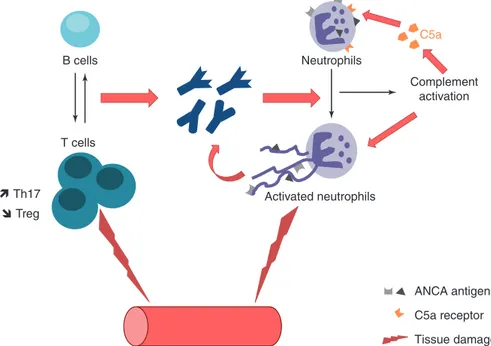
3 Etiopathogenesis of ANCA-Associated Vasculitis . . . 33
Delphine Sterlin, Alexis Mathian, and Makoto Miyara
4 ANCA: Methods and Clinical Significance . . . 47
Elena Csernok and Antonella Radice
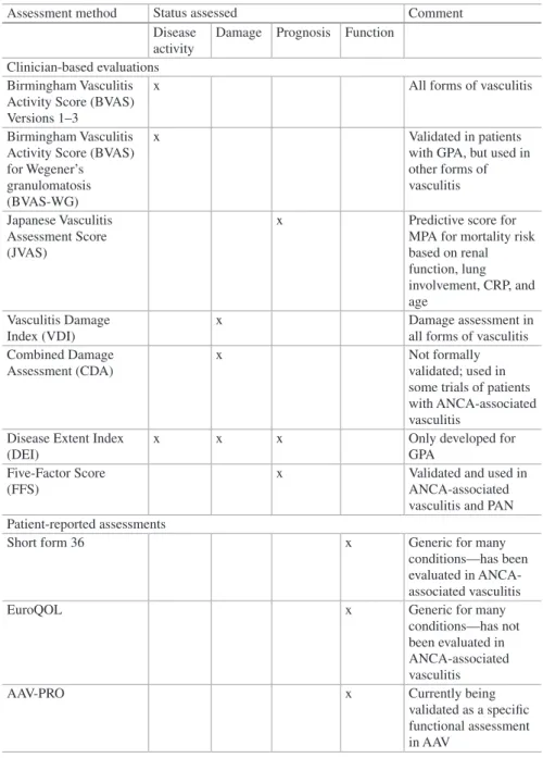
5 Activity and Damage . . . 57
Raashid A. Luqmani
6 Eosinophilic Granulomatosis with Polyangiitis
(EGPA, Churg–Strauss) . . . 77
Yann Nguyen and Loïc Guillevin
7 Granulomatosis with Polyangiitis . . . 97
Christian Pagnoux
8 Microscopic Polyangiitis . . . 131
Renato Alberto Sinico, Filippo Maria Sala, Maria Rosa Pozzi, Paolo Fabbrini, and Federico Pieruzzi
Part II Vasculitis/Organs Involvement
9 ANCA-Associated Vasculitis—ENT Involvement . . . 147
Trimarchi Matteo, Galli Andrea, and Roberto Teggi
10 Lung Involvement in ANCA-Associated Vasculitis . . . 163
Marta Casal Moura and Ulrich Specks
x
11 Kidney Involvement . . . 177
Renato Alberto Sinico, Fabio Pagni, Vincenzo L’Imperio, Valentina Binda, Paolo Fabbrini, Federico Pieruzzi, and Gabriella Moroni
12 Peripheral Nervous System Involvement . . . 193
Michael P. Collins and P. James B. Dyck
13 Central Nervous System Involvement in ANCA-Associated
Vasculitis . . . 239
Hubert de Boysson
14 Skin Involvement . . . 251
Angelo Valerio Marzano, Simona Tavecchio, and Emilio Berti
15 Miscellaneous Organ Involvement in ANCA-Associated Vasculitis . . . 269
Giorgio Trivioli and Augusto Vaglio
16 Prognosis and Outcomes of ANCA- Associated Vasculitis . . . 293
David Jayne
17 Treatment of ANCA-Associated Vasculitides . . . 313
Loïc Guillevin, Loïc Raffray, Yann Nguyen, Benjamin Chaigne, and Benjamin Terrier
Index . . . 329 Contents
Part I
ANCA-Associated Vasculitis
3 © Springer Nature Switzerland AG 2020
R. A. Sinico, L. Guillevin (eds.), Anti-Neutrophil Cytoplasmic Antibody (ANCA) Associated Vasculitis, Rare Diseases of the Immune System,
https://doi.org/10.1007/978-3-030-02239-6_1 J. C. Jennette (*)
Department of Pathology and Laboratory Medicine, School of Medicine, University of North Carolina at Chapel Hill, Chapel Hill, NC, USA e-mail: [email protected]
R. J. Falk
Department of Medicine, School of Medicine, University of North Carolina at Chapel Hill, Chapel Hill, NC, USA
e-mail: [email protected]
1
Introduction: Nomenclature
and Classification
J. Charles Jennette and Ronald J. Falk
1.1
AAV Pathologic Features
Vasculitis is inflammation in blood vessel walls. Antineutrophil cytoplasmic anti-body (ANCA)-associated vasculitis (AAV) is vasculitis accompanied by circulating ANCAs, or phenotypically identical disease without detectable ANCA (ANCA- negative AAV). AAV is a necrotizing vasculitis with few or no immune deposits that affects predominantly small vessels (i.e., capillaries, venules, arterioles, and small arteries) (Table 1.1) [1–3].
An understanding of the pathologic features of AAV is required to understand the historical and contemporary nomenclature and classification of AAV. Acute AAV lesions in blood vessels are characterized by segmental neutrophil-rich inflamma-tion and necrosis (Fig. 1.1). Vessel wall necrosis allows plasma constituents, includ-ing coagulation factors, to spill from vessels into perivascular tissue or adjacent spaces, for example, the urinary space adjacent to glomerular capillaries (Fig. 1.1a) and the air space adjacent to alveolar capillaries (Fig. 1.2a). In perivascular tis-sue, the coagulation factors contact thrombogenic substances (e.g., tissue factor) and form fibrin. The accumulation of fibrin at sites of vascular necrosis is called fibrinoid necrosis (Fig. 1.1a, b). The infiltrating neutrophils and other leukocytes undergo nuclear fragmentation (leukocytoclasia) as a result of cell death produc-ing a pattern of injury called leukocytoclastic vasculitis, which is seen most often
4
in inflamed venules and arterioles (Fig. 1.1c). At a given site of inflammation, the acute necrotizing lesions evolve within several days or weeks into chronic inflam-matory lesions with a predominance of lymphocytes and monocytes, followed by progressive scarring. In patients with active disease, there is ongoing onset of new self-limited acute lesions concurrent with the evolution of chronic lesions. This is
Table 1.1 Vasculitis names adopted by the 2012 International Chapel Hill Consensus Conference on the Nomenclature of Vasculitides (2012 CHCC) [1]
Large-vessel vasculitis (LVV) Takayasu arteritis (TAK) Giant cell arteritis (GCA) Medium-vessel vasculitis (MVV) Polyarteritis nodosa (PAN) Kawasaki disease (KD) Small-vessel vasculitis (SVV)
Antineutrophil cytoplasmic antibody (ANCA)–associated vasculitis (AAV)
Microscopic polyangiitis (MPA)
Granulomatosis with polyangiitis (Wegener’s) (GPA) Eosinophilic granulomatosis with polyangiitis
(Churg-Strauss) (EGPA) Immune complex SVV
Anti-glomerular basement membrane (anti-GBM) disease
Cryoglobulinemic vasculitis (CV) IgA vasculitis (Henoch- Schönlein) (IgAV) Hypocomplementemic urticarial vasculitis (HUV)
(anti-C1q vasculitis) Variable vessel vasculitis (VVV) Behcet’s disease (BD) Cogan’s syndrome (CS) Single-organ vasculitis (SOV) Cutaneous leukocytoclastic angiitis Cutaneous arteritis
Primary central nervous system vasculitis - Isolated aortitis Others
Vasculitis associated with systemic disease Lupus vasculitis
Rheumatoid vasculitis Sarcoid vasculitis Others
Vasculitis associated with probable etiology
Hepatitis C virus–associated cryoglobulinemic vasculitis Hepatitis B virus–associated vasculitis
Syphilis-associated aortitis
Drug-associated immune complex vasculitis Drug-associated ANCA-associated vasculitis Cancer-associated vasculitis
Others
5
observed in renal biopsy specimens that have different glomeruli with necrotizing, mixed necrotizing and sclerotic, or purely sclerotic lesions.
Because AAV can affect vessels in every organ and tissue of the body, the clini-cal manifestations of disease are extremely variable. These are classified into one of several clinicopathologic categories, including microscopic polyangiitis (MPA), granulomatosis with polyangiitis (GPA) (formerly Wegener’s granulomatosis), eosinophilic granulomatosis with polyangiitis (EGPA) (formerly Churg-Strauss syndrome), and organ limited AAV (e.g., renal limited vasculitis, RLV) [1].
MPA has AAV with no granulomatous inflammation, whereas GPA and EGPA have necrotizing granulomatous inflammation, which is most frequent in the respi-ratory tract (Table 1.1) [1]. Only EGPA is associated with asthma and blood eosino-philia [1]. Pathologically indistinguishable vasculitis and glomerulonephritis occurs in MPA, GPA, and EGPA (Fig. 1.1).
a b c
Fig. 1.1 Necrotizing small-vessel vasculitis in patients with AAV. (a) Glomerulus with necrotiz-ing and crescentic glomerulonephritis with segmental fibrinoid necrosis (long arrow) and cellular crescent formation (short arrow) (Masson trichrome stain). (b) Necrotizing arteritis with circum-ferential fibrinoid necrosis (arrow) and associated infiltration by leukocytes (Masson trichrome stain). (c) Leukocytoclastic angiitis in the renal medulla with transmural accumulation of leuko-cytes, including neutrophils, and multiple nuclear fragments (arrows) indicative of leukocytoclasia (hematoxylin and eosin stain)
a b c
Fig. 1.2 Pulmonary inflammation in patients with AAV. (a) Hemorrhagic capillaritis with alveolar air spaces filled with blood (hematoxylin and eosin stain). (b) Necrotizing granulomatous inflam-mation in a GPA patient with infiltrating neutrophils, lymphocytes, monocytes, and macrophages, including multinucleated giant cells (arrows) (hematoxylin and eosin stain). (c) Inflammatory infil-trate in an EGPA patient showing conspicuous eosinophils (arrows) (hematoxylin and eosin stain) 1 Introduction: Nomenclature and Classification
6
Lung lesions exemplify the diversity of AAV inflammatory lesions. MPA, GPA, and EGPA all can have hemorrhagic capillaritis (Fig. 1.2a), which evolves to inter-stitial fibrosis. GPA and EGPA can have pulmonary granulomatous inflammation (Fig. 1.2b) that begins as neutrophil-rich necrotizing lesions. These lesions have progressive replacement of neutrophils by lymphocytes, monocytes, and macro-phages (including multinucleated giant cells), often surrounding a central zone of necrosis. These lesions may form cavities and may become fibrotic with minimal or no residual inflammation. Vasculitis and granulomatosis in EGPA typically have numerous eosinophils and neutrophils in active inflammatory lesions (Fig. 1.2c).
AAV is characterized immunopathologically by few or no immune deposits of immunoglobulin and complement in vessel walls, which distinguishes AAV from immune complex-mediated vasculitis and anti-glomerular basement membrane antibody (anti-GBM)-mediated vasculitis (Fig. 1.3) [1].
AAV also can be classified based on ANCA antigen specificity, for example, ANCA specific for proteinase 3 (PR3-ANCA AAV) or myeloperoxidase (MPO- ANCA AAV) or with negative serology for ANCA (ANCA-negative AAV) [1, 4, 5]. ANCA-negative AAV has clinical and pathologic features identical to those in ANCA-positive AAV. The most informative classification and diagnosis of AAV include both the clinicopathologic phenotype and the serotype [1]. The correla-tion of ANCA serotypes with clinicopathologic variants and clinical outcomes is reviewed later in this chapter.
1.2
Historical Background
The investigation of two different manifestations of vasculitis, which eventually intersected, led to the discovery of AAV (Fig. 1.4). These two manifestations are skin purpura caused by inflammation in small vessel in the dermis and segmental inflammation of arteries (arteritis).
Purpura (purple spots on the skin) is caused by segmental inflammation of small vessels in the skin resulting in localized hemorrhage. Robert Willian, a
a b c
Fig. 1.3 Patterns of glomerular staining for IgG indicative of immune complex glomerulonephri-tis (a), anti-GBM glomerulonephriglomerulonephri-tis (b), and pauci-immune ANCA glomerulonephriglomerulonephri-tis (c) (FITC anti-IgG stain)
7
dermatologist, reported in 1808 that purpura could be associated with systemic manifestation, such as pain in the extremities and in the abdomen, suggesting that the pathologic process causing purpura in the skin was also causing injury in other organs [6]. The pediatricians, Johann Schönlein and Eduard Henoch, collectively reported the association of purpura with arthralgias, abdominal pain and bleeding, and nephritis [7–9]. The children seen by Schönlein and Henoch most likely had IgA vasculitis (Henoch-Schönlein purpura) rather than AAV, because IgA vasculitis occurs most often in children and AAV occurs most often in adults. On the other hand, the internist William Osler described adults with purpura that was associ-ated with arthritis, peripheral neuropathy, abdominal pain, pulmonary hemorrhage, epistaxis, iritis, and nephritis [10, 11]. Many of his patients had nephritis and some had rapid progression of uremia, and at autopsy had glomeruli “compressed” by “crescentic” masses of cells in Bowman’s space [11]. Undoubtedly, some of Osler’s patients had AAV.
In 1919, the pathologist Ernest Goodpasture reported the results of an autopsy on a patient who died of pulmonary hemorrhage and rapidly progressive glomerulone-phritis [11]. He observed vasculitis affecting small arteries in the spleen, arterioles in the gut, and capillaries in the pulmonary alveoli and glomeruli. Because of this
Microscopic
polyangiitis Granulomatosiswith polyangiitis Polyarteritisnodosa
Churg-Strauss syndrome Kawasaki disease Eosinophilic granulomatosis with polyangiitis Cryoglobulinemic vasculitis IgA vasculitis Microscopic
polyangiitis granulomatosisWegener’s
Cryoglobulinemic vasculitis Henoch-Schönlein purpura Kawasaki
disease Polyarteritisnodosa
CHCC (1994)
CHCC (2012)
(2012) Necrotizing arteritis
Churg and Strauss (1951) Godman and Churg
(1954)
Wegener’s granulomatosis
Kussmaul and Maier (1866) Cryoglobulins Meltzer (1966) Klinger (1931) Wegener (1939) IgA Faille-Kuyper et al. (1973) Kawasaki (1967) ANCA Falk and Jennette
(1989)
ANCA van der Wouda
et al. (1985) Unknown Unknown ANCA Tervaert et al. (1990)
Immune complex SVV ANCA associated SVV
Periarteritis nodosa
Churg-Strauss
syndrome Kawasaki
disease Polyarteritisnodosa
Wohlwill (1923) Purpura Schönlein (1837) Henoch (1868) Osler (1914) Henoch-Schönlein purpura Microscopic periarteritis Biomarker discovery
Clinical and pathologi
c
descriptions
Fig. 1.4 Historical sequence of advances in the recognition and classification of vasculitides that cause purpura and/or necrotizing arteritis. These two pathways of discovery intersect and overlap with AAV (shaded area) because AAV causes both purpura and necrotizing arteritis
8
report, his name became associated with pulmonary–renal syndrome caused by GBM, although the presence of arteritis in his patient is not consistent with anti-GBM disease and indicates that the patient probably had AAV, most likely MPA.
At the same time, Schönlein and Henoch were investigating purpura caused by small-vessel vasculitis, Adolf Kussmaul (an internist) and Rudolf Maier (a pathologist) published the first detailed report of a patient with systemic necrotiz-ing arteritis [12]. They coined the name periarteritis nodosa because of the grossly visible focal nodular lesions along medium-sized arteries caused by inflammation extending through vessel walls and into the perivascular tissue [12]. The preferred diagnostic term later became polyarteritis nodosa because the inflammation is trans-mural rather than perivascular [13]. For over 50 years, virtually any patient who was found to have arteritis was lumped under a diagnosis of periarteritis nodosa or polyarteritis nodosa.
In 1923, Friedrich Wohlwill described two patients with a “microscopic form” of periarteritis nodosa [14]. His observations were confirmed and extended by Davson [15, 16]. This recognition that arteritis can occur along with small-vessel vasculitis began to link studies of arteritis [14–19] with studies of small-vessel vasculitis with purpura, glomerulonephritis, and pulmonary capillaritis [6–11] (Fig. 1.4).
From the 1920s to the 1960s, multiple variants of vasculitis were identified that had arteritis resembling polyarteritis nodosa, but also had distinctive features that warranted specific diagnoses, including microscopic polyarteritis [14], Wegener’s granulomatosis [17, 18], Churg-Strauss syndrome [19], and Kawasaki disease [20, 21]. The recognition of specific variants of arteritis resembling polyarteritis nodosa is ongoing (e.g., the discovery of adenosine deaminase-2 deficiency vasculitis) [22].
In 1954, Gabriel Godman and Jacob Churg published a breakthrough article that astutely concluded that “microscopic periarteritis,” Wegener’s granulomatosis, and Churg-Strauss syndrome were related and were distinct from polyarteritis nodosa [23]. They also suggested that these three variants are probably related pathogeneti-cally. This was a harbinger of the discovery of ANCA, which confirmed the relat-edness of ANCA-positive MPA, GPA, and EGPA, and their distinctiveness from ANCA-negative polyarteritis nodosa [24].
Multiple biomarkers related to specific pathogenic mechanisms that cause vas-culitis facilitated the classification and diagnosis of vasvas-culitis based on laboratory results, such as vessel wall IgA-dominant immune deposits in Henoch-Schönlein purpura (IgA vasculitis) [25], cryoglobulins in the circulation in cryoglobulinemic vasculitis [26], and circulating ANCA in what is now called MPA, GPA, and EGPA (Fig. 1.4) [27–29] (Fig. 1.4). This set the stage for establishing consensus names and definitions for systemic vasculitides, including AAV [1, 30].
1.3
Vasculitis Nomenclature and Classification
Nomenclature, classification, and diagnosis of vasculitis is difficult because of the broad spectrum of types and locations of vessels affected, multiple patterns of injury, diverse known etiologies and pathogenic mechanisms, absence of known
9
etiologies and pathogenic mechanisms in some forms of vasculitis, and the myriad overlapping and nonspecific signs and symptoms caused by vasculitides.
A nomenclature system provides names and definitions for diseases. A classifi-cation system organizes patients into well-defined groups (classes). Classificlassifi-cation allows selection of standardized patient groups (classes) to study disease character-istics or perform clinical trials. A diagnostic system uses validated criteria to make a clinically actionable diagnosis in an individual patient. As stated by Hasan Yazici, “diagnosis is nothing different than classification in the individual patient” [31].
Classification criteria are observations or data used to place groups of patients into standardized classes. Diagnostic criteria are observations or data used to confi-dently predict the presence of the defining features of a disease in a specific patient. Diagnostic criteria allow diagnosing (classifying) a single patient in a specific class.
If useful, names and definitions can remain the same indefinitely. Classification criteria and diagnostic criteria evolve more quickly, driven in part by advances in diagnostic technologies and development of new clinical laboratory tests that were not available when earlier criteria were established.
The goals of nomenclature, classification, and diagnostic systems are to enable effective communication among biomedical investigators and healthcare providers, guide clinical and basic research on well-defined cohorts (classes) of patients, and, most importantly, facilitate diagnosis and effective treatment of individual patients. Nomenclature, classification, and diagnostic systems should be under constant scru-tiny and adjusted as new knowledge emerges. Homer Smith, a nephrologist, warns us that “Though we name the things we know, we do not necessarily know them because we name them” [32].
Two overarching etiologic categories of vasculitis are infectious vasculitis caused by proliferation of microorganisms in vessel walls and noninfectious vas-culitis not caused by proliferation of microorganisms in vessel walls. The latter nevertheless may be caused indirectly by an infection that initiates a sequence of event that results in vascular inflammation, for example, hepatitis C virus infection secondarily causing cryoglobulinemic vasculitis.
A widely used approach to classifying vasculitis uses three categories based on the types of vessel that are involved, that is, large-vessel vasculitis, medium-vessel vasculitis, and small-vessel vasculitis. This approach was adopted in 1994 at an international consensus conference on the nomenclature of systemic vasculitides [30] and revised in 2012 by at a second consensus conference [1]. These Chapel Hill Consensus Conferences (1994 CHCC and 2012 CHCC) provide standard-ized names and definitions for different classes of vasculitis (Tables 1.1 and 1.2) (Fig. 1.5), but do not provide validated criteria for classifying cohorts of patients into these classes, or for diagnosing (classifying) individual patients.
According to 2012 CHCC, large-vessel vasculitis (LVV) is vasculitis affecting large arteries more often than other vasculitides [1]. Large arteries are the aorta and its major branches. Any size artery may be affected. Medium-vessel vascu-litis (MVV) is vascuvascu-litis predominantly affecting medium arteries defined as the main visceral arteries and their branches [1]. Any size artery may be affected. Inflammatory aneurysms and stenoses are common. Small-vessel vasculitis (SVV)
10
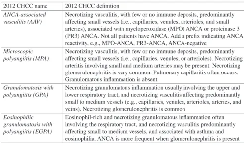
Table 1.2 AAV and AAV variant definitions from the 2012 International Chapel Hill Consensus
Conference on the Nomenclature of Vasculitides (2012 CHCC) [1] 2012 CHCC name 2012 CHCC definition
ANCA-associated vasculitis (AAV)
Necrotizing vasculitis, with few or no immune deposits, predominantly affecting small vessels (i.e., capillaries, venules, arterioles, and small arteries), associated with myeloperoxidase (MPO) ANCA or proteinase 3 (PR3) ANCA. Not all patients have ANCA. Add a prefix indicating ANCA reactivity, e.g., MPO-ANCA, PR3-ANCA, ANCA-negative
Microscopic polyangiitis (MPA)
Necrotizing vasculitis, with few or no immune deposits, predominantly affecting small vessels (i.e., capillaries, venules, or arterioles). Necrotizing arteritis involving small and medium arteries may be present. Necrotizing glomerulonephritis is very common. Pulmonary capillaritis often occurs. Granulomatous inflammation is absent
Granulomatosis with polyangiitis (GPA)
Necrotizing granulomatous inflammation usually involving the upper and lower respiratory tract, and necrotizing vasculitis affecting predominantly small to medium vessels (e.g., capillaries, venules, arterioles, arteries, and veins). Necrotizing glomerulonephritis is common
Eosinophilic granulomatosis with polyangiitis (EGPA)
Eosinophil-rich and necrotizing granulomatous inflammation often involving the respiratory tract, and necrotizing vasculitis predominantly affecting small to medium vessels, and associated with asthma and eosinophilia. ANCA is more frequent when glomerulonephritis is present
Large vessel vasculitis Takayasu arteritis Giant cell arteritis
Medium vessel vasculitis Polyarteritis nodosa
Kawasaki disease
ANCA-associated small vessel vasculitis Microscopic polyangiitis
Granulomatosis with polyangiitis
Eosinophilic granulomatosis with polyangiitis Immune complex small vessel vasculitis
IgA vasculitis
Cryoglobulinemic vasculitis
Hypocomplementemic urticarial vasculitis
Anti-GBM disease Capillary Arteriole Artery Artery Aorta Venule Vein
Fig. 1.5 Diagram depicting the predominant vessel involvement by large-vessel vasculitis, medium-vessel vasculitis, and small-vessel vasculitis. Note that AAV is a form of SVV that has a greater diversity of vessel involvement than SVV caused by immune complex disease or anti-GBM disease. Also note that arteritis caused by AAV overlaps with arteritis caused by medium-vessel vasculitis. Reproduced with permission from [1]
11
is vasculitis predominantly affecting small vessels, defined as small intraparenchy-mal arteries, arterioles, capillaries, and venules [1]. Medium arteries and veins may be affected. The two pathogenic categories of SVV are immune complex SVV and ANCA- associated SVV (AAV).
1.4
Classification and Diagnosis of AAV
The current names for the three major categories of AAV are microscopic polyangi-itis (MPA), granulomatosis with polyangipolyangi-itis (GPA), and eosinophilic granulomato-sis with polyangiitis (EGPA) (Table 1.2) [1]. The descriptive names GPA and EGPA were adopted by 2012 CHCC and replaced the names Wegener’s granulomatosis and eosinophilic granulomatosis that were used in 1994 CHCC.
Except for the preposition (i.e., with), all of the words in the names for MPA, GPA, and EGPA refer to pathologic features. However, importantly, this does not mean that direct histopathologic observation of pathologic features is required for diagnosis or classification. Histopathologic observations may not be practicable or necessary if validated noninvasive surrogate criteria are available. For example, in an appropriate clinical context, destructive nodular or cavitary pulmonary lesions, or destructive lesions in nasal cartilage or bone, can suffice to reasonably conclude that an ANCA-positive patient with pauci-immune necrotizing and crescentic glo-merulonephritis should be classified or diagnosed as GPA rather than MPA.
Clinical evidence for SVV that raises the possibility of AAV includes skin pur-pura, petechiae, or small ulcers; glomerulonephritis with dysmorphic erythrocy-turia, erythrocyte cylindruria, or proteinuria >1 g/day; pulmonary hemorrhage, radiographic consolidation, or hemoptysis; or ocular scleritis, uveitis, or ulcerative keratitis. Pathologic findings that confirm SVV include dermal venulitis, necrotizing glomerulonephritis (Fig. 1.1a), renal medullary angiitis (Fig. 1.1c), and pulmonary capillaritis (Fig. 1.2a). Immunohistologic identification of few or no immune depos-its in vessel walls supports a diagnosis of AAV (Fig. 1.3c). However, immunohisto-logic identification of moderate to marked vessel wall deposits of immunoglobulin and/or complement does not rule out AAV, because AAV can be concurrent with anti-GBM disease or immune complex disease [33]. Positive serology for ANCA has strong positive predictive value for AAV, but a negative result does not rule out AAV because a minority of patients with a clinical and pathologic phenotype that is identical to ANCA-positive AAV are ANCA-negative (i.e., ANCA-negative AAV). For example, at least 10% of patients with pauci-immune crescentic glomerulone-phritis are ANCA-negative [34].
AAV patients may have arteritis (Fig. 1.1b), but this alone does not distinguish between medium-vessel vasculitis (e.g., polyarteritis nodosa and Kawasaki disease) and SVV with arterial involvement. Clinical evidence for arteritis includes skin ery-thematous nodules or ulcers >1 cm; peripheral neuropathy (mononeuritis multiplex or asymmetrical polyneuropathy); or imaging showing arterial aneurysms, visceral infarcts, or gut perforation.
Importantly, classification or diagnosis of AAV clinicopathologic variants requires consideration of both inclusion criteria and exclusion criteria. For example,
12
classification or diagnosis of RLV and MPA requires exclusion of evidence for GPA and EGPA (i.e., no evidence of granulomatous inflammation, blood eosinophilia, or asthma). A patient can have every possible positive (inclusion) classification or diagnostic criterion for MPA, but these criteria alone will not be sufficient for classi-fication or diagnosis unless they are paired with negative (exclusion) criteria to rule out other diseases that share the positive criteria, for example, absence of evidence for granulomatous inflammation to rule out GPA.
Classification or diagnosis of GPA and EGPA requires clinical or pathologic evi-dence for granulomatous inflammation (Fig. 1.2b). Clinical evidence for granulo-matous inflammation includes pulmonary nodules or cavities, or destructive bone or cartilage lesions in the upper respiratory tract. Pathologic confirmation of granulo-matous inflammation includes identification of either active necrotizing granuloma-tous inflammation (especially in the respiratory tract) or chronic changes consistent with earlier necrotizing granulomatous inflammation. EGPA is distinguished from GPA by the presence of asthma and blood eosinophilia. The vasculitic and granu-lomatous lesions of EGPA typically contain conspicuous eosinophils (Fig. 1.2c); however, this is not specific for EGPA because MPA and GPA, as well as polyarteri-tis nodosa, may have numerous eosinophils in inflammatory lesions.
To our knowledge, there are no widely accepted, well validated, classification or diagnostic criteria for AAV and its variants. However, a major effort is underway to remedy this deficiency. The Diagnostic and Classification Criteria in Vasculitis Study (DCVAS) is an international, multicenter, observational study that has col-lected data on over 1000 AAV patients from more than 100 sites, as well as data from patients with other forms of vasculitis and patients with diseases that mimic vasculitis [35, 36]. The DCVS goal is to develop and validate diagnostic and clas-sification criteria for systemic vasculitides, including AAV [35].
1.5
Both Serotype and Phenotype Are Useful
for Classification and Diagnosis
2012 CHCC requires that the name (diagnosis) of AAV should include a prefix indicating ANCA serotype, for example, MPO-ANCA, PR3-ANCA, and ANCA- negative [1]. This is because both the clinicopathologic phenotype and the sero-type are useful for classification, diagnosis, and patient management. Classifying or diagnosing a patient as only MPO-ANCA AAV or only GPA is less informa-tive and less valuable than classifying or diagnosing a patient as MPO-ANCA GPA. Undoubtedly, identifying the serotype is much easier than confidently identi-fying the clinicopathologic phenotype; and the phenotype may change over time as more data are available or as the disease process evolves in a given patient.
Classifying patients based on PR3-ANCA versus MPO-ANCA serotype corre-lates with clinical and pathologic features, and with clinical course and outcome [4, 5, 37, 38], thus ANCA serotype is important component of an AAV diagnosis. Figure 1.6 shows data from an inception cohort of ANCA-positive AAV patients
13
from the Southeastern USA (excluding EGPA patients) patients [37]. The relative frequency of MPO-ANCA and PR3-ANCA varies based on the clinicopathologic phenotype (Fig. 1.6). For example, renal-limited vasculitis (RLV) patients have the highest frequency of MPO-ANCA, whereas patients with pulmonary nodules, nasal mucosal ulcers, and inflammatory destruction of nasal cartilage causing saddle nose deformity have a predominance of PR3-ANCA. This relationship indicates that ANCA antigen specificity modulates the targets and nature of pathogenic events. However, the serotype is not specific for a given clinicopathologic phenotype.
Table 1.3 shows data from the same patient cohort used in Fig. 1.6 [4, 37]. In the Southeastern USA, 81% of ANCA-positive RLV patients have MPO-ANCA, whereas 74% of GPA patients have PR3-ANCA. MPA patients have a more equal distribution of serotypes. However, even though PR3-ANCA is more frequent in GPA patients, because of the higher frequency of MPO-ANCA in this region, PR3- ANCA patients more often have MPA (50%) than GPA (40%) (Table 1.3) [4].
100.00 Renal limited Lung with no nodules % PR3-ANCA positive % MPO-ANCA positive Lung with nodules Nasal ulcers Saddle nose 90.00 80.00 70.00 % of all ANCA-P ositiv e patients 60.00 50.00 40.00 30.00 20.00 10.00 0.00 Renal limited GN
No lung and no ENTlung with no ENTlung with no nodules Plus skinAny lung
ENT no Lung Plus GISinusitis Plus ner ves Any ENT Otitis media Lung with nodules
Hear ing lossEpistaxis Subglottic stenosisAny g
ranu lomat
osis Nasal ulcersNasal cr
usting
Bloody nasal discharge Lung with ca vities Destr uctiv e bon y disease
Saddle nose def ormilty
Fig. 1.6 Correlation between serotype and clinical phenotype in an inception cohort of ANCA- positive AAV patients from the Southeastern USA evaluated at the UNC Kidney Center (excluding EGPA patients). Reproduced with permission from [4, 38]
14
The relative association of serotype with phenotype in a classification or diagnostic system for AAV varies based on geography and ethnicity. This will impact the positive and negative predictive value of a serotype for a given phe-notype in a given location or ethnic group. An interim analysis of DCVAS AAV patient data indicates that PR3-ANCA AAV is the predominant type of vascu-litis in patients with Northern Europeans, Middle Eastern/Turkish and Indian subcontinent ethnicity, whereas MPO-ANCA AAV is the predominant serotype of vasculitis in Japanese and Chinese populations [36]. MPO-AAV is more common in Caucasian Americans and Southern Europeans than in Northern Europeans.
ANCA-positive patients with EGPA usually have MPO-ANCA; however, less than 50% of patients with EGPA have ANCA [39–42]. Patients with clinical fea-tures of EGPA, such as asthma and blood eosinophilia, who are ANCA-positive, are more likely to have phenotypic features of vasculitis including glomerulonephritis, skin lesions, alveolar capillaritis, and peripheral neuropathy [42]. ANCA-positive EGPA appears to be a subset of patients with asthma and eosinophilia, which may be a separate disease process, or a distinct variant that develops over time in some but not all patients.
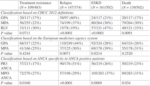
Table 1.4 uses the same 502 AAV patient cohort shown in Table 1.3 and Fig. 1.6 to compare the correlation of three different classification systems for AAV with clinical outcomes [4, 37]. The three classification approaches are as follows: (1) classification based on the 2012 CHCC names and definitions [1]; (2) the 2007 European Medicines Agency (EMA) classification system [43], which blends ele-ments of the 1990 American College of Rheumatology classification system [44], 1994 CHCC definitions [30], and ANCA serotypes; and (3) classification based on serotype alone. The clinical outcomes are the result of different treatment regimens from 1985 to 2007, and thus, the responses are not in line with current optimum therapy; however, all classes of patients were treated similarly. Additional outcome correlations are in the publication by Lionaki et al. [37]. Table 1.4 indicates that
Table 1.3 ANCA serotype (MPO-ANCA+ versus PR3-ANCA+) and clinicopathologic pheno-type (RLV, MPA, GPA) of an inception cohort of ANCA- positive vasculitis patients with high-frequency renal disease evaluated at the UNC Kidney Center (excluding EGPA patients)
Phenotype MPO- ANCA+ (%) PR3- ANCA+ (%)
RLV (n = 121) 81 19
MPA (n = 264) 59 41
GPA (n = 117) 26 74
Serotype RLV (%) MPA (%) GPA (%)
All ANCA+ (n = 502) 24 53 23
MPO-ANCA+ (n = 283) 35 55 11
PR3-ANCA+ (n = 219) 10 50 40
Additional features of this cohort were published in refs. [4, 38]
15
classification based on phenotype as well as classification based on serotype cor-relate with disease outcomes.
1.6
Concluding Remarks
As noted earlier in this chapter, the goals of classification and diagnostic systems are to enable effective communication among biomedical investigators and health-care providers, guide clinical and basic research on well-defined cohorts (classes) of patients, and, most importantly, facilitate diagnosis and effective treatment of individual patients. Physicians and scientists have made many advances in the clas-sification and diagnosis of vasculitides since the early seminal observational studies of Schönlein and Henoch [7, 8], Kusmal and Maier [12], and Godman and Churg [23], but validated and widely applied classification criteria and diagnostic crite-ria that are sufficiently accurate and precise for clinical research and patient care, respectively, remain elusive.
We agree with Homer Smith that “Though we name the things we know, we do not necessarily know them because we name them” [32]. But we also believe that being able to accurately name (i.e., diagnose) the disease in a patient will help us know the disease better and provide better care to the patient.
Table 1.4 ANCA vasculitis outcomes based on different classification systems evaluated in the same cohort by Lionaki et al. [37] shown in Table 1.3 and Fig. 1.6
Treatment resistance (N = 109/483) Relapse (N = 147/374) ESKD (N = 161/502) Death (N = 139/502) Classification based on CHCC 2012 definitions
GPA 20/117 (17%) 58/97 (60%) 24/117 (21%) 20/117 (17%) MPA 56/255 (22%) 74/199 (37%) 80/264 (30%) 79/264 (30%) RLV 33/111 (30%) 15/78 (19%) 57/121 (47%) 40/121 (33%)
P value 0.0711 <0.0001 <0.0001 0.0091
Classification based on the European medicines agency system
GPA 68/317 (22%) 110/249 (44%) 92/324 (28%) 84/324 (26%) MPA 41/166 (25%) 37/125 (30%) 69/178 (39%) 55/178 (31%)
P value 0.4244 0.0071 0.0214 0.2520
Classification based on ANCA specificity in ANCA-positive patients PR3 ANCA 37/213 (17%) 90/176 (51%) 56/219 (26%) 50/219 (23%) MPO ANCA 72/270 (27%) 57/198 (29%) 105/283 (37%) 89/283 (31%) P value 0.0160 <0.0001 0.0069 0.034
Treatment Resistance = persistence or new appearance of extrarenal manifestations and/or pro-gressive decline in renal function with active urine sediment in spite of immunosuppressive ther-apy. Relapse = reactivation of vasculitis in any organ after initial response to treatment. ESKD = chronic need for dialysis or transplantation. Death = death from any cause
16
References
1. Jennette JC, Falk RJ, Bacon PA, et al. 2012 revised international Chapel Hill consensus confer-ence nomenclature of vasculitides. Arthritis Rheum. 2013;65:1–11.
2. Jennette JC, Falk RJ. Pathologic classification of vasculitis. Pathol Case Rev. 2007;12:179–85. 3. Jennette JC, Thomas DB. Pauci-immune and antineutrophil cytoplasmic autoantibody glo-merulonephritis and vasculitis. In: Jennette JC, Olson JL, Silva FG, D’Agati V, editors. Heptinstall’s pathology of the kidney. 7th ed. Philadelphia: Wolters Kluwer; 2015, Chapter 16. p. 685–714.
4. Jennette JC, Nachman PH. ANCA glomerulonephritis and vasculitis. Clin J Am Soc Nephrol. 2017;12:1680–91.
5. Cornec D, Cornec-Le Gall E, Fervenza FC, Specks U. ANCA-associated vasculitis - clinical utility of using ANCA specificity to classify patients. Nat Rev Rheumatol. 2016;12:570–9. 6. Willan R. On cutaneous diseases, vol. I. London: J. Johnson; 1808.
7. Schönlein JL. Allegemeine und specielle Pathologie und Therapie, vol. 2. 3rd ed. Herisau: Literatur-Comptoir; 1837. p. 48.
8. Henoch E. Uber den zusammenhang von purpura und intestinal-stoerungen. Berl Klin Wochenschur. 1868;5:517–9.
9. Henoch E. Lectures on diseases of children: a handbook for physicians and students. New York: W. Wood and Co; 1882.
10. Osler W. The visceral lesions of purpura and allied conditions. Br Med J. 1914;1:517–25. 11. Goodpasture WE. The significance of certain pulmonary lesions in relation to the etiology of
influenza. Am J Med Sci. 1919;158:863–70.
12. Kussmaul A, Maier R. Über eine bisher nicht beschreibene eigenthümliche Arterienerkrankung (Periarteriitis nodosa), die mit Morbus Brightii und rapid fortschreitender allgemeiner Muskellähmung einhergeht. Dtsch Arch Klin Med. 1866;1:484–518.
13. Dickson W. Polyarteritis acuta nodosa and periarteritis nodosa. J Pathol Bacteriol. 1908;12:31–57.
14. Wohlwill F. Uber die mur mikroskopisch erkenbarre form der periarteritis nodosa. Arch Pathol Anat. 1923;246:377–411.
15. Davson J, Ball M, Platt R. The kidney in periarteritis nodosa. QJM. 1948;17:175–202. 16. Wainwright J, Davson J. The renal appearance in the microscopic form of periarteritis nodosa.
J Pathol Bacteriol. 1950;62:189–96.
17. Klinger H. Grenzformen der Periarteriitis nodosa. Frankf Ztschr Pathol. 1931;42:455–80. 18. Wegener F. Über eine eigenartige rhinogene Granulomatose mit besonderer Beteiligung des
Arteriensystems unter den Nieren. Beitr Pathol Anat. 1939;102:36–68.
19. Churg J, Strauss L. Allergic granulomatosis, allergic angiitis, and periarteritis nodosa. Am J Pathol. 1951;27:277–94.
20. Kawasaki T. MLNS showing particular skin desquamation from the finger and toe in infants. Allergy. 1967;16:178–89.
21. Tanaka N, Naoe S, Kawasaki T. Pathological study on autopsy cases of mucocutaneous lymph node syndrome. J Jpn Red Cross Central Hosp. 1971;2:85–94.
22. Karadag O, Jayne DJ. Polyarteritis nodosa revisited: a review of historical approaches, subphe-notypes and a research agenda. Clin Exp Rheumatol. 2018;36 Suppl 111(2):135–42.
23. Godman G, Churg J. Wegener’s granulomatosis. Pathology and review of the literature. Arch Pathol Lab Med. 1954;58:533–53.
24. Guillevin L1, Lhote F, Amouroux J, Gherardi R, Callard P, Casassus P. Antineutrophil cyto-plasmic antibodies, abnormal angiograms and pathological findings in polyarteritis nodosa and Churg-Strauss syndrome: indications for the classification of vasculitides of the polyarteritis Nodosa group. Br J Rheumatol. 1996;35:958–64.
25. Faille-Kuyber EH, Kater L, Kooiker CJ, Dorhout Mees EJ. IgA-deposits in cutaneous blood- vessel walls and mesangium in Henoch-Schönlein syndrome. Lancet. 1973;1:892–3.
17 26. Meltzer M, Franklin EC, Elias K, McCluskey RT, Cooper N. Cryoglobulinemia-a clinical and
laboratory study. II. Cryoglobulins with rheumatoid factor activity. Am J Med. 1966;40:837–56. 27. van der Woude FJ, Rasmussen N, Lobatto S, et al. Autoantibodies against neutrophils and
monocytes: tool for diagnosis and marker of disease activity in Wegener’s granulomatosis. Lancet. 1985;1:425–9.
28. Falk RJ, Jennette JC. Anti-neutrophil cytoplasmic autoantibodies with specificity for myelo-peroxidase in patients with systemic vasculitis and idiopathic necrotizing and crescentic glo-merulonephritis. N Engl J Med. 1988;318:1651–7.
29. Tervaert JW, Elema JD, Kallenberg CG. Clinical and histopathological association of 29kD- ANCA and MPO-ANCA. APMIS Suppl. 1990;19:35.
30. Jennette JC, Falk RJ, Andrassy K, et al. Nomenclature of systemic vasculitides: the proposal of an international consensus conference. Arthritis Rheum. 1994;37:187–92.
31. Yazici H. Diagnostic versus classification criteria - a continuum. Bull NYU Hosp Jt Dis. 2009;67:206–8.
32. Smith HW. Renal physiology. In: Fishman AP, Richards DW, editors. Circulation of the blood: men and ideas. New York: Springer; 1982, Chapter 9. p. 581.
33. Jennette JC. Rapidly progressive and crescentic glomerulonephritis. Kidney Int. 2003;63:1164–72.
34. Chen M, Kallenberg CG, Zhao MH. ANCA-negative pauci-immune crescentic glomerulone-phritis. Nat Rev Nephrol. 2009;5:313–8.
35. Craven A, Robson J, Ponte C, et al. ACR/EULAR-endorsed study to develop diagnostic and classification criteria for vasculitis (DCVAS). Clin Exp Nephrol. 2013;17:619–21.
36. Pearce FA, Craven A, Merkel PA, et al. Global ethnic and geographic differences in the clini-cal presentations of anti-neutrophil cytoplasm antibody-associated vasculitis. Rheumatology. 2017;56:1962–9.
37. Lionaki S, Blyth ER, Hogan SL, et al. Classification of antineutrophil cytoplasmic autoantibody vasculitides: the role of antineutrophil cytoplasmic autoantibody specificity for myeloperoxi-dase or proteinase 3 in disease recognition and prognosis. Arthritis Rheum. 2012;64:3452–62. 38. Yates M, Watts R. ANCA-associated vasculitis. Clin Med (Lond). 2017;17:60–4.
39. Scott DG, Watts RA. Epidemiology and clinical features of systemic vasculitis. Clin Exp Nephrol. 2013;17:607–10.
40. Sinico RA, Di Toma L, Maggiore U, et al. Prevalence and clinical significance of antineutro-phil cytoplasmic antibodies in Churg-Strauss syndrome. Arthritis Rheum. 2005;52:2926–35. 41. Sokolowska BM, Szczeklik WK, Wludarczyk AA, et al. ANCA-positive and ANCA-negative
phenotypes of eosinophilic granulomatosis with polyangiitis (EGPA): outcome and long-term follow-up of 50 patients from a single Polish center. Clin Exp Rheumatol. 2014;32:S41–7. 42. Cottin V, Bel E, Bottero P, et al. Revisiting the systemic vasculitis in eosinophilic
granuloma-tosis with polyangiitis (Churg-Strauss): a study of 157 patients by the Groupe d’Etudes et de Recherche sur les Maladies Orphelines Pulmonaires and the European Respiratory Society Taskforce on eosinophilic granulomatosis with polyangiitis (Churg-Strauss). Autoimmun Rev. 2017;16:1–9.
43. Watts R, Lane S, Hanslik T, Hauser T, et al. Development and validation of a consensus meth-odology for the classification of the ANCA-associated vasculitides and polyarteritis nodosa for epidemiological studies. Ann Rheum Dis. 2007;66:222–7.
44. Fries JF, Hunder GG, Bloch DA, et al. The American College of Rheumatology 1990 criteria for the classification of vasculitis. Summary. Arthritis Rheum. 1990;33:1135–6.
19 © Springer Nature Switzerland AG 2020
R. A. Sinico, L. Guillevin (eds.), Anti-Neutrophil Cytoplasmic Antibody (ANCA)
Associated Vasculitis, Rare Diseases of the Immune System,
https://doi.org/10.1007/978-3-030-02239-6_2
F. Alberici
Nephrology and Immunology Unit, ASST Santi Paolo e Carlo, San Carlo Borromeo Hospital, Milan, Italy
P. A. Lyons
Department of Medicine, University of Cambridge School of Clinical Medicine, Cambridge, UK
e-mail: [email protected]
D. Martorana (*)
Unit of Medical Genetics, University Hospital of Parma, Parma, Italy e-mail: [email protected]
2
Genetics of ANCA-Associated Vasculitis
Federico Alberici, Paul Anthony Lyons,
and Davide Martorana
2.1
Introduction
2.1.1 Why Study the Genetics of AAV
Antineutrophil cytoplasmic antibody (ANCA)-associated vasculitis (AAV) is a multisystem inflammatory-autoimmune disease including granulomatosis with polyangiitis (GPA) (formerly Wegener’s granulomatosis), microscopic polyangiitis (MPA), and eosinophilic granulomatosis with polyangiitis (EGPA, formerly Churg- Strauss syndrome) [1].
AAV pathogenesis is complex with a proposed role for environmental and infec-tious factors as well as dysregulation of the immune system; rare familial cases also suggested a potential role for genetic predisposition supporting further the theory of a multifactorial nature for the disease [2]. Such diseases are usually defined as “complex” meaning that both genetic and environmental factors contribute to the risk of their development.
Several families with GPA have been described although the increased risk for the development of disease in relatives of patients with GPA has been shown to be low compared to other autoimmune disorders [3], such as systemic lupus
20
erythematosus (SLE), inflammatory bowel disease (IBD), and multiple sclerosis (MS). Of interest, in a family containing a father with EGPA and a son with GPA, a shared HLA haplotype known to be a marker of autoimmunity was detected in the two affected [4]; other studies have explored the role of genetic predisposition in familial cases of AAV although results have been mainly negative probably due to the genotyping approaches employed.
While these studies were not conclusive, and in some cases contradictory, they provided a rational for exploring further the possible role of genetic factors in the development of AAV.
2.2
Molecular Genetic Approaches
Genetic studies in complex diseases require large numbers of subjects from which to estimate the prevalence of a disease in the population. However, many complex diseases are rare; thus, scientists must rely on case-controlled studies comparing a group of patients who harbor the disease in question with a nonaffected patient group to identify factors that may contribute to the disease. In complex diseases, the most commonly investigated genetic markers are single-nucleotide polymorphisms (SNPs). SNPs are variants in the genome that if located in the coding space may impact directly on gene function or if located in the noncoding space gene expres-sion may be involved in gene expression; despite their causal role differences in allele or genotype, frequencies between patients with a given disease and controls may suggest that they may be associated with the disease itself.
Despite SNPs being the most widely tested markers in case–control association studies, they are not the only ones used. In recent years, copy-number variants (CNVs) have also been investigated, with a number of studies demonstrating their potential to underlie susceptibility to complex diseases [5]. CNVs are areas of the human genome that may be repeated a variable number of times potentially impact-ing on gene expression and on the amount of protein produced.
Irrespective of the genetic marker studied, what is usually investigated is the rela-tive frequency of a genetic variant in cases and controls or its association with disease. It should be noted that the concept of association does not necessarily mean “causal-ity” with the latter requiring more complex follow-up studies in order to be assumed as true. As discussed previously, we should keep in mind that these are “complex” diseases, and therefore, several genetic variants are expected to contribute to the dis-ease itself with each variant only playing a small effect on the final phenotype.
Several genotyping techniques may be employed; noninclusive approaches are ones testing a specific hypothesis; in other words, they explore the association between a gene thought to play a role in the pathogenesis of a disease and the dis-ease itself. Inclusive approaches are techniques that enable the exploration of com-mon variation across the whole genome or at least a very big proportion of it. These are hypothesis free studies and are, therefore, able to identify novel, unexpected associations. The downside of the latter approach is the easy identification of
21
spurious associations and hence the requirement for a strict p-value threshold to control for this.
The candidate gene approach is the hallmark of noninclusive approaches, usu-ally it investigates genetic variants belonging to a specific biological pathway; in this case, the threshold of significance from the statistical point of view is usually represented by the p value of 0.05 (the one usually employed in any statistical analysis). In order to reduce the risk of associations identified by chance, a correc-tion is strongly suggested with the Bonferroni one being the most commonly used.
Genome-wide association studies (GWASs) are the most used and the hallmark nowadays of possible inclusive approaches; these use a simple case–control design but rely on genotyping techniques able to investigate millions of SNPs usually tag-ging 90% of the human genome. In order to avoid spurious associations, a very high level of statistical significance (p < 5 × 10⁻⁸) is required for an association to be
considered true. Although very robust, such studies have limitations as well, for example, large sample sizes are generally required; the number of identified asso-ciations is in fact proportional to the cohort dimension, and therefore, the bigger the study, the higher the probability of obtaining useful information, and this may be problematic in the field of rare diseases.
In both candidate gene and GWAS studies, replication in independent cohorts plays a central role to ensure the finding is robust; this is a key step to avoid findings that, for some reason, may be restricted only to the population initially explored. Irrespective of the genetic approach employed, the study design stage is of great importance in order to maximize the probability for a successful study; in particular, the cases should represent a well-defined phenotype of disease and controls should be matched for age, gender, and geographical area.
Other examples of inclusive genetics approaches are whole-exome/genome sequencing; these are approaches not yet routinely employed in the field of genetics of AAV but may have a big potential especially in the investigation of familial cases or clusters of extreme phenotypes; the employment of such approaches to large cohorts is still not clear, but the potential is significant allowing the exploration of the whole genome.
A genetic design halfway between inclusive and noninclusive approaches may relay on the use of chips exploring a big set of SNPs identified a priori as of poten-tial interest. The main example in the field of rare immunological diseases is the Immunochip that investigates SNPs and CNVs previously found as associated with inflammatory and autoimmune diseases [6] including a dense coverage of the HLA area. Due to relatively lower costs compared to conventional GWAS chips, this is a feasible and economically sustainable approach able to provide useful information probably more meaningful than candidate gene approach studies but yet lacking the wide exploratory nature of the ones provided by GWASs.
Genetic studies in the field of AAV have so far mainly been restricted to candi-date gene approaches, with only a few GWAS carried out; we will critically discuss the results of such studies performed so far.
22
2.3
Genetics of GPA and MPA
Rare diseases genetics is always a balance between the reliability of the information obtained and the challenges in performing the studies themselves. The size of the cohorts analyzed impacts significantly on the findings and on the analytical methods that may be employed. Only large cohorts of patients may be studied with inclusive genetic methods to obtain solid results with the bigger the population studied, the higher the probability of finding associations with small effect sizes. On the other hand, due to the rarity of some diseases, the cohorts available may not always allow identification of associations employing inclusive genetic methods of analysis, and in these cases, a candidate gene approach may be required. In the field of AAV, three GWAS studies [7–9] have provided four strong biologically plausible associations; in addition, some focused candidate gene studies have also identified other associa-tions of potential interest but, with the exception of one, the level of evidence under-pinning the result is lower and this should be taken into account when these studies are considered.
The first piece of information that genetics has provided to the field of AAV con-firms the autoimmune nature of the disease. Several genetic studies have described an association with the HLA Class II region even in relatively small cohorts [7]. Of interest, the HLA association with GPA and MPA is different being respectively
HLA-DP and the HLA-DQ. The association is stronger when the population is strati-fied according to ANCA specificity rather than clinical syndrome with HLA-DP being restricted to patients with PR3-ANCA and HLA-DQ restricted to patients with MPO-ANCA [7]. The mechanisms by which a predisposing HLA may facili-tate the development of autoimmunity are several and may include abnormal anti-gen presentation, positive selection of autoreactive T cells, or prevention of their negative selection [10]. A further mechanism recently described in Japanese MPA patients carrying the HLA–DRB1∗09:01, which is in strong linkage disequilibrium with HLA-DQ, is the ability to process and present the antigen MPO at the cell surface; this complex appears to be recognized by MPO-ANCA providing an important pathogenetic insight [11].
In the case of MPA, GWASs have not been able to so far identify other genetic associations largely due to the small samples size of the cohorts analyzed; further studies with larger samples will be required to shed further light on the genetic architecture of MPA. On the other hand, in case of GPA, the larger sample sizes have allowed the identification of three further associations at PRTN3, SERPINA1, and PTPN22.
PRTN3 encodes the serum protease PR3, the fact that patients with GPA are more often PR3-ANCA positive already pointed toward this enzyme playing a cen-tral in the pathogenesis. PR3 is expressed on the surface of a proportion of neutro-phils, and it has been described that only this membrane bound form is able to interact with ANCA facilitating neutrophil activation. In addition, PR3 may mediate direct tissue damage once it is released by the neutrophils [12]. Of interest, the SNP associated with GPA has been found to be linked to the levels of PR3 expression with the carriers of the susceptibility genotype characterized by higher expression
23
of the protein [9]. This is in keeping with studies that had previously described that neutrophils with high expression of membrane bound PR3 were found more fre-quently in GPA patients compared to controls [13] and suggest that higher levels of PR3 is an important step in the pathogenesis of the disease.
SERPINA1 encodes alpha-1 antitrypsin which is a protease inhibitor, with PR3 being one of its targets. The SNP associated with the risk of developing GPA has been identified as one encoding a variant of the enzyme with null activity (the so- called Z allele) [9]. The importance of reduced activity of alpha-1 antitrypsin in the pathogenesis of AAV had been already suggested by studies from the pre-GWAS era; in a cohort of 433 GPA cases and 421 controls, the presence of two of the alleles with null or reduced enzymatic activity (“Z” and “S” allele) increased the probabil-ity of developing disease with an odds-ratio of 14.58 [14].
PTPN22 is a genetic hallmark of autoimmunity, and it encodes the lymphoid tyrosine phosphatase (Lyp) which may be aberrant in the form encoded by the genetic variant associated with autoimmunity, causing abnormal CD4 T-cell (Treg)
activity, increased humoral activity, and enhanced neutrophil functions [15–17]. Interestingly, Ptpn22 knock-out mice have higher IgG levels, further supporting the significant impact of this gene on the B-cell compartment [18]. The central role of B cells in the pathogenesis of AAV is now well established as confirmed by the therapeutic efficacy of the anti-CD20 monoclonal antibody rituximab [19], provid-ing a further biological rationale for the important role of this gene in AAV. Some candidate gene studies in well-replicated cohorts have suggested a role for the 620W variant in the development of GPA. Importantly, recently this has been fur-ther confirmed at the level of GWAS significance. Of interest, this association was observed after combining GPA and MPA cases while, when stratifying the cohort according to the diagnoses, the magnitude of the association was similar in both GPA and MPA, but with lower level of significance probably due to the reduction of case numbers when analyzing subgroups; this suggests that, differently from PRTN3 and SERPINA1, PTPN22 might actually be a shared association between GPA and MPA [9].
Although still lacking confirmation at genome-wide significance, CTLA4 is likely to play a central role in the pathogenesis of AAV as well as other autoimmune diseases. The inhibitory glycoprotein encoded by this gene is expressed on activated T cells and competes with the co-stimulatory molecule CD28 for the binding of CD80 and CD86 preventing the stimulatory drive induced by CD28 itself. The cen-tral role in the immunological response of this immune checkpoint is nicely demon-strated by the fact that the CTLA4 inhibitors are now in use in the field of oncology to enhance immune system activity while a fusion protein CTLA4-Ig (abatacept) is now in use in autoimmune diseases, including GPA with nonsevere manifestations, aiming to achieve the opposite result. So far, several candidate gene studies have proposed a role for this gene as risk factor in AAV [20, 21]; however, bigger cohorts will be required to confirm its association at genome-wide significance. Table 2.1
summarizes the genes robustly described as associated with GPA and/or MPA. Several other genes have been identified as potentially associated with GPA and/ or MPA by smaller genetic studies. These relied on a candidate gene approach
24
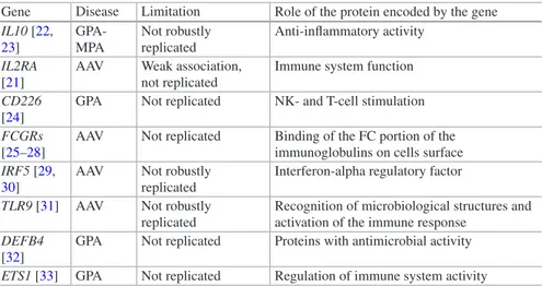
design, meaning that some genes thought a priori to be involved in the disease have been explored. The threshold of the association required to be considered significant is lower than that of GWAS; on one hand, this allows easier identification of asso-ciations; on the other hand, it may increase the rate of spurious ones. Findings obtained by such studies may be of potential interest but should be considered pre-liminary as long as confirmation in bigger cohorts is lacking. Among these associa-tions, the most potentially significant are reported in Table 2.2.
The genetics of GPA and MPA is still at a relative early stage of development compared to other diseases. However, it has already contributed in a significant way to improving our understanding of the disease. First of all, it has provided signifi-cant insight into a better classification of AAV which will be discussed below. Second, it has confirmed and provided further rational for potential therapeutic tar-gets: the confirmation of the central role of B-cell hyperactivity as well as of the
Table 2.1 Genes robustly associated with the risk of developing AAV
Gene Disease Phenotype resulting from the variant associated with the disease
HLA- DPB1 GPA Abnormal antigen presentation
HLA-DQ MPA Abnormal antigen presentation
PRTN3 GPA Increased expression of PR3 on neutrophils surface
SERPINA1 GPA Reduced activity of alpha1-antitrypsine, main inhibitor of PR3 activity
PTPN22 GPA Abnormal Treg function, increase B cells and neutrophils function
CTLA4 GPA Increased T-cell activity
GPA granulomatosis with polyangiitis, MPA microscopic polyangiitis, PR3 proteinase 3
Table 2.2 Genes proposed as associated with the risk of developing AAV with a high degree of
uncertainty
Gene Disease Limitation Role of the protein encoded by the gene
IL10 [22, 23] GPA- MPA Not robustly replicated Anti-inflammatory activity IL2RA [21]
AAV Weak association, not replicated
Immune system function
CD226
[24]
GPA Not replicated NK- and T-cell stimulation
FCGRs
[25–28]
AAV Not replicated Binding of the FC portion of the immunoglobulins on cells surface
IRF5 [29,
30]
AAV Not robustly replicated
Interferon-alpha regulatory factor
TLR9 [31] AAV Not robustly replicated
Recognition of microbiological structures and activation of the immune response
DEFB4
[32]
GPA Not replicated Proteins with antimicrobial activity
ETS1 [33] GPA Not replicated Regulation of immune system activity
GPA granulomatosis with polyangiitis, MPA microscopic polyangiitis, FCGRs Fc gamma receptors
25
immune checkpoint CTLA4 supports, respectively, the use of B-cell depleting strat-egies and abatacept among the treatment armamentarium of the disease.
Another important contribution of genetics is an increased understanding of the underlying pathogenesis. The central role of an HLA association has been the defin-itive confirmation of the autoimmune nature of the disease. Moreover in GPA, increased PR3 expression or activity has been recognized as key. As expected in a disease with a multifactorial pathogenesis, other factors play a role such as an abnormal and increased function of the T- and B-cell compartments as supported by the associations with PTPN22 and CTLA4.
However, what we know so far is still the tip of the iceberg; bigger studies and combined analyses of the studies performed so far will provide further insight on this topic and contributing to an even deeper understanding of AAV.
2.4
Genetics of EGPA
The challenges of performing genetic studies in EGPA are even more striking than in GPA and MPA mainly due to the lower frequency of the disease as well as diffi-culties from the classification point of view. ANCA-positive EGPA is more often characterized by vasculitic manifestations, such as lung hemorrhage, neuropathy, glomerulonephritis, and purpura, and is the subset of EGPA most easy to diagnose having a clear-cut clinical presentation and frequently relying on a biomarker such as ANCA. In the ANCA-negative cases, the diagnostic challenges have more sig-nificant difficulties in terms of differential diagnoses with other immunological dis-eases such as allergy/atopy or hypereosinophilic syndrome. These difficulties have been the major obstacles to the running of genetics studies with an inclusive approach in EGPA; so far, in fact, no GWAS has been published in this disease, although one is now ongoing by the European Vasculitis Genetic Consortium (EVGC).
What data exist in terms of the genetics of EGPA come from relatively small candidate gene studies; however, despite providing some insight, these data are probably still insufficient in order to draw firm conclusions.
The first set of candidate genes that were chosen to be explored were within the HLA area: two studies identified a robust association with the genes DRB4 and DRB3 [34, 35]; of interest, the likelihood of having this association increased with the increasing of the number of vasculitic manifestation. The identification of an associa-tion in the HLA area pointed also in EGPA toward an autoimmune nature of the dis-ease, providing an important step forward in the understanding of the disease.
Unfortunately, these findings have been the most robust so far in EGPA; other small studies have been performed with, however, relatively inconsistent results. One of them regarded the gene encoding IL10; IL-10 is a cytokine produced by the T-helper cells characterized by anti-inflammatory properties, including the inhibi-tion of immune mediator secreinhibi-tion, antigen presentainhibi-tion, and phagocytosis. A poly-morphism in this gene has been found to be associated with EGPA, although this association was restricted to the ANCA-positive subtype [23].
26
CNVs represent a significant source of genetic heterogeneity in addition to SNPs; CNVs refer to the intrinsic characteristics of some genes being potentially represented in a variable number of copies; this may induce phenotypic heterogene-ity influencing protein expression. In patients with autoimmune diseases, CNVs involving Fcγ receptor (FcγR) genes have reported an association of CNVs of the Fcγ-receptor 3B (FCGR3B) with systemic lupus erythematosus (SLE). Since carri-ers of low copies of the FCGR3B were shown to express lower protein levels, the pathogenetic mechanism proposed for this association was a reduced ability to clear immune complexes in subjects at risk [36]. The role of CNVs of FCGR3B has been investigated also in EGPA, and it has been found that being carrier of one copy of
FCGR3B was a risk factor for the disease; this is relatively surprising since the role of immune complexes in the pathogenesis of the disease may not be considered as relevant as in SLE. However, of interest, this association was stronger in the sub-group of patients showing vasculitic manifestations, the subsub-group more likely to be ANCA positive, suggesting that in some way an altered interaction with the autoan-tibody ANCA might be the explanation for this association.
Data on genetics in EGPA are still scanty; what we may so far conclude is a likely autoimmune nature for the disease and a possible different genetic back-ground between subsets of the disease with and without vasculitic manifestations; these conclusions are, however, preliminary and the results of the ongoing GWAS run by the EVGC might be able to provide further insight.
2.5
Contribution of Genetics to Better AAV Classification
AAV has been historically classified into three different clinical entities: GPA, MPA, and EGPA. Significant overlap exists between these leading to difficulties in clinical classification, which may impact on the therapeutic approach and disease monitoring with important consequences for the single patient. Of interest, ANCAs are a shared biomarker, and this, together with the important clinical overlap, may suggest a pathogenetic overlap between GPA, MPA, and EGPA. Of note, only 40% of EGPA cases are ANCA-positive and the ANCA-negative subgroup is more fre-quently represented by a variant of the disease usually without vasculitic manifesta-tions and more often characterized by clinical characteristics that arise as a consequence of hypereosinophilia. In this context of clinical uncertainty, genetics has played a big role in improving our understanding of AAV classification.First of all, the GWASs in GPA and MPA provided the insight that the genetic background is more clearly associated with the ANCA specificity rather than the clinical diagnoses. The associations were in fact stronger when the population was stratified according to the ANCA specificity rather that the clinical phenotype. This provides the rational for the suggestion of the probable need of re-think GPA and MPA more as PR3+ and MPO+ AAV. The idea that ANCA specificity might be more robustly able to differentiate disease subtypes rather than clinical syndrome was not new, and some clinical observations already pointed toward this direction; the contribution of genetics has been to confirm that giving a very strong rational to the observation. Table 2.3 reports the clinical and genetic characteristics supporting
![Table 1.1 Vasculitis names adopted by the 2012 International Chapel Hill Consensus Conference on the Nomenclature of Vasculitides (2012 CHCC) [1]](https://thumb-eu.123doks.com/thumbv2/123dokorg/5511889.63843/12.659.251.580.81.782/vasculitis-adopted-international-chapel-consensus-conference-nomenclature-vasculitides.webp)
![Table 1.3 shows data from the same patient cohort used in Fig. 1.6 [4, 37]. In the Southeastern USA, 81% of ANCA-positive RLV patients have MPO-ANCA, whereas 74% of GPA patients have PR3-ANCA. MPA patients have a more equal distribution of serotypes](https://thumb-eu.123doks.com/thumbv2/123dokorg/5511889.63843/21.659.86.578.365.825/patient-southeastern-positive-patients-patients-patients-distribution-serotypes.webp)
![Table 1.4 uses the same 502 AAV patient cohort shown in Table 1.3 and Fig. 1.6 to compare the correlation of three different classification systems for AAV with clinical outcomes [4, 37]](https://thumb-eu.123doks.com/thumbv2/123dokorg/5511889.63843/22.659.85.576.138.287/patient-compare-correlation-different-classification-systems-clinical-outcomes.webp)