ABSTRACT 5
RIASSUNTO 8
1. INTRODUCTION 12
1.1 - Biological membranes 12
1.2 - Transport systems 12
1.3 - Amino acid transport 14
1.4 - Mammalian amino acid transporters 15
1.4.1 - Na+- dependent transporters 16
1.4.2 - Na+- independent transporters 17
1.5 - The solute carrier (SLC) superfamily 18
1.6 - The SLC1 family 19
1.7 - ASCT2 (SLC1A5) 21
1.8 - SLC7 and SLC3 families 23
1.9 - LAT1 (SLC7A5) 26
1.10 - 4F2hc (SLC3A2) 28
1.11 - hASCT2 and hLAT1 in cancer 29
1.12 - Heterologous protein production 32
1.13 - Heterologous systems 33
1.13.1 - Bacteria 34
1.13.2 - Yeast 34
1.13.3 - Insect cells 35
1.13.4 - Mammalian cells 36
1.14 - Escherichia coli and Pichia pastoris 36
2.1.1 - Low Salt LB (Luria-Bertani) medium 43
2.1.2 - YT 2X medium 43
2.1.3 - Yeast Extract Peptone Dextrose Medium (YPD) 43
2.1.4 - Yeast Extract Peptone Dextrose Medium (YPDS) 44
2.1.5 - Buffered Glycerol-complex Medium (BMGY) 44
2.1.6 - Buffered Methanol-complex Medium (BMMY) 44
2.1.7 - Fermentation Basal Salts Medium 45
2.1.8 - Plates 45
2.1.9 - TAE (Tris/Acetate/EDTA) 50 X 46
2.1.10 - IPTG 46
2.1.11 - Protease Inhibitor Cocktail 46
2.1.12 - PMSF 46
2.1.13 - Running buffer for SDS-PAGE 10X 47
2.1.14 - MES running buffer for SDS-PAGE 20X 47
2.1.15 - Coomassie Brilliant Blue 47
2.2 - Experimental procedures 48
2.2.1 - Polymerase chain reaction (PCR) 48
2.2.2 - Agarose gel electrophoresis 49
2.2.3 - Purification of DNA fragments from agarose gel 50
2.2.4 - Cloning 50
2.2.5 - E. coli transformation 51
2.2.6 - DNA extraction by QIAprep Spin Miniprep kit 52
2.2.10 - Polyacrylamide gel electrophoresis (PAGE) 54
2.2.11 - Western blot 56
2.2.12 - Protein purification by affinity chromatography 57
2.3 - Cloning 58
2.3.1 - Cloning of hASCT2 58
2.3.2 - Cloning of hLAT1 59
2.3.3 - Cloning of hCD98 59
2.4 - Recombinant production 60
2.4.1 - Recombinant production of hASCT2 protein 60
2.4.2 - Recombinant production of hLAT1 protein 61
2.4.3 - Recombinant production of hCD98 protein 62
2.5 - Protein purification 62 2.5.1 - hASCT2 purification 62 2.5.2 - hLAT1 purification 63 2.5.3 - GST-hCD98 Purification 64 3. RESULTS 66 4. CONCLUSION 83 REFERENCES 87 ACKNOWLEDGEMENTS 94 PUBLICATIONS 95
5
Amino acid transport across the plasma membrane in mammalian cells is mediated by
different transport systems such as the Na+-dependent systems A, ASC and N and the Na+
-independent system L. Very interestingly some of these transporters such as ASCT2 and LAT1 are over-expressed in many tumors. Cancer cells, in fact, display enhanced need for amino acids and altered amino acid metabolism. Thus, structural and functional studies of these transporters are very important not only for characterization but also for applications in human therapy. Over-expression of the transport proteins is the starting point for obtaining purified transporters. Bacterial and/or yeast cell systems have been employed for this purpose, so far.
LAT1 (SLC7A5) belongs to the system L which catalyze the transport of branched chain and aromatic amino acids. LAT1 is an heterodimer and its counterpart, CD98 (SLC3A2) protein, is probably involved in substrate recognition and membrane localization.
Bacterial over-expression of the hLAT1 transporter has been performed using a screening strategy of E. coli strains transformed with several plasmid constructs. The best expression of the hLAT1 protein was achieved after cloning of the cDNA into pH6EX3 vector and
transformation of Rosetta(DE3)pLysS cells. The hLAT1 protein was purified by Ni2+
-chelating chromatography with a yield of about 3.5 mg/L. The cDNA coding for hCD98 was cloned in the pGEX-4T1 vector containing a N-terminal GST tag. Protein expression was obtained using the same bacterial strain above described.
Differently from hLAT1, the GST-CD98 protein was soluble. hCD98 was obtained after thrombin treatment and separation by size exclusion chromatography, with a yield of 2 mg/L. ASCT2 (SLC1A5) belongs to the system ASC and has high affinity for Ala, Ser, Cys, Gln and Asn. E. coli revealed not suitable for expressing this protein. Thus, a different approach using P. pastoris was performed to produce the recombinant hASCT2 protein. After codon
6
optimization for P. pastoris, the hASCT2 cDNA was cloned in the pPICZB expression vector carrying a C-terminal 6His-tag. For large protein production, the recombinant P. pastoris strain was grown in fermentors. The recombinant proteins was mainly localized to the
membrane. After purification using Ni2+-NTA resin a yield of at least 10 mg/L was obtained.
The procedure described can be now used for producing the three proteins in appropriate amounts for crystallization trials and functional studies.
8
Il trasporto degli amminoacidi attraverso la membrana plasmatica nelle cellule umane è
mediato da differenti sistemi di trasporto come i sistemi Na+-dipendenti A, ASC e N, ed il
sistema Na+-indipendente L. In maniera molto interessante, alcuni di questi trasportatori come
ASCT2 e LAT1 sono over-espressi in molti tumori. Le cellule tumorali, infatti, mostrano un aumentato ed alterato trasporto di amminoacidi. Dunque, studi strutturali e funzionali di questi trasportatori sono molto importanti, non solo per la loro caratterizzazione, ma anche per applicazioni utili alle terapie umane.
L’over-espressione di proteine di trasporto è il punto di partenza per ottenere trasportatori purificati. Sistemi cellulari batterici e/o di lievito sono stati utilizzati per questo scopo.
LAT1 (SLC7A5) appartiene al sistema L che catalizza il trasporto di amminoacidi con catena ramificata e aromatici. LAT1 è un etero-dimero e la sua controparte, la proteina CD98 (SLC3A2), è probabilmente coinvolta nel riconoscimento del substrato e nella localizzazione in membrana. L’over-espressione batterica del trasportatore LAT1 è stata ottenuta mediante una strategia che prevedeva lo screening di diversi ceppi cellulari di E. coli, trasformati con diversi costrutti plasmidici. Il miglior risultato di espressione della proteina hLAT1 è stato raggiunto dopo clonaggio del cDNA nel vettore pH6EX3 e trasformazione delle cellule Rosetta(DE3)pLysS.
La proteina hLAT1 è stata purificata mediante cromatografia di chelazione al Ni2+ con una
resa finale di circa 3.5 mg/L.
Il cDNA codificante per hCD98 è stato clonato nel vettore pGEX-4T1 contenente il tag GST nella porzione N-terminale. L’espressione proteica è stata ottenuta utilizzando lo stesso ceppo batterico sopra descritto.
9
Differentemente da hLAT1, la proteina GST-CD98 era solubile. hCD98 è stata ottenuta dopo taglio con la trombina e separazione tramite cromatografia per esclusione dimensionale, con una resa finale di 2 mg/L.
ASCT2 (SLC1A5) appartiene al sistema ASC ed ha alta affinità per Ala, Ser, Cys, Gln ed Asn. E. coli non si è dimostrato adatto per l’espressione di questa proteina. Dunque, un differente approccio utilizzando P. pastoris è stato messo a punto per produrre la proteina ricombinante hASCT2. Dopo l’ottimizzazione dei codoni, il cDNA di hASCT2 è stato clonato nel vettore di espressione pPICZB con il tag 6His al C-terminale.
Per la produzione proteica su larga scala, P. pastoris ricombinante è stato coltivato nei fermentatori. La proteina ricombinante era localizzata principalmente nelle frazioni di
membrana. hASCT2 è stata poi purificata utilizzando la resina Ni2+-NTA ottenendo una resa
di almeno 10 mg/L. La procedura decritta può essere usata, ora, per la produzione dei tre polipeptidi in quantità sufficiente per test di cristallizzazione e studi funzionali.
Abbreviations and Symbols
AOX Alcohol Oxidase
ABC ATP-binding cassette
ASCT Alanine serine cysteine transporter
BCH 2-aminobicyclo-(2,2,1)-heptane-2-carboxylic acid
C12E8 Octaethylene glycol monododecyl ether
CAI Codon adaptation index
DDM N-Dodecyl β-D-maltopyranoside
DTT Dithiothreitol
E. coli Escherichia coli
EDTA Ethylenediaminetetraacetic acid
GST Glutathione-S-transferase
IPTG Isopropyl β-D-1-thiogalactopyranoside
LAT Large amino acid transporter
LDAO N,N-Dimethyldodecylamine N-oxide
MPs Membrane proteins
mTOR Mammalian target of rapamycin
P. pastoris Pichia pastoris
PMSF Phenylmethanesulfonyl fluoride
PTMs Post translation modifications
PVDF Polyvinylidene difluoride
S. cerevisiae Saccharomyces cerevisiae
SLC Solute carrier
TEMED Tetramethylethylenediamine
TMD Transmembrane domain
CHAPTER 1
INTRODUCTION
12
1.1 - Biological membranes
Biological membranes consist of a continuous lipid bilayer in which many membrane proteins are included. In general, there are three main kind of lipid molecules in the membrane fraction: phospholipids, cholesterol, and glycolipids. Moreover, the lipid compositions of the inner and outer monolayers is different, that is because the two faces of a cell membrane have different functions.
Some proteins, included in the membrane, need specific lipid head groups to work properly; this can explain, in part at least, why eukaryotic biological membranes contain many different kinds of lipid molecules (1).
Fig. 1: Schematic drawing showing three-dimensional view of a cell membrane. (Adapted from
http://facstaff.cbu.edu).
1.2 - Transport systems
In the past, it was believed that many physiological or xenobiotic compounds cross biological membranes by simple diffusion. This observation is not properly correct. Since that time, membrane transporter studies exponentially increased. Transport systems serve the cell in different ways.
13
They are essential for uptake, elimination, and intracellular trafficking of all nutrients and metabolites; they play a very important role for maintaining and regulating cell homeostasis. Then, many of these membrane transporter systems are essential for life (2).
These systems allow entry of all essential nutrients into the cytoplasmic compartment, allowing metabolism of exogenous sources of carbon, nitrogen, sulfur, and phosphorus, regulate metabolite concentrations by catalyzing the elimination of end products of metabolic pathways from organelles and cells. These transporters allow elimination of drugs and other toxic compounds from either the cytoplasmic compartment or the plasma membrane. They mediate uptake and efflux of ionic species that must be maintained at different concentrations between internal and external environment. This is really important to maintain a membrane potential, a ion concentration gradients, and appropriate cytoplasmic concentrations of all essential trace minerals that are involved as cofactors in metabolic pathways. Transporters are also directly involved in the elimination of many physiologic molecules such as lipids, proteins, and complex carbohydrates into and beyond the cytoplasmic membrane. All these macromolecules play an important role to protect against environmental insult and predation and also in pathogenesis.
Therefore, it seems clear that integral membrane proteins mediate almost all transmembrane transport processes. Sometimes, they work in conjunction with extra cytoplasmic receptors or with cytoplasmic energy-coupling and regulatory proteins forming protein complex (3).
Transporters can be classified into two main categories: carriers and channels that are fundamentally different.
Carriers bind their substrates with high stereospecificity. They are saturable in the same
14
Channels are mainly oligomeric complexes of several, often identical, subunits. They show
less stereospecificity than carriers and are not saturable.
Transport systems represent a significant fraction of all proteins encoded in the genomes of
both simple and complex organisms. There are probably a thousand or more different transporters in the human genome. Only a few hundred transporters from different species have been studied with biochemical and genetic tools, but the three-dimensional structures for only a handful of these have been determined.
Studying many transporters it has been shown sequence similarities among them and in general similar amino acid sequences in proteins reflect similar three-dimensional structures and mechanisms of action. Then, by determining the structure and function of at least one member of each protein family, we could obtain information about structures, substrate specificities and function of other proteins belonging to the same family (4).
1.3 - Amino acid transport
Proteins are introduced with the diet and forms up to 30% of the typical western human diet. After their digestion, the resulting peptides and amino acids are efficiently absorbed by the enterocytes of the small intestine. Enterocytes are specialized cells, where peptides are metabolized, and the resulting amino acids are conveyed by amino acid transporters.
Amino acids are necessary for protein and bioactive molecules synthesis as well as for energy metabolism; they are delivered to all tissues through the blood (5).
The flow of these important nutrients, across the plasma membrane, is mediated and strictly controlled by amino acid transporters. Sometimes, when amino acids act as neurotransmitters or synaptic modulators the transporters allow reuptake from the synaptic cleft.
15
After the work of Christensen's group, many initial studies with mammalian cells were carried out and different transport systems for amino acids as well as general properties of mammalian amino acid transport were identified.
The main criteria used to classify amino acid transporters on the basis of their function have been, the type of amino acid (acidic, zwitterionic and characteristics of its side chain) and the thermodynamic properties of the transport. To date, this classification is still considered effective, since structural data on higher eukaryote amino acid transporters are incomplete. In the early 1990s, the identification of the first brain GABA transporter and of the first cationic amino acid transporter represent the starting points for the study of mammalian amino acid transporter genes (6).
1.4 - Mammalian amino acid transporters
Christensen and colleagues using radiolabeled amino acids and amino acid analogs studied the functional characteristics, such as substrate specificity, kinetic and regulatory properties, ion dependence and pH sensitivity, to distinguish between specific transporters (7). On the basis of these functional characteristics, the amino acid transporters were classified in different “systems”.
Christensen’s work identified system L in which are included amino acid transporters that prefer leucine and other large hydrophobic neutral amino acids, system A (alanine and other small and polar neutral amino acids) and system ASC (alanine, serine, and cysteine).
For amino acid transporters of cationic (system y+) and anionic amino acids (system X- AG) a
16
In general, amino acid transporters are divided into two categories, Na+-dependent and Na+
-independent. The Na+-dependent amino acid transporters utilize the potential energy present
across the membrane established by Na+ electrochemical gradient; this gradient is maintained
mainly by the Na+/K+-ATPase, to drive the uptake of amino acids across the membrane
against their concentration gradient. On the other hand, Na+-independent transporters drive
the selective movement of amino acids across the plasma membrane independently of Na+.
The nomenclature used for mammalian amino acid transporters terms Na+-dependent systems
in uppercase letters and Na+-independent systems in lowercase letters. The only exception is
the Na+-independent transporter System L which has mantained its uppercase designation for
historical purposes (8).
1.4.1 - Na
+-dependent transporters
System ASC
System ASC includes two Na+-dependent antiporters (exchangers) termed ASCT1 and
ASCT2 (System ASC amino acid transporters 1 and 2, respectively) (9).
System ASC was initially so termed for three of its preferred substrates (alanine, serine, cysteine) to distinguish it from System A (10).
A transport activity similar to ASC system, was previously described in intestinal and kidney
epithelia (11,12); it was known as “neutral brush border” (13), and later named B0. Initially,
ASC and B0 activities could be distinguished by threonine selectivity or the uptake of anionic
amino acids at acidic pH values, but more recent studies have not supported this kind of distinction (8).
17
System N drives Na+-coupled influx transport of neutral amino acids, including glutamine,
asparagine, and histidine in exchange with H+. To date, two human isoforms
(SN1/SNAT3/SLC38A3 and SN2/SNAT5/SLC38A5) are known, which have a different tissue localization (14). Kilberg described for the first time System N in rat hepatocytes, demonstrating that transport activity of System N had a substrate specificity for all substrates containing nitrogen in their side chain, such as glutamine, histidine and asparagine (15). This transport system has served as the focus of several studies in liver and muscle. System N-like activities were also described in skeletal muscle and neurons, and termed Nm and Nb, respectively, to distinguish their transport activities from the liver systems (8).
System A
System A catalyzes transport in almost all cell types, and mediates the symport of most small
neutral amino acids, including alanine, serine, and glutamine, with Na+ ion. There are three
different isoforms of system A: ATA1/SNAT1/SLC38A1, ATA2/SNAT2/SLC38A2, and ATA3/SNAT4/SLC38A4 (sodium-coupled neutral amino acid transporters 1,2 and 4, respectively); their main difference is the tissue distribution. (14).
1.4.2 - Na
+-independent transporters
System L
System L was one of the first transport activities to be identified and was designated as such for its leucine-preferring transport; in particular, this system is involved for entry of large neutral amino acids with bulky side chains such as leucine, isoleucine, and phenylalanine. System L is also involved in glutamine transport, but its rate almost always represents a minority of total uptake (8).
18
1.5 - The solute carrier (SLC) superfamily
On the basis of functional properties, transporter proteins are divided into two main superfamilies: the solute carrier (SLC) and the ATP-binding cassette (ABC) transporters. In general, SLC members function as influx transporters for nutrients and compounds essential for cell survival, such as sugars, digested peptides, amino acids, nucleosides, and inorganic ions. On the other hand, ABC proteins serve as efflux transporters for unwanted metabolites and toxins, including many anticancer drugs of clinical use (14).
The human SLC superfamily comprises 386 members that catalyze the transport of a broad spectrum of substances. The superfamily transporters are classified into 52 families.
The classification is based on the number of predicted or observed transmembrane α-helices
(usually 10–14) and sequence similarity, in which members of each family share sequence identity of at least 20% with at least one other family member. Although their common evolutionary origin, sometimes transporters within an SLC family can have substrates with different physicochemical properties. For example, the SLC22 family includes transporters of organic anions, cations, or zwitterions. On the other hand, SLC families such as the amino acid transporter families SLC1 and SLC7 can be unrelated evolutionarily but still have substrates with very similar physicochemical properties (16).
The members of the SLC families have different biochemical properties. Some of these are coupled transporters or exchangers, often driven by the cellular sodium gradient, and some are passive transporters. Their cellular localization also varies; most of them are localized to the plasma membrane while others are specifically localized in mitochondria, synaptic vesicles or peroxisomes. The SLC family is one of the largest families of membrane proteins in human together with G protein-coupled receptors (GPCRs), voltage gated ion channels and tyrosine kinase receptors. The GPCR family is the largest with about 800 members followed by SLC
19
family with 386 members, voltage gated ion channels with 143 members and transmembrane protein kinases with 105 members.
The entire SLC content of a single vertebrate genome has not been determined and analyzed completely because phylogenetic relationship between the SLCs is very complex and genes coding for SLC transporters have complex genomic structure, generally with a high number of introns. On the basis of the type of substrate and the number of transmembrane domains, Fredriksson et al. in 2008 identified 10 major classes of substrates that are transported by SLCs as well as classes for orphans (substrate is unknown).
Almost 40% of all SLCs are still orphans. The largest class of substrates is inorganic ions, with in total 58 SLCs. This class constitutes formerly metal and sulfate ion transporters. The amino acid transport across plasma membrane is strongly regulated, and for this there are over 60 known transporters for amino acids found in different SLC families (SLC4, 6, 7, 16, 25, 36, 38 and 43). In these families, there are also 54 orphan transporters, and many of those could also be amino acid transporters. Therefore, there could be almost 100 SLCs amino acid transporters in the human genome which would account for over 25% of the SLC repertoire (17).
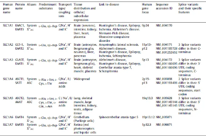
1.6 - The SLC1 family
The solute carrier family 1 (SLC1) includes five high-affinity glutamate transporters, EAAC1/SLC1A1, GLT-1/SLC1A2, GLAST/SLC1A3, EAAT4/SLC1A6 and EAAT5/SLC1A7 and the two neutral amino acid transporters, ASCT1/SLC1A4 and ASCT2/SLC1A5.
Each of these transporters exhibits different transport activities despite they have similar predicted structures.
20
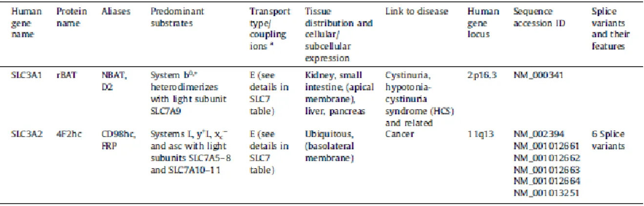
In humans, the five glutamate transporters possess 44–55% amino acid sequence identity with each other, while ASCT1 and ASCT2 exhibit 57% identity with each other (18). In Table 1, type and mechanism of transport, substrates, tissue localization and links to disease are summarized:
Table 1: SLC1: the high-affinity glutamate and neutral amino acid transporter family. (Adapted from Kanai Y.
et al, The SLC1 high-affinity glutamate and neutral amino acid transporter family, 2013)
21
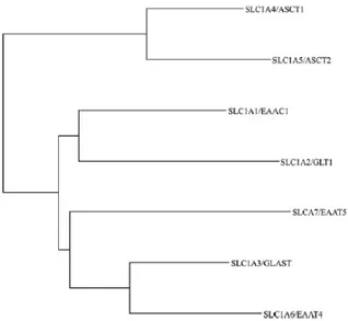
Fig. 2: Phylogenetic tree of the seven human SLC1 family members. (Adapted from Kanai Y. et al, The SLC1
high-affinity glutamate and neutral amino acid transporter family, 2013)
1.7 - ASCT2 (SLC1A5)
SLC1A5 gene was located to human chromosome 19q13.3 by chromosomal assignment studies using somatic cell hybrid analysis and fluorescent in situ hybridization.
The human SLC1A5 cDNA is 2,885 bp long with an open reading frame of 1,626 bp (including termination codon).
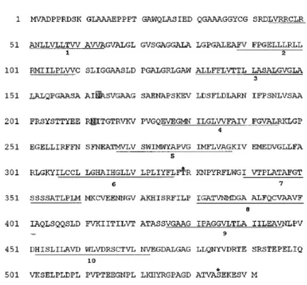
The open reading frame of 1,626 bp encodes a polypeptide of 541 amino acids that has a molecular mass of 57 kDa, called ASCT2 and also known as AAAT or hATB0. Hydrophobicity analysis indicated that the ASCT2 protein contains ten putative transmembrane domains (Fig. 3). There are also two potential N-glycosylation sites between the transmembrane domains 3 and 4 and two potential sites for protein kinase C-dependent phosphorylation in putative intracellular domains.
22
Fig. 3: Amino acid sequence of hASCT2.Putative trans membrane domains are underlined. Sites for N-linked glycosylation (shaded) and protein kinase C-dependent phosphorylation (asterisk) are indicated. (Adapted from Kekuda R. et al., Cloning of the Sodium-dependent, Broad-scope, Neutral Amino Acid Transporter B0 from a Human Placental Choriocarcinoma Cell Line, 1996).
The amino acid sequence of hASCT2 shows 61% identity and 77% similarity to human ASCT1. hASCT2 amino acid sequence shows 40% identity and 65% similarity to human glutamate transporters (19).
hASCT2 transports L-alanine, L-serine, L-cysteine and L-threonine, but also L-glutamine and L-asparagine at high affinity, and some other neutral amino acids with lower affinity. hASCT2 catalyzes also the glutamate transport but with low affinity; this activity is higher at
low pH. ASCT2 mediates Na+-dependent obligatory exchange of substrate amino acids, and it
has been shown to be present in the brush-border membranes of proximal tubule cells (kidney) and enterocytes (intestine). ASCT2 has been found also to be a retrovirus receptor (20). hASCT2 mRNA (2.9 kb) has been shown to be expressed in placenta, lung, kidney,
23
pancreas, skeletal muscle and human colon carcinoma cell lines (19). Further genomic studies by Northern blot analysis showed its expression in a human kidney proximal tubule cell line and reverse transcriptase-polymerase chain reaction analysis showed its expression in human intestinal epithelia (8,21).
1.8 - SLC7 and SLC3 families
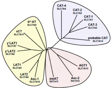
The SLC7 family is divided into two subgroups, the cationic amino acid transporters (CATs, SLC7A1–4 and SLC7A14) and the light chains or catalytic subunits (L-type amino acid transporters (LATs), SLC7A5-13 and SLC7A15) of the heteromeric amino acid transporters (HATs) (Fig. 4).
Fig. 4: Phylogenetic tree of SLC7 family members. The SLC7 family is composed of the CATs and the light
subunits of HATs. (Adapted from Fotiadis D. et al., The SLC3 and SLC7 families of amino acid transporters, 2013).
LATs are also named glycoprotein-associated amino acid transporters. The SLC3 family, instead, includes the associated heavy subunits (glycoproteins) 4F2hc (SLC3A2) or rBAT (SLC3A1) of HATs.
24
CATs are facilitated diffusers for cationic amino acids and play an important role in nitric oxide synthesis. These transporters are N-glycosylated and have 14 putative transmembrane domains (TMDs) (22).
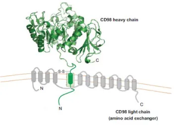
On the other hand, LATs are not N-glycosylated and only have 12 TMDs (23). CATs and HATs, originate from an ancestral protein with 12 transmembrane domains and that duplication of the last two domains of this protein, happened about 2.6 billion years ago, is the origin of the CAT structure with 14 domains, according to sequence analyses. Homologous CAT and LAT proteins are also found in prokaryotes, but the cysteine residue of the LATs that is involved in the disulfide bridge with the heavy subunit is not conserved. The cysteine residue involved in the disulfide bridge between the light chain and the heavy chain is located between TMD III and IV of LATs (Fig. 5). HATs are mostly exchangers with a wide spectrum of substrates and they are disulfide-linked heterodimers of SLC3 members and eukaryotic LATs from the SLC7 family.
Six different LATs form heterodimers with 4F2hc (the heavy chain of the 4F2 antigen): LAT1 (SLC7A5), LAT2 (SLC7A8), y+LAT1 (SLC7A7), y+LAT1-2 (SLC7A6) and the cystine/glutamate antiporter xCT (SLC7A11) and ASC-1 (SLC7A10).
25
Fig. 5: Model of human heterodimer 4F2hc/LAT1 proteins. (Adapted from Fotiadis D. et al., The SLC3 and
SLC7 families of amino acid transporters, 2013).
On the contrary, only the amino acid transporter b0,+AT (SLC7A9) forms heterodimers with
rBAT. The two members of the SLC3 family (Table 2): rBAT (SLC3A1, also named D2 and NBAT) and 4F2hc (SLC3A2, also named CD98hc and FRP, for fusion regulatory protein) share about 20% identity.
Table 2: SLC3 – heavy subunits of the heteromeric amino acid transporters. (Adapted from Fotiadis D. et al.,
26
4F2hc and rBAT are N-glycosilated and molecular weight for the mature glycosylated forms is ~85 kDa for 4F2hc and ~94 kDa for rBAT. They have an intracellular N-terminus, a single TMD, and a large extracellular C-terminus (50–60 kDa) (24); the ectodomain of human 4F2hc has been solved at 2.1 Å resolution (25). The cysteine involved in the disulfide bridge is four to five amino acids away from the TMD. The main function of the heavy subunit is the trafficking of the transporter to the plasma membrane, but 4F2hc can also be involved into β-integrin signaling, cell fusion and cell proliferation (26).
1.9 - LAT1 (SLC7A5)
LAT1 (SLC7A5) was the first cloned light chain of heteromeric amino acid transporters (27,28). The hLAT1 (NM 003486.5) amino acid transporter consists of 507 amino acids (Fig.
6) with a molecular weight of ∼55 kDa (28), but its apparent molecular mass in SDS gels is
~40 kDa due to its increased hydrophobicity (5).
Fig. 6: Amino acid sequence of hLAT1.Potential tyrosine kinase-dependent phosphorylation site and protein kinase C-dependent phosphorylation sites are labeled with # and *, respectively. (Adapted from Kanai Y. et al., Expression Cloning and Characterization of a Transporter for Large Neutral Amino Acids Activated by the Heavy Chain of 4F2 Antigen (CD98), 1998).
27
Heteromeric transporter 4F2hc/LAT1 drives the sodium-independent obligatory exchange with 1:1 stoichiometry of large neutral amino acids such as leucine, isoleucine, valine, phenylalanine, tyrosine, tryptophan, methionine, and histidine. The LAT1 affinity for large neutral and aromatic amino acids is up to 100-fold higher on the cytosolic side of the transporter compared to the extracellular side. LAT1 transfers one amino acid out of the cell and at the same time another amino acid molecule is transported into the cell and is sensitive to 2-aminobicyclo-(2,2,1)-heptane-2-carboxylic acid (BCH), a specific inhibitor of the system L (24). LAT1 is involved in the transport of several drugs such as L-DOPA, melphalan, baclofen, 3-O-methyldopa, alpha-methyltyrosine, gabapentin, alphamethyldopa, thyroid hormones, pregabalin and gabapentin (29). Its activity also not influenced by extracellular pH (30).
SLC7A5 gene coding for hLAT1 is expressed in the placenta > the brain > the spleen > the testes and the colon (27). Experiments using anti-LAT1 antibodies demonstrated that LAT1 is mainly expressed in microvessels of the central nervous system, where it is involved in the transport of L-3,4-dihydroxyphenylalanine (L-DOPA) across the blood–brain barrier (31), in the inner blood-retinal barrier, where it plays an important role in maintaining large neutral amino acids and neurotransmitters (32), as well as in placental membranes feeding with thyroid hormones and amino acids the fetus and the placenta (24).
No splice variants of LAT1 transporter have been identified. However, 352 single nucleotide polymorphisms (SNPs) have been described in the SLC7A5 gene. Only five SNPs are in the coding region; three are synonymous SNPs and two non-synonymous (rs1060250 and rs17853937). The non-synonymous SNP rs1060250 (N230K) didn’t show any functional implication (33) while is still not clear the relationship between rs17853937 (D223V) polymorphism and the protein activity. In 2006, an amino acidic substitution in position 41
28
(G41D) of the LAT1 gene has been described in a phenylketonuric patient population. This mutation could be involved in the exceptionally mild clinical course of the disease in some patients (30).
1.10 - 4F2hc (SLC3A2)
The 4F2 heavy chain (4F2hc, also called CD98 in mice) gene has been identified on chromosome 11 and seems to be more ubiquitously expressed than other human heavy chain rBAT (6). The 4F2hc glycoprotein can bind with many light chains to form different transporters, and its main role is the trafficking of the complex to the membrane. The 4F2hc protein is comprised of 630 amino acid residues, it is heavily glycosylated, resulting in an apparent molecular mass of ~85 kDa. The light subunits are connected to the 4F2hc heavy subunit by a disulfide bridge (27).
The two cysteines involved in the disulfide bridge, are located just outside the plasma membrane of the 4F2hc protein and in the extracellular loop between transmembrane helix 3 and 4 of all light subunits (Fig. 7) (34).
Fig. 7: schematic representation of CD98/light chain. (Adapted from Cantor J.M. et al., CD98 at the crossroads
29
1.11 - hASCT2 and hLAT1 in cancer
A constant nutrients supply is required by tumors to support their characteristic unabated growth. In fact, tumor cells can consume more nutrients than required for their own metabolic needs, and exhibit different metabolic profiles compared to normal cells. These differences provide the overarching theme in the “tumor metabolome” definition. The nutrients supply of tumors occurs through the collective processes of angiogenesis and the portentous increased expression of nutrient transporters in the plasma membranes of constituent cells. Compared to normal cells or tissues, cancer cells display improved and altered amino acids channeling. Amino acids are the primary source of cellular nitrogen, used for a wide range of cellular biosynthesis mechanisms such as nucleotide, glutathione and protein synthesis. In addition to their metabolic utility, amino acids also play a regulatory role in modulate growth, mainly through signaling to the energy, nutrient and growth factor integrating kinase mammalian target-of-rapamycin (mTOR). Given their metabolic importance, it is not surprising that amino acids are taken up at accelerated rates by growing tumors (9).
In 1990, before amino acid transporter isolation, Christensen proposed that specific amino acid carriers could be upregulated to support the high levels of protein synthesis necessary for growth and proliferation of cancer cells (35).
In 2005, Fuchs B.C. and Bode B.P., using the “cDNA Virtual Northern” tool of the Cancer
Genome Anatomy Project (CGAP) website (http://cgap.nci.nih.gov), found ASCT2 and LAT1
expression levels upregulated three-fold (collectively) in a variety of cancerous tissues where their expression pattern is almost identical (Table 3).
30
Table 3: expression level of ASCT2 and LAT1 mRNA (ESTs) in normal and cancerous specific human tissues.
(Adapted from Fuchs B.C. et al., Amino acid transporters ASCT2 and LAT1 in cancer: Partners in crime?, 2005).
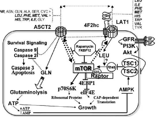
Based on the available data , both LAT1 and ASCT2 are portentously expressed in primary human cancers and several cancer cell lines, where they have been shown to be a key role in growth and survival. To date it is still unknown why these amino acid transporters are craved by tumors cells. Fuchs B.C. and Bode B.P. hypothesized that LAT1 is so important to the transformed cells because it provides the essential amino acids to enhance growth in cancer cells via mTOR-stimulated translation, whereas ASCT2 is a key role player in maintaining the cytoplasmic amino acid pool and it is required to drive LAT1 function, suppresses apoptosis, and supplies energetic fuel via glutamine delivery (Fig. 8).
31
Fig. 8: relationship of ASCT2, LAT1 and mTOR in the growth and survival of cancer cells. (Adapted from
Fuchs B.C. et al., Amino acid transporters ASCT2 and LAT1 in cancer: Partners in crime?, 2005).
Among the ASCT2 high affinity substrates, represented by small neutral amino acid, there is glutamine which is highly consumed by tumor cells. Both glutamine and ASCT2 have been shown to inhibit apoptosis through independent but overlapping pathways. While, among intermediate affinity ASCT2 substrates, a lot of LAT1 essential amino acids are included (in
bold; Fig. 9). LAT1 functions as a heterodimer with 4F2hc delivering mostly essential amino
acids to the cytoplasm. Both ASCT2 and LAT1 are obligate exchangers, and may share substrates for import/export in a transport cycle (double headed dashed arrows) necessary to equilibrate cytoplasmic amino acid pools. The mTOR kinase integrates signaling from growth factors, energy metabolism, and nutrients (especially LAT1 amino acids), to regulate translation via phosphorylation of key regulatory proteins (9). Recent data suggest reciprocal stimulatory links between ASCT2, LAT1 and mTOR. Loss of ASCT2 expression or
32
deprivation of LAT1 essential amino acids inhibit mTOR signaling; reciprocally, rapamycin inhibits mTOR activity and down-regulates ASCT2 and LAT1 expression.
Target-of-rapamycin (TOR) is a member of the phosphatidylinositol-3-kinase-related kinase (PIKK) family and represents a serine/threonine kinase, evolutionarily high conserved, that integrates signaling from growth factors, energy status and nutrients, especially amino acids (36). Rapamycin is an immunosuppressant that blocks T-cell proliferation by arresting cells in G1 phase. When complexed with the prolyl isomerase FK506 binding protein (FKBP12), rapamycin binds to mammalian TOR (mTOR) inhibiting its activity. In mammalian cells, mTOR senses intracellular amino acid status through an unknown mechanism, and regulates translation by phosphorylating key regulatory proteins. Both mTOR-mediated phosphorylation events stimulate the translation of specific classes of mRNA into growth-related proteins. Thus, mTOR main function is to regulate growth in cells. Because of its role in growth, rapamycin and related analogs able to inhibit mTOR, are being developed as potential cancer therapies. However, recent evidence suggests that not all mTOR-regulated functions are rapamycin-sensitive, including the trafficking of 4F2hc-associated amino acid transporters to the plasma membrane (9).
1.12 - Heterologous protein production
Heterologous protein production is a particular approach used to amplify the yield of a desired protein target. To date, this is not a well established process, because an optimal protein production experiment is based on different parameters and many of these are still poorly understood. This kind of experiment is much more challenging for eukaryotic proteins and membrane proteins. Many eukaryotic membrane proteins are targets for different drugs and they are implicated in diseases as cancer, cystic fibrosis, epilepsy, hyperinsulinism, heart
33
failure, hypertension and Alzheimer’s disease. All this makes them potential target for structural and functional characterization. For this purpose, large amounts of pure protein, and hence high production levels in a particular host, are required (37).
To understand how membrane proteins function and how their activity can be modulate by specific drugs, experiments on their structures and function are essential prerequisites.
The pharmaceutical industry is directly involved in this kind of study, to produce new drugs that can bind membrane proteins modulating their activity.
To date, high-resolution structures are available for a wide spectrum of soluble proteins, but three-dimensional structures have been described for only 34 membrane proteins, and most of them have prokaryotic origin, with only five being mammalian membrane proteins. These particular MPs were crystallized for their natural abundance, avoiding all the difficulties associated with heterologous overexpression. Furthermore, the most of medically and pharmaceutically relevant MPs are present in natural tissues at very low concentration, making heterologous overexpression in host cells an important prerequisite for large-scale production and structural studies. In general, mammalian MPs are more difficult to purify in large scale compared to prokaryotic MPs, due to the need to produce them in heterologous systems to achieve large amounts of protein (38).
1.13 - Heterologous systems
Studying the structure and the function of membrane proteins, the first aspect to clarify is which heterologous system should be used for the production of the protein target (39). There are many heterologous systems used for the production of eukaryotic integral membrane proteins; commonly one is the prokaryotic system (bacteria) and three are eukaryotic systems
34
(yeast, insect cells, and mammalian cells). The choice of the best expression system for the protein target remains highly empirical (40).
1.13.1 - Bacteria
Often prokaryotic homologues proteins can be expressed in bacteria in large amount, thus they have been most amenable for obtaining structural data on membrane proteins. However, this approach can not easily be applied to mammalian proteins, since these proteins are mainly expressed in inclusion bodies, from which they need to be purified under denaturing conditions.
For this reason, derivatives of E. coli BL21(DE3) strain, CD41(DE3) and CD43(DE3), selected to grow to high cell density and to overproduce proteins without any toxic effect for the host cell, have been successfully exploited for mitochondrial MPs production (38).
In general, among many systems available for heterologous protein production, the Gram-negative bacterium Escherichia coli is one of the most used. E. coli can grow rapidly and at high density on inexpensive media, has well-characterized genetics and an large number of cloning plasmids and mutant host strains are available. A lot of work has been directed at improving the performance and versatily of this system, despite there is no guarantee that a recombinant protein product will be found in E. coli at high level in a full-length and active form (41).
1.13.2 - Yeast
The two yeast systems most commonly used for protein production are Saccharomyces
35
parameters necessary for membrane protein expression, and a greater variety of different strains is available. On the other hand, P. pastoris offers a tightly regulated inducible expression system. In general, yeast has several advantages as host system for the eukaryotic protein expression compared to prokaryotic system. Like bacteria, yeast can be genetically manipulated, its genome is very well characterized, can be easily cultured, and can be grown at very high density. On the other hand, yeast is an eukaryotic host and have protein processing and post-translational modification mechanisms related to those found in mammalian cells (40).
S. cerevisiae has been successfully used to functionally produce and purify several
mammalian membrane proteins. The methylotrophic yeast, P. pastoris, has been used for the production of more than 300 heterologous proteins and as a tool for large-scale recombinant soluble protein production (42). In high cell density cultures, ethanol (a product of S.
cerevisiae fermentation) becomes toxic and limits further growth and foreign protein
production. P. pastoris, can be cultured at very high densities (500 OD600 U/mL) in the
controlled environment of a fermenter thanks to its preference for respiratory growth. Furthermore, using P. pastoris, foreign genes are stably integrated in single or multiple copy behind the AOX1 (alcohol oxidase 1) promoter, one of the strongest, most regulated promoters known (38).
1.13.3 - Insect cells
The baculovirus expression system (BVES) was found studying insect pests. To date, this system is commonly used as a method for protein expression in insect cells, while agricultural applications are limited. The advent of serum-free media and the use of intermediate-scale
36
shaker suspension systems have made BVES a very accessible system, despite it is more expensive than yeast.
However, insect cells are simpler to grow compared to mammalian cells and at the same time, they offer a membrane composition and post translation modifications closer to those of mammalian cells than yeast (40).
1.13.4 - Mammalian cells
Mammalian, and mainly human, cells offer the most native cellular environment for the production of membrane proteins that are associated with human physiology and disease. In the past, their use in structural studies had been limited by the high costs and experimental approaches of culturing and transfecting.
Traditionally, mammalian expression systems are based on the isolation of stable transformants and on the use of viral vectors to transfect the host cells (40).
1.14 - Escherichia coli and Pichia pastoris
To date, two different heterologous systems are particularly well represented in the scientific literature reporting on recombinant protein production.
Escherichia coli was the first host to be used for this purpose almost 40 years ago. Within the
past 15 years, Pichia pastoris has successfully entered the scene and is now the second most-used host for recombinant protein production. On the basis of the PubMed citation database, the use of P. pastoris as an heterologous system has increased from 4% to 17%, from 1995 to
2009. Within the same time period, the usage of E. coli as an expression host remained
37
tend to form in E. coli inclusion bodies and the protein yield is low. This because of the rate of gene translation in E. coli is 4- to 10-fold higher than in eukaryotes. E. coli should not be used as heterologous system if posttranslational modifications (PTMs) are important for the study because these microorganisms are unable to incorporate PTMs, such as N-linked glycan chains (43).
Many important elements are essential in the design of recombinant expression systems. Heterologous expression is normally induced from a plasmid with some genetic elements: origin of replication (ori), an antibiotic resistance marker, transcriptional promoters, translation initiation regions (TIRs) as well as transcriptional and translational terminators. A strong transcriptional promoter to control gene expression is required for protein production in a heterologous system. Promoter can be activated thermal or chemical and the most common inducer is the sugar molecule isopropyl-beta-D-thiogalactopyranoside (IPTG) (Fig.
9).
Fig. 9: isopropyl-beta-D-thiogalactopyranoside (IPTG).
One of the most used expression system in E. coli is the pET expression system. More than 40 different pET plasmids are available. The system consists of hybrid promoters, multiple cloning sites for the incorporation of different fusion tags and protease cleavage sites. Protein production of the target requires a host strain lysogenized by a DE3 phage fragment, encoding the T7 RNA polymerase, under the control of the IPTG inducible lacUV5 promoter (Fig. 10). LacI represses the lacUV5 promoter and the T7/lac hybrid promoter on the expression
38
plasmid. T7 RNA polymerase is transcribed when IPTG binds and triggers the release of tetrameric LacI from the lac operator. Transcription of the target gene from the T7/lac hybrid promoter (repressed by LacI as well) is, at this point, started by T7 RNA polymerase (Fig. 10) (44).
Fig. 10: The pET expression system. A general pET plasmid configuration is shown on the left. The
macromolecular situations prior to and after induction are on the right. (Adapted from Sørensen H. P. et al., Advanced genetic strategies for recombinant protein expression in Escherichia coli, 2004).
E. coli has a wide spectrum of advantages and many of these are also offered by P. pastoris, a
methylotropic yeast that can metabolize methanol and use it as its only carbon source. On the other hand, P. pastoris folds most eukaryotic proteins more efficiently and forms disulfide bonds correctly.
Pichia pastoris is an eukaryotic system and it has many of the advantages of higher
eukaryotic expression systems such as protein processing, folding, and posttranslational modification; at the same time it is easy to manipulate as Escherichia coli or Saccharomyces
cerevisiae and generally gives higher expression levels. All these properties make Pichia very
useful as a protein expression system.
When P. pastoris grows in methanol-containing medium the promoter of the alcohol oxidase I (AOX1) gene is upregulated. This strong and tightly regulated promoter is incorporated into the majority of vectors used for expression of recombinant genes in P. pastoris (43).
39
The metabolism of methanol starts with the oxidation of methanol to formaldehyde using molecular oxygen by the enzyme alcohol oxidase. This first reaction generates also hydrogen peroxide. This compound is toxic, and for this reason, methanol metabolism takes place within a specialized cell organelle, named peroxisome. Alcohol oxidase enzyme has low
affinity for O2, and Pichia pastoris compensates by producing large amounts of the enzyme.
The promoter involved in alcohol oxidase production is the one used to drive heterologous protein expression in Pichia.
In particular, two genes in Pichia pastoris code for alcohol oxidase, and they are called AOX1 and AOX2. The alcohol oxidase activity is mainly due to the product of the AOX1 gene and its production is tightly regulated and induced by methanol. The AOX1 gene has been isolated and a plasmid with AOX1 promoter is used to drive expression of the gene target. AOX2 gene is about 97% homologous to AOX1, and when only AOX2 is expressed growth on methanol is much slower than with AOX1.
Linear DNA obtained after plasmid digestion, can generate very stable transformants of
Pichia pastoris via homologous recombination between the plasmid DNA and regions of
homology within the genome. Multiple insertion events occur spontaneously during the
homologous recombination and the Fig. 11 shows the insertion mechanism into the genome and the result of multiple insertions to the AOX1 locus (45).
40
Fig. 11: Recombination and integration in Pichia pastoris genome. (Adapted from Invitrogen EasySelect Pichia
Expression Kit, 2010).
1.15 - Codon bias
The genetic code uses 61 nucleotide triplets (codons) to encode 20 amino acids and three codons to terminate translation (STOP codons); it means that each amino acid could be encoded by more than one different but synonymous codons (methionine and tryptophan being the only exception). These codons are ‘read’ in the ribosome by complementary tRNAs that have been charged with the appropriate amino acid. The ability of the genetic code to encode the same amino acid with different codons is defined as “degeneracy of the genetic code”. The degeneration of the genetic code allows the same protein to be encoded by alternative nucleic acid sequences (46). Although the same amino acid could be encoded by different codons, each organism has its own preferred codon for each individual amino acid (37). This is called codon bias.
41
There is a high variability in the frequencies with which different codons are used between different organisms, between proteins expressed at high or low levels within the same organism, and sometimes even within the same operon (47).
Andersson and coworkers have hypothesized that codon biases reduce the diversity of isoacceptor tRNAs reducing the metabolic load. This results in a benefit for the organisms that spend part of their lives under rapid growth conditions (48). The reasons for codon bias are still unknown, but it has become increasingly clear that codon biases can have a profound impact on the heterologous expression of proteins (46).
In fact, more efficient translation can be obtained when adapting a gene sequence using the preferred codons of the host organism. Basically, there are two different ways to increase the final protein yield: optimize the consensus sequence around the starting ATG to ensure efficient translation initiation as well as optimize all the codons of the gene target to be host specific (37).
The gene expression levels are modulate and influenced by a wide variety of factors. The
native gene coding for the protein target, during heterologous expression, contains tandem rare codons that can reduce the efficiency of translation or block the translational machinery. For this reason, to improve the heterologous protein production, can be useful obtaining and using the optimezed gene to be host specific, that can achieve the highest possible level of production. During codon optimization many parameters are considered such as codon usage, GC content, inhibition of splicing and prevention of stable mRNA secondary structures.
CHAPTER 2
MATERIALS AND METHODS
43
2.1 - Materials
2.1.1 - Low Salt LB (Luria-Bertani) medium
• 1% Tryptone • 0.5% Yeast extract • 0.5% NaCl
These compounds were dissolved in water and the pH of the solution was adjusted to 7.5 with 1 N NaOH.
2.1.2 - YT 2X medium
• 1.6% Tryptone • 1% Yeast extract • 0.5% NaCl
These compounds were dissolved in water, the pH of the solution was adjusted to 7.5 with 1 N NaOH and autoclaved for 20 minutes on liquid cycle.
2.1.3 - Yeast Extract Peptone Dextrose Medium (YPD)
1% Yeast extract 2% Peptone from meat 2% Dextrose (glucose)
44
2.1.4 - Yeast Extract Peptone Dextrose Medium (YPDS)
1% Yeast extract 2% Peptone from meat 2% Dextrose (glucose) 1 M Sorbitol
These compounds were dissolved in water and autoclaved for 20 minutes on liquid cycle.
2.1.5 - Buffered Glycerol-complex Medium (BMGY)
1% Yeast extract 2% Peptone from meat
100 mM Potassium phosphate, pH 6.0 1.34% YNB (Yeast nitrogen base) 4 × 10⁻⁵% Biotin
1% Glycerol
Dissolve peptone from meat and yeast extract in water. Autoclave 20 minutes on liquid cycle. Cool to room temperature, then add YNB (filter sterilized), biotin (filter sterilized), glycerol (autoclaved), potassium phosphate (autoclaved) and mix well.
2.1.6 Buffered Methanol-complex Medium (BMMY)
1% Yeast extract 2% Peptone from meat
100 mM Potassium phosphate, pH 6.0 1.34% YNB (Yeast nitrogen base)
45
4 × 10⁻⁵% Biotin 0.5% Methanol
Dissolve peptone from meat and yeast extract in water. Autoclave 20 minutes on liquid cycle. Cool to room temperature, then add YNB (filter sterilized), biotin (filter sterilized), methanol (filter sterilized), potassium phosphate (autoclaved) and mix well.
2.1.7 - Fermentation Basal Salts Medium
Phosphoric acid, 85% 26.7 mL Calcium sulfate 0.93 grams Potassium sulfate 18.2 grams Magnesium sulfate-7H2O 14.9 grams Potassium hydroxide 4.13 grams
Glycerol 40 grams
Water to 1 L
These compounds were dissolved in water, added to fermentor and autoclaved for 20 minutes on liquid cycle.
2.1.8 - Plates
To make LB, YPD or YPDS plates 2% agar was added. When the temperature is about 60°C the specific antibiotic was added and plates were stored at 4°C.
46
2.1.9 - TAE (Tris/Acetate/EDTA) 50 X
• 242 grams Tris base
• 18.6 grams EDTA (Ethylenediaminetetraacetic acid) • 60 mL Glacial acetic acid
Use acetic acid to adjust pH 8 and bring up the volume to 1 liter with water.
2.1.10 - IPTG
The isopropyl β-D-1-thiogalactopyranoside (IPTG) was resuspended in sterile water and used
in a concentration range between 0.1 mM and 1 mM.
2.1.11 - Protease Inhibitor Cocktail
Inhibits serine, cysteine, aspartic and thermolysin-like proteases, and aminopeptidases.
One mL of cocktail was used for the inhibition of proteases extracted from 20 g of
Escherichia coli cells.
2.1.12 - PMSF
The PMSF (Phenylmethanesulfonyl fluoride) inhibits serine proteases such as trypsin, chymotrypsin and cysteine proteases. Its effective concentration is between 0.1-1 mM. It was used in Pichia pastoris lysate at a final concentration of 0.5 mM.
47
2.1.13 - Running buffer for SDS-PAGE 10X
• 250 mM Tris base • 14,4% glycine • 1% SDS
These compounds were dissolved in water and the pH was adjusted to 8.3. The 1X buffer was used during the running.
2.1.14 - MES running buffer for SDS-PAGE 20X
• 1M MES (2-N-morpholino ethanesulfonic acid) • 1M Tris base
• 20 mM EDTA • 2% SDS
These compounds were dissolved in water and the pH should be 7.3. The 1X buffer was used during the running.
2.1.15 - Coomassie Brilliant Blue
• 0.25 g Coomassie Brilliant Blue • 45 mL Methanol
• 45 mL Distilled H2O • 10 mL Acetic acid
48
2.2 - Experimental procedures
2.2.1 - Polymerase chain reaction (PCR)
The polymerase chain reaction (PCR) is a biochemical technology used in many field to
amplify a single or a few copies of a piece of DNA, generating thousands to millions of copies
of a particular DNA fragment. Short DNA fragments called primers, containing sequences
complementary are designed to bind to the start and end of the DNA target, along with a DNA polymerase are key components to enable selective and repeated amplification. As PCR progresses, the DNA generated is itself used as a template for replication in which the DNA template is amplified. To perform a PCR reaction, the DNA template that contains the target sequence is mixed to primers, free nucleotides, and an enzyme called DNA polymerase. The thermocycler, a particular machine, increases and decreases the temperature of the sample in automatic. In the first step, the mixture is heated to separate the double-stranded DNA template into single strands. The mixture is then cooled so that the primers can bind to the DNA template. At this point, the DNA polymerase begins to synthesize new strands of DNA starting from the primers. Following synthesis and at the end of the first cycle, each double-stranded DNA molecule consists of one new and one old DNA strand. PCR then continues with additional cycles (usually 25-35 cycles) that repeat the previous steps. The reaction mixture consist of:
• 10 μL buffer 5 X (containing MgCl2 1.5 mM);
• 1 μL dNTP mix (0.2 mM dATP, 0.2 mM dCTP, 0.2 mM dGTP, 0.2 mM dTTP);
• 0.3 μM Forward primer;
• 0.3 μM Reverse primer;
49
• 0.6 μL di Phusion High Fidelity Finnzymes 2 u/μL
• H2O nuclease-free to 50 μL
Tipical thermal cycling conditions :
Initial denaturation
Denaturation Annealing Extension Final
extension 96 °C 96 °C 45-65 °C 72 °C 72 °C 4 °C 5' 30'' 30'' 90'' 10' ∞ 30-40 Cicli
2.2.2 - Agarose gel electrophoresis
Agarose gel electrophoresis is a method used in molecular biology, to separate a population of
DNA in a matrix of agarose. The DNA can be separated by length applying an electric fieldto
move the negatively charged molecules through an agarose matrix. Most agarose concentration used is between 0.7% (gives good separation or resolution of large 5–10kb DNA fragments) and 2% (gives good resolution for small 0.2–1 kb fragments). Agarose is dissolved in a suitable electrophoresis buffer such as Tris/Acetate/EDTA (TAE) or Tris/Borate/EDTA (TBE).
Agarose gels are normally stained with ethidium bromide, which intercalates into the major grooves of the DNA and fluoresces under UV light. Other safer methods of staining are SYBR Green or methylene blue. The gel stained with ethidium bromide is viewed with an
50
ultraviolet (UV) transilluminator while SYBR Green requires the use of a blue-light transilluminator.
2.2.3 - Purification of DNA fragments from agarose gel
The DNA fragments separated by agarose gel were isolated and purified using the gel extraction kits Wizard SV Gel and PCR Clean-Up System (Promega). These quick protocol are simple to perform, and the PCR products are purified from contaminants, including primer dimers, PCR additives and amplification primers. Protocols included in these kits generally call for the dissolution of the gel-slice in 3 volumes of chaotropic agent at 50-60°C to dissolve the agarose, freeing the DNA for binding to the silica membrane. Then, the solution is applied to a spin-column (DNA remains in the column). Washing with ethanol 70% (the DNA remains in the column, salt and impurities are washed out), and elution of the DNA in a small volume (20 µL) of nuclease-free water or elution buffer free of salt or macromolecular contaminants, are performed. These kits can also be used to purify DNA from enzymatic reactions such as restriction digestion and alkaline phosphatase treatment.
2.2.4 - Cloning
DNA for cloning experiments can be obtained from RNA using reverse transcriptase (cDNA), or in the form of synthetic DNA. The DNA is treated with restriction enzymes to generate fragments with ends capable of being linked to those of the vector. DNA and vector, treated with same enzymes, are simply mixed together at appropriate concentrations (a molar ratio of 1:3 vector to insert) and exposed to DNA Ligase (T4 DNA ligase in our case) that covalently links the ends together. This reaction is termed ligation.
51
A typical ligation protocol is shown below:
COMPONENT VOLUMES
Vector DNA (3 kb) 50 ng
Insert DNA (1 kb) 50 ng
Buffer T4 DNA Ligase 10X 2 µL
T4 DNA Ligase 1 µL
Nuclease-free water to 20 µL
2.2.5 - E. coli transformation
Transformation is the process of getting the recombinant vector or empty vector into host cells. To enable the cells to take up circular vector DNA they have to be made competent. The method for the preparation of competent cells depends on the transformation method.
E. coli cells were grown in 100 mL LB medium overnight at 37°C to OD600~ 0.4-0.6. The
cells were centrifuged at 4000 rpm for 15 minutes at 4°C and the pellet resuspended with 50
mL ice cold CaCl2 50 mM and stored on ice for 30 minutes. At this point, the resuspension
was again centrifuged and the pellet resuspended with 2 mL of CaCl2 50 mM.
170 µL of competent cells were mixed with ligation mix (10 µL) or plasmidic DNA (100 ng) and incubated for 30 minutes on ice. The mixture was heat shocked at 42°C for 90 seconds and returned on ice for 2 minutes. At this point, 170 µL LB medium were added and the mixture was incubated for 1 hour at 37°C with shaking. The cells were plated on LB agar plates containing appropriate antibiotics.
52
2.2.6 - DNA extraction by QIAprep Spin Miniprep kit
The QIAprep miniprep procedure (Qiagen) is based on alkaline lysis of bacterial cells followed by adsorption of DNA onto silica membrane in the presence of high concentration of chaotropic salt. Bacteria are lysed under alkaline conditions, and the lysate is subsequently neutralized and adjusted to high-salt binding conditions. After lysate clearing, the sample is ready for purification on the QIAprep silica membrane. The protocol is designed for
purification of up to 20 μg of high-copy plasmid DNA from 1–5 mL overnight cultures of E.
coli in LB medium. The bacterial cells pellet is resuspended in 250 μL Buffer P1 and transfer
to a microcentrifuge tube. 250 μL Buffer P2 were added and mixed by inverting the tube 4–6
times.Then, 350 μL Buffer N3 were added and mixed immediately by inverting the tube 4–6
times. The mixture was centrifuged for 10 min at 13,000 rpm in a table-top microcentrifuge.
The supernatant was loaded to the QIAprep spin column by pipetting and centrifuged for 60 seconds. The column was first washed by adding 0.5 mL Buffer PB and centrifuged for 60 seconds and then washed by adding 0.75 mL Buffer PE and centrifuged for 60 seconds. The column was centrifuged at full speed for an additional 1 min to remove residual wash buffer
containing ethanol. The DNA was eluted adding 50 μL Buffer EB or nuclease-free water to
column and centrifuging for 3 minutes.
2.2.7 - P. pastoris transformation
P. pastoris cells were transformed by electroporation. To prepare cells for transformation,
Pichia pastoris X33 strain was grown in 10 mL YPD at 30°C overnight. This culture was
used to inoculate 250 mL of fresh YPD medium and grown to an OD600 = 1.3–1.5. The cells
53
ice-cold sterile water. The cells were centrifuged as in previously step and resuspended with 125 mL of ice-cold sterile water. At this point, the cells were centrifuged at 1,500 × g for 5 minutes at 4°C and the pellet was resuspended with 10 mL of ice-cold sterile 1M sorbitol. The
cells were centrifuged again and resuspended with 400 μL of ice-cold sterile 1M sorbitol. The
competent cells were kept on ice and used the same day.
80 μL of the cells were mixed with 5–10 μg of linearized DNA (in 5–10 μL sterile water) and
transfered to an ice-cold 0.2 cm electroporation cuvette. The cuvette was incubated with the
cells on ice for 5 minutes. The cells were pulsed using 2.2 kV, 25 μF and 200 ohm by
electroporator and immediately 1 mL of ice-cold 1 M sorbitol was added to the cuvette. The cuvette content was transferred to a sterile 15-mL tube and inbubated at 30°C without shaking for 1 to 2 hours.
50, 100, 200 and 450 μL each were spread on separate, labeled YPDS plates containing 100
μg/mL Zeocin™ and incubated 3 days at 30°C until colonies form. A total of 52 transformants were screened for growth on YPDS plates containing a high concentration of
Zeocin™ (2000 μg/mL) and analyzed after 2 days.
2.2.8 - Sonication
Sonication is the process of converting an electrical signal into a physical vibration. Sonication is usually performed to break apart compounds or cells for further examination. The vibration has a very powerful effect on solutions, causing their molecules to break apart
and cells to rupture. The laboratory method for cell disruption applies ultrasound (typically
20–50 kHz) to the sample. In principle, the high-frequency is generated electronically and the mechanical energy is transmitted to the sample via a metal probe that oscillates with high frequency. The sonication probe transmits the vibration to the sample. This probe is a
54
carefully constructed tip that moves in time with the vibration, transmitting it into the solution. The high-frequency oscillation causes a localized lot pressure region resulting in cavitation and impaction, ultimately breaking open the cells.
In general, a sonication device consists of an ultrasonic electric generator, a transducer that converts the electric signal to mechanical vibration by using piezoelectric crystals and a sonication probe.
2.2.9 - The French press
The French press, or, French pressure cell press, is an apparatus used in biological experimentation to break the plasma membrane by passing the cells through a narrow valve under high pressure. A French press is commonly used to break the plasma membrane and cell wall of yeast, bacteria and other microorganisms for isolation of proteins and other cellular components.
The press uses an external pump to drive a piston within a larger cylinder that contains the liquid sample. The motor-driven piston develops pressures up to 40,000 psi.
The highly pressurized sample is then pressed past a small valve. As the sample passes through the valve, the shear stress and decompression of the sample, cause cellular disruption. The major components of a French press are made of stainless steel to prevent sample contamination.