“International PhD program in Cardiovascular Pathophysiology and Therapeutics – CardioPaTh”
CARDIOVASCULAR RISK ASSESSMENT AND OUTCOMES: FROM CHILDHOOD TO ADULTHOOD
“Senectus ipsa est morbus?”
CARDIOVASCULAR RISK ASSESSMENT AND OUTCOMES: FROM CHILDHOOD TO ADULTHOOD
“Senectus ipsa est morbus?”
Fabio Marsico, 28/01/1984, Stigliano (Matera, Italy)
Naples, 21/01/2019, University Federico II of Naples, Faculty of Medicine, Via Pansini n. 5, 80131 Naples (Italy)
1 TABLE OF CONTENTS
Chapter 1 1. Introduction 6
1.1.Cardiovascular disease could start from childhood 6 1.2.Congenital heart disease: from childhood to GUCH 8
References 11
PART 1 16
Cardiovascular risk in adults could start from childhood: how to detect early cardiovascular risk starting from conventional and non-conventional risk factors
Chapter 2 NSAIDs and cardiovascular risk 17
Marsico F, Paolillo S, Perrone Filardi P J Cardiovasc Med 2017;18:e40-e43
Chapter 3 Multicentre multi-device hybrid imaging study of 22 coronary artery disease: results from the EValuation
of INtegrated Cardiac Imaging for the Detection and Characterization of Ischaemic Heart Disease (EVINCI) hybrid imaging population
Liga R, Vontebol J, Rovai D, Marinelli M, Caselli C, Pietila M, Teresinska A, Aguadé-Bruix S, Pizzi MN, Todiere G, Gimelli A,
Chiappino D, Marraccini P, Schroeder S, Drosch T, Poddighe R, Casolo G, Anagnostopoulos C, Pugliese F, Rouzet F, Le Guludec D, Cappelli F, Valente S, Gensini GF, Zawaideh C, Capitanio S, Sambuceti G, Marsico F, Perrone Filardi P, Fernàndez-Golfin C, Rincon LM, Graner FP, de Graaf MA, Stehli J, Reyes E, Nkomo S, Mäki M, Lorenzoni V, Turchetti G, Carpeggiani C, Puzzuoli S, Mangione M, Marcheschi P, Giannessi D, Nekolla S, Lombardi M, Sicari R, Scholte AJ, Zamorano JL, Underwood SR, Knuuti J, Kaufmann PA, Neglia D, Gaemperli O; EVINCI study Investigators
2 Chapter 4 Glucose metabolism abnormalities in heart failure 33
patients: insights and prognostic relevance Marsico F, Gargiulo P, Parente A, Paolillo S Heart Fail Clinics (in press)
Chapter 5 Efficacy and safety of glucagon-like peptide-1 59 agonists on macrovasular and microvascular
events in type 2 diabetes mellitus: a meta-analysis
Gargiulo P, Savarese G, D’Amore C, De Martino F, Lund LH, Marsico F, Dellegrottaglie S, Marciano C, Trimarco B, Perrone Filardi P
Nutr Metab Cardiovasc Dis 2017;27:1081-1088
Chapter 6 Vitamin D deficiency and clinical outcome in 68 patients with chronic heart failure: a review
D’Amore C, Marsico F, Parente A, Paolillo S, De Martino F, Gargiulo P, Ferrazzano F, De Roberto AM, La Mura L, Marciano C,
Dellegrottaglie S, Trimarco B, Perrone Filardi P Nutr Metab Cardiovasc Dis 2017;27:837-849
PART 2 82
Cardiovascular risk in children: role of HIV in cardiovascular disease
Chapter 7 Left ventricular function, epicardial adipose 83 tissue and carotid intima-media thickness in
children and young adults with vertical HIV infection
Marsico F, Lo Vecchio A, Paolillo S, D’Andrea C, De Lucia V, Bruzzese E, Vallone G, Dellegrottaglie S, Marciano C, Trimarco B, Guarino A, Perrone Filardi P
3
PART 3 104
Congenital heart disease: from childhood to adulthood
Chapter 8 Hypertrophic cardiomyopathy in mitochondrial 105 disorders: description of an uncommon clinical case
Marsico F, D’Andrea C, Parente A, De Martino F, Capasso L, Raimondi F, Paolillo S, Dellegrottaglie S, Marciano C, Trimarco B, Perrone Filardi P
Eur J Heart Fail 2017;19:1201-1204
Chapter 9 Cardiac imaging for prognostication in patients 110 with pulmonary hypertension: the accuracy of
magnetic resonance imaging versus echocardiography and baseline values versus interval changes
Marsico F, Wustmann K, Wahl A, Schwitz F, Goulouti E, Schenker S, Mülchi K, Pichler Hefti J, Geiser T, Schwerzmann M
Draft
Chapter 10.1 Predictors of cardiovascular outcomes in adult 151 patients with repaired coarctation
Marsico F, Tobler D, Greutmann M, Schwerzmann M, Wustmann K Draft
Chapter 10.2 Blood pressure profile evaluated by ambulatory 190 measurement and ambulatory blood pressure
monitoring in repaired CoA adult patients
Marsico F, Tobler D, Greutmann M, Schwerzmann M, Wustmann K Draft
Chapter 10.3 Comparison of echocardiographic and cardiac 215 magnetic resonance aortic stiffness assessment
in adult patients with repaired CoA
Marsico F, Tobler D, Greutmann M, Schwerzmann M, Wustmann K Draft
4 Curriculum vitae 240 List of publications 243 Acknowledgments 250
6 CHAPTER 1.
1. Introduction
Although the major part of cardiovascular (CV) events in childhood are characterized by congenital heart disease (CHD), there is a wide spectrum of CV events that start from conventional CV risk factors that are present also in children, arriving to atherosclerotic events, most of all in some particular populations (i.e. patients with rheumatic diseases, obese patients, patients with HIV, patients with kidney disease, etc.). On the other hand, the large use of some pharmacological agents can play a role in the development of CV events, generally atherosclerotic. This aspect, that normally involve adult patients, could affect also children. There is also a spectrum of causes related to heart failure, that generally involve adults, but that can start from childhood. However, CHD remain the most common CV presentation in childhood. Thanks to progress in surgery, a great part of these patients arrives to adulthood, where they are included in another classification called grown-up congenital heart disease (GUCH).
1.1. Cardiovascular diseases could start from childhood
Atherosclerotic CV disease remains the leading cause of morbidity and mortality worldwide, but manifest disease in childhood and adolescence is rare (1). By contrast, risk factors responsible of atherosclerosis are present since childhood. The autopsy findings in The Bogalusa Heart Study and Pathobiological Determinants of Atherosclerosis in Youth showed a strong relationship of vascular disease to traditional CV risk factors (2,3). This
7 observation provided irrefutable evidence of the importance of act on modifiable CV risk factors, such as cigarette smoking, hypercholesterolemia, hypertension, obesity and diabetes (4–6). On the other hand, there are several emerging CV risk factors, that have not been widely studied (i.e. C reactive protein, homocysteine, small dense low density lipoprotein, oxidized low density lipoprotein, apoliproteins, lipoprotein a, fibrinogen) (7), and also some non-modifiable atherosclerosic risk factors (i.e. age, gender, CV family history), that don’t permit to reduce completely the CV burden in childhood, resulting in a great CV risk in adulthood. Considered a relatively recently discovered CV risk factor, common carotid artery intima-media thickness (IMT) measured by ultrasound imaging, represents a marker of preclinical atherosclerosis, correlating with vascular risk factors (8– 10), relating to the severity and extent of coronary artery disease (8). Several studies, in particular in some specific conditions (i.e. obesity, HIV), have shown an increase of IMT also in childhood (11,12). Finally, a more recent parameter of CV risk has been studied, epicardial adipose tissue (EAT), that is part of the visceral fat deposited within the pericardic sac around the heart. EAT shares a common embryological origin with the intra-abdominal visceral adipose tissue and, as such, it is a metabolically active adipose tissue (13,14). Hence, although EAT represents only 1% of the total body fat mass, it may be important in the pathophysiology of CV disease, in particular in obese patients and in HIV patients (13,14). Well known is the role of EAT in adult patients, instead weak is known about its possible role in CV risk in childhood, in particular in HIV young patients. These CV markers play an important role in a subclinical atherosclerosis, which can lead to
8 coronary artery disease. For this reason, very important is also the role of imaging techniques to detect very early subclinical coronary artery disease, to treat as soon as possible CV disease, for a better prognosis. There is also a large part of iatrogenic CV events, that could start from childhood, but that generally are more frequent in adult patients, due to the large use of some medications in adulthood. In particular, in patients with rheumatic diseases, that could be present also in childhood, there is a wide use of non-steroid anti-inflammatory drugs (NSAIDs), that have many side effects, not only on gastrointestinal system, but also at cardiac level (15), increasing the risk of ischemic disease and heart failure. Although heart failure is typically an “older disease”, there are many situations related to its development that can start since childhood. First of all, many CHD could lead to heart failure, also HIV infection could be related to left ventricular dysfunction, but there are also many other causes of heart failure development, that go from some CV risk factors implicated not only in atherosclerotic events (i.e. diabetes) (16– 18), to a deficiency of some factors (one of the most important is vitamin D) (19). There are two points to define in these cases: first of all, these two causes can start since childhood, leading to CV events very early, and secondary, there are new treatment approach for diabetes (i.e. inhibitors of sodium glucose co-transporter-2, and glucagon-like peptide-1 agonists) (16–18) and also vitamin D supplementation (20,21), that could be responsible of a regression of CV events in these patients.
1.2. Congenital heart disease: from childhood to GUCH
9 congenital anomalies consist of heart defects (22,23). Reported birth prevalence of CHD varies widely among studies worldwide. The estimate of 8 per 1,000 live births is generally accepted as the best approximation (22,24). A reliable classification of CHD should be the one that define a cyanotic pattern, or ductal-dependent classification, considering an important role of ductus arteriosus in the survival of these patients. In the group of congenital heart disease, there are also some known mutations, that involve the heart, leading to some pattern of cardiomyopathy that sometimes are incompatible with survival. One of these are the mitochondrial disorders, which are generally associated with congenital hypertrophic cardiomyopathy, with a fatal prognosis. Without a treatment, most defects of moderate or great complexity have a bleak prognosis (25). Thanks to the progress in cardiac surgery, since many years, a great part of patients born with CHD, reach adulthood, creating a completely new and steadily growing patient population: patients with grown-up congenital heart disease (GUCH) (22). It is estimated that about 90% of affected children are expected to survive to adulthood (26), with an estimated prevalence of CHD of 4 per 1,000 adults (27). Patients with GUCH often need long-term expert medical care and healthcare-related costs are high (28). In fact, it is recognized that CHD is associated with lifelong comorbidity that affect healthcare costs. The impact of ongoing disease burden includes atrial arrhythmias, pulmonary hypertension, arterial hypertension, heart failure, cerebrovascular events, coronary events, and a repeated need for surgery, which results in significant increase in health services utilization during childhood, transition years, adulthood, and geriatric age group (29). A particular mention
10 is due to pulmonary hypertension, that is present in most part of shunt-defects, and that is responsible of a bad prognosis in these patients and arterial hypertension, that is a common complication in patients with coarctation of the aorta (CoA), and that worsen the prognosis in these patients.
11 References
1. Articles S. Expert Panel on Integrated Guidelines for Cardiovascular Health and Risk Reduction in Children and Adolescents: Summary Report. Pediatrics [Internet].
2011;128(Supplement):S213–56. Available from:
http://pediatrics.aappublications.org/cgi/doi/10.1542/peds.2009-2107C
2. Berenson GS. Cardiovascular Risk Begins in Childhood. A Time for Action. Am J Prev Med [Internet]. 2009;37(1 SUPPL.):S1–2. Available from: http://dx.doi.org/10.1016/j.amepre.2009.04.018
3. Berenson GS, Srinivasan S, Bao W, Newman WP, Tracy Ri, Wattingney W. Association Between Multiple Cardiovascular Risk Factors and Atherosclerosis in Children and Young Adults. N Engl J Med. 1998;338:1650–6.
4. Libby P. Changing concepts of atherogenesis. J Intern Med. 2000;247(3):349–58. 5. Dawber TR, Kannel WB. The Framingham study. An epidemiological approach to coronary artery disease. Circulation. 1966;34:553–555.
6. Kannel WB, McGee D, Gordon T. A general cardiovascular risk profile: The Framingham study. Am J Cardiol. 1976;38(1):46–51.
7. Fruchart J-C. New Risk Factors for Atherosclerosis and Patient Risk Assessment. Circulation [Internet]. 2004;109(23_suppl_1):III-15-III-19. Available from: http://circ.ahajournals.org/cgi/doi/10.1161/01.CIR.0000131513.33892.5b
8. Raitakari OT, Juonala M, Kahonen M, Taittonen L, Laitinen T, Maki-Torkko N, et al. Cardiovascular risk factors in childhood and carotid artery intima-media thickness in adulthood. Jama. 2003;290(17):2277–83.
12 9. Poli A., Tremoli E., Colombo A., Sirtor M., Pignoli P., \emphet al. Ultrasonographic measurement of the common carotid artery wall thickness in hypercholesterolemic patients. A new model for the quantitation and follow-up of preclinical atherosclerosis in living human subjects. Atherosclerosis. 1988;70(3):253–61.
10. Desk R, Williams L, Health K. Clinical Investigation Carotid Arteriosclerosis in Identical Twins Discordant for Cigarette Smoking. Circulation. 1989;80:10–6.
11. Charakida M, Donald AE, Green H, Storry C, Clapson M, Caslake M, et al. Early structural and functional changes of the vasculature in HIV-infected children: Impact of disease and antiretroviral therapy. Circulation. 2005;112(1):103–9.
12. Idris NS, Grobbee DE, Burgner D, Cheung MMH, Kurniati N, Uiterwaal CSPM. Effects of paediatric HIV infection on childhood vasculature. Eur Heart J. 2016;37(48):3610–6.
13. Manco M, Morandi A, Marigliano M, Rigotti F, Manfredi R, Maffeis C. Epicardial fat, abdominal adiposity and insulin resistance in obese pre-pubertal and early pubertal children. Atherosclerosis [Internet]. 2013;226(2):490–5. Available from: http://dx.doi.org/10.1016/j.atherosclerosis.2012.11.023
14. Iacobellis G, Bianco AC. Epicardial adipose tissue: emergin physiological, pathophysiological and clinical features. Trends Endocrinol Metab 2011;22:450-457. 15. Scott PA, Kingsley GH, Smith CM, Choy EH, Scott DL. Non-steroidal anti-inflammatory drugs and myocardial infarctions: Comparative systematic review of evidence from observational studies and randomised controlled trials. Ann Rheum Dis. 2007;66(10):1296–304.
13 16. Zinman B, Wanner C, Lachin JM, Fitchett D, Bluhmki E, Hantel S, et al. Empagliflozin, Cardiovascular Outcomes, and Mortality in Type 2 Diabetes. N Engl J Med
[Internet]. 2015;373(22):2117–28. Available from:
http://www.nejm.org/doi/10.1056/NEJMoa1504720
17. Savarese G, D’Amore C, Federici M, De Martino F, Dellegrottaglie S, Marciano C, et al. Effects of Dipeptidyl Peptidase 4 Inhibitors and Sodium-Glucose Linked coTransporter-2 Inhibitors on cardiovascular events in patients with type coTransporter-2 diabetes mellitus: A meta-analysis. Int J Cardiol [Internet]. 2016;220:595–601. Available from: http://dx.doi.org/10.1016/j.ijcard.2016.06.208
18. Chamberlain JJ, Rhinehart AS, Shaefer CF, Neuman A. Diagnosis and management of diabetes: Synopsis of the 2016 American diabetes association standards of medical care in diabetes. Ann Intern Med. 2016;164(8):542–52.
19. Anderson JL, May HT, Horne BD, Bair TL, Hall NL, Carlquist JF, et al. Relation of vitamin D deficiency to cardiovascular risk factors, disease status, and incident events in a general healthcare population. Am J Cardiol [Internet]. 2010;106(7):963–8. Available from: http://dx.doi.org/10.1016/j.amjcard.2010.05.027
20. Donneyong MM, Hornung CA, Taylor KC, Baumgartner RN, Myers JA, Eaton CB, et al. Risk of heart failure among postmenopausal women a secondary analysis of the randomized trial of Vitamin D plus calcium of the women’s health initiative. Circ Hear Fail. 2015;8(1):49–56.
21. Scragg R, Stewart AW, Waayer D, Lawes CMM, Toop L, Sluyter J, et al. Effect of Monthly High-Dose Vitamin D Supplementation on Cardiovascular Disease in the Vitamin
14 D Assessment Study. JAMA Cardiol [Internet]. 2017;2(6):608. Available from: http://cardiology.jamanetwork.com/article.aspx?doi=10.1001/jamacardio.2017.0175
22. Van Der Linde D, Konings EEM, Slager MA, Witsenburg M, Helbing WA, Takkenberg JJM, et al. Birth prevalence of congenital heart disease worldwide: A systematic review and meta-analysis. J Am Coll Cardiol [Internet]. 2011;58(21):2241–7. Available from: http://dx.doi.org/10.1016/j.jacc.2011.08.025
23. Dolk H, Loane M, Garne E. Congenital heart defects in Europe: Prevalence and perinatal mortality, 2000 to 2005. Circulation. 2011;123(8):841–9.
24. Bernier PL, Stefanescu A, Samoukovic G, Tchervenkov CI. The challenge of congenital heart disease worldwide: Epidemiologic and demographic facts. Semin Thorac Cardiovasc Surg Pediatr Card Surg Annu [Internet]. 2010;13(1):26–34. Available from: http://dx.doi.org/10.1053/j.pcsu.2010.02.005
25. Warnes CA, Liberthson R, Danielson GK, Dore A, Harris L, Hoffman JI, et al. Task force 1: the changing profile of congenital heart disease in adult life. J Am Coll Cardiol [Internet]. 2001;37(5):1170–5. Available from: http://dx.doi.org/10.1016/S0735-1097(01)01272-4
26. Moons P, Bovijn L, Budts W, Belmans A, Gewillig M. Temporal trends in survival to adulthood among patients born with congenital heart disease from 1970 to 1992 in Belgium. Circulation. 2010;122(22):2264–72.
27. Khairy P, Ionescu-Ittu R, MacKie AS, Abrahamowicz M, Pilote L, Marelli AJ. Changing mortality in congenital heart disease. J Am Coll Cardiol [Internet]. 2010;56(14):1149–57. Available from: http://dx.doi.org/10.1016/j.jacc.2010.03.085
15 28. Somerville J. Grown-up congenital heart disease medical demands look back, look forward. 2000;
29. Marelli AJ, Ionescu-Ittu R, Mackie AS, Guo L, Dendukuri N, Kaouache M. Lifetime prevalence of congenital heart disease in the general population from 2000 to 2010. Circulation. 2014;130(9):749–56.
16
PART 1
Cardiovascular risk in adults could start from childhood: how to detect early cardiovascular risk starting from conventional and non-conventional risk factors.
17 CHAPTER 2.
NSAIDs and cardiovascular risk
Marsico F, Paolillo S, Perrone Filardi P J Cardiovasc Med 2017;18:e40-e43
22 CHAPTER 3.
Multicentre multi-device hybrid imaging study of coronary artery disease: results from the EValuation of INtegrated Cardiac Imaging for the Detection and
Characterization of Ischaemic Heart Disease (EVINCI) hybrid imaging population
Liga R, Vontebol J, Rovai D, Marinelli M, Caselli C, Pietila M, Teresinska A, Aguadé-Bruix S, Pizzi MN, Todiere G, Gimelli A, Chiappino D, Marraccini P, Schroeder S, Drosch T, Poddighe R, Casolo G, Anagnostopoulos C, Pugliese F, Rouzet F, Le Guludec D, Cappelli F, Valente S, Gensini GF, Zawaideh C, Capitanio S, Sambuceti G, Marsico F, Perrone Filardi P, Fernàndez-Golfin C, Rincon LM, Graner FP, de Graaf MA, Stehli J, Reyes E, Nkomo S, Mäki M, Lorenzoni V, Turchetti G, Carpeggiani C, Puzzuoli S, Mangione M, Marcheschi P, Giannessi D, Nekolla S, Lombardi M, Sicari R, Scholte AJ, Zamorano JL, Underwood SR, Knuuti J, Kaufmann PA, Neglia D, Gaemperli O; EVINCI study Investigators
33 CHAPTER 4.
Glucose metabolism abnormalities in heart failure patients: insights and prognostic relevance
Marsico F, Gargiulo P, Parente A, Paolillo S Heart Fail Clinics (in press)
34 Glucose metabolism abnormalities in heart failure patients: insights and prognostic
relevance
Fabio Marsicoa, MD, Paola Gargiuloa, MD, PhD, Antonio Parentea, MD, Stefania Paolilloa, MD, PhD
a
Department of Advanced Biomedical Sciences, University Federico II, Via Pansini 5, 80131, Naples, Italy
Email addresses:
Fabio Marsico: [email protected]
Paola Gargiulo: [email protected]
Antonio Parente: [email protected]
Stefania Paolillo: [email protected]
Address for correspondence:
Fabio Marsico, MD Via Pansini, 5
I-80131, Naples, Italy
35 Conflict of interest: None
Financial Disclosure: None
Synopsis
Heart failure is a clinical syndrome characterized by left ventricular dysfunction and/or elevated intracardiac pressures, with a prevalence of about 1 – 2%. In the last decades, many metabolic disorders have been studied as linked with heart failure, in particular glucose metabolism abnormalities. Diabetes mellitus and insulin resistance are strictly related with heart failure, with a bidirectional link, where each one can influence the other. The aim of this review is to report the role of glucose metabolism abnormalities in the development of HF, defining the epidemiology, and assessing pathophysiology and prognosis of HF related to glucose metabolism disorders.
Key words: Heart failure; Glucose metabolism abnormalities; Diabetes mellitus; Insuline resistance; Hyperglycemia; Insulin sensitivity.
36 Key points
Heart failure is a clinical syndrome characterized by left ventricular dysfunction and/or elevated intracardiac pressures, with a prevalence of about 1 – 2%
There are several factors involved in HF development, that go from ischemic heart disease to metabolic disorders, passing through genetic etiology
Although the most known cardiac nosological entity related to glucose metabolism disorders is diabetic cardiomyopathy, insulin resistance is a very frequent finding among HF patients
the homeostasis model assessment (HOMA) index has proved to be robust and reliable tool for the assessment of insulin resistance
Heart failure can be considered the major cause of hospitalization in patients with glucose metabolism abnormalities
37 Introduction
Heart failure (HF) is a clinical syndrome caused by structural and/or functional cardiac abnormality, resulting in a reduced left ventricular function, and/or elevated intracardiac pressures.1 The prevalence of HF is approximately 1 – 2% of the adult population, rising to ≥ 10% over 70 years of age.1–5
Today it is not possible to define a precise etiology of HF, but there are several factors involved in HF development, that go from ischemic heart disease to metabolic disorders, passing through genetic etiology.1 Focusing on metabolic disorders responsible of HF development, there is a wide spectrum of metabolic abnormalities that can lead to HF.
In the last years there was an increase of prevalence in many metabolic disorders, in particular in glucose metabolism abnormalities rate,6 in the context of westernized lifestyles, high-fat diets and decreased exercise, leading to increasing levels of obesity, insulin resistance (IR), compensatory hyperinsulinemia and ultimately, type 2 diabetes mellitus (T2DM).6 Although the most known cardiac (not ischemic) nosological entity related to glucose metabolism disorders is diabetic cardiomyopathy, IR, whether or not associated with T2DM, is a very frequent finding among HF patients, with a prevalence ranging from 33 to 70%.7–9 There is a bidirectional link between IR and HF, where although IR can predict HF, it often develops in HF patients, with more severe symptoms and worse clinical outcome.10–13 The degree of IR is significantly related with worsen clinical presentation and a poor prognosis in patients with HF, and it is known that metformin can prevent HF progression, improving exercise capacity.7,10,12–14
38 The scope of this appraisal is to report the role of glucose metabolism abnormalities in the development of HF, defining a classification and diagnosis of glucose metabolism defects, which can lead to T2DM, and assessing pathophysiology and prognosis of HF related to glucose metabolism abnormalities.
Classification of glucose metabolism abnormalities
Glucose metabolism disorders are a wide spectrum of abnormalities, characterized by elevated levels of blood glucose and impaired levels of circulating insulin. The actual classification of glucose metabolism abnormalities is based on recommendations from the World Health Organization (WHO) and the American Diabetes Association (ADA)6,15–17 (table 1).
Beyond the classical four main etiological categories of DM (table 1) identified as type 1 DM (due to destruction of pancreatic beta-cells, progressing to absolute insulin deficiency. Although is typical of young age, it can occur at any age), T2DM (characterized by a combination of IR and beta-cells failure), gestational DM, and other specific types of DM (this entity includes single genetic mutation forms, DM secondary to other diseases, drug- or chemically induced DM, and infective forms), there is group of entities called “pre-diabetic disorders”, which are strictly related to cardiovascular (CV) events and in particular to HF development, and that includes a variety of disorders to be discussed.6,17
39 Different types of “pre-diabetic disorders”
All the types of glucose metabolism disorders (table 1), can often be considered as precursors of blown DM (generally T2DM), but in many cases are isolated and not related to the presence of T2DM. Between these entities, there are impaired fasting glucose (IFG) and impaired glucose tolerance (IGT), that are often referred to as “pre-diabetes”, reflecting the natural history of progression from normoglycaemia to T2DM.6 Considered the normal oscillation of fasting plasma glucose values day by day, many times these forms of glucose metabolism abnormalities could pass misdiagnosed. For this reason, IFG and IGT often can only be recognized by the results of an oral glucose tolerance test (OGTT).6 However, the most important form of glucose metabolism disorders, that, although could lead to T2DM, is common also in non-diabetic patients, and that is strongly related bi-directionally to HF, is IR. The concept of IR was proposed first time in 1936, and is generally defined as reduced biological action of insulin, such as inhibition of hepatic glucose production and insulin-mediated glucose disposal.18–20 There is a known correlation between obesity and IR development. In particular, patients with IR who are not obese by traditional weight criteria, may have an increased percentage of body fat distributed predominantly in the abdominal region.17 This type of glucose metabolism abnormality frequently goes undiagnosed for many years because in the beginning there is not a clear hyperglycemia, which develops gradually, without showing the classic symptoms of diabetes.17 Nevertheless, a great part of patients, experience CV events, including also the developing of HF, explaining the role of IR in HF pathogenesis also in
40 non-diabetic patients.17 As result of normal or elevated levels of insulin in patients with IR, the higher blood glucose levels cannot be compensated by high levels of circulating insulin, due to low effect of insulin. Thus, insulin secretion is defective in a first step in these patients and insufficient to compensate for IR. In a second step, considering the possible development of T2DM, also levels of circulating insulin become insufficient, needing a supplement of them.17
Diagnosis of glucose metabolism abnormalities
Diabetes mellitus, impaired fasting glucose and impaired glucose tolerance
As general rule, DM is defined by an elevated level of blood glucose. Based on this assumption, the WHO criteria for diagnosis of glucose metabolism abnormalities are based on fasting plasma glucose (FPG) and 2-hour post–load plasma glucose (2hPG) (when there isn’t an overt hyperglycemia (OGTT)) concentrations6,21
(table 2). On the other hand, in addition to these parameters of glucose metabolism disorders diagnosis, ADA recommend to use also glycated hemoglobin A1C (HbA1C). Therefore, for the diagnosis of DM, IFG
and IGT there are several parameter to use, different between WHO and ADA. In particular, for WHO, the cut-points are the following:6
1. Diabetes mellitus:
- HbA1C can be used, with a cut-point ≥ 6,5%;
41 - 2hPG is recommended, with a cut-point ≥ 200 mg/dL;
2. Impaired glucose tolerance:
- FPG is recommended, with a cut-point < 126 mg/dL;
- 2hPG is recommended, with a cut-point ≥ 140 - < 200 mg/dL; 3. Impaired fasting glucose:
- FPG is recommendend, with a cut-point of 110 – 125 mg/dL.
Regarding ADA, the cut-points are the following:6 1. Diabetes mellitus:
- HbA1C is recommended, with a cut-point ≥ 6,5%;
- FPG is recommended, with a cut-point ≥ 126 mg/dL; - 2hPG is recommended, with a cut-point ≥ 200 mg/dL; 2. Impaired glucose tolerance:
- FPG is recommended, with a cut-point < 126 mg/dL; 3. Impaired fasting glucose:
- FPG is recommendend, with a cut-point of 110 – 125 mg/dL.
Insulin resistance
Regarding IR, the homeostasis model assessment (HOMA) index has proved to be a robust and reliable tool for the assessment of IR (table 2). This method is based on a homeostatic mathematic model considering FPG and fasting plasma insulin. Several studies18,20,22–28 have assessed a normal range value for HOMA-index that is considered now between 0,23 and 2,5.
42 Strategy for early detection of impaired glucose metabolism
As known, DM does not cause specific symptoms for many years, which could explain the great number of T2DM undiagnosed over several years. Overall, this could be the reason of several CV disorders DM related which occur before the diagnosis of DM. On the other hand, as known, IR (a frequent precursor of T2DM) is always asymptomatic and is not associated with hyperglycemia, for this reason, it is more difficult to detect with routinely exams IR6. However, screening of hyperglycemia (or of IR), should be targeted to high-risk individuals of CV disease and HF.6 So, according to current guidelines6, the approaches for early detection of glucose metabolism abnormalities are:
- Measuring PG or HbA1C;
- Using demographic and clinical characteristics to determine the likelihood of impaired glucose metabolism (for the eventual evaluation of HOMA-index);
- Evaluate the presence of possible risk factors of glucose metabolism disorders, CV events and HF (for the eventual evaluation of HOMA-index).
Pathophysiological insights of heart failure related to glucose metabolism disorders
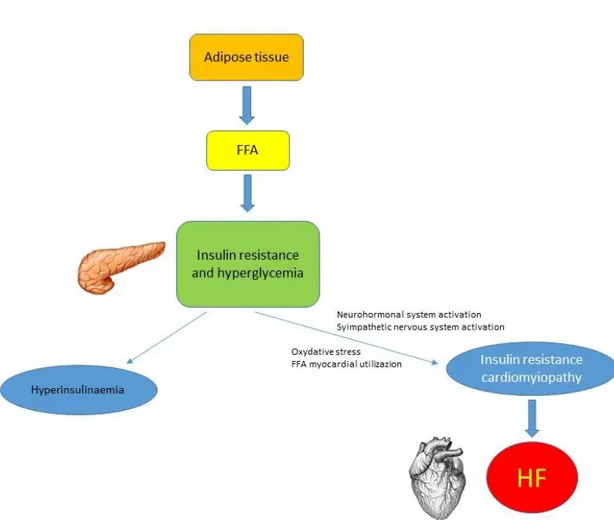
As previously described, IR is a direct precursor of T2DM, and the consequent compensatory hyperinsulinemia peculiar of IR, with consequent elevated levels of PG, associated with clustering of CV risk, can lead to several CV disease.6 As known, T2DM patients are generally obese (or with higher levels of fat abdominally distributed), and the
43 release of free fatty acid (FFA) from adipose tissue, directly impairs insulin sensitivity6,29 (figure 1). So the “primum movens” is IR, with several consequent mechanism involved in HF presentation and progression. However, well known is the bidirectional link between IR and HF,11 where several studies indicate that DM and IR are not only causative factors of HF,30–32 but patients with HF and DM or IR showed a more aggressive form of left ventricular (LV) dysfunction, with a higher mortality rate.33 Insulin resistance is the entity with higher prevalence in HF patients (up to 60%),34 with a complex pathophysiological interaction between these two conditions, since IR may be the cause and consequence of HF at the same time.30 Similarly, DM has a prevalence of 10 – 40% in patients with HF,35 showing a quite high prevalence, but lower than IR. This could explain the more important and potential role of IR in HF development (and vice versa), compared to DM, that was widely discussed in the years.8,9,30
Pathophysiology of heart failure in IR and DM
Well known is the role of IR and DM in several functional, metabolic and structural alterations that involve myocardial tissue and that can lead to HF (figure 1). In the initial stage of HF, there is a change in substrate utilization, where glucose becomes the primary substrate oxidized.9 Hyperglycemia is responsible for several cellular pathway abnormalities, going from increased polyol, modification of proteins, and formation of advanced glycation endoproducts, to increased protein kinase C expression, phenomenon leading to overproduction of superoxide and consequent oxidative stress30,36 (figure 1). On the other hand, the increase of FFA myocardial uptake, typical of diabetes and obesity,
44 leads to long-chain FFA oxidation and to a disproportionate oxidative request to mitochondria with uncoupling of mitochondrial oxidative phosphorylation30,37 (figure 1). In addition, the impaired expression of contractile proteins, is responsible for depressed myofibrillar ATP activities and abnormalities of the sarcoplasmic reticular and sarcolemmal calcium transport process, with consequent calcium overload and impaired diastolic function.30,38
Hyperactivation of adrenergic system
Another consideration to do is on the impairment of cardiac sympathetic innervation, commonly observed in HF patients affected by DM and/or IR. Paolillo et al,34 in a recent study, showed in HF patients with DM, or without DM, but with IR, a more impaired cardiac sympathetic innervation, compared with non-diabetic and non-IR patients, indicating a chronic adrenergic hyperactivity, that correlates with high levels of hyperglycemia and HF development and worsening. In the same year, Rengo et al,39 reported that levels of GRK2, a protein kinase involved in the desensitization of cardiac beta-receptors, are significantly more elevated in HF patients with DM compared with non-diabetic patients with HF, meaning a stronger adrenergic activation, known to be involved in progression and worsening of HF. This is a peculiarity of DM and IR patients with HF, where it is well known that because of adrenergic overactivity, HF is complicated by DM and IR.8
45 Epidemiology of heart failure in glucose metabolism abnormalities and vice versa
Since now, it was widely discussed the bidirectional link between HF and glucose metabolism disorders. Well known is the prevalence of HF in the general population, that is about 1 – 2%,1–5 rising to 12 – 30% in diabetic patients.40,41 Glucose metabolism abnormalities (in particular DM and IR) are independent risk factors for the development of HF. In the Framingham study, the relative risk of HF in patients with T2DM was doubled for men and six times as high in women6,42. These data were confirmed by the National Health and Nutrition Examination Survey, where T2DM was showed to be an independent predictor of HF (HR 1.85, 95% CI 1.51 – 2.28).43
On the other hand, the prevalence of DM in the general population is about 6 – 8 %,6 but as described by MacDonald et al,44 rises to 12 – 30% in HF patients. However, HF patients are older than general population. This could be considered a bias selection, although is widely known (also in the general population) not only the role of glucose metabolism disorders in development of HF, but also the major prevalence of glucose metabolism abnormalities (in particular DM and IR, as previously described) in HF patients. Confirming these data, the TOSCA registry,7 a recent Italian registry, made on 526 patients (81% male, age 62.5 ± 12.2 years) has shown a prevalence of IR in HF patients of 30 – 35%, in line with previous studies, demonstrating the great impact of glucose metabolism abnormalities on HF.
46 Prognosis of heart failure related to glucose metabolism abnormalities
Great trials
Today, HF can be considered the major cause of hospitalization in patients with glucose metabolism abnormalities. These data were confirmed by the Hypertension, Microalbuminuria or Proteinuria, Cardiovascular Events and Ramipril (DIABHYCAR) trial,45 with a mortality 12-fold higher in patients with HF and glucose metabolism disorders, compared to patients without HF (36% vs 3%). On the other hand, in the BEta blocker STroke (BEST) trial, impaired glucose metabolism increased the risk of hospitalization in HF patients, with T2DM as independent predictor of mortality, mostly for HF.46 More recently, the Metoprolol CR/XL Randomized Intervention Trial in Congestive Heart Failure (MERIT-HF),47 showed HF patients with glucose metabolism alterations, to be more hospitalized than patients free from DM.
Previous studies
A previous study of Suskin et al,12 studied 663 patients with abnormalities in glucose metabolism (DM or IR) and HF, assessing prognostic role of glucose metabolism, in particular, evaluating functional class, six minute walking performance and LV function. In this cohort of patients, they found a greater proportion of diabetic patients compared to non-diabetic patients in NYHA class III/IV (161 vs 77, p = 0.011), with a higher value of insulin in non-diabetic patients in NYHA class III/IV compared to NYHA class I/II (19.6 ± 2.3 vs 10.2 ± 0.6 mU, p < 0.005). In addition, among non-diabetic patients, significantly more NYHA class III/IV patients had elevated HOMA-index levels (44% vs 28%, p <
47 0.005) compared to NYHA class I/II patients. These data were confirmed also for six minute walking distance, that was significantly shorter in diabetic patients compared to non-diabetic patients (369 ± 7 vs 385 ± 4 meters, p = 0.03). Furthermore, patients with impaired HOMA-index had a significantly shorter six minute walking distance than those with normal values (372 ± 7 vs 391 ± 5 meters, p = 0.02).
A more recent study of Doehner et al,13 evaluated insulin sensitivity in 105 male patients with HF. After a mean follow up of 44 ± 4 months, patients with an insulin sensitivity below the median value had a worse survival (61% at two years) compared to patients with an insulin sensitivity above the median value (83% at two years) (RR 0.38, 95% CI 0.21 – 0.67, p = 0.001). Furthermore, insulin sensitivity resulted independent predictor of mortality in the study cohort.
Therapeutic possibilities
Considering the impact on prognosis of glucose metabolism abnormalities, it is possible affirm that DM and IR can be considered as potential target of HF. Although is known the role of several new pharmacological agents in the reduction of mortality in HF diabetic patients,48 poor is known about the potential treatment of IR and its impact on HF prognosis. Considering this assumption, Wong et al,14 randomized in a double-blind, placebo-controlled study 62 non-diabetic HF patients, to receive either four months of metformin or matching placebo. Compared with placebo, metformin decreased HOMA-index, improving also the secondary endpoint of the slope of the ratio of minute ventilation
48 to carbon dioxide production (VE/VCO2 slope). These data confirm the hypothesis that
treatment of IR should be protective in patients with HF.
Summary
There is a strong correlation between glucose metabolism abnormalities and HF, with a bidirectional link between them, where glucose metabolism abnormalities at every step can affect HF. On the other hand, DM and IR are more prevalent in HF patients, and patients with both HF and DM have a worst prognosis, due to a combination of effects of both the components. Well known is the role of some new drugs in reducing the mortality in HF patients with DM, but more studies are warranted to know the real effect of treatment of IR in patients with HF.
Acknowledgements
Dr. Fabio Marsico has been supported by a research grant provided by the Cardiovascular Pathophysiology and Therapeutics PhD program.
References
1. Ponikowski P, Voors AA, Anker SD, et al. 2016 ESC Guidelines for the diagnosis and treatment of acute and chronic heart failure. Eur Heart J. 2016;37(27):2129-2200m.
49 doi:10.1093/eurheartj/ehw128
2. Mosterd A, Hoes AW. Clinical epidemiology of heart failure. Heart. 2007;93(9):1137-1146. doi:10.1136/hrt.2003.025270
3. Redfield MM, Jacobsen SJ, Burnett, Jr JC, et al. Burden of Systolic and Diastolic Ventricular Dysfunction in the Community. Jama. 2003;289(2):194. doi:10.1001/jama.289.2.194
4. Bleumink GS, Knetsch AM, Sturkenboom MCJM, et al. Quantifying the heart failure epidemic: Prevalence, incidence rate, lifetime risk and prognosis of heart failure - The Rotterdam Study. Eur Heart J. 2004;25(18):1614-1619. doi:10.1016/j.ehj.2004.06.038 5. Ceia F, Fonseca C, Mota T, et al. Prevalence of chronic heart failure in Southwestern Europe: the EPICA study. Eur J Heart Fail. 2002;4(4):531-539.
6. Rydén L, Grant PJ, Anker SD, et al. ESC guidelines on diabetes, pre-diabetes, and cardiovascular diseases developed in collaboration with the EASD. Eur Heart J. 2013;34(39):3035-3087. doi:10.1093/eurheartj/eht108
7. Bossone E, Arcopinto M, Iacoviello M, et al. Multiple hormonal and metabolic deficiency syndrome in chronic heart failure: rationale, design, and demographic characteristics of the T.O.S.CA. Registry. Intern Emerg Med. 2018;13(5):661-671. doi:10.1007/s11739-018-1844-8
8. Saccà L. Heart failure as a multiple hormonal deficiency syndrome. Circ Hear Fail. 2009;2(2):151-156. doi:10.1161/CIRCHEARTFAILURE.108.821892
50 9. Arcopinto M, Salzano A, Isgaard J, et al. Hormone replacement therapy in heart failure. Curr Opin Cardiol. 2015;30(3):277-284. doi:10.1097/HCO.0000000000000166 10. Cittadini A, Napoli R, Monti MG, et al. Metformin prevents the development of chronic heart failure in the SHHF rat model. Diabetes. 2012;61(4):944-953. doi:10.2337/db11-1132
11. Ingelsson E, Sundström J, Ärnlöv J, et al. Insulin Resistance and Risk of Congestive Heart Failure. Jama. 2005;294(3):334. doi:10.1001/jama.294.3.334
12. Suskin N, McKelvie RS, Burns RJ, et al. Glucose and insulin abnormalities relate to functional capacity in patients with congestive heart failure. Eur Heart J. 2000;21(16):1368-1375. doi:10.1053/euhj.1999.2043
13. Doehner W, Rauchhaus M, Ponikowski P, et al. Impaired insulin sensitivity as an independent risk factor for mortality in patients with stable chronic heart failure. J Am Coll Cardiol. 2005;46(6):1019-1026. doi:10.1016/j.jacc.2005.02.093
14. Wong AKF, Symon R, Alzadjali MA, et al. The effect of metformin on insulin resistance and exercise parameters in patients with heart failure. Eur J Heart Fail. 2012;14(11):1303-1310. doi:10.1093/eurjhf/hfs106
15. The Expert Committee on the Diagnosis and Classification of Diabetes Mellitus. Report of the expert committee on the diagnosis and classification of diabetes mellitus. Diabetes Care. 1997;20(January):1183-1197. doi:10.2337/diacare.25.2007.S5
51 Diabetes Mellitus. Diabetes Care. 2003;26(11):3160-3167. doi:10.2337/diacare.26.11.3160
17. Diabetes DOF. Diagnosis and classification of diabetes mellitus. Diabetes Care. 2012;35(SUPPL.1). doi:10.2337/dc12-s064
18. Tang Q, Li X, Song P, Xu L. Optimal cut-off values for the homeostasis model assessment of insulin resistance (HOMA-IR) and pre-diabetes screening: Developments in research and prospects for the future. Drug Discov Ther. 2015;9(6):380-385. doi:10.5582/ddt.2015.01207
19. Himsworth HP. Diabetes mellitus: its differentiation into sensitive and insulin-insensitive types. 1936. Int J Epidemiol. 2013;42(6):1594-1598. doi:10.1093/ije/dyt203 20. Alebić MŠ, Bulum T, Stojanović N, et al. Definition of insulin resistance using the homeostasis model assessment (HOMA-IR) in IVF patients diagnosed with polycystic ovary syndrome (PCOS) according to the Rotterdam criteria. Endocrine. 2014;47(2):625-630. doi:10.1007/s12020-014-0182-5
21. Diabetes DOF. Diagnosis and classification of diabetes mellitus. Diabetes Care. 2010;33(SUPPL. 1). doi:10.2337/dc10-S062
22. Marques-Vidal P, Mazoyer E, Bongard V, et al. Prevalence of insulin resistance syndrome in Southwestern France and its relationship with inflammatory and hemostatic markers. Diabetes Care. 2002;25(8):1371-1377. doi:10.2337/diacare.25.8.1371
cut-52 off points for simple identification of insulin-resistant subjects. Exp Clin Endocrinol Diabetes. 2006;114(5):249-256. doi:10.1055/s-2006-924233
24. Geloneze B, Repetto EM, Geloneze SR, et al. The threshold value for insulin resistance (HOMA-IR) in an admixtured population. IR in the Brazilian Metabolic Syndrome Study. Diabetes Res Clin Pract. 2006;72(2):219-220. doi:10.1016/j.diabres.2005.10.017
25. Esteghamati A, Ashraf H, Khalilzadeh O, et al. Optimal cut-off of homeostasis model assessment of insulin resistance (HOMA-IR) for the diagnosis of metabolic syndrome: third national surveillance of risk factors of non-communicable diseases in Iran (SuRFNCD-2007). Nutr Metab (Lond). 2010;7:26. doi:10.1186/1743-7075-7-26
26. Yamada C, Moriyama K, Takahashi E. Optimal cut-off point for homeostasis model assessment of insulin resistance to discriminate metabolic syndrome in non-diabetic Japanese subjects. J Diabetes Investig. 2012;3(4):384-387. doi:10.1111/j.2040-1124.2012.00194.x
27. Yin J, Li M, Xu L, et al. Insulin resistance determined by Homeostasis Model Assessment (HOMA) and associations with metabolic syndrome among Chinese children and teenagers. Diabetol Metab Syndr. 2013;5(1):71. doi:10.1186/1758-5996-5-71
28. Timoteo AT, Miranda F, Carmo MM, et al. Optimal cut-off value for homeostasis model assessment (HOMA) index of insulin-resistance in a population of patients admitted electively in a Portuguese cardiology ward. Acta Med Port. 2014;27(4):473-479.
53 29. Hossain P, Kawar B, El Nahas M. Obesity and Diabetes in the Developing World — A Growing Challenge. N Engl J Med. 2007;356(3):213-215. doi:10.1056/NEJMp068177 30. Perrone-Filardi P, Paolillo S, Costanzo P, et al. The role of metabolic syndrome in heart failure. Eur Heart J. 2015;36(39):2630-2634. doi:10.1093/eurheartj/ehv350
31. Barzilay JI, Kronmal RA, Gottdiener JS, et al. The association of fasting glucose levels with congestive heart failure in diabetic adults ≥65 years: The Cardiovascular Health Study. J Am Coll Cardiol. 2004;43(12):2236-2241. doi:10.1016/j.jacc.2003.10.074
32. Moller DE, Flier JS. Insulin resistance--mechanisms, syndromes, and implications. N Engl J Med. 1991;325(13):938-948
33. Pocock SJ, Wang D, Pfeffer MA, et al. Predictors of mortality and morbidity in patients with chronic heart failure. Eur Heart J. 2006;27(1):65-75. doi:10.1093/eurheartj/ehi555
34. Paolillo S, Rengo G, Pellegrino T, et al. Insulin resistance is associated with impaired cardiac sympathetic innervation in patients with heart failure. Eur Heart J Cardiovasc Imaging. 2015;16(10):1148-1153. doi:10.1093/ehjci/jev061
35. Soläng L, Malmberg K, Rydén L. Diabetes mellitus and congestive heart failure. Further knowledge needed. Eur Heart J. 1999;20(11):789-795. doi:10.1053/euhj.1998.1472
54 36. Stratmann B, Tschoepe D. Heart in diabetes: Not only a macrovascular disease. Diabetes Care. 2011;34(SUPPL. 2). doi:10.2337/dc11-s208
37. Stanley WC, Lopaschuk GD, Mccormack JG. Regulation of energy substrate metabolism in the diabetic heart. 1997:25-33.
38. Dhalla NS, Liu X, Panagia V, et al. Subcellular remodeling and heart dysfunction in chronic diabetes. Cardiovasc Res. 1998;40(2):239-247. doi:10.1016/S0008-6363(98)00186-2
39. Rengo G, Pagano G, Paolillo S, et al. Impact of diabetes mellitus on lymphocyte GRK2 protein levels in patients with heart failure. Eur J Clin Invest. 2015;45(2):187-195. doi:10.1111/eci.12395
40. Thrainsdottir I, Aspelund T, Thorgeirsson G, et al. Abnormalities and Heart Failure in the. Diabetes Care. 2005;28:612-616.
41. Bertoni AG, Hundley WG, Massing MW, et al. Heart Failure Prevalence, Incidence, and Mortality in the Elderly with Diabetes. Diabetes Care. 2004;27(3):699-703. doi:10.2337/diacare.27.3.699
42. Kengne AP, Turnbull F, MacMahon S. The Framingham Study, Diabetes Mellitus and Cardiovascular Disease: Turning Back the Clock. Prog Cardiovasc Dis. 2010;53(1):45-51. doi:10.1016/j.pcad.2010.02.010
43. He J, Ogden LG, Bazzano LA, et al. Risk Factors for Congestive Heart Failure in US Men and Women. Arch Intern Med. 2001;161(7):996-1002.
55 doi:10.1001/archinte.161.7.996
44. MacDonald MR, Petrie MC, Hawkins NM, et al. Diabetes, left ventricular systolic dysfunction, and chronic heart failure. Eur Heart J. 2008;29(10):1224-1240. doi:10.1093/eurheartj/ehn156
45. Vaur L, Gueret P, Lievre M, et al. Development of congestive heart failure in type 2 diabetic patients with microalbuminuria or proteinuria: observations from the DIABHYCAR (type 2 DIABetes, Hypertension, CArdiovascular Events and Ramipril)
study. Diabetes Care. 2003;26(3):855-860.
http://www.ncbi.nlm.nih.gov/pubmed/12610049.
46. Domanski M, Krause-Steinrauf H, Deedwania P, et al. The effect of diabetes on outcomes of patients with advanced heart failure in the BEST trial. J Am Coll Cardiol. 2003;42(5):914-922. doi:10.1016/S0735-1097(03)00856-8
47. Deedwania PC, Giles TD, Klibaner M, et al. Efficacy, safety and tolerability of metoprolol CR/XL in patients with diabetes and chronic heart failure: Experiences from MERIT-HF. Am Heart J. 2005;149(1):159-167. doi:10.1016/j.ahj.2004.05.056
48. Gargiulo P, Savarese G, D’Amore C, et al. Efficacy and safety of glucagon-like peptide-1 agonists on macrovascular and microvascular events in type 2 diabetes mellitus: A meta-analysis. Nutr Metab Cardiovasc Dis. 2017;27(12):1081-1088. doi:10.1016/j.numecd.2017.09.006
56 Tables
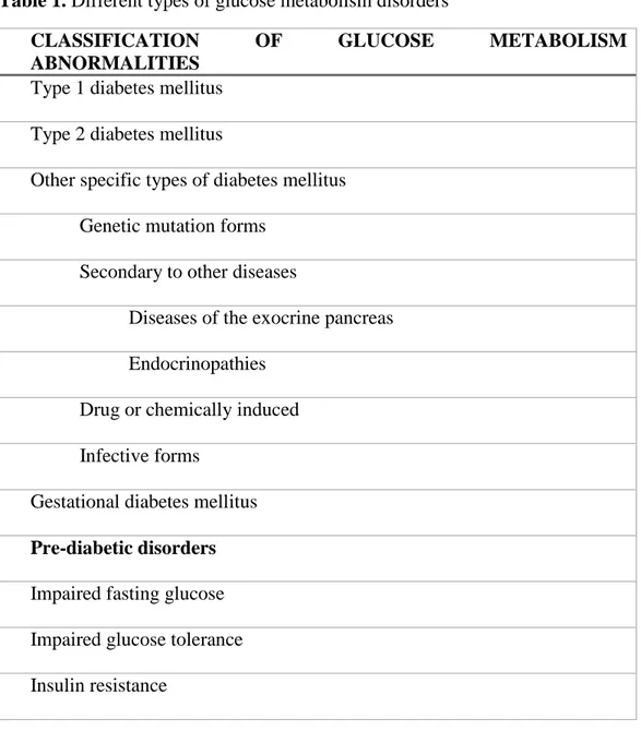
Table 1. Different types of glucose metabolism disorders
CLASSIFICATION OF GLUCOSE METABOLISM
ABNORMALITIES Type 1 diabetes mellitus Type 2 diabetes mellitus
Other specific types of diabetes mellitus Genetic mutation forms
Secondary to other diseases Diseases of the exocrine pancreas Endocrinopathies
Drug or chemically induced Infective forms
Gestational diabetes mellitus Pre-diabetic disorders Impaired fasting glucose Impaired glucose tolerance Insulin resistance
Table 2. Diagnostic evaluation of glucose metabolism disorders (according to WHO and ADA)
57 Diabetes mellitus
Glycated hemoglobin A1C (HbA1C)
Fasting plasma glucose
2-hour post–load plasma glucose (OGTT) Impaired glucose tolerance
Fasting plasma glucose
2-hour post–load plasma glucose (OGTT)
Impaired fasting glucose
Fasting plasma glucose
Insuline resistance
58 Figures
Figure 1. Pathophysiology of HF development related to insulin resistance. FFA = free fatty acid; HF = heart failure.
59 CHAPTER 5.
Efficacy and safety of glucagon-like peptide-1 agonists on macrovasular and microvascular events in type 2 diabetes mellitus: a meta-analysis
Gargiulo P, Savarese G, D’Amore C, De Martino F, Lund LH, Marsico F, Dellegrottaglie S, Marciano C, Trimarco B, Perrone Filardi P
Nutr Metab Cardiovasc Dis 2017;27:1081-1088
68 CHAPTER 6.
Vitamin D deficiency and clinical outcome in patients with chronic heart failure: a review
D’Amore C, Marsico F, Parente A, Paolillo S, De Martino F, Gargiulo P, Ferrazzano F, De Roberto AM, La Mura L, Marciano C, Dellegrottaglie S, Trimarco B, Perrone Filardi P Nutr Metab Cardiovasc Dis 2017;27:837-849
82
PART 2
83 CHAPTER 7.
Left ventricular function, epicardial adipose tissue and carotid intima-media thickness in children and young adults with vertical HIV infection
Marsico F, Lo Vecchio A, Paolillo S, D’Andrea C, De Lucia V, Bruzzese E, Vallone G, Dellegrottaglie S, Marciano C, Trimarco B, Guarino A, Perrone Filardi P
84 Left ventricular function, epicardial adipose tissue and carotid intima-media thickness in children and young adults with vertical HIV infection
Fabio Marsicoa,b*, MD, Andrea Lo Vecchioc*, MD, PhD, Stefania Paolilloa, MD, PhD, Claudia D’Andreaa
, MD, PhD, Vittoria De Luciaa, MD, Eugenia Bruzzesec, MD, PhD, Gianfranco Valloned, MD, Santo Dellegrottagliee, MD, PhD, Caterina Marcianof, MD, Bruno Trimarcoa, MD, Alfredo Guarinoc, MD, Pasquale Perrone Filardia, MD, PhD
*The two authors equally contributed to the study and are considered as first authors
a
Department of Advanced Biomedical Sciences, University of Naples Federico II
b
Center for Congenital Heart Disease, University Hospital Inselspital, Bern, Switzerland
c
Department of Translational Medical Sciences, Section of Pediatrics, University of Naples Federico II
d
Department of Advanced Biomedical Sciences, Section of Radiology, University of Naples Federico II
e
Villa dei Fiori, Acerra, Naples, Italy
f
85 Abstract
BACKGROUND: Life expectancy of HIV patients has increased considerably as a result of antiretroviral therapy, and cardiovascular (CV) disease has emerged as an important late concern. HIV infection may affect systolic function either in adults or children, however data on diastolic function and markers of CV risk, such as epicardial adipose tissue (EAT) and intima-media thickness (IMT), are lacking. Aim of the present study is to evaluate left ventricular function, EAT and IMT in children and young adults with vertically-acquired HIV infection.
METHODS and RESULTS: We enrolled 29 subjects on antiretroviral therapy (ART) (13, 45% male; median age of 14.0 and IQR 8.7), and 29 age-matched controls. All patients and controls underwent echocardiographic evaluation, with study of the systo-diastolic function and measurement of the EAT, and a carotid ultrasound study for IMT measurement. Comparing HIV-infected patients to healthy controls, we found a statistical significant increase of EAT and IMT (EAT: 3,16 ± 1,05 vs 1,24 ± 0,61 mm; p < 0,0001. IMT: 0,77 ± 0,15 vs 0,51 ± 0,11 mm; p < 0,0001), and a significant reduction of ejection fraction, evaluated with biplane Simpson method (52,3 ± 17,49% vs 66 ± 4,24%; p = 0,029). These results are not related with age, gender, degree of lipodystrophy, dyslipidemia, hyperinsulinism and ART duration or the use of single antiretroviral classes.
CONCLUSIONS: Vertically-infected HIV children and young adults show an increased thickness of the EAT and IMT, expression of potentially increased CV risk. They also show an impaired systolic function.
86 Introduction
Human immunodeficiency virus (HIV) infection is a major cause of morbidity and mortality worldwide. In developed countries, where life expectancy has increased considerably as a result of antiretroviral therapy (ART), cardiovascular diseases (CVD) have emerged as an important late comorbidity in HIV patients [1,2].
Results from the Pediatric Pulmonary and Cardiovascular Complications of Vertically Transmitted HIV Infection (P2C2 HIV) study have shown that subclinical cardiac abnormalities develop early in HIV1-infected children, and that cardiac alterations are frequent, persistent, and often progressive [3-6]. Common abnormalities include dilated cardiomyopathy (decreased left ventricular [LV] contractility and LV dilation) and inappropriate LV hypertrophy (high LV mass with decreased height and weight) [3].
Echocardiographic abnormalities can be found in up to 44% of patients infected with HIV [7]. These findings include pericardial effusion, LV dysfunction, dilated cardiomyopathy, infective endocarditis, pulmonary arterial hypertension, and cardiac masses such as lymphoma and Kaposi’s sarcoma of the heart. In addition, ART has been associated with the development of ischemic heart disease and LV diastolic abnormalities [8].
Although most data come from adult populations, evidence is also available for an association between HIV infection and systolic and diastolic dysfunction in children [9,10], and it has been demonstrated that children undergoing ART show decreased incidence of CVD [11,12].
87 In recent years new markers of increased CV risk have emerged. Epicardial adipose tissue (EAT), that has the same embryogenic origin of the visceral fat [13], has been reported to be associated with CV risk and metabolic syndrome [14,15]. Increased EAT thickness in HIV-infected adults compared to healthy controls has been reported [16,17], with an association between EAT and lipodystrophy [18]. However, data on children and young adults with vertically acquired HIV infection are lacking.
Similarly, the increase in intima-media thickness (IMT) is associated with higher CV risk, and a higher IMT has been reported in HIV-infected children, compared to non-infected patients [19]. The IMT thickness appears higher in naïve children than in HIV-patients undergoing ART therapy [20].
The aim of this study was to assess EAT, IMT and left ventricle function in young patients with vertically-acquired HIV infection, to correlate these parameters to metabolic profile and anti-retroviral treatment.
Methods
This cohort study was carried out between January 1st, and November 30th 2017 at the Regional Reference Center for Pediatric HIV/AIDS of the University of Naples Federico II. The Referral Center covers a territory of about 5 million inhabitants in the most populous region of the Southern Italy, and manages about 30 HIV-infected children and adolescents with 1-2 new diagnosis/year of HIV infection in the last years.
88 All patients currently in follow-up were enrolled in the study, and a group of age-matched subjects was enrolled as controls. The study was conducted according to the principles of the Helsinki declaration and the study protocol was approved by the Ethical Committee of the University Federico II of Naples (protocol number 153/16). All patients and caregivers, according to age, signed an informed consent after receiving specific information from the study coordinators.
Anamnestic evaluation
All patients underwent anamnestic and lifestyle evaluation (physical activity, smoking, drinking, drug addiction, eating habits), clinical and viro-immunological assessment with measurement of HIV viral load and CD4+ count. In addition, current ART, history of ART regimens and duration of single antiretroviral classes were reviewed.
Evaluation of the metabolic and CV risk
All patients underwent clinostatic and orthostatic blood pressure measurement, followed by the evaluation on blood sample of lipid profile (total cholesterol, high density lipoprotein (HDL), low density lipoprotein (LDL), triglycerides) and glucose
profile (fasting glucose, basal insulin, homeostatic model assessment (HOMA) index). We also evaluated the presence of metabolic syndrome, considering the International Diabetes Federation (IDF) diagnostic criteria and the modified National Cholesterol Education Program Adult Treatment Panel III (NCEP-ATP III) criteria [21]. Lipodystrophy was
89 classified as absent, mild (mild lipodystrophy of face and arms) and severe (severe lipodystrophy of face and arms, with involvement of abdomen and legs).
Echocardiography and carotid assessment
For all echocardiographic measurements, the final values were obtained after averaging over two cardiac cycles. The evaluation of LV ejection fraction was made with Simpson biplane mode, tracing tele diastolic and tele systolic volume both in four and two chamber apical view (n.v. 52-70%).
The evaluation of LV diastolic function was made evaluating the E/A ratio and the E/E’ ratio. The E/A ratio was evaluated in a four chambers apical view, using pulsed Doppler, applied on the coaptation point of the mitral flaps. The E wave is the early component of the left ventricle diastolic filling, meanwhile the A wave corresponds to the atrial systole. The E/A ratio was considered normal for a value higher than 1. Values lower than 1 (first type of diastolic dysfunction) or higher than 2 (third or fourth type of diastolic dysfunction) were considered abnormal. The E’ wave was obtained applying a tissue Doppler on the mitral annulus (medial and lateral). A E’ mean value lower than 8 indicate a diastolic dysfunction, but, considering the E/E’ ratio, the normal value is lower
than 8. A value between 8 and 13 can be considered as first type of diastolic dysfunction, meanwhile a value higher than 13 can be considered as a third or fourth type of diastolic dysfunction.
The EAT was measured in a parasternal long-axis view, on the top of the right ventricle free wall, considering the maximum thickness, as end-systolic measurement