AC Electrophoresis of Microdroplets in Anisotropic Liquids:
Transport, Assembling and Reaction
Journal: Soft Matter
Manuscript ID: SM-ART-06-2013-051705 Article Type: Paper
Date Submitted by the Author: 21-Jun-2013
Complete List of Authors: Hernandez-Navarro, Sergi; Universitat de Barcelona, Química Física tierno, pietro; university of Barcelona, Departament d'Estructura i Constituents de la Matèria
Ignes, Jordi; Universitat de Barcelona, Química Física; Sagues, Francesc; Universidad de Barcelona, Quimica Fisica
TOC:
Controlled motion of microdoplets in anisotropic fluids is a rich field of research which unveils new perspectives in the transport of water miscible chemicals or drugs. We demonstrate several strategies to transport, react and assemble in a controlled way microdroplets of aqueous solution in a nematic liquid medium. As an example, the images show the mixing of two different water soluble dyes (red and green) to give rise to a final droplet having brownish color.
Journal Name
Cite this: DOI: 10.1039/c0xx00000x
www.rsc.org/xxxxxx
Dynamic Article Links
►
ARTICLE TYPE
This journal is © The Royal Society of Chemistry [year] [journal], [year], [vol], 00–00 | 1
AC Electrophoresis of Microdroplets in Anisotropic Liquids: Transport,
Assembling and Reaction
Sergi Hernàndez-Navarro,
aPietro Tierno
,*bJordi Ignés-Mullol,
aFrancesc Sagués
a Received (in XXX, XXX) Xth XXXXXXXXX 20XX, Accepted Xth XXXXXXXXX 20XXDOI: 10.1039/b000000x
5
We describe the realization and controlled transport of water-based microreactors dispersed in a nematic liquid crystal and transported by application of an alternating electric field through the mechanism of induced charge electrophoresis. We characterize the propulsion speed of these microreactors in terms of droplet size, strength and frequency of the applied field and show how to use them to transport and coalesce microscale colloidal cargoes or water miscible chemicals. Controlled motion of microdoplets in
10
anisotropic fluids is a rich field of research which unveils new perspectives in the transport of water miscible chemicals or drugs.
Introduction
Water based microemulsions in oil phases are of fundamental importance in chemical and analytical sciences, due to the
15
possibility to encapsulate and deliver chemical or biological entities otherwise immiscible in the dispersion medium. The controlled transport of these microdroplets is of crucial importance for many applications related with lab-on-a-chip,1 drug delivery,2 or the realization of remotely addressable
20
micromotors.3 In contrast to simple oils as dispersion media, anisotropic fluids like liquid crystals offer the advantage of showing a variety of phases and structures which can be easily controlled and changed via temperature or external fields. These features, in turn, could be used to manipulate the physical or
25
chemical properties of the dispersed droplets.4
In this context, nematic liquid crystals (NLC) are complex fluids massively used in display technology, and characterized by rod shaped molecules which tend to orient their long axis along one direction, called director field. When a microscale particle is
30
dispersed in a nematic medium, the distortion of the director field caused by the inclusion leads to the formation of defects around the particle, which will mainly depend on the anchoring conditions at the particle surface.5 For homeotropic anchoring, i.e. when the nematic molecules align preferentially perpendicular
35
to the surface of the particle, two main defects can arise: a point defect known as hyperbolic hedgehog,6 or an equatorial Saturn- ring like defect7 [Fig. 1(a)-(c)]. A recent breakthrough in electric field transport of colloidal microspheres8 showed that colloidal
particles with point defects can be transported in a NLC by using
40
alternating current (AC) electric fields through the mechanism known as induced-charge electrophoresis (ICEP).9 The electrophoretic motion of the particles is due to the fact that the point defect, in contrast to the symmetric Saturn ring, breaks the spatial symmetry, unbalancing the ionic flows created by the field
45
around the particle. The main advantage of this technique,
compared to the standard capillary electrophoresis methods based on the use of a direct current (DC) field, is the absence of ion migration, avoiding electrochemical processes and steady flows,10 thus making it suitable to transport also liquid rather than solid
50
inclusions. It is worth remarking that for ICEP to occur, the system must feature broken symmetries. For instance, this phenomenon has been also reported for Janus particles in aqueous solution, where the contrast between metallic and dielectric half surfaces results in unbalanced ionic flow and in particle motion
55
under AC fields.9 In this respect, the use of NLC provides a more
versatile strategy, compatible with droplets of any nature, since the introduction of structural anisotropies in liquid droplets is a technically challenging process.11
We here demonstrate the controlled motion of water
60
microdroplets dispersed in a NLC and propelled in this medium via AC electric fields. Point defects generated by the microdroplets in the NLC break the fore-aft symmetry and enable controlled motion of the water droplets in the direction of the defect, even if the field is symmetric in time and space.
65
Moreover, we employ a NLC with negative dielectric anisotropy, which induces motion of the inclusions perpendicular to the electric field. We characterize the droplets velocity in terms of droplet size, frequency and strength of the applied field, and show that these droplets can be used as microreactors to transport in the
70
nematic medium nanoscale particles or to mix femtosized volumes of chemicals, ranging from 10-14 to 10-16 L.
Experimental part
The experimental cell was composed of two 0.7 mm thick microscope slides of size 15 × 25 mm2, coated with a layer of
75
indium-tin oxide (ITO), with surface resistivity = 100Ω/square (VisionTeck Systems). The slides were cleaned with a Micro-90 (Sigma-Aldrich) solution, dried and spin-coated with polyimide (PI-2555, HD-Microsystems), baked at 300 oC, and then rubbed
Page 2 of 12
Soft Matter
2 | Journal Name, [year], [vol], 00–00 This journal is © The Royal Society of Chemistry [year]
with a velvet cloth to obtain planar alignment in one direction. The two plates were then separated by a polyethylene terephtalate spacer (Mylar, Goodfellow, nominal thickness 23 µm) and glued together along two sides with the ITO layers facing inwards.
5
Figure 1. (a,c) Optical microscope images showing water microdroplets
characterized by a hyperbolic hedgehog defect (a), and a Saturn-ring defect (c). In (a) the double arrow indicates the direction of the director field. (b) and (d) show schematics of the corresponding configurations of the nematic liquid crystal around the water droplets. (e) Histogram
10
showing the typical population of particles with hedgehog defects in an experimental cell. The fraction of observed particles featuring a Saturn-ring was below 1%. Subsequent measurements were done for droplet size between 1 - 6 µm, as indicated in the image.
Actual thickness of cells was measured via interferometric
15
techniques. Electrical contacts were made by eliminating part of the polyimide coating on the slides by using a suitable solvent (T9039, HD-Microsystems) and gluing wires with colloidal silver. Dispersions of aqueous microdroplets were created by adding 1 µL of Milli-Q water-based aqueous solution to 10 µL of
20
a nematic liquid crystal (MLC-7029, Merck) with a negative dielectric anisotropy, ∆ε = -3.6 at 1 kHz, followed by vortex agitation (IKA MS3 basic) for 3 minutes. Some tests were performed also by using the nematic liquid crystal MLC-2037 from Merk (∆ε = = -3.1 at 1kHz) and MBBA from Synthon
25
Chemicals (∆ε = = -0.53 at 1kHz). The emulsions were stabilized using 0.01-0.05 M Sodium Dodecylsulphate (SDS, Sigma-Aldrich) which, in turn, induced homeotropic alignment of the nematic molecules at the LC/water interface. The experimental
cells were then filled by capillarity with freshly prepared
30
dispersions and sealed with glue. With this protocol we obtained suspensions of polydispersed water droplets with sizes ranging from 1 to 20 µm in diameter and a distribution peaked at a diameter of 3µm (Fig. 1). Sinusoidal electric fields were applied to the experimental cell by using a function generator
(ISO-35
TECH IFA 730) feeding a power amplifier (TREK model PZD700). The voltages were monitored by a multimeter (HP 34401A) and were in the range 0 to 30 Volts peak to peak, while the applied range of frequencies varied from 0 to 100Hz. Experimental observations were performed with an optical
40
microscope (Nikon Eclipse 50iPol). Images and videos were captured with an AVT Marlin F-131C CMOS camera controlled with the software AVT SmartView 1.10.2. They were then treated and analyzed using the software packages ImageJ and IgorPro.
45
Results and discussion
In Fig. 1(a) and 1(c), we show optical micrographs of droplets featuring hedgehog (a) or Saturn-ring (b) defects. As shown in the corresponding schematics [Fig. 1(b) and 1(d)], the hedgehog defect is observed as a dark circular spot which appears on one
50
side of the microdroplet, and oriented along the nematic director. As a result, the symmetry of the nematic director resembles that of an electric dipole. In contrast, the Saturn-ring defect displays a circular ring surrounding the microdroplet and features quadrupolar symmetry. In most of the experimental cells used in
55
this work, we find that the distribution of defects around the microdroplets was predominantly of the hedgehog-type, while only a small fraction of inclusions (<1%) feature Saturn-ring defects. For the studies reported below, we restrict our analysis to droplets of diameter less than 6 µm. These constitute the majority
60
of hedgehog particles [Fig. 1(e)] and are less prone to buoyancy effects, which tend to pin larger droplets.
In Fig. 2(a), we show the geometry of our experimental system. Water microdroplets were stabilized in the NLC matrix with sodium dodecyl sulphate (SDS). This surfactant provides strong
65
homeotropic anchoring at the water/NLC interface, i.e. the NLC molecules tend to orient perpendicular to the surface of the inclusions. We observe that, once dispersed in the NLC, most of the water droplets display a point defect (hedgehog), arranged on one side of the particle, while few feature Saturn rings. The
70
suspensions of SDS stabilized aqueous microdroplets were enclosed between two transparent electrodes, treated in order to induce planar (i.e. parallel) anchoring of the nematic molecules at the surface of the cell along a direction determined by the buffing of the polyimide layer. The cell spacing (23 µm) was
75
chosen as a compromise to minimize the interactions with the cell substrates and to guarantee that most of the droplets where in the same observation plane.
In order to completely decouple the AC electrophoresis from any residual linear (DC) electrophoretic contribution, we choose to
80
use a NLC characterized by a negative dielectric anisotropy, so the nematic director orients perpendicular to the direction of the applied field.12,13 With this strategy, we transport the microdroplets always perpendicularly to the direction of the oscillating field. This has the further advantage that the motion
This journal is © The Royal Society of Chemistry [year] Journal Name, [year], [vol], 00–00 | 3
can be controlled along any arbitrary predesigned in-plane track in the NLC matrix.
Figure 2. (a) Schematic of the experimental cell composed of water
microdroplets dispersed in a nematic liquid crystal and enclosed between
5
two transparent glass electrodes treated to induce surface planar anchoring. The water droplets are propelled by application of an AC electric field between the electrodes. (b) Optical micrographs taken every 4.5 s of a water droplet having 6.5 µm of diameter and propelled at a speed of 1.7 µm/s by an AC field with amplitude, E = 0.7 V/µm and
10
frequency, f = 10 Hz. The corresponding video (Video 1) can be found in the Supporting Information. (c) Positions (x,y) versus time of the droplet shown in panel (b).
In our system application of an external AC electric field between both electrodes sets the droplets in motion always in the direction
15
towards the point defect, Fig. 2(a). As it was reported by Lavrentovich and co-workers,8 depending on the LC used the motion could be towards or against the point defect for the same particles and under the same conditions of applied field. After testing different liquid crystals, we found that the motion of the
20
microdroplets was towards the point defects for MLC-7029 or MLC-2037, and against the point defect for MBBA.
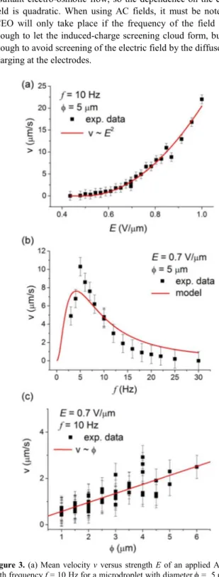
We characterize the transport properties of the microdroplets by measuring their average speed as a function of the strength E (Figure 3(a)) and frequency f (Figure 3(b)) of the applied field,
25
and as a function of the diameter φ for a fixed value of Ε and f (Figure 3(c)). In the first case, we observe that the droplets start to move above a threshold field E0 ~ 0.5 V/µm and that the speed
increases quadratically with the field, v ~ E2. This electrophoretic behavior in a nematic medium is in agreement with previous
30
work,8,12 where it was found that the first nonlinear term in the speed was quadratic rather than cubic like in isotropic fluids. A quadratic term allows using a symmetric AC field to move the particles in the medium, since changing the field polarity does not affect the particle speed. We confirm this quadratic behavior by
35
fitting the experimental values with v=β(Ε−E0)
2
, and we find E0 =
0.49 V/µm. Also, we find that the dependence of the droplets velocity on the frequency of the applied AC field can be well described by invoking the induced-charge electro-osmosis (ICEO) model, which was originally developed for isotropic
40
fluids.14 This model is based in the electro-osmotic flow that appears when a tangential electric field is applied to a charged surface. If the surface charge is fixed, the electro-osmotic flow is linear with the applied field. In contrast, if the surface charge is induced by the same applied electric field, the nonlinear
45
phenomenon of ICEO takes place. In ICEO, the first nonlinear term is quadratic with the electric field, thus allowing the use of AC and not only DC fields to generate electro-osmotic flow. If
we have a spherical polarizable inclusion immersed in a liquid electrolyte and subjected to an electric field, a quadrupolar
50
electro-osmotic flow is formed around the inclusion, from its poles to its equator. The electric field first generates the induced-charge screening cloud on the inclusion, and then it drives the resultant electro-osmotic flow, so the dependence on the electric field is quadratic. When using AC fields, it must be noted that
55
ICEO will only take place if the frequency of the field is low enough to let the induced-charge screening cloud form, but high enough to avoid screening of the electric field by the diffuse-layer charging at the electrodes.
60
Figure 3. (a) Mean velocity v versus strength E of an applied AC field
with frequency f = 10 Hz for a microdroplet with diameter φ = 5 µm. (b) v versus frequency f of a φ = 5 µm water droplet subjected to an applied AC field with strength E = 0.7 V/µm. (c) v versus size (φ) for microdroplets driven by an applied field with f = 10 Hz and E = 0.7
65
V/µm. For images (a) and (b), the continuous red lines denote fits following the equations described in the text, while in (c) it denotes a linear relationship between speed and size.
Page 4 of 12
Soft Matter
4 | Journal Name, [year], [vol], 00–00 This journal is © The Royal Society of Chemistry [year]
Following the model developed in Ref. [14], the velocity of the induced flow can be described by the following equation: ݒሺ߱ሻ ൌ ݒ ఠమఛమ
ሺଵାఠమఛ
మሻሺଵାఠమఛమሻ
(1)
5
being ω = 2πf. As proposed in Ref. [8], this equation can be also used to describe the velocity of a particle in ICEP, where v0
contains a quadratic dependence with the amplitude of the applied electric field, consistently with the data trend shown in Fig. 3(a). In Eq. (1), τc = λDφ/2D is the droplet charging time and
10
τe = λDL/2D is the electrode charging time. Here φ = 5 µm, and
the distance between the electrodes is L = 23 µm, which gives the ratio τe/τc =L/φ = 4.6. Unknown parameters are λD , which is the
Debye screening length of the droplet in the NLC, and D the diffusion coefficient of the ions. From the nonlinear fit of the
15
experimental data, we find v0 = 11.5 µm/s, τe ~ 0.09 s, and τc ~
0.02 s, which gives a ratio λD/D = 0.008 s/µm. We estimate the
Debye length λD, considering εr=5.4, T=298K, and a number
density of monovalent ions in the LC of the order of [1019 – 1020] m-3,15 which leads to λD ~ [0.3 – 0.9] µm. Our fitted value λD/D is
20
consistent with the previous range of values of λD, giving typical
values of D in the range of [10-10 – 10-11 ] m2s−1 .16
By varying the size of the microdroplets, we observe a linear increase of v with φ, at fixed strength and frequency of the applied field, which is consistent with the fact that larger droplets
25
induce stronger distortion of the nematic director and thus enable larger electrophoretic speed.
We next demonstrate that these liquid droplets can be effectively used as microcarriers to transport, mix or disperse solid particles or femtoliter volumes of aqueous solutions. First, we observe that
30
microdroplets with Saturn ring defects, although immobile, distort the motion of neighbouring droplets with point defects. The attraction between dipolar and quadrupolar droplets is long range and mediated by the elastic distortion of the nematic medium.17 We use this property to realize a localized cargo
35
release operation, as shown in Fig. 4(a). In particular, one microdroplet was previously loaded with polystyrene particles having 1.7 µm in diameter, and driven by an AC field towards a larger droplet characterized by a Saturn ring defect. The large droplet attracts the small one, and they coalesce releasing the
40
colloidal cargo into a larger volume of fluid.
Droplets with point defects feature highly anisotropic interactions. In particular, a microdroplet with a point defect can be considered as a dipole, with one charge located on the point defect and the
45
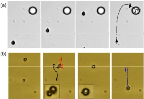
Figure 4. (a) Sequence of images showing the absorption of a microdroplet (φ = 7.3 µm), filled with 11 polystyrene particles (1.7 µm diameter), by a larger droplet (φ = 18 µm) with a Saturn ring defect (E = 0.7 V/µm, f = 10 Hz). Time interval between images is 17.2 s. (b) Microscope images showing two water microdroplets (φ = 2.7 µm and 3.7 µm) driven in opposite directions by an AC field (E = 0.70 V/µm, f = 10 Hz) and containing the separated reactants, potassium ferrocyanide [hexacyanoferrate (II)] (0.2 M) and ferric ions [Fe3+] solution (0.3 M). The droplets approach and coalesce in a larger
50
one (φ = 4.3 µm), forming Prussian blue as a precipitate. In this particular example, the field was momentarily switched off to facilitate the assembly of the droplets. The AC field was then used to prompt coalescence and to drive the resulting droplet away from the reaction zone. The corresponding videos (Video 2 and Video 3) can be found in the Supporting Information
other, of opposite sign, located inside the droplet. In general, two approaching inclusions in the NLC will thus try to assemble with
55
the defects in opposite sides.18 In our case, we find that when driven towards each other, the microdroplets deviate from their collision trajectory. After crossing, their dipolar defects are in antiparallel configuration, and the two droplets attract and can
coalesce into a larger one. In Fig. 4(b) we use this feature to
60
realize a chemical reaction by coalescing two water droplets doped with aqueous solutions of the reactants, potassium ferrocyanide [hexacyanoferrate (II)] and ferric ions [Fe3+] solution, in order to obtain Prussian blue,
This journal is © The Royal Society of Chemistry [year] Journal Name, [year], [vol], 00–00 | 5
4FeCl3(aq) + 3K4[Fe(CN)6](aq) → Fe4[Fe(CN)6]3 ↓ + 12KCl(aq)
The resulting droplet shows the typical dark-blue precipitate of ferric ferrocyanide, and initially features a Saturn-ring defect, which quickly transforms back into a hedgehog defect, allowing
5
to drive the reaction product to a desired location of the experimental platform. The process shown in Figure 4(b) is only an example of localized microreactions which can be performed with this technique. Our system also allows to explore a self-assembly scenario under AC fields. In particular, by modifying
10
the experimental protocol by introducing the ultrasonication of the NLC emulsions, we are able to further reduce the size of the microdroplets. We find that smaller water droplets, with diameters below 1 µm, were less prone to coalesce. In particular, we show in the Supporting Information (Video 4) that application
15
of the AC field was able to accumulate smaller droplets, while decreasing the frequency releases them in the nematic medium covering a larger area.
Conclusions
In conclusion, we have shown several strategies to transport,
20
react and assemble in a controlled way microdroplets of aqueous solutions in a nematic liquid medium driven by AC electric fields. Although the process can be described in a semiquantitative way by the model of induced charge electroosmosis, the actual mechanism driving the liquid
25
inclusions has still some pending questions, such as how the nature of the NLC determines the direction of motion of the dipolar droplets with respect to the position of the hedgehog defect. Using NLC with negative dielectric anisotropy allows driving the liquid inclusions in the direction perpendicular to the
30
cell electrodes, which can include any predesigned track in the NLC matrix. For example, one could establish the direction of motion by locally altering the in-plane orientation of the NLC by means of an optical field in the presence of photosensitive confining layers. Controlled motion of microdoplets in
35
anisotropic fluids is a rich field of research which unveils new perspectives in the transport of water miscible chemicals or drugs.
Acknowledgements
We acknowledge financial support by MICINN (Projects No.
40
FIS2010-21924C02, FIS2011-15948-E, FIS2011-13771-E) and by DURSI (Project No. 2009 SGR 1055). S.H.-N. acknowledges the support from the FPU Fellowship (AP2009-0974). P.T. was supported by the program ‘‘Ramon y Cajal’’.
Notes
45a Universitat de Barcelona, Departament de Química Física, Av.Diagonal
647, Barcelona Spain.
b Universitat de Barcelona, Departament d'Estructura i Constituents de la
Matéria, Av. Diagonal 647, Barcelona Spain. E-mail: [email protected]
50
† Electronic Supplementary Information (ESI) available: four videos files (3 .AVI 1 . WMV format). See DOI: ...
References
1 (a) H. A. Stone, A. D. Stroock and A. Ajdari, Annu. Rev. Fluid Mech., 2004, 36, 381-411. (b) T. M. Squires, S. R. Quake, Rev. Mod. Phys.
55
2005, 77, 977–1026. (c) H. Song, D. L. Chen, R. F. Ismagilov, Angew. Chem. Int. Ed., 2006, 45, 7336-7356. (d) J.-U. Shim, G. Cristobal, D. R. Link, T. Thorsen, Y. Jia, K. Piattelli and S. Fraden, J. Am. Chem. Soc. 2007, 129,8825–8835.
2 D. L. Buhr, F. E. Acca, E. G. Holland, K. Johnson, G. M. Maksymiuk,
60
A. Vaill, B. K. Kay, D. A. Weitz, M. P. Weiner and M. M. Kiss, Methods, 2012, 58, 28-33.
3 (a) W. F. Paxton, K. C. Kistler, C. C. Olmeda, A. Sen, S. K. St. Angelo, Y. Y. Cao, T. E. Mallouk, P. E. Lammert and V. H. Crespi, J. Am. Chem. Soc. 2004, 126, 13424-13431. (b) S. Fournier-Bidoz, 65
A. C. Arsenault, I. Manners and G. A. Ozin, Chem. Commun. 2005, 441-443. (c) S. Sanchez, A. A. Solovev, Y. Mei and O. G. Schmidt, J. Am. Chem. Soc. 2010, 132, 13144-13145. (d) J. R. Howse, R. A. L. Jones, A. J. Ryan, T. Gough, R. Vafabakhsh and R. Golestanian, Phys. Rev. Lett. 2007, 99, 048102. (e) W. Gao, A. Pei and J. Wang, 70
ACS Nano 2012, 6, 8432-8438.
4 (a) P. Poulin, H. Stark, T. C. Lubensky and D. A. Weitz, Science 1997,
275, 1770-1773. (b) J.-C. Loudet, P. Barois and P. Poulin, Nature
2000, 407, 611-613. (c) I. Muševič, M. Škarabot, U. Tkalec, M. Ravnik and S. Žumer, Science 2006, 313, 954-958. (d) C. P.
75
Lapointe, T. G. Mason, and I. I. Smalyukh, Science 2009, 326, 1083- 1086. (e) G. M. Koenig, I.-H. Lin, N. L. Abbott, Proc. Natl. Acad. Sci. USA 2010, 107, 3998-4003. (f) T. A. Wood, J. S. Lintuvuori, A. B. Schofield, D. Marenduzzo and W. C. K. Poon, Science 2011, 334, 79-83.
80
5 (a) S. V. Burylov, Y. L. Raikher, Phys. Rev. E 1994, 50, 358–367. (b) H. Stark, Physics Reports 2001, 351, 387-474.
6 P. Poulin, D. A. Weitz, Phys. Rev. E 1998, 57, 626-637.
7 (a) E. M. Terentjev, Phys. Rev. E 1995, 51, 1330-1337. (b) Y. D. Gu, N. L. Abbott, Phys. Rev. Lett. 2000, 85, 4719-4722.
85
8 O. D. Lavrentovich, I. Lazo, O. P. Pishnyak, Nature 2010, 467, 947– 950.
9 S. Gangwal, O. Cayre, M. Bazant and O. Velev, Phys. Rev. Lett., 2008,
100, 058302.
10 A. S. Dukhin, S. S. Dukhin, Electrophoresis 2005, 26, 2149-2153.
90
11 J. Guzowski, P. M. Korczyk, S. Jakiela and P. Garstecki, Soft Matter, 2012, 8, 7269.
12 I. Lazo and O. D. Lavrentovich, Phil. Trans. A, 2013, 371, 20120255. 13 S. Hernàndez-Navarro, P. Tierno, J. Ignés-Mullol and F. Sagués, Soft
Matter 2011, 7, 5109-5112. 95
14 T. M. Squires and M. Z. Bazant, J. Fluid Mech. 2004, 509, 217-252. 15 H. Naito, K. Yoshida, M. Okuda, A. Sugimura, Jpn. J. Appl. Phys.,
1994, 33, 5890.
16 D.A. Dunmur, A. Fukuda, G.R. Luckhurst, Physical properties of liquid crystals: nematics, p. 460-461, INSPEC, 2001
100
17 (a) U. Ognysta, A. Nych, V. Nazarenko, M. Škarabot and I. Muševič, Langmuir 2009, 25, 12092-12100. (b) Z. Eskandari, N. M. Silvestre, M. Tasinkevych and M. M. Telo da Gama, Soft Matter 2012, 8, 10100-10106.
18 M. Yada, J. Yamamoto and H. Yokoyama, Phys. Rev. Lett. 2004, 92,
105
185501.
Page 6 of 12
Soft Matter
SUPPORTING VIDEO FILES.
With the article there are four video files as support of the experimental results.
Video 1 (.AVI): Movie showing a water droplet with 6.5 µm of diameter and propelled at a speed of 1.7 µm/s by an AC field with amplitude, E = 0.7 V/µm and frequency, f = 10 Hz, Fig. 1(b) of the article.
Video 2 (.AVI): Movie showing the absorption of a microdroplet (φ = 7.3 µm), filled with 11 polystyrene particles (1.7 µm diameter), by a larger droplet (φ = 18 µm) with a Saturn ring defect (E = 0.7 V/µm, f = 10 Hz), Fig. 3(a) of the article.
Video 3 (.AVI): Movie showing two water microdroplets (φ = 2.7 µm and 3.7 µm) driven in opposite directions by an AC field (E = 0.70 V/µm, f = 10 Hz) and containing the separated reactants, potassium ferrocyanide [hexacyanoferrate (II)] (0.2 M) and ferric ions [Fe3+] solution (0.3 M). The droplets approach and coalesce in a larger one (φ = 4.3 µm), forming Prussian blue as a precipitate. In this particular example, the field was momentarily switched off to facilitate the assembly of the droplets. The AC field was then used to drive the resulting droplet away from the reaction zone, Fig. 3(b) of the article.
Video 4 (.WMV): Movie showing several small microdroplets (φ ~ 1 µm) which are accumulated without coalescence around a larger one (φ ~ 7 µm) with a Saturn ring defect via application of an AC field with strength E = 0.65 V/µm and frequency f = 20 Hz. Later on they are dispersed by decreasing the frequency of the field to f = 3 Hz. To better visualize the small droplets, the video was recorded with crossed polarizers.