POLITECNICO DI MILANO
FACOLTA’ DI INGEGNERIA DEI SISTEMI
Corso di Laurea Magistrale in Ingegneria Biomedica
Analysis of Respiratory Parameters in COPD
Patients Monitored at Home by a Novel
Telemedicine System
Relatori: Prof. Raffaele Dellacà
Prof. Marco Parvis
Correlatori: Ing. Alessandro Gobbi, Ph.D.
Ing. Pasquale Pio Pompilio, Ph.D.
Tesi di Laurea di:
Ivan Cenci Matr. 751565
Contents
Summary 1
Sommario 7
Introduction 13
1 Chronic Obstructive Pulmonary Disease and assessment of lung
func-tion 17
1 Introduction . . . 17
2 Definition of Chronic Obstructive Pulmonary Disease . . . 18
3 Pathogenesis, Pathology and Pathophysiology of COPD . . . 19
3.1 Pathogenesis . . . 20 3.2 Pathology . . . 21 3.3 Pathophysiology . . . 23 4 Epidemiology of COPD . . . 25 4.1 Prevalence . . . 25 4.2 Morbidity . . . 26 4.3 Mortality . . . 27 4.4 Risk factors . . . 28 5 Exacerbations of COPD . . . 29
6 Diagnosis and assessment of COPD . . . 32
7 Assessment of temporal variability of airflow limitation and disease pro-gression . . . 34
8 The Forced Oscillation Technique (FOT) . . . 36
9 Conclusions . . . 38
2 Validation of a novel telemedicine system for chronic respiratory dis-eases’ home monitoring 41 1 Introduction . . . 41
2 Experimental setup . . . 42
2.1 Commercial device . . . 42
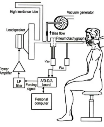
2.2 Reference FOT system . . . 46
3 Experimental protocols . . . 48
3.1 Systems comparison . . . 50
3.2 Test objects’ measurement . . . 53
3.3 Breathing pattern comparison . . . 54
4 Results . . . 55
4.1 Calibration stability . . . 55
4.2 Validation of the front-end hardware . . . 58
4.3 Breathing pattern comparison . . . 62
4.4 Validation of the real-time processing algorithm . . . 62
4.5 Overall validation . . . 66
5 Conclusions . . . 69
3 Home monitoring of COPD and data analysis 73 1 Introduction . . . 73
2 Methods . . . 75
2.1 Data collection and processing . . . 75
2.2 Automatic outlier detection . . . 80
2.3 Methods for missing data estimate . . . 83
2.4 Ad-hoc Locally Weighted Regression method . . . 91
2.5 Analysis of the CAT-based questionnaire . . . 99
2.6 Principal component analysis . . . 102
2.7 Variability analysis at different time scales . . . 103
3 Results . . . 104
3.1 Outlier automatic detection . . . 104
3.2 Missing data estimate . . . 108
3.3 Analysis of the CAT-based questionnaire . . . 117
3.4 Principal component analysis . . . 121
3.5 Variability analysis at different time scales . . . 126
4 Conclusions . . . 128
General Conclusions 131
Summary
Chronic obstructive pulmonary disease (COPD) represents a major public health prob-lem. It is the fourth leading cause of chronic morbidity and mortality in the United States and by 2020, according to recent studies, it is projected to rank third among the leading causes of death and fifth in economical burden caused worldwide [1–3]. The increased mortality is driven by the expanding epidemic of smoking and the changing demographics in most countries, with an increase in population’s life expectancy. Of these two forces, demographics is the stronger driver of the trend. Furthermore, al-though COPD has received increasing attention from the medical community in recent years, it is still relatively unknown or ignored by the public as well as public health and government officials.
From a pathophysiologic perspective, COPD is characterized by chronic airflow limi-tation and a range of pathological changes in the lung, some significant extra-pulmonary effects, and important comorbidities which may contribute to the severity of the dis-ease in individual patients. Thus, COPD should be regarded as a pulmonary disdis-ease, but these significant comorbidities must be taken into account in a comprehensive diagnostic assessment of severity and in determining appropriate treatment.
The progression of the pathological condition is described by stable periods dis-rupted by sudden exacerbations of the symptoms during which a severe inflammatory process occurs. Such aggravations recur in a periodic fashion and often require hospital-ization. A speculation proposed is that a long-term observation of objective parameters may help to characterize the progression of the disease over time and, possibly, early detect the onset of extreme future events.
Unfortunately, the diagnosis of this syndrome still relies on the measurements of spirometric indices that, given the effort-dependent maneuvers required, are not suitable for self-testing of airway obstruction. Therefore, a long-term and accurate observation of respiratory parameters is still missing.
A different and innovative approach for the characterization of the progression of COPD over time, for the association of clinical symptoms to quantitative parameters and for the early detection of acute events could be based on the measurement by
forced oscillation technique (FOT) of indices related to the mechanical properties of the respiratory system.
Accordingly, the present work aims at advancing knowledge about the pathology by employing a novel commercial telemedicine system based on FOT. To do so, the accuracy of this device is firstly evaluated in order to validate its functionality. Secondly, clinical data is acquired and analyzed thanks to the employment of the device in the largest and most detailed clinical trial ever defined for the observation of COPD. The work is presented in three chapters and their content is summarized as follows.
Chapter 1 - Chronic Obstructive Pulmonary Disease and assessment of lung function
As the present work is based on experimental studies involving COPD patients, the majority of Chapter 1 focuses on the pathophysiologic and epidemiologic characteristics of COPD. This is to provide a comprehensive background for the interpretation of the validation process of Chapter 2 and the data analysis of Chapter 3.
The disease is first defined, then processes characterizing pathophysiology, epidemi-ology and exacerbations of COPD are discerned in their main components and phases. Finally, clinical methods for the diagnosis of obstructive diseases and the assessment of their temporal variability are discussed. There is now a good understanding of how the underlying disease process in COPD leads to the characteristic physiologic abnormalities and symptoms. For example, decreased FEV1 primarily results from inflammation and
narrowing of peripheral airways, while decreased gas transfer arises from the parenchy-mal destruction of emphysema. Also, the peak expiratory flow (PEF) is the clinical tool recommended in most modern guidelines as a monitoring instrument for the mea-surement of variability of symptoms over time [4–6].
Since the just mentioned spirometric indexes are effort dependent and provide data with poor reproducibility when performed without supervision, it is speculated that more meaningful clinical outcomes could be obtained by leaving the spirometric approach and concentrating on the analysis of the temporal fluctuations of reliable and accurate parameters that do not require the execution of forced respiratory maneuvers, as the respiratory mechanical indices measured by FOT.
Chapter 2 - Validation of a novel telemedicine system for chronic respiratory diseases’ home monitoring
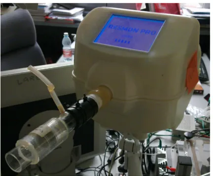
In Chapter 2 a novel telemedicine system (RESMON R PRO) developed and
Summary
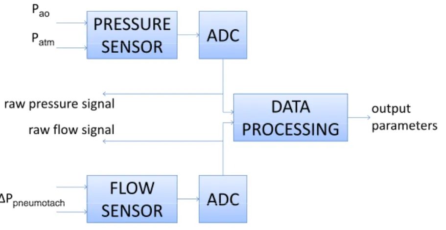
with the Laboratory of Biomedical Research (TBM Lab) of the Politecnico di Milano, is presented and analyzed. The device detects flow and pressure signals at the airway openings and calculates the value of impedance of the tracheobronchial tree thanks to the application of the FOT. In addition, it assesses many other respiratory parameters and provides additional information about each test performed.
The analysis of the accuracy of this novel FOT device is based on comparisons between measurements performed by it and measurements performed by a reference FOT system for laboratory use which is taken as a gold standard. Through properly designed experimental protocols the accuracy of the macro-components of the device (i.e, front-end hardware and real-time processing algorithm) is investigated. Also, analyses are provided for assessing calibration stability and efficiency of the aspirator needed to expel the exhaled carbon dioxide. Every experimental protocol is supported by the employment of numerical tools of statistical significance.
Results of calibration stability indicate that the front-end maintains the same char-acteristics over a time span of at least two months, and that calibration is expected to remain valid for much longer periods. The aspirator, on the contrary, does not show very positive performances as it is not sufficient to completely extract the carbon dioxide present in the high-inertance path after exhalation. Therefore an alternative solution able to guarantee a greater flow rate should be taken into account.
Results of accuracy analysis fully validate the signal processing software installed on the device, whereas the accuracy of the front-end hardware cannot be assessed in absolute terms. This is due to the physiologic inter-test variability characterizing every subject tested by the device. In other words, physiologic inter-test variability “masks” the actual difference in accuracy between the two systems used for making comparisons. Several clues suggest that the actual difference in precision is rather small, but this is not directly provable.
Chapter 3 - Home monitoring of COPD and data analysis
Chapter 3 offers an analysis of clinical data acquired by the validated FOT device. The observational clinical study utilized as source of data moves from the speculation that a long-term observation of objective parameters may help to characterize the progression of the COPD over time and, possibly, early detect the onset of extreme future events. The purpose of the clinical trial is to assess daily variability of FOT data measured at home of a group of COPD patients in order to identify possible correlations between symptoms change, breathing pattern, lung mechanical impedance and occurrence of exacerbation. Every day, patients are requested to make a test at the
same time, usually in the morning and two hours after taking medications. It consists in administering a preliminary questionnaire that is a version of the COPD Assessment Test (CAT) purposely adapted to be shown on the screen of the FOT device [99]. The CAT is designed to measure the impact of COPD on a person’s life, and how this changes over time. Successively, the actual FOT assessment is performed by providing multifrequency stimuli for 3 minutes. The main outputs of every test are represented by the total score of the CAT-based questionnaire and 13 respiratory parameters. The estimated enrollment of 80 patients and the time frame consisting in daily assessments for 6 to 8 months make this study the largest and most detailed clinical trial ever defined for the observation of COPD.
Methods designed for managing and checking the quality of the large amount of data produced by the clinical trial, such as tools for automatic outlier detection and missing
data estimate, provide very positive performances. The user can quickly navigate
through the acquisitions of the various patients and effectively visualize and correct acquisition suspected to be outliers. Whenever a test is missing, an algorithm of locally weighted regression estimates values for the 13 respiratory parameters in a way that leaves unaltered the original statistical properties of the time series. Despite the very good functioning of the mentioned methods, possible future improvements are identified and elucidated.
Results of the analyses performed on the parameters, namely a study of signifi-cance of the CAT-based questionnaire, the principal component analysis (PCA) and an analysis of variability at different temporal scales, supply interesting insights into the pathology despite the modest amount of patients currently available.
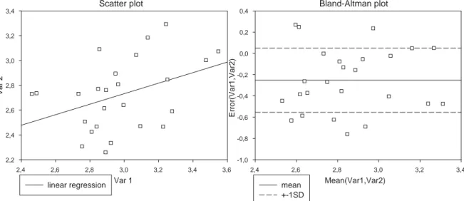
First, results firmly demonstrate that the CAT-based questionnaire cannot be used as a tool for the punctual assessment of the degree of lung obstruction in COPD. In-deed, linear regression ascertains that the questionnaire is not systematically correlated neither with any of the respiratory parameters taken singularly, nor with any of the 8192 different sets drawn from them. This confirms the typical criticism made about questionnaire-based measures consisting in the fact that the gathered information is by its nature subjective.
Second, PCA indicates that two variables, opportunely defined as linear combina-tions of the original set of 14 parameters, suffice to explain a fair percentage (64.8%) of the total information content represented by the complete set of parameters. Further-more, the first principal component is very much related to the parameters of respiratory mechanics1 whereas the second principal component is somewhat aligned with
breath-1Parameters describing the lung mechanical impedance are referred to as parameters of respiratory
Summary
ing pattern parameters2. This means that the parameters of respiratory mechanics are
the most sensitive to any deterioration or improvement of the health status and that parameters of respiratory mechanics and parameters of breathing pattern might be used to represent different aspects of the pathology.
Third, the variability analysis at different temporal scales illustrates how short-term fluctuations of parameters of respiratory mechanics might constitute a measure of the severity of the disease and, at the same time, represent the effects of pathophysio-logic mechanisms on both the central and peripheral airways. In fact, parameters of respiratory mechanics show fluctuations that are minimum in healthy subjects, mod-est in mild asthmatic patients and very strong in severe COPD patients. Differently, the long-term variability confirms that asthma and COPD are characterized by similar pathological changes in the central airways, whereas inflammatory processes in the pe-ripheral airways and in lung parenchyma are much more pronounced in COPD patients. This is suggested by the fact that fluctuations for temporal scales greater than one day are comparable in asthma and COPD only for parameters of resistance. Parameters of reactance, on the contrary, are characterized by much larger fluctuations in COPD patients.
In conclusion, this work indicates how a home daily monitoring of respiratory me-chanics may contribute to advance the understanding of COPD and to improve the clinical management of patients suffering from this disease.
2Parameters describing respiratory volumes and respiratory frequencies are referred to as parameters
Sommario
La broncopneumopatia cronica ostruttiva (COPD) rappresenta uno dei principali prob-lemi di salute pubblica. Essa `e la quarta causa causa di morbilit`a e mortalit`a negli USA e, secondo recenti studi, nel 2020 diventer`a la terza causa di morte e il quinto onere economico sanitario a livello mondiale [1–3]. L’aumentata mortalit`a `e guidata dalla sempre maggiore diffusione del fumo di tabacco a da cambiamenti nella demografia di molti paesi, con associato un incremento dell’aspettativa di vita delle popolazioni. Tra questi due aspetti, la demografia `e la causa pi`u forte di questo trend. In aggiunta,
sebbene la COPD ha ricevuto sempre maggiore attenzione da parte della comunit`a
medica negli anni recenti, questa `e ancora relativamente sconosciuta o ignorata sia dalla sanit`a pubblica che da quella privata.
Da un punto di vista patofisiologico, la COPD `e caratterizzata da una limitazione cronica del flusso respiratorio e una serie di cambiamenti patologici del tessuto pol-monare, alcuni significativi effetti extra-polmonari e importanti comorbilit`a che possono contribuire alla gravit`a della malattia. Perci`o la COPD dovrebbe essere considerata una malattia polmonare con comorbilit`a che devono essere tenute in considerazione in una valutazione diagnostica globale della sua gravit`a, e nella determinazione di un tratta-mento appropriato
L’evoluzione della condizione patologica `e descritta da periodi stabili interrotti da improvvise riacutizzazioni dei sintomi durante le quali si hanno seri processi infiamma-tori. Queste ricadute ricorrono periodicamente e spesso richiedono una ospedaliz-zazione. Una speculazione proposta indica che una osservazione a lungo-termine di parametri oggettivi potrebbe aiutare a caratterizzare la progressione temporale della malattia e, possibilmente, rilevare con il dovuto anticipo l’insorgenza di eventi estremi. Sfortunatamente la diagnosi di questa sindrome fa ancora affidamento sulla mis-urazione di indici spirometrici che, date le manovre sforzo-dipendenti richieste, non sono adatti ad un testing non supervisionato del livello di ostruzione delle vie aeree. Di conseguenza un’accurata osservazione a lungo-termine dei parametri respiratori non `e ancora disponibile nella pratica clinica.
COPD, l’associazione dei sintomi clinici con dei parametri quantitativi e l’individuazione anticipata dell’insorgenza di eventi acuti potrebbe essere basato sulla misurazione di indici legati alle propriet`a meccaniche del sistema respiratorio tramite la tecnica delle oscillazioni forzate (FOT).
Lo scopo di questo lavoro `e incrementare la conoscenza della patologia tramite l’utilizzo di un nuovo sistema commerciale di telemedicina basato sulla FOT. A questo fine l’accuratezza del dispositivo `e inizialmente valutata e validata. In seconda istanza dati clinici sono acquisiti e analizzati grazie all’impiego del sistema nel pi`u grande e pi`u dettagliato studio clinico mai definito per l’osservazione della COPD. La tesi `e divisa in tre capitoli il cui contenuto `e riassunto nelle seguenti Sezioni.
Chapter 1 - Chronic Obstructive Pulmonary Disease and assessment of lung function
Siccome questo lavoro `e basato su studi sperimentali riguardanti pazienti COPD, la maggior parte del Capitolo 1 `e focalizzato sulle caratteristiche patofisiologiche ed epi-demiologiche della COPD. In questo modo `e possibile fornire un ampio background per l’interpretazione del processo di validazione affrontato nel Capitolo 2 e l’analisi dati del Capitolo 3.
La malattia `e inizialmente definita. Successivamente, i processi che caratterizzano patofisiologia, epidemiologia e riacutizzazioni della COPD sono distinti nelle loro fasi e componenti principali. Infine, metodi clinici per la diagnosi delle malattie ostruttive e per la valutazione della loro variabilit`a temporale sono discussi. Attualmente si ha una buona comprensione del modo in cui i processi fondanti la COPD portano ai caratter-istici sintomi e anormalit`a fisiologiche. Per esempio, un ridotto FEV1 risulta causato
principalmente da una infiammazione e un restringimento delle vie aeree periferiche, mentre un ridotto scambio gassoso `e generato dalla distruzione del parenchima per via dell’enfisema. In aggiunta, il picco di flusso espiratorio (PEF) `e il tool clinico maggior-mente raccomandato nelle moderne linee guida come strumento di monitoraggio della variabilit`a temporale dei sintomi [4–6].
Poich`e gli indici spirometrici appena introdotti sono sforzo-dipendenti e forniscono dati con una povera riproducibilit`a quando questi sono eseguiti senza supervisione, si specula che risultati clinici pi`u significativi sarebbero ottenibili tramite la sostituzione dell’approccio spirometrico con l’analisi delle fluttuazioni temporali di parametri affid-abili e accurati che non richiedono l’esecuzione di manovre respiratorie forzate, come gli indici di meccanica respiratoria ottenuti tramite FOT.
Sommario
Chapter 2 - Validation of a novel telemedicine system for chronic respiratory diseases’ home monitoring
Nel Capitolo 2 un nuovo sistema di telemedicina (RESMON R PRO) sviluppato e
com-mercializzato da una societ`a spin-off del Politecnico di Milano (Restech Srl), in col-laborazione con il Laboratorio di Tecnologie Biomediche (TBM Lab) del Politecnico di Milano, `e presentato e analizzato. Il dispositivo rileva segnali di flusso e pressione all’apertura delle vie aeree e calcola il valore di impedenza dell’albero tracheobronchiale grazie all’applicazione della FOT. In aggiunta, esso valuta molti altri parametri respi-ratory e fornisce informazioni aggiuntive su ogni test effettuato.
L’analisi di accuratezza di questo dispositivo `e basata su confronti tra misure ef-fettuate da esso e misure efef-fettuate da un sistema FOT di riferimento per uso di laboratorio che `e considerato come gold standard. Tramite protocolli sperimentali op-portunamente definiti, l’accuratezza dei macro-componenti del dispositivo (front-end hardware e algoritmo di elaborazione real-time) `e valutata. Inoltre si propongono analisi per stimare la stabilit`a della calibrazione e l’efficienza dell’aspiratore che ha il compito di espellere il diossido di carbonio esalato. Ogni protocollo sperimentale `e supportato dall’applicazione di strumenti numerici di significativit`a statistica.
I risultati di stabilit`a della calibrazione indicano che il front-end mantiene le stesse caratteristiche per un lasso di tempo di almeno due mesi, e che verosimilmente la cal-ibrazione rimanga valida per periodi molto pi`u lunghi. L’aspiratore, al contrario, non mostra prestazioni molto positive in quanto non `e sufficiente ad eliminare completa-mente il diossido di carbonio presente a fine espirazione nel tubo ad alta inertanza. Di conseguenza una soluzione alternativa in grado di garantire una portata maggiore dovrebbe essere presa in considerazione.
I risultati dell’analisi di accuratezza validano pienamente il software di elaborazione dei segnali installato sul dispositivo, mentre l’accuratezza del front-end hardware non pu`o essere quantificata in termini assoluti. Ci`o `e dovuto alla variabilit`a fisiologica inter-test caratterizzante tutti i soggetti acquisiti tramite il dispositivo. In altre parole, la variabilit`a fisiologica inter-test “maschera” la vera differenza in accuratezza tra i due sistemi usati per fare confronti. Diversi indizi portano ad affermare che la vera differenza in precisione `e piuttosto piccola, ma questo non `e direttamente provabile.
Chapter 3 - Home monitoring of COPD and data analysis
Il Capitolo 3 offre una analisi di dati clinici acquisiti tramite il dispositivo FOT validato. Lo studio clinico osservazionale usato come sorgente di dati nasce dalla speculazione che un’osservazione a lungo-termine di parametri oggettivi potrebbe aiutare nel
carat-terizzare la progressione temporale della COPD e, possibilmente, rilevare con anticipo l’insorgenza di futuri eventi estremi. Lo scopo dello studio clinico `e quello di misurare la variabilit`a giornaliera di dati FOT acquisiti a casa di un gruppo di pazienti COPD al fine di identificare possibili correlazioni tra cambiamenti nei sintomi, pattern respirato-rio, impedenza meccanica polmonare e occorrenza delle riacutizzazioni. Ogni giorno, ai pazienti `e richiesto di effettuare un test alla stessa ora, possibilmente di mattina e due ore dopo l’assunzione dei farmaci. Il test consiste nella somministrazione di un
questionario che `e una versione del COPD Assessment Test (CAT) opportunamente
adattato per essere visualizzato sullo schermo del dispositivo FOT [99]. Il CAT `e ideato per misurare l’impatto della COPD sulla vita di una persona, e come questa cambia col passare del tempo. Successivamente il vero test FOT `e eseguito tramite l’invio di stimoli a multifrequenza per 3 minuti. Gli output principali di ogni test sono rappre-sentati dal total score del questionario e da 13 parametri respiratori. Il reclutamento stimato di 80 pazienti e la valutazione giornaliera da 6 a 8 mesi fa di questo studio il pi`u esteso e dettagliato trial clinico mai definito per l’osservazione della COPD.
I metodi progettati per gestire e verificare la qualit`a della grande mole di dati prodotta dallo studio clinico, come gli strumenti per la individuazione automatica degli outliers e per la stima dei dati mancanti, forniscono prestazioni molto elevate. L’utente pu`o velocemente navigare tra le acquisizioni dei vari pazienti e pu`o efficacemente visu-alizzare e correggere le acquisizioni sospette di essere outliers. Ogni qual volta un test `
e mancante, un algoritmo di regressione locale pesata stima i valori dei 13 parametri respiratori in modo da lasciare inalterate le propriet`a statistiche delle serie temporali originali. Nonostante i metodi presentati dimostrano un funzionamento molto valido, possibili miglioramenti futuri sono identificati ed elucidati.
I risultati delle analisi effettuate sui parametri, ovvero uno studio di significativit`a del questionario, l’analisi delle componenti principali (PCA) e una analisi di variabilit`a a diverse scale temporali, forniscono interessanti spunti sulla patologia nonostante il modesto numero di pazienti attualmente disponibile.
Innanzi tutto, i risultati dimostrano fermamente come il questionario non pu`o essere usato come strumento per la valutazione puntuale del grado di ostruzione polmonare nella COPD. Infatti, la regressione lineare accerta che il questionario non `e sistemati-camente correlato n`e con nessun parametro respiratorio preso singolarmente, n`e con nessuno dei 8192 diversi gruppi estratti dal set totale di parametri. Ci`o conferma la comune critica mossa verso le misure basate su questionario che consiste nel fatto che l’informazione ottenuta `e per sua natura soggettiva.
Inoltre la PCA indica che due variabili, opportunamente definite come combinazioni lineari dell’insieme originale di 14 parametri, sono sufficienti a spiegare una buona
per-Sommario
centuale (64.8%) del contenuto informativo totale rappresentato dal set completo di parametri. La prima componente principale `e molto legata ai parametri di mecca-nica respiratoria3, mentre la seconda componente principale `e piuttosto allineata con i
parametri di pattern respiratorio4. Ci`o significa che i parametri di meccanica
respirato-ria sono i pi`u sensibili a qualsiasi deterioramento o miglioramento dello stato di salute e che i parametri di meccanica respiratoria e quelli di pattern respiratorio potrebbero essere usati per rappresentare diversi aspetti della patologia.
Infine, l’analisi di variabilit`a a diverse scale temporali illustra come le fluttuazioni a breve-termine dei parametri di meccanica respiratoria potrebbero costituire una misura della gravit`a della malattia e, allo stesso tempo, rappresentare gli effetti di meccanismi patofisiologici su entrambe le via aeree centrali e periferiche. A conferma di ci`o, i parametri di meccanica respiratoria mostrano fluttuazioni che sono minime in soggetti sani, modeste in pazienti lievemente asmatici e molto forti in pazienti COPD gravi. Al contrario, la variabilit`a lungo-termine conferma che asma e COPD sono caratterizzate da simili cambiamenti patologici nella vie aeree centrali, mentre i processi infiammatori delle vie aeree periferiche e nel parenchima sono molto pi`u pronunciate nei pazienti COPD. Questo `e suggerito dal fatto che le fluttuazioni per scale temporali maggiori di un giorno sono confrontabili in asma e in COPD solo per i parametri di resistenza. I parametri di reattanza, al contrario, sono caratterizzati da fluttuazioni molto pi`u pronunciate nei pazienti COPD.
In conclusione, questo lavoro indica come un monitoraggio domiciliare della mec-canica respiratoria potrebbe contribuire ad avanzare la comprensione della COPD e migliorare la gestione clinica dei pazienti che soffrono di questa malattia.
3I parametri che descrivono l’impedenza meccanica polmonare sono chiamati parametri di
mecca-nica respiratoria
4I parametri che descrivono volumi polmonari e frequenze respiratorie sono chiamati parametri di
Introduction
Complex physiological rhythms and fluctuations characterize nearly all aspects of life [8] and appear at different spatial scales, ranging from the molecular dimension to the organ and organism level. Examples of the latter include rhythmic heart beat, daily cycle of sleep and wakefulness and respiration. Interestingly, most biological fluctuations are not strictly periodic but show irregular patterns over many time scales [9]. Moreover, they interact not only with each other but with the external environment, which itself also exhibits substantial irregular fluctuations.
During the entire course of life, each organism tries to maintain a constant inter-nal state by regulating its physiological processes. This dynamic self control is usually called homeostasis. However, the maintenance of homeostasis requires some controlled parameters have a range of permitted values, and these parameters continuously oscil-late within its boundaries. Therefore, a more appropriate term is homeokinesis, that is the ability of an organism interacting with a variable external environment to main-tain an organized internal environment, which fluctuates within acceptable limits by dissipating energy in a state far from thermodynamic equilibrium [10]. This definition implies that, while continuous fluctuations are part of normal life, alterations in the fluctuations always signify abnormal physiology. The disruption of such rhythms often leads to collapse or even the death of the organism [11, 12].
If homeokinesis is healthy and pathology is lack or excess of fluctuations in some observed parameters, it is clear that variability itself contains an encoded message that needs to be deciphered, quantified, analyzed and understood. Understanding this message should shed light into what is considered health and which are the mechanisms that bring to illness.
Due to the complexity of natural and biological feedbacks and the presence of un-known environmental factors, it is becoming clear that variability can not be analyzed using a classic reductionist approach. This is noticeably true also for the altered vari-ability observed in the principal chronic respiratory pathologies, bronchial asthma and chronic obstructive pulmonary disease (COPD).
that 300 millions of people have asthma (Global Initiative for Asthma 2009) and 80 millions suffer from COPD (World Health Organization 2006). Asthma has become more common in recent decades; its prevalence has been associated with an increase in atopic sensitization and is in parallel with the increase of other allergic disorders such as rhinitis. COPD is the fourth leading cause of death in the world and increases in its prevalence and mortality are expected in the next future [13].
Many features of these diseases, including pathogenesis and progression, are not fully understood. Indeed, COPD and asthma are characterized by high variability of symptoms over time and a proper and accurate evaluation of patients’ condition at any point in time would require frequent, at least daily, assessment of their disease. However, new objective parameters for the follow up of their severity and variability were still needed. A strategy with more than one observed index or including statisti-cal measures of objective parameters with time should provide a more comprehensive picture of the pathology and its progression and it would eventually help improving patients’ quality of life and, potentially, their life expectancy.
Telemedicine and home-monitoring devices have been addressed as possible solu-tions for the chronic stabilization of patients suffering from chronic respiratory dis-eases. However, despite recent advances in medical technology, the assessment and management of asthma and COPD still relied on tests performed with spirometry or peakflow meters, which did not provide data consistent with the quality requirements of international guidelines when the measurements are performed in unsupervised en-vironments [14, 15].
A different and innovative multidimensional approach for the characterization of the variability and the progression of asthma and COPD over time could be based on the measurement of parameters related to the mechanical properties of the respiratory sys-tem by forced oscillation technique (FOT). FOT constitutes a valid solution to develop a new model for a better management of such patients: as a tool for the investiga-tion of respiratory mechanics in clinical practice, it is well supported theoretically and has the advantage of being a non-invasive, versatile method and demanding minimal cooperation of the patient. For these reasons, FOT has been indicated as a potential alternative to spirometry for home assessment of respiratory function [16, 17].
Accordingly, the purpose of the work here presented is to:
• validate a new strategy for the analysis of the temporal variability of asthma and COPD, based on the development of a novel FOT telemedicine system for chronic respiratory diseases’ home monitoring;
• analyze clinical data from COPD patients at stage 3 and 4 of GOLD classification measured with the device.
Introduction
The main outcomes are positive feedback about the reliability of the device and new insights into the pathology despite the modest amount of patients currently available.
Chapter 1
Chronic Obstructive Pulmonary
Disease and assessment of lung
function
1
Introduction
Chronic obstructive pulmonary disease (COPD) is a chronic respiratory disorder char-acterized by the presence of airflow limitation [13]. The pathology consists by a chronic inflammation of the respiratory tract that is mediated by increased expression of mul-tiple inflammatory proteins, including cytokines, chemokines, adhesion molecules, in-flammatory enzymes and receptors.
The progression of the pathological condition is described by stable periods dis-rupted by sudden exacerbations of the symptoms during which a severe inflammatory process occurs. Such aggravations recur in a periodic fashion and often require hospi-talization.
Worldwide, cigarette smoking is the most commonly encountered risk factor for COPD, even though in many countries air pollution resulting from the burning of wood and other biomass fuels has also been identified as a COPD risk factor.
As the present work is based on experimental studies involving COPD patients, the majority of this Chapter will focus on the pathophysiologic and epidemiologic character-istics of COPD. Successively, a separate section will introduce a non-invasive methods useful to monitor the progression of the pathology on a daily, namely the forced oscil-lation technique (FOT).
2
Definition of Chronic Obstructive Pulmonary
Dis-ease
Internationally accepted opinion has defined the Chronic Obstructive Pulmonary Dis-ease (COPD) as a disDis-ease state characterized by chronic airflow limitation due to chronic bronchitis and emphysema [18]. Chronic bronchitis has been defined in clinical terms: the presence of chronic productive cough for at least 3 consecutive months in 2 consecutive years. Other causes of chronic productive cough must be ruled out. Emphysema, on the other hand, has been defined by its pathologic description: an abnormal enlargement of the air spaces distal to the terminal bronchioles accompa-nied by destruction of their walls and without obvious fibrosis. The latest ATS1/ERS2
guidelines, like the GOLD3 guidelines, have parted from this traditional description of
COPD. Similar to the changes in the definition of asthma by the NHLBI4, the defini-tion of COPD has undergone major revision. COPD, like asthma, is now recognized as an inflammatory disease of the airways [22]. This is supported by extensive clinical and basic science research showing that asthma and COPD have different and distinct cellular and inflammatory mediator profiles. The current ATS/ERS definition reflects these scientific advances:
Chronic obstructive pulmonary disease (COPD) is a preventable and treatable dis-ease characterized by airflow limitation that is not fully reversible. The airflow limitation is usually progressive and is associated with an abnormal inflammatory response of the lungs to noxious particles or gases, primarily caused by cigarette smoking. Although COPD affects the lungs, it also produces significant systemic consequences [20].
While the current guidelines do not specifically include chronic bronchitis and em-physema in the definition of COPD, it is made clear that they are considered the predominant causes of COPD.
1American Thoracic Society 2European Respiratory Society
3Global Initiative for Chronic Obstructive lung Disease 4National Heart, Lung, and Blood Institute
1.3 Pathogenesis, Pathology and Pathophysiology of COPD
3
Pathogenesis, Pathology and Pathophysiology of
COPD
The chronic airflow limitation characteristic of COPD is caused by a mixture of small airway disease (obstructive bronchiolitis) and parenchymal destruction (emphysema), the relative contributions of which vary from subject to subject (see Figure 1.1). Chronic inflammation causes structural changes and narrowing of the small airways. Destruction of the lung parenchyma, also by inflammatory processes, leads to the loss of alveolar attachments to the small airways and decreases lung elastic recoil. In turn, these changes diminish the ability of the airways to remain open during expiration.
Figure 1.1: Mechanisms underlying airflow limitation in COPD.
Emphysema, or destruction of the gasexchanging surfaces of the lung (alveoli), is a pathological term that is often (but incorrectly) used clinically and describes only one of several structural abnormalities present in patients with COPD. Chronic bronchitis, or the presence of cough and sputum production for at least 3 months in each of two con-secutive years, remains a clinically and epidemiologically useful term as well. However, it does not reflect the major impact of airflow limitation on morbidity and mortality in COPD patients. It is also important to recognize that cough and sputum produc-tion may precede the development of airflow limitaproduc-tion but, conversely, some patients develop significant airflow limitation without chronic cough and sputum production.
3.1
Pathogenesis
The inflammation in the respiratory tract of COPD patients appears to be an amplifi-cation of the normal inflammatory response of the respiratory tract to chronic irritants such as cigarette smoke. The mechanisms for this amplification are not yet understood but may be genetically determined. Some patients develop COPD without smoking, but the nature of the inflammatory response in these patients is unknown [21]. Lung inflammation is further amplified by oxidative stress and an excess of proteinases in the lung. Together, these mechanisms lead to the characteristic pathological changes in COPD exhibited in Figure 1.2.
Figure 1.2: Pathogenesis of COPD.
Inflammation
COPD is characterized by a specific pattern of inflammation involving neutrophils, macrophages and lymphocytes [22]. These cells release inflammatory mediators and interact with structural cells in the airways and lung parenchyma.
The wide variety of inflammatory mediators that have been shown to be increased in COPD patients attract inflammatory cells from the circulation (chemotactic factors), amplify the inflammatory process (proinflammatory cytokines), and induce structural changes (growth factors) [23].
1.3.2 Pathology
Oxidative stress
Oxidative stress may be an important amplifying mechanism in COPD [24]. Biomarkers of oxidative stress (e.g., hydrogen peroxide, 8-isoprostane) are increased in the exhaled breath condensate, sputum, and systemic circulation of COPD patients. Oxidative stress is further increased in exacerbations. Oxidants are generated by cigarette smoke and other inhaled particulates, and released from activated inflammatory cells such as macrophages and neutrophils. There may also be a reduction in endogenous antiox-idants in COPD patients. Oxidative stress has several adverse consequences in the lungs, including activation of inflammatory genes, inactivation of antiproteases, stim-ulation of mucus secretion, and stimstim-ulation of increased plasma exudation. Many of these adverse effects are mediated by peroxynitrite, which is formed via an interaction between superoxide anions and nitric oxide. In turn, the nitric oxide is generated by inducible nitric oxide synthase, which is expressed in the peripheral airways and lung parenchyma of COPD patients. Oxidative stress may also account for a reduction in histone deacetylase activity in lung tissue from COPD patients, which may lead to en-hanced expression of inflammatory genes and also a reduction in the antiinflammatory action of glucocorticosteroids [25].
Protease-antiprotease imbalance
There is compelling evidence for an imbalance in the lungs of COPD patients between proteases that break down connective tissue components and antiproteases that protect against this. Several proteases, derived from inflammatory cells and epithelial cells, are increased in COPD patients. There is increasing evidence that they may interact with each other. Protease-mediated destruction of elastin, a major connective tissue component in lung parenchyma, is an important feature of emphysema and is likely to be irreversible.
3.2
Pathology
Pathological changes characteristic of COPD are found in the central airways, peripheral airways, lung parenchyma, and pulmonary vasculature [26]. In patients with chronic bronchitis, an inflammatory exudate of fluid and cells infiltrates the epithelium lining the central airways and the associated glands and ducts.
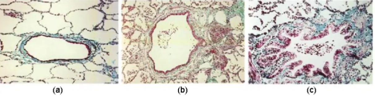
Figure 1.3 shows how the early decline in lung function in COPD is correlated with inflammatory changes in the peripheral airways similar to those that occur in the central airways [27]: exudate of fluid and cells in the airway wall and lumen, goblet
and squamous cell metaplasia of the epithelium, edema of the airway mucosa due to inflammation and excess mucus in the airways due to goblet cell metaplasia. However, the most characteristic change in the peripheral airways of patients with COPD is airway narrowing.
Figure 1.3: Histological section of peripheral airways. (a) Section from a sigarette smoker with normal lung function showing a nearly normal airway with small number of inflammatory cells. (b) Section from a patient with small airway disease showing inflammatory exudate in the wall and lumen of the airway. (c) Section showing more advanced small airway disease, with reduced lumen causing structural reorganization of the airway wall, increased smooth muscle and deposition of peribronchial connective tissue.
Inflammation leads to repeated cycles of injury and repair of the walls of the periph-eral airways. This injury then initiates repair processes. Although airway repair is only partly understood, it seems likely that disordered repair processes can lead to tissue remodeling with altered structure and function [28]. This injury-and-repair process re-sults in a structural remodeling of the airway wall, with increasing collagen content and scar tissue formation that narrows the lumen and produces fixed airways obstruction. The peripheral airways become the major site of airways obstruction in COPD [29].
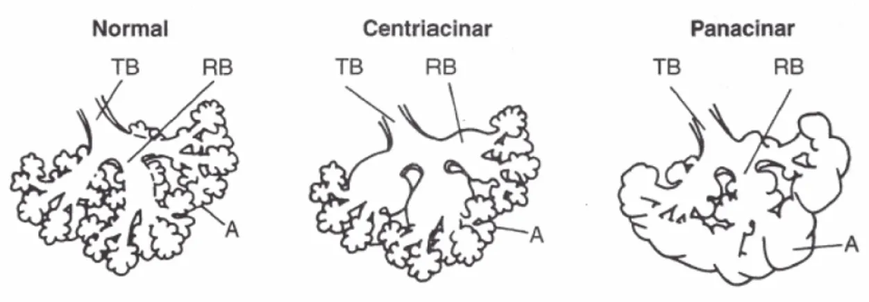
The most common type of parenchymal destruction in COPD patients is the cen-triacinar form of emphysema (see Figure 1.4), which involves dilatation and destruction of the respiratory bronchioles [30]. These lesions occur more frequently in the upper lung regions in milder cases. Panacinar emphysema, which extends throughout the acinus, involves dilatation and destruction of the alveolar ducts and sacs as well as the respiratory bronchioles. It tends to affect the lower more than the upper lung regions. Pulmonary vascular changes in COPD are characterized by a thickening of the ves-sel wall that begins early in the natural history of the disease, followed by an increase in vascular smooth muscle and the infiltration of the vessel wall by inflammatory cells. Since endothelium plays an important role in regulating vascular tone and cell
prolif-1.3.3 Pathophysiology
Figure 1.4: Centriacinar and panacinar emphysema. Notice that in centriacinar em-physema the destruction is confined to the terminal and respiratory bronchioles (TB and RB). In panacinar emphysema the peripheral alveoli (A) are also involved.
eration, it is likely that endothelial dysfunction might initiate the sequence of events that results ultimately in structural changes. These structural changes are correlated with an increase in pulmonary vascular pressure that develops first with exercise and then at rest. As COPD worsens, greater amounts of smooth muscle, proteoglycans, and collagen further thicken the vessel wall. In advanced disease, the changes in the muscular arteries may be associated with emphysematous destruction of the pulmonary capillary bed.
3.3
Pathophysiology
There is now a good understanding of how the underlying disease process in COPD leads to the characteristic physiologic abnormalities and symptoms. For example, decreased FEV1 primarily results from inflammation and narrowing of peripheral airways, while
decreased gas transfer arises from the parenchymal destruction of emphysema.
Airflow limitation and air trapping
The extent of inflammation, fibrosis, and luminal exudates in small airways is correlated with the reduction in FEV1 and FEV1/FVC ratio, and probably with the accelerated
decline in FEV1 characteristic of COPD [31]. This peripheral airway obstruction
pro-gressively traps air during expiration, resulting in hyperinflation.
Although emphysema is more associated with gas exchange abnormalities than with reduced FEV1, it does contribute to air trapping during expiration. This is especially so
as alveolar attachments to small airways are destroyed when the disease becomes more severe. Hyperinflation reduces inspiratory capacity such that functional residual capac-ity increases, particularly during exercise (when this abnormalcapac-ity is known as dynamic hyperinflation), and this results in dyspnea and limitation of exercise capacity. It is now thought that hyperinflation develops early in the disease and is the main mechanism for exertional dyspnea [32]. Bronchodilators acting on peripheral airways reduce air trap-ping, thereby reducing lung volumes and improving symptoms and exercise capacity. Gas exchange abnormalities
Gas exchange abnormalities result in hypoxemia and hypercapnia, and have several mechanisms in COPD. In general, gas transfer worsens as the disease progresses. The severity of emphysema correlates with arterial PO2 and other markers of
ventilation-perfusion (VA/Q) imbalance. Peripheral airway obstruction also results in VA/Q
im-balance, and combines with ventilatory muscle impaired function in severe disease to reduce ventilation, leading to carbon dioxide retention. The abnormalities in alveolar ventilation and a reduced pulmonary vascular bed further worsen the VA/Q
abnormal-ities.
Mucus hypersecretion
Mucus hypersecretion, resulting in a chronic productive cough, is a feature of chronic bronchitis and is not necessarily associated with airflow limitation. Conversely, not all patients with COPD have symptomatic mucus hypersecretion. When present, it is due to mucous metaplasia with increased numbers of goblet cells and enlarged submucosal glands in response to chronic airway irritation by cigarette smoke and other noxious agents. Several mediators and proteases stimulate mucus hypersecretion and many of them exert their effects through the activation of epidermal growth factor receptor (EGFR) [33].
Pulmonary hypertension
Mild to moderate pulmonary hypertension may develop late in the course of COPD and is due to hypoxic vasoconstriction of small pulmonary arteries, eventually resulting in structural changes that include intimal hyperplasia and later smooth muscle hyper-trophy/hyperplasia [34]. There is an inflammatory response in vessels similar to that seen in the airways and evidence for endothelial cell dysfunction.
The loss of the pulmonary capillary bed in emphysema may also contribute to increased pressure in the pulmonary circulation. Progressive pulmonary hypertension
1.4 Epidemiology of COPD
may lead to right ventricular hypertrophy and eventually to right-side cardiac failure (cor pulmonale).
Systemic features
It is increasingly recognized that COPD involves several systemic features, particularly in patients with severe disease, which have a major impact on survival and comorbid diseases [35, 36]. These are:
• Cachexia: loss of fat free mass.
• Skeletal muscle wasting: apoptosis, disuse atrophy. • Osteoporosis.
• Depression.
• Normochromic normocytic anemia.
• Increased risk of cardiovascular disease: associated with increase in C-reactive protein (CRP).
4
Epidemiology of COPD
In the past, imprecise and variable definitions of COPD have made it difficult to quantify prevalence, morbidity and mortality. Furthermore, the underrecognition and underdiag-nosis of COPD lead to significant underreporting. The extent of the underreporting varies across countries and depends on the level of awareness and understanding of COPD among health professionals, the organization of health care services to cope with chronic diseases, and the availability of medications for the treatment of COPD [37].
4.1
Prevalence
Existing COPD prevalence data show remarkable variation due to differences in survey methods, diagnostic criteria and analytic approaches [38, 39]. Despite these complex-ities, data are emerging that enable some conclusions to be drawn regarding COPD prevalence. A systematic review and meta-analysis of studies carried out in 28 countries between 1990 and 2004 [38], and an additional study from Japan [40], provide evidence that the prevalence of COPD (Stage I: Mild COPD and higher) is appreciably higher in smokers and ex-smokers than in nonsmokers, in those over 40 years than those under 40, and in men than in women.
The Latin American Project for the Investigation of Obstructive Lung Disease (PLATINO) examined the prevalence of post-bronchodilator airflow limitation (Stage
I: Mild COPD and higher) among persons over age 40 in five major Latin American cities each in a different country (Brazil, Chile, Mexico, Uruguay, and Venezuela). In each country, the prevalence of Stage I: Mild COPD and higher increased steeply with age, with the highest prevalence among those over 60 years, ranging from a low of 18.4% in Mexico City, Mexico to a high of 32.1% in Montevideo, Uruguay. In all cities/countries the prevalence was appreciably higher in men than in women. The reasons for the differences in prevalence across the five Latin American cities are still under investigation [41].
In 12 Asia-Pacific countries and regions a study based on a prevalence estimation model indicated a mean prevalence rate for moderate to severe COPD among individ-uals 30 years and older of 6.3% for the region. The rates varied twofold across the 12 Asian countries and ranged from a minimum of 3.5% (Hong Kong and Singapore) to a maximum of 6.7% (Vietnam) [42].
4.2
Morbidity
Morbidity measures traditionally include physician visits, emergency department visits, and hospitalizations. Although COPD databases for these outcome parameters are less readily available and usually less reliable than mortality databases, the limited data available indicate that morbidity due to COPD increases with age and is greater in men than in women [43–45]. In these data sets, however, COPD in its early stages (Stage I: Mild COPD and Stage 2: Moderate COPD) is usually not recognized, diagnosed, or treated, and therefore may not be included as a diagnosis in a patient’s medical record. Morbidity from COPD may be affected by other comorbid chronic conditions [46] (e.g., musculoskeletal disease, diabetes mellitus) that are not directly related to COPD but nevertheless may have an impact on the patient’s health status, or may negatively interfere with COPD management. In patients with more advanced disease (Stage III: Severe COPD and Stage IV: Very Severe COPD), morbidity from COPD may be misattributed to another comorbid condition.
Another way of estimating the morbidity burden of disease is to calculate years of living with disability (YLD). The Global Burden of Disease Study estimates that COPD results in 1.68 YLD per 1,000 population, representing 1.8% of all YLDs, with a greater burden in men than in women (1.93% vs. 1.42%) [1–3].
1.4.3 Mortality
4.3
Mortality
The World Health Organization (WHO) publishes mortality statistics for selected causes of death annually for all WHO regions. Data must be interpreted cautiously, however, because of inconsistent use of terminology for COPD. Nowadays the problem of labeling the disease has been partly solved, but underrecognition and underdiagnosis of COPD still affect the accuracy of mortality data. Although COPD is often a primary cause of death, it is more likely to be listed as a contributory cause of death or omitted from the death certificate entirely, and the death attributed to another condition such as cardiovascular disease.
Despite the problems with the accuracy of the COPD mortality data, it is clear that COPD is one of the most important causes of death in most countries. The Global Burden of Disease Study has projected that COPD, which ranked sixth as the cause of death in 1990, will become the third leading cause of death worldwide by 2020 [1–3]. This increased mortality is driven by the expanding epidemic of smoking and the changing demographics in most countries, with more of the population living longer. Of these two forces, demographics is the stronger driver of the trend.
Trends in mortality rates over time provide further important information but, again, these statistics are greatly affected by terminology, awareness of the disease, and po-tential gender bias in its diagnosis. COPD mortality trends generally track several decades behind smoking trends. Trends in age-standardized death rates for the six leading causes of death in the United States from 1970 through 2002 [47] indicates that while mortality from several of these chronic conditions declined over that period, COPD mortality increased (refer to Figure 1.5). Death rates for COPD in Canada, in both men and women, have also been increasing since 1997. In Europe, however, the trends are different, with decreasing mortality from COPD already being seen in many countries [48]. There is no obvious reason for the difference between trends in North America and Europe, although presumably factors such as awareness, changing terminology, and diagnostic bias contribute to these differences.
The mortality trends for COPD have been particularly striking for women. In
Canada, the death rate from COPD among women accelerated in the 1990s and is expected to soon overtake the rate among men [45]. In the United States, COPD deaths among women have been rising steeply since the 1970s. In 2000, the number of deaths from COPD in the United States was greater among women than men (59,936 vs. 59,118), although the mortality rates among women remain somewhat lower than among men [49].
Figure 1.5: Trends in Age-standardized Death Rates for the 6 Leading Causes of Death in the United States, 1970-2002 [47].
Burden of Disease Study [1–3] projected baseline, optimistic, and pessimistic models for COPD mortality from 1990 to 2020 that take into account the expected aging of the world’s population, projected increases in smoking rates, and projected declines in other causes of death such as diarrheal and HIV-related diseases.
4.4
Risk factors
The identification of risk factors is an important step towards the development of strate-gies for prevention and treatment of any disease. The division into host factors and exposures reflects the current understanding of COPD as resulting from an interaction between the two types of factors.
A list of host factors is:
• Genes (e.g., alpha-1 antitrypsin deficiency). • Airway hyperresponsiveness.
• Lung growth. A list of exposures is:
• Direct and environmental tobacco smoke. • Occupational dust and chemicals.
• Indoor and outdoor air pollution. • Infections.
1.5 Exacerbations of COPD
The best-documented host factor is a severe hereditary deficiency of alpha-1 antit-rypsin, a major circulating inhibitor of serine proteases. This severe deficiency causes premature and accelerated development of panlobular emphysema and decline in lung function. Asthma and airway hyperresponsiveness, identified as risk factors that con-tribute to the development of COPD, are complex disorders related to a number of ge-netic and environmental factors. Reduced maximal attained lung function (as measured by spirometry) may identify individuals who are at increased risk for the development of COPD.
The major environmental risk factors are tobacco smoke [50], occupational dusts and chemicals (vapors, irritants, fumes), and indoor and outdoor air pollution. However, it is very difficult to demonstrate that a given risk factor is sufficient to cause the disease.
5
Exacerbations of COPD
As already mentioned COPD is characterized by a progressive and irreversible decline in lung function, breathlessness and other respiratory symptoms (e.g., cough and sputum production), and a deterioration of health status [51]. In addition to their chronic disease, patients with COPD often experience regular acute exacerbations (typically around 2-3 per year) that increase in frequency with increased disease severity [51, 52]. These debilitating exacerbations have a substantial impact on patients and on healthcare systems. COPD exacerbations are also associated with considerable phys-iologic deterioration and increased airway inflammatory changes that are caused by a variety of factors such as viruses, bacteria, common pollutants and low tempera-tures [53]. COPD exacerbations are more common in the winter months and there may be important interactions between cold temperatures and exacerbations caused by viruses or pollutants.
Definition of a COPD exacerbation
Despite their importance, there is no standardized, universally accepted definition for
COPD exacerbations. This situation reflects the multifactorial, heterogenous and
poorly understood pathophysiology of exacerbations and the difficulty in differentiating true exacerbations from normal day-to-day variations of COPD. As a result, exacerba-tions remain poorly recognized and often poorly treated.
One of most widely known definitions of COPD exacerbations is that proposed by Anthonisen et al. in 1987 [54]. This definition is based on the presence of three spe-cific symptoms in patients with COPD, namely increased dyspnoea, sputum volume
and sputum purulence. Three sub-types of exacerbations were defined: type 1, occur-rence of all three symptoms; type 2, occuroccur-rence of two out of the three symptoms; and type 3, occurrence of one of the three symptoms in addition to at least one of the following: recent upper respiratory infection, fever, increased wheezing, increased cough, or increased respiratory or heart rate.
Epidemiology of COPD exacerbation
Exacerbations are an important cause of hospital admission and have a considerable impact on quality of life and activities of daily living of the patients.
Patients with COPD are accustomed to frequent symptom changes and this may explain their tendency to under report exacerbations to physicians. Some studies of patient with acute infective exacerbations of chronic bronchitis found that exacerbation frequency is an important determinant of health status in COPD and is thus one of the important outcome measures in COPD. Factors predictive of frequent exacerbations included daily cough, sputum and frequent exacerbations in the previous year [55]. Falls in peak flow and FEV1 at exacerbation are generally small and not useful in
predicting exacerbations, but larger falls in peak flow are associated with symptoms of dyspnoea, common colds and related to a longer recovery time from exacerbations. The combination of the symptoms of increased dyspnoea and the common cold at exacerbation with a prolonged exacerbation recovery suggests that viral infections may lead to more prolonged exacerbation.
The reasons for the incomplete recovery of symptoms and lung function are not clear, but may involve inadequate treatment or persistence of the causative agent. The incomplete physiologic recovery after an exacerbation could contribute to the decline in lung function with time. A study reveals that patients with a history of frequent exacerbations had a faster decline of FEV1 compared to patients with a history of
infrequent exacerbations [56].
Mechanisms underlying exacerbations
Although it has been assumed that exacerbations are associated with increased airway inflammation, there has been little information available on the nature of inflammatory markers, especially when studied closed to an exacerbation, as performing bronchial biopsies at exacerbation is difficult in patients with moderate to severe COPD.
Exacerbations may be triggered by a variety of factors, including viral or bacterial infection and air pollution. COPD exacerbations are frequently associated with upper respiratory tract infections and these are more common in the winter months, when
1.5 Exacerbations of COPD
there are more respiratory viral infections in the community. Recent studies have shown that around half of COPD exacerbations were associated with viral infections and that the majority of these were due to rhinovirus, that is, viral exacerbations were associated with symptomatic colds and prolonged recovery [55, 57]. This finding is in agreement with the data that respiratory viruses produce longer and more severe exacerbations and have a major impact on healthcare utilization [58].
Airway bacterial colonizations have been found in approximately 30% of COPD patients, and these colonizations have been shown to be related to the degree of airflow obstruction and current cigarette smoking status. Bacterial colonization in COPD may be an important determinant of airway inflammation and therefore further long-term studies are required to determine whether bacterial colonization predisposes to decline in lung function, characteristic of COPD.
Pathophysiologic changes at COPD exacerbation
As seen before, the mechanical performance of respiratory muscles is reduced in patients with moderate to severe COPD. The airflow obstruction leads to hyperinflation, so that the respiratory muscles work far from the optimal extension and generate reduced inspi-ratory pressure. The load on the respiinspi-ratory muscles is also increased in patients with airflow obstruction given by the presence of intrinsic positive end-expiratory pressure (iPEEP). With an exacerbation of COPD, the increase in airflow obstruction will further increase the load on the respiratory muscles and increase the work of breathing, precip-itating respiratory failure in more severe cases. Minute ventilation may be normal, but the respiratory pattern will be irregular with increased frequency and decreased tidal volume. The resultant hypercapnia and acidosis will then reduce inspiratory muscles function, contributing to further deterioration of the respiratory failure.
Hypoxemia in COPD usually occurs due to a combination of ventilation-perfusion mismatch and hypoventilation, although arteriovenous shunting can also contribute to the acute setting. This causes an increase in pulmonary artery pressure, which can lead to salt and water retention and the development of edema. The degree of the ventilation-perfusion abnormalities increases during acute exacerbations and resolves over the following few weeks. Acidosis is an important prognostic factor in survival from respiratory failure during a COPD exacerbation and thus early correction of acidosis is an essential goal of therapy.
6
Diagnosis and assessment of COPD
COPD is a typical mid-to-late age pathology, especially observed in smokers or ex-smokers [59]. Chronic cough may precede the development of airway limitation by many years [13], even if some patients with limited airflow do not present any form of cough. The majority of patients seek medical attention after the development of dyspnea, which is usually persistent and becomes progressively more serious during everyday activities or at rest.
Airflow limitation is best measured by spirometry, as this is the most widely avail-able, reproducible test of lung function. Although it does not fully capture the impact of COPD on a patient’s health, it remains the gold standard for diagnosing the disease and monitoring its progression in specialized labs.
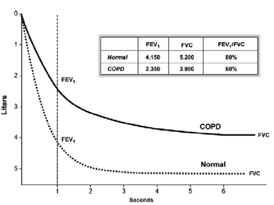
The main spirometric indices include the volume of air forcibly exhaled from the point of maximal inspiration (forced vital capacity, FVC), the volume of air exhaled during the first second of this maneuver (forced expiratory volume in one second, FEV1),
the peak of expiratory flow (PEF) and the FEV1/FVC ratio. Spirometry measurements
are evaluated by comparison with reference values based on age, height, sex and race. Figure 1.6 shows a normal spirogram and a spirogram typical of patients with mild
to moderate COPD. Patients with COPD typically show a decrease in both FEV1 and
FVC. The degree of spirometric abnormality generally reflects the severity of COPD. The presence of a postbronchodilator FEV1<80% of the predicted value in combination
with an FEV1/FVC<70% confirms the presence of airflow limitation that is not fully
reversible [13]. The FEV1/FVC on its own is a more sensitive measure of airflow
limitation and an FEV1/FVC<70% is considered an early sign of airflow limitation in
patients whose FEV1 remains normal (≥80% predicted). This approach in defining
airflow limitation is considered pragmatic in view of the fact that universally applicable reference values for FEV1 and FVC are not available.
Assessment of COPD severity is based on the patient’s level of symptoms, the sever-ity of the spirometric abnormalsever-ity, and the presence of complications such as respiratory failure, right heart failure, weight loss and arterial hypoxemia. The characteristic symp-toms of COPD are cough, sputum production, and dyspnea upon exertion. COPD has a variable natural history and not all individuals follow the same course, however, it is generally a progressive disease, especially if a patient’s exposure to noxious agents continues. A simple classification of disease severity into five stages is presented [60]:
• Stage 0: At Risk - Characterized by chronic cough and sputum production. Lung function, as measured by spirometry, is still normal.
1.6 Diagnosis and assessment of COPD
Figure 1.6: Normal spirogram and spirogram typical of patients with moderate COPD.
• Stage I: Mild COPD - Characterized by mild airflow limitation (FEV1/FVC<70%
but FEV1>80% predicted) and usually, but not always, by chronic cough and
sputum production. At this stage, the individual may not even be aware that his or her lung function is abnormal.
• Stage II: Moderate COPD - Characterized by worsening airflow limitation (50%≤FEV1<80%
predicted) and usually progression of symptoms, with shortness of breath typi-cally developing on exertion. This is the stage at which patients typitypi-cally seek medical attention because of dyspnea or an exacerbation of their disease. • Stage III: Severe COPD - Characterized by further worsening of airflow
limita-tion (30%≤FEV1<50% predicted), increased shortness of breath, and repeated
exacerbations which have an impact on patients’ quality of life.
• Stage IV: Very Severe COPD - Characterized by severe airflow limitation (FEV1<30%
predicted) or the presence of chronic respiratory failure.
The staging is based on airflow limitation as measured by spirometry. Specific FEV1
cut-points (e.g., <80% predicted) are used for purposes of simplicity.
Respiratory failure may also lead to effects on the heart such as cor pulmonale (right heart failure). At this stage, quality of life is very appreciably impaired and exacerbations can threaten the life of the patient.
The diagnosis and staging of chronic respiratory diseases with spirometry have some limitations. As asthma and COPD are characterized by abnormal temporal variability of symptoms and fluctuations in the inflammatory state of the airways, the one-time punctual assessment of lung function in hospitals do not provide a comprehensive evaluation of the severity and progression. Second, all the spirometric indices are obtained using effort-dependent maneuvers that do require the supervision of clinicians or trained technician for the evaluation of their correctness and accuracy. However, also in this condition, some elderly patients and children may have difficulty completing spirometry with the desired quality standards [14, 15].
7
Assessment of temporal variability of airflow
limi-tation and disease progression
Asthma and COPD are characterized by a high and abnormal variability of symptoms over time. An elegant mechanicistic definition of life considers an organism as an entity interacting with a complex and fluctuating external environment, continuously trying to maintain a state of internal dynamic equilibrium known as homeokinesis [10]. While fluctuations of physiological parameters are part of normal life, excess or lack of variability is normally associated to pathology or, eventually, to death. This is true also for asthma and COPD, where abnormal temporal variability of airway caliber and significant changes in functional and structural parameters are often observed [61, 62]. These observations suggest that a correct understanding of the disease and an accurate evaluation of its severity can not be based just on one-time assessment of clinical symptoms and on a punctual measurement of pulmonary function. A more comprehensive strategy, including statistical measures of objective parameters with time should provide a more comprehensive and clear picture of the pathology and its progression.
The clinical tool recommended in the most modern guidelines as a monitoring in-strument for the measurement of such variability is the peak expiratory flow (PEF) meter [63]. PEF is the highest flow obtained during a forced expiration, starting im-mediately after a deep inspiration to the total lung capacity. However, PEF meters have some limitations that have prevented the development of accurate self-testing at home, a desirable goal in the chronic stabilization of patients suffering from airways obstruction [64, 65]. First, even if PEF meters provide data with good temporal reso-lution, many patients may have difficulty in executing correctly the forced maneuvers without the supervision of nurses, clinicians or trained technicians. Second, PEF is

![Figure 1.5: Trends in Age-standardized Death Rates for the 6 Leading Causes of Death in the United States, 1970-2002 [47].](https://thumb-eu.123doks.com/thumbv2/123dokorg/7508959.105022/32.892.125.751.139.426/figure-trends-standardized-death-leading-causes-united-states.webp)