Università di Pisa
Scuola di Dottorato in Tecnologie per la Salute: Valutazione
e Gestione delle Innovazioni nel Settore Biomedicale
Presidente: Prof. Mauro Ferrari
Tesi di Dottorato di Ricerca
Settore Scientifico-Disciplinare: MED/36
CT Coronary Angiography with 100kV tube voltage and a low
noise reconstruction filter in non-obese patients: evaluation of
radiation dose and diagnostic quality of 2D and 3D image
reconstructions using open source software (OsiriX)
Tutor:
Dr. Emanuele Neri
Candidato:
Dr. Lorenzo Faggioni
CONTENTS
Abstract... pp. i-ii Introduction... p. 1 Materials and Methods... p. 4
Patient selection... p. 4 CTCA acquisition protocol... p. 5 Image analysis... p. 6
CORONARY SEGMENTATION AND EVALUATION OF IMAGE QUALITY OF 2D AND 3D RECONSTRUCTIONS....p. 6 CALCULATION OF INTRAVASCULAR DENSITY, SNR, CNR, AND EFFECTIVE DOSE... p. 11
Statistical analysis... p. 13
Results... p. 14 Discussion... p. 18 References... p. 24
CT Coronary Angiography with 100kV tube voltage and a low noise reconstruction filter in non-obese patients: evaluation of radiation dose and diagnostic quality of 2D
ABSTRACT
INTRODUCTION AND PURPOSE. Computed tomography coronary angiography (CTCA) has seen a dramatic evolution in the last decade owing to the availability of multislice CT scanners with 64 detector rows and beyond. However, this evolution has been paralleled by an increase in radiation dose to patients, that can reach extremely high levels (>20mSv) when retrospective ECG-gating techniques are used. On CT angiography, reduction of tube voltage allows to cut radiation dose with improved contrast resolution due to the lower energy of the X-ray beam and increased photoelectric effect. Our purpose is twofold: 1) to evaluate the radiation dose of CTCA studies carried out using a tube voltage of 100kV and a low noise reconstruction filter, compared with a conventional tube voltage of 120kV and a standard reconstruction kernel; 2) to assess the impact of the 100kV acquisition technique on the diagnostic quality of 2D and 3D image reconstructions performed with open source software (OsiriX).
MATERIALS AND METHODS. Fifty-one non-obese patients underwent CTCA on a 64-row CT scanner. Out of them, 28 were imaged using a tube voltage of 100kV and a low noise reconstruction filter, while in the remaining 23 patients a tube voltage of 120kV and a standard reconstruction kernel were selected. All CTCA datasets were exported via PACS to a Macintosh™ computer (iMac™) running OsiriX 4.0 (64-bit version), and Maximum Intensity Projection (MIP), Curved Planar Reformation (CPR), and Volume Rendering (VR) views of each coronary artery were generated using a dedicated plug-in (CMIV CTA; Linköping University, Sweden). Diagnostic
radiologists with experience in cardiac CT using a three-point score (1=poor, 2=good, 3=excellent). Signal-to-noise ratio (SNR), contrast-to-noise ratio (CNR), intravascular CT density, and effective dose for each group were also calculated.
RESULTS. Image quality of VR views was significantly better with the 100kV than with the 120kV protocol (2.77±0.43 vs 2.21±0.85, p=0.0332), while that of MIP and CPR reconstructions was comparable (2.59±0.50 vs 2.32±0.75, p=0.3271, and 2.68±0.48 vs 2.32±0.67, p=0.1118, respectively). SNR and CNR were comparable between the two protocols (16.42±4.64 vs 14.78±2.57, p=0.2502, and 13.43±3.77 vs 12.08±2.10, p=0.2486, respectively), but in the 100kV group aortic root density was higher (655.9±127.2 HU vs 517.2±69.7 HU, p=0.0016) and correlated with VR
image quality (rs=0.5409, p=0.0025). Effective dose was significantly lower with the
100kV than with the 120kV protocol (7.43±2.69 mSv vs 18.83±3.60 mSv, p<0.0001).
CONCLUSIONS. Compared with a standard tube voltage of 120kV, usage of 100kV and a low noise filter leads to a significant reduction of radiation dose with equivalent and higher diagnostic quality of 2D and 3D reconstructions, respectively in non-obese patients.
KEYWORDS: Coronary Artery Disease, Computed Tomography, CT Coronary
INTRODUCTION
In the last decade the dramatic evolution of computed tomography coronary angiography (CTCA) has led to a great improvement of image quality, owing to the availability of faster multislice CT (MSCT) scanners with 64 detector rows and beyond that can generate thousands of images of the heart with submillimetric resolution in all spatial directions. Compared with previous generation CT scanners, the increased temporal resolution and the overall shortening of imaging time have led to a substantial reduction of motion artifacts, as images are reconstructed from fewer heartbeats by using a wider total X-ray beam collimation [1-6]. Moreover, the shorter imaging time (less than 10 seconds with the most recent MSCT equipment) has enabled a marked reduction of the total amount of intravenous iodinated contrast material (CM) needed for CTCA, together with improved efficiency of the CM bolus [3, 7-9]. These improvements have resulted into the possibility to display the coronary tree down to its distal branches with minimal invasiveness and to perform plaque characterization with unprecedented accuracy [1-6].
Unfortunately, this evolution has been paralleled by an increase in radiation dose to patients, that can reach extremely high levels (>20mSv) [1-6, 10-11]. Such a heavy radiation burden is mainly caused by the retrospective ECG-gating technique, by which very low (in the order of 0.2) beam pitch values are used to acquire highly oversampled spiral CT data that are subsequently interpolated to generate fixed images of the moving heart in any desired phase of the R-R cycle. This way, data are acquired throughout the entire duration of the cardiac cycle, while only a fraction of
it (typically from 40% to 70% of the R-R cycle) would need to be sampled for CTCA purposes, thus leading to a substantial waste of dose [1-5].
Several dose-limiting techniques have been developed that allow to substantially reduce radiation dose in CTCA with retrospective ECG-gating, such as ECG-based tube current modulation (also known as ECG-sensing) or prospective ECG gating [1-5, 12-19]. With ECG-sensing, tube current is set to vary from a maximum value corresponding to the cardiac phases in which CTCA images are intended to be reconstructed, down to a minimum outside such range, resulting in a dose reduction up to 40% [1-5]. Another strategy is to acquire CTCA images using prospective ECG-gating, by which data are directly acquired in axial mode at predefined time intervals depending on the patient’s heart rhythm. This technique ensures excellent image quality with lowest radiation dose (in the order of 1-4mSv), but it is extremely sensitive to arrhythmia and its usage on a routine basis is still often limited to patients with heart rate less than 70bpm [1-5, 12-17].
On CT angiography, reduction of tube voltage allows to cut radiation dose and to improve contrast resolution due to the lower energy of the X-ray beam and increased photoelectric effect, which also allows reducing the amount of iodinated contrast material (CM) required to achieve adequate arterial enhancement [20-21]. On the other hand, the lower penetration of X-rays generated by a low tube potential leads to worsening of the signal-to-noise ratio, which may be problematic when larger patients are to be imaged and/or in contrast-unenhanced CT studies, but can be compensated for by the increased density of contrast-enhanced vessels in CT angiography examinations. To this latter respect, there is evidence that CT
angiography of peripheral and coronary arteries can be successfully performed using lower tube voltages on non-obese patients with substantially reduced radiation dose and amount of intravenous CM [22-29]. However, to our knowledge no extensive data exist in the literature about the diagnostic quality of 2D and 3D reconstructions from low voltage CTCA datasets, that represent an essential tool for diagnosis by radiologists and communication with referring physicians and specialists outside the radiological area [3, 30-33].
Our purpose is twofold:
1) to evaluate the radiation dose of CTCA studies carried out using a tube voltage of 100kV and a low noise reconstruction filter, compared with a conventional tube voltage of 120kV and a standard reconstruction kernel;
2) to assess the impact of the 100kV acquisition technique on the diagnostic quality of 2D and 3D image reconstructions performed with open source software (OsiriX).
MATERIALS AND METHODS PATIENT SELECTION
From August 2010 to November 2011, fifty-seven non-obese patients (32 male, 25 female) underwent CTCA using a 64-row CT scanner (LightSpeed VCT, GE Medical Systems, Milwaukee, WI). Out of them, 30 were imaged using a tube voltage of 100kV and a low noise reconstruction filter (Soft), while in the remaining 27 patients a tube voltage of 120kV and a standard reconstruction kernel were selected. Patients were assigned to the 100kV or the 120kV protocol depending on their weight being less or equal to/exceeding 70kg, respectively. All patients had intermediate pre-test probability of coronary artery disease, as defined by the American Heart Association criteria for appropriate use of cardiac CT [6]. Exclusion criteria were serum creatinine levels less than 60mL/min, elevated TSH levels (>3.5mU/L), known allergy to iodinated CM, and presence of coronary artery stents or bypass grafts. Following fast review of 2D axial CTCA images, six CTCA datasets (2 obtained with the 100kV and 4 with the 120kV protocol) were discarded due to sudden onset of arrhythmia during the acquisition causing severe motion artifacts that compromised image quality. Finally, 51 CTCA datasets were evaluated, of which 28 had been
acquired at 100kV (BMI 24.2±2.5 kg/m2, mean ± standard deviation) and 23 at
CTCA ACQUISITION PROTOCOL
In all patients, CTCA was performed in spiral mode ranging from the proximal third of the ascending aorta to 1cm below the diaphragmatic dome, as identified on scout view images. A very tight retrospective gating was used (70-80% of the R-R interval) with ECG-based tube current modulation (range 150-500mA). Other scanning parameters were: sampling field of view Small (32cm), Display Field of View 20-25cm, detector configuration 64x0.625mm, slice thickness 0.625mm, reconstruction interval 0.4mm, beam pitch 0.18-0.24 depending on heart rate.
80mL of nonionic iodinated CM (iodixanol 320mgI/mL; Visipaque 320, GE Healthcare, Oslo, Norway) were administered intravenously via a 18-gauge needle through the antecubital vein at a flow rate of 6.5mL/s, followed by a flush of 40mL of saline (0.9% NaCl) injected at the same flow rate. CM was preheated at 37°C in order to reduce its viscosity, thus favoring high speed injection and reducing patient discomfort and extravasation risk. Immediately prior to CTCA, patients also received intravenous propranolol to keep heart rate under 70bpm (5mg, up to a maximum of 10mg in case of persistent tachycardia), together with 1mg isosorbide dinitrate to achieve optimal dilation of the coronary arteries.
IMAGE ANALYSIS
CORONARY SEGMENTATION AND EVALUATION OF IMAGE QUALITY OF 2D AND 3D RECONSTRUCTIONS
All CTCA datasets were exported via PACS to a Macintosh™ computer (iMac™, Apple Inc, Cupertino, CA) running OsiriX 4.0 (64-bit version;
www.osirix-viewer.com) [34-35] and anonymized. One radiologist [BLINDED] experienced in
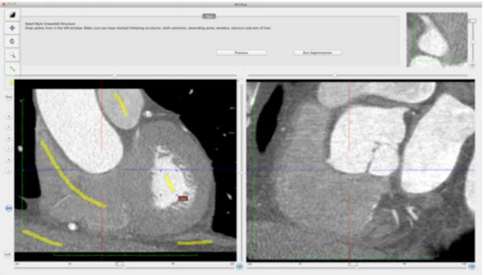
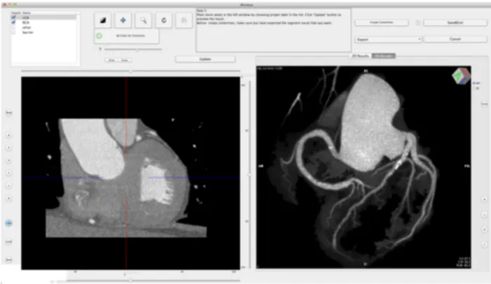
cardiac CT performed Maximum Intensity Projection (MIP), Curved Planar Reformation (CPR), and Volume Rendering (VR) reconstructions of each CTCA dataset on the iMac™ using the CMIV CTA plugin (Linköping University, Sweden) [36-37]. The plugin can be freely downloaded from the OsiriX plugin manager and allows semiautomatic 2D and 3D analysis of CTCA images loaded in the OsiriX viewer. After selecting an appropriate volume of interest (VOI) encompassing the entire heart, the user is required to place two seed points by means of ‘virtual catheters’ at the origin of the left main and right coronary arteries on orthogonal multiplanar reformat (MPR) views (Fig. 1a-b). As a second step, extracoronary structures are marked manually on MPR views and identified as veins, bones or other tissues through a region-growing algorithm (Fig. 1c). Subsequently, both coronary arteries and extracoronary voxels are segmented based on the landmarks previously placed by the operator. The result of the segmentation process is shown in a separate window in either MIP or VR mode. It is therefore possible to check the segmentation output and add or remove objects to the segmentation model by marking or unmarking one or more selected structures, or to refine the segmentation by placing or removing seeds or redrawing segmentation traces (Fig. 1d). Once accepted, final
MIP or VR images can be saved in DICOM format, or the segmented VOI can be exported back into the main OsiriX viewer window and used for further analysis outside the CMIV CTA plugin (Fig. 1e).
Fig. 1a. The CTA CMIV plugin: virtual catheter placed at the origin of the LCA.
Fig. 1c. The CTA CMIV plugin: semiautomatic removal of extracoronary structures.
Fig. 1e. The CTA CMIV plugin: panoramic MIP image of the coronary arteries (frontal view) obtained from automatic segmentation inside the plugin environment.
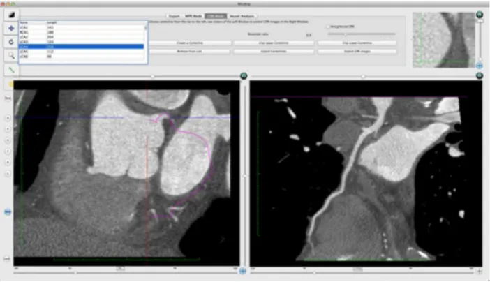
The plugin also allows performing vessel analysis of each segmented coronary artery by generating CPR views, either stretched (Fig. 2a) or straightened (Fig. 2b). Compared with MIP and VR reconstructions, CPR images do not provide a panoramic assessment of the entire coronary artery tree, but have the advantage of depicting the finest details of the vessel lumen and wall, thus enabling accurate measurement of cross-sectional vessel size and quantification of atherosclerotic plaques [30-33].
Fig. 2a. The CTA CMIV plugin: stretched CPR of the circumflex artery.
CPR views of each coronary artery (right coronary artery [RCA], left main [LM] continuing into the circumflex artery [Cx], and LM continuing into the left anterior descendent [LAD] artery) were generated, as well as panoramic MIP and VR reconstructions of the entire coronary tree. Subsequently, all reconstructions obtained from CTCA datasets were anonymized and saved in DICOM format in a separate folder of the OsiriX database. Two radiologists [BLINDED] experienced in cardiac CT (other than the one who performed 2D and 3D reconstructions), blinded to the imaging protocol and to each other, assessed the diagnostic quality of MIP, CPR and VR views through a three-point score (1=poor, 2=good, 3=excellent).
CALCULATION OF INTRAVASCULAR DENSITY, SNR, CNR, AND EFFECTIVE DOSE
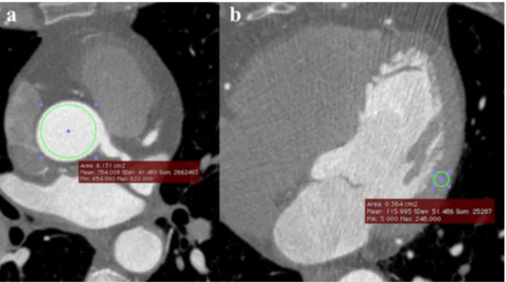
Intravascular CT density was measured as an index of the degree of overall coronary enhancement by placing a circular ROI inside the aortic root on source CTCA axial images, at the level of the LM origin. This ROI was as large as possible, but reached a distance no less than 1mm from the inner vessel wall, in order to include the largest number of voxels while minimizing partial volume phenomena for an accurate and reproducible density measurement. SNR was then calculated as the ratio between mean ROI density and its standard deviation (Fig. 3a).
To compute CNR, another circular ROI was traced inside the mid left ventricular (LV) myocardium on source CTCA axial images with the same criteria used for the aortic ROI, taking care to avoid epicardial vessels. CNR was calculated as the ratio
positioned by two additional radiologists with experience in vascular and cardiac CT [BLINDED].
Finally, effective doses were calculated by multiplying dose-length product (DLP)
values by a conversion coefficient for cardiac CT (0.014mSv·mGy-1·cm-1) [40].
STATISTICAL ANALYSIS
Image quality scores from the 100kV and 120kV protocols were compared using the two-tailed Mann-Whitney test, while continuous variables (i.e. intravascular density, SNR, CNR, and effective dose) were compared with the two-tailed unpaired Student t test.
The correlation between image quality and intravascular density was calculated using the Spearman rank test.
For each measurement involving two independent radiologists, inter-rater agreement was calculated using the Cohen’s kappa coefficient. Agreement was considered to be poor for κ less than 0.20, fair for κ between 0.21 and 0.40, moderate for κ between 0.41 and 0.60, good for κ between 0.61 and 0.80, and very good for κ between 0.81 and 1 [41].
Data are expressed as mean ± standard deviation. CT densities are expressed in Hounsfield units (HU).
Statistical analysis was carried out using software (GraphPad Prism version 5;
RESULTS
Image quality scores at 100kV and 120kV are listed in Tab. 1, while intravascular density, SNR, CNR, and effective dose are shown in Tab. 2.
Image quality of VR views was significantly better with the 100kV than with the 120kV protocol (2.77±0.43 vs 2.21±0.85, p=0.0332), while that of MIP and CPR reconstructions was comparable (2.59±0.50 vs 2.32±0.75, p=0.3271, and 2.68±0.48 vs 2.32±0.67, p=0.1118, respectively) (Fig. 4 and 5).
MIP CPR VR
100kV, low noise 2.59±0.50 2.68±0.48 2.77±0.43
120kV, standard 2.32±0.75 2.32±0.67 2.21±0.85
p value 0.3271 0.1118 0.0332*
Tab. 1. Image quality of MIP, CPR, and VR reconstructions with protocols A and B. *p<0.05 indicates statistical significance.
Ao (HU) SNR CNR ED (mSv)
100kV, low noise 655.9±127.2 16.42±4.64 13.43±3.77 7.43±2.69
120kV, standard 517.2±69.7 14.78±2.57 12.08±2.10 18.83±3.60
p value 0.0016* 0.2502 0.2486 <0.0001*
Tab. 2. Intravascular density (Ao=aortic root), SNR, CNR, and effective dose (ED) with the 100kV and 120kV protocols. *p<0.05 indicates statistical significance.
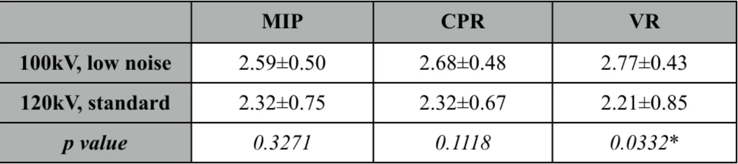
Fig. 4. Image quality of CTCA examination with the 100kV, low noise filter protocol in patient with mild focal soft plaque of the RCA (dashed arrow) and segmental mixed plaque of the LAD (arrow). MIP (a), CPR of the RCA (b), Cx (c), LAD [d (straightened), e (stretched)], and VR views (f).
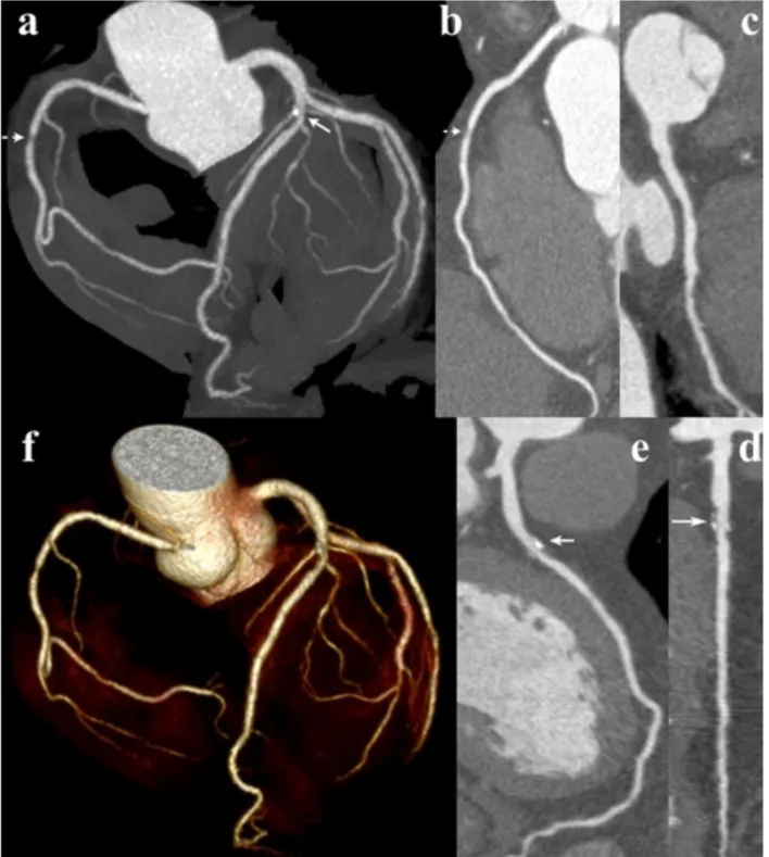
Fig. 5. Image quality of CTCA examination with the 120kV, standard filter protocol in patient with mixed, mainly calcified plaque of the LAD (arrow). MIP (a), CPR of the RCA (b), Cx (c), and LAD (d), and VR views (e).
SNR and CNR were comparable between the two protocols (16.42±4.64 vs 14.78±2.57, p=0.2502, and 13.43±3.77 vs 12.08±2.10, p=0.2486, respectively), while image noise was higher at 100kV than at 120kV (41.5±8.2 HU vs 35.5±5.2 HU, respectively, p=0.0362). However, in the 100kV group intravascular CT density was higher (655.9±127.2 HU vs 517.2±69.7 HU, p=0.0016) and correlated with VR
image quality (rs=0.5409, p=0.0025).
Effective dose was significantly lower with the 100kV than with the 120kV protocol (7.43±2.69 mSv vs 18.83±3.60 mSv, p<0.0001).
Inter-rater agreement was very good for both visual assessment of 2D and 3D reconstructions (κ=0.85) and for ROI measurements (κ=0.82).
DISCUSSION
Our findings show that, by reducing tube voltage from 120kV to 100kV while keeping the remaining parameters unchanged, it is possible to obtain a marked reduction of radiation dose with equivalent or superior quality of 2D and 3D reconstructions, respectively, of CTCA examinations performed in nonobese patients. CT angiography of coronary [22-24] and extracoronary arteries [25-29] has been shown to be feasible at 100kV with equivalent or better image quality of source axial images compared with 120kV, as X-rays generated by a lower tube voltage have a significantly lower average energy, closer to the k-edge of iodine (33.2keV), which also maximizes photoelectric effect. As a result, higher contrast resolution (and in particular, greater iodine conspicuity) can be achieved with dramatically reduced radiation exposure. This scenario is confirmed by our finding of a significantly higher intravascular CT density and equivalent CNR with the 100kV protocol.
The increased aortic and coronary enhancement at 100kV, together with usage of a low noise reconstruction kernel, can also explain the comparable SNR among the two CTCA protocols under evaluation. Indeed, at 100kV the reduction of mean X-ray energy (and therefore, of radiation exposure) is paralleled by a higher image noise due to lower tissue penetration, which could affect visibility of the coronary arteries especially in their distal portion and/or if stenosed or obstructed. To overcome this drawback, in low voltage CT angiography protocols tube current is usually increased to keep noise within acceptable levels and thus improve SNR. However, increasing tube current leads to a linear increase in radiation dose, which is of course
undesirable and can only partially compensate for the higher noise (as noise varies with the square of dose) [1-3].
To offset the greater image noise without increasing radiation dose, we kept tube current constant in both the 120kV and 100kV groups and reconstructed raw CT data at 100kV with a low noise convolution kernel. However, low noise kernels are low pass filters that attenuate the higher frequencies of the image space (i.e. those encoding information about morphological details, such as object contours), which may pose a penalty in terms of in-plane spatial resolution [1-4, 42]. However, while this could be problematic for depiction of stent struts and in-stent restenosis (for which highest spatial resolution in all planes is mandatory) [6, 43], the evaluation of native coronary arteries and bypass grafts is less likely to be affected, as the borders of plaques and normal vessel walls are relatively smooth and maximization of SNR is the main priority. Moreover, a very high spatial resolution with voxel isotropy was available natively owing to the selection of the smallest achievable sampling field-of-view (32cm, i.e. restricted to the heart for CTCA purposes), ultrathin detector collimation (0.625mm) and overlapped image reconstruction (0.4mm).
Besides, the increased conspicuity of the coronary arteries at 100kV suggests that CTCA could be performed with a smaller amount of iodinated CM compared with standard 120kV CTCA protocols, with clear benefits in terms of healthcare costs and patient safety. To this latter respect, it is worth mentioning that many patients with coronary artery disease have impaired renal function, as well as several comorbitidies that raise the risk of contrast-induced nephropathy [44-45], for which reducing CM
While most studies have focused on the efficacy of 100kV CTCA scanning in terms of quality of native axial images [38, 46-47], we evaluated 2D (MIP, CPR) and 3D (Volume Rendering) views as quality indexes of CTCA studies. In fact, 2D and 3D reconstructions play a fundamental role in the interpretation of CTCA datasets (as well as in overall modern multislice CT) [3, 30-33], but to our knowledge, no extensive data exist in the literature about quality of 2D and 3D views obtained from 100kV CTCA datasets. In this context, we found that 3D reconstructions obtained from 100kV CTCA studies have a significantly better quality than those from 120kV CTCA images, which correlates with aortic enhancement. This finding supports the key role of contrast resolution between the vessel lumen and wall for optimal 3D depiction of the coronary arteries, even despite a significantly higher image noise at 100kV than at 120kV. Indeed, the CMIV CTA plugin requires that vessel tracking be performed prior to generation of 2D and 3D views, and vessel segmentation is done automatically after placing seed points at the origin of the right and left coronary arteries. In this context, a higher contrast resolution between iodine-filled vessel lumen, vessel wall (either normal or with plaques), and extracoronary fat tissue should improve region growing-based automatic detection of vessel boundaries, as a higher density threshold exists between them [48-50]. Also image quality of 2D reconstructions (MIP and CPR) was not inferior at 100kV than at 120kV, but no significant difference was detected between them supposedly because unlike VR techniques, CPR and especially MIP algorithms do not rely on CT density-based transfer curves for differentiation of the selected structures and are therefore less dependent on CT density gradients [3, 30-33].
Our study has some limitations. First, all of our CTCA examinations were performed on a 64-slice CT scanner with retrospective ECG-gating, which is associated with a higher radiation dose for CTCA compared with prospective gating [1-5]. However, this latter was not available on our CT equipment at the time of our study, and brings the risk of obtaining poor quality or even non-diagnostic images if the patient’s heart rhythm is not regular during the acquisition [1-5, 12-17]. Nonetheless, in an attempt to reduce radiation dose as much as possible, we used ECG sensing with a full-dose (i.e. maximum tube current) temporal window limited to the 70-80% R-R interval (corresponding to the end-diastolic phase), while tube current was kept to a minimum of 150mA outside this phase. Still, 6/57 patients (10.5%) had to be discarded because of sudden acceleration of heart rhythm during the CTCA acquisition, that could not be predicted during patient monitoring before scanning and led to compromised image quality due to motion artifacts. Besides, our findings should be valid irrespective of using prospective or retrospective ECG-gating, and usage of prospective ECG-gating should lead to an even lower radiation dose with the 100kV, low noise filter protocol.
Another limitation is the fact that patients with stents or bypass grafts were excluded from our study. Indeed, depiction of stents with CT is hurdled by beam hardening and blooming artifacts due to their metal structure and confined geometry within a restricted area [43], that are inherent to filtered back-projection reconstruction (FBP) algorithms working on current CT equipment and tend to be more severe at lower tube voltages [1-3]. Therefore, we elected to exclude patients with coronary stents in order to avoid compromising in-stent lesion detectability and introducing a potential
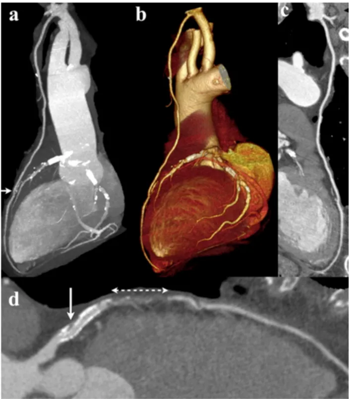
bias in data interpretation. Yet, CTCA is currently not recommended for the evaluation of coronary stents narrower than 3mm due to the above pitfalls [6]. These latter are expected to be overcome by the introduction of more accurate and dose-efficient CT image reconstruction techniques, such as iterative reconstruction algorithms, allowing for artifact minimization and dose reduction compared with conventional FBP [51]. As for bypass grafts, their investigation would have implied extending the upper limit of scanning up to the thoracic inlet (especially in case of internal mammary artery grafts), requiring a different CTCA protocol with higher radiation dose and longer duration of the CM bolus, which would have been difficult to compare with those used for the depiction of native coronary arteries. However, the validity of our findings should be preserved for the evaluation of bypass grafts too, as the interpretation of our results is likely to be extendable to such cases (Fig. 6). Still, the routine application of our 100kV, low reconstruction filter CTCA protocol in patients with coronary stents or bypass grafts deserves further investigation, and it is likely that the radiation dose delivered to patients (as well as image quality) can be further improved by the widespread dissemination of prospective ECG-gating and iterative reconstruction methods.
Fig. 6. Image quality of CTCA examination with the 100kV, low noise filter protocol in patient with LIMA-LAD bypass graft (short arrow indicates distal anastomosis) and stenting of proximal LAD and RCA. Although the evaluation of coronary bypass grafts was beyond the aim of our study, MIP (a), VR (b), and CPR (c) reconstructions provide optimal depiction of graft patency through its entire course. (d) CPR view of the LAD, which is patent distal to the bypass anastomosis, but is occluded in its middle third (dashed double-headed arrow). Also notice mild in-stent restenosis (long arrow).
REFERENCES
1. Gerber TC, Kantor B, Williamson EE (eds) Computed tomography of the
cardiovascular system. Informa Healthcare, London, 2007
2. Ohnesorge B. Future technical developments in cardiac CT. In Ohnesorge BM et
al (eds) Multi-slice and dual-source CT in cardiac imaging. 2nd ed. Springer-Verlag, Berlin - Heidelberg - New York, 2007
3. Faggioni L, Cerri F, Giustini D. Cardio-TC e TC dual source. In Faggioni L,
Paolicchi F, Neri E (eds) Elementi di tomografia computerizzata. Springer-Verlag, Milan, 2010
4. Earls JP, Leipsic J. Cardiac computed tomography technology and
dose-reduction strategies. Radiol Clin North Am 2010;48:657-674
5. Mahesh M, Cody DD. Physics of cardiac imaging with multiple-row detector
CT. Radiographics 2007;27:1495-1509
6. Taylor AJ, Cerqueira M, Hodgson JM et al. ACCF/SCCT/ACR/AHA/ASE/
ASNC/NASCI/SCAI/SCMR 2010 Appropriate Use Criteria for Cardiac Computed Tomography. A Report of the American College of Cardiology Foundation Appropriate Use Criteria Task Force, the Society of Cardiovascular Computed Tomography, the American College of Radiology, the American Heart Association, the American Society of Echocardiography, the American Society of Nuclear Cardiology, the North American Society for Cardiovascular Imaging, the Society for Cardiovascular Angiography and Interventions, and the
S o c i e t y f o r C a r d i o v a s c u l a r M a g n e t i c R e s o n a n c e . C i r cu l a t i o n 2010;122:e525-555
7. Weininger M, Barraza JM, Kemper CA, Kalafut JF, Costello P, Schoepf UJ.
Cardiothoracic CT angiography: current contrast medium delivery strategies. AJR Am J Roentgenol 2011;196:W260-W272
8. Hein PA, May J, Rogalla P, Butler C, Hamm B, Lembcke A. Feasibility of
contrast material volume reduction in coronary artery imaging using 320-slice volume CT. Eur Radiol 2010;20:1337-1343
9. Bae KT. Optimization of contrast enhancement in thoracic MDCT. Radiol Clin
North Am 2010;48:9-29
10. Lauer MS. Elements of danger – the case of medical imaging. N Engl J Med 2009;361:841-843
11. Thomas KE. CT utilization - trends and developments beyond the United States’ borders. Pediatr Radiol 2011;41 Suppl 2:562-566
12. Raff GL. Radiation dose from coronary CT angiography: five years of progress. J Cardiovasc Comput Tomogr 2010;4:365-374
13. Xu L, Zhang Z. Coronary CT angiography with low radiation dose. Int J Cardiovasc Imaging 2010;26 Suppl 1:17-25
14. Torres FS, Crean AM, Nguyen ET, Paul N. Strategies for radiation-dose reduction and image-quality optimization in multidetector computed tomographic coronary angiography. Can Assoc Radiol J 2010;61:271-279
15. Alkadhi H, Leschka S. Radiation dose of cardiac computed tomography - what has been achieved and what needs to be done. Eur Radiol 2011;21:505-509
16. Earls JP, Berman EL, Urban BA et al. Prospectively gated transverse coronary CT angiography versus retrospectively gated helical technique: improved image quality and reduced radiation dose. Radiology 2008;246:742-753
17. Scheffel H, Alkadhi H, Leschka S et al. Low-dose CT coronary angiography in the step-and-shoot mode: diagnostic performance. Heart 2008;94:1132-1137 18. Ketelsen D, Fenchel M, Buchgeister M et al. Estimation of radiation exposure of
different dose saving techniques in 128-slice computed tomography coronary angiography. Eur J Radiol 2011, doi:10.1016/j.ejrad.2011.01.052
19. Qin J, Liu LY, Meng XC et al. Prospective versus retrospective ECG gating for 320-detector CT of the coronary arteries: comparison of image quality and patient radiation dose. Clin Imaging 2011;35:193-197
20. Kalva SP, Sahani DV, Hahn PF, Saini S. Using the K-edge to improve contrast conspicuity and to lower radiation dose with a 16-MDCT: a phantom and human study. J Comput Assist Tomogr 2006;30:391-397
21. Kalender WA, Buchenau S, Deak P, et al. Technical approaches to the optimisation of CT. Phys Med 2008;24:71-79
22. Bischoff B, Hein F, Meyer T et al. Impact of a reduced tube voltage on CT angiography and radiation dose: results of the PROTECTION I study. JACC Cardiovasc Imaging 2009;2:940-946
23. Ripsweden J, Brismar TB, Holm J et al. Impact on image quality and radiation exposure in coronary CT angiography: 100 kVp versus 120 kVp. Acta Radiol 2010;51:903-909
24. Zhang C, Zhang Z, Yan Z, Xu L, Yu W, Wang R. 320-row CT coronary angiography: effect of 100-kV tube voltages on image quality, contrast volume, and radiation dose. Int J Cardiovasc Imaging 2011;27:1059-1068
25. Heyer CM, Mohr PS, Lemburg SP, Peters SA, Nicolas V. Image quality and radiation exposure at pulmonary CT angiography with 100- or 120-kVp protocol: prospective randomized study. Radiology 2007;245:577-583
26. Faggioni L, Neri E, Sbragia P et al. CT pulmonary angiography with 80kV tube voltage and 40mL of iodinated contrast material in lean patients: comparison of vascular enhancement with iodixanol 320mgI/mL and iomeprol 400mgI/mL. AJR Am J Roentgenol 2012 (in press)
27. Schindera ST, Graca P, Patak MA et al. Thoracoabdominal-aortoiliac multidetector-row CT angiography at 80 and 100 kVp: assessment of image quality and radiation dose. Invest Radiol 2009;44:650-655
28. Kayan M, Köroğlu M, Yeşildağ A et al. Carotid CT-angiography: Low versus standard volume contrast media and low kV protocol for 128-slice MDCT. Eur J Radiol 2011, doi 10.1016/j.ejrad.2011.05.006
29. Waaijer A, Prokop M, Velthuis BK, Bakker CJ, de Kort GA, van Leeuwen MS. Circle of Willis at CT angiography: dose reduction and image quality--reducing tube voltage and increasing tube current settings. Radiology 2007;242:832-839 30. Lipson SA. MDCT and 3D workstations. A practical guide and teaching file.
Springer-Verlag, New York, 2006
31. Faggioni L, Neri E, Castellana C, Caramella D, Bartolozzi C. The future of PACS in healthcare enterprises. Eur J Radiol 2011;78:253-258
32. Faggioni L, Lazzarini R, Paolicchi F. Tecniche di elaborazione delle immagini. In Faggioni L, Paolicchi F, Neri E (eds) Elementi di tomografia computerizzata. Springer-Verlag, Milan, 2010
33. Sirineni GK, Kalra MK, Pottala KM et al. Visualization techniques in computed tomographic coronary angiography. Curr Probl Diagn Radiol 2006;35:245-257 34. Rosset A, Spadola L, Ratib O. OsiriX: an open-source software for navigating in
multidimensional DICOM images. J Digit Imaging 2004;17:205-216
35. Rosset A, Spadola L, Pysher L, Ratib O. Informatics in radiology (infoRAD): navigating the fifth dimension: innovative interface for multidimensional multimodality image navigation. Radiographics 2006;26:299-308
36. Quick Guide of CMIV CTA Plug-in. http://www.osirix-viewer.com/CTA_Plugin/
quickguide.htm
37. Petersson H, Sinkvist D, Wang C, Smedby O. Web-based interactive 3D visualization as a tool for improved anatomy learning. Anat Sci Educ 2009;2:61-68
38. Park EA, Lee W, Kang JH, Yin YH, Chung JW, Park JH. The image quality and radiation dose of 100-kVp versus 120-kVp ECG-gated 16-slice CT coronary angiography. Korean J Radiol 2009;10:235-243
39. Hausleiter J, Meyer T, Hadamitzky M et al. Radiation dose estimates from cardiac multislice computed tomography in daily practice: impact of different scanning protocols on effective dose estimates. Circulation 2006;113:1305-1310 40. International Commission on Radiological Protection. 2007 recommendations of
the International Commission on Radiological Protection. Ann ICRP 2007; 37:publication no. 103
41. Landis JR, Koch GG. The measurement of observer agreement for categorical data. Biometrics 1977;33:159-174
42. Goldman LW. Principles of CT: radiation dose and image quality. J Nucl Med Technol 2007;35:213-225
43. Pugliese F, Cademartiri F, van Mieghem C et al. Multidetector CT for visualization of coronary stents. Radiographics 2006;26:887-904
44. Chinnaiyan KM, McCullough PA. Optimizing outcomes in coronary CT imaging. Rev Cardiovasc Med 2008;9:215-224
45. Maliborski A, Zukowski P, Nowicki G, Bogusławska R. Contrast-induced nephropathy--a review of current literature and guidelines. Med Sci Monit 2011;17:RA199-204
46. Feuchtner GM, Jodocy D, Klauser A et al. Radiation dose reduction by using 100-kV tube voltage in cardiac 64-slice computed tomography: a comparative study. Eur J Radiol 2010;75:e51-e56
47. Leschka S, Stolzmann P, Schmid FT et al. Low kilovoltage cardiac dual-source CT: attenuation, noise, and radiation dose. Eur Radiol 2008;18:1809-1817
48. Oda S, Utsunomiya D, Funama Y et al. A low tube voltage technique reduces the radiation dose at retrospective ECG-gated cardiac computed tomography for anatomical and functional analyses. Acad Radiol 2011;18:991-999
49. Marquering HA, Dijkstra J, de Koning PJ, Stoel BC, Reiber JH. Towards quantitative analysis of coronary CTA. Int J Cardiovasc Imaging 2005;21:73-84 50. D’Errico L, Salituri F, Ciardetti M et al. CT-derived epicardial fat volume:
influence of scanning protocol and reproducibility of measurements. RSNA 2011, Chicago, IL
51. Leipsic J, Labounty TM, Heilbron B et al. Estimated radiation dose reduction using adaptive statistical iterative reconstruction in coronary CT angiography: the ERASIR study. AJR Am J Roentgenol 2010;195:655-660