408 Surg Neurol 1989;32:408-17
Cerebral Vasospasm in a Double-Injection Model in Rabbit
Aldo Spallone, M.D., and Francesco Saverio Pastore, M.D.
Division of Neurosurgery, Second University of Rome, "Tor Vergata," Rome, Italy
Spallone A, Pastore FS. Cerebral vasospasm in a double-injection model in rabbit. Surg Neurol 1989;32:408-17.
T h e present study was designed to assess the occurrence of cerebral vasospasm following an experimental sub- arachnoid h e m o r r h a g e model in rabbits. Sixty-nine N e w Zealand albino rabbits were used in this study. One milliliter of fresh arterial blood was injected t h r o u g h the surgically exposed atlanto-occipital m e m b r a n e over a period of 20 seconds. T h e procedure was then repeated 24 hours later. Fifty animals underwent digital subtraction angiography at one of the following prefixed intervals: 1, 3, or 8 days after the second injection hemorrhage. Nineteen animals underwent one angiographic examina- tion prior to the instillation of the intracisternal blood. This procedure was followed by a repeated angiography 3 days after the second experimental subarachnoid hem- orrhage. For the purpose of evaluation, the films were magnified 10-fold and the diameter of the basilar artery as well as that of the extracranial vertebral artery at three different levels were measured. We assessed the diameter of the basilar artery as well as the mean ratio extracranial vertebral artery/basilar artery diameters. This ratio was considered to minimize anatomical and technical vari- abilities. T h e results in the first 50 animals showed a trend suggesting that spasmogenic activity reaches a peak at about the third day after subarachnoid hemorrhage. These results were confirmed in the latter 19 animals. However, mortality in this g r o u p was high: 50%. This double-injection model of subarachnoid hemorrhage in rabbits consistently reproduced cerebral vascular spasm 3 days after repeated subarachnoid hemorrhage. However, its usefulness as an experimental model for subarachnoid hemorrhage is limited practically by the high animal mortality in the protocols where repeated angiographic studies are necessary.
KEY WORDS: Experimental subarachnoid hemorrhage; Cere- bral spasm; Angiographic study; Rabbit
Address reprint requests to: Aldo Spallone, M.D., Cattedra di Neurochirurgia, Seconda Universit~ degli Studi di Roma, "Tor Verga- ta," Via Orazio Raimondo, 8, 1-00173 Rome, Italy.
Received January 11, 1988; accepted April 14, 1989.
I n t r o d u c t i o n
Ischemic complications, p r e s u m a b l y related to cerebral vasospasm following subarachnoid h e m o r r h a g e (SAH), are a m a j o r source o f disappointing results seen in the m a n a g e m e n t o f r u p t u r e d intracranial aneurysms [11]. Extensive research in cerebral vasospasm following S A H is necessary in the h o p e o f improving the results o f surgical intervention. Several animal models of S A H have b e e n r e p o r t e d in the literature. T h e aim o f the p r e s e n t study was to assess the validity o f a new model o f S A H in rabbit, an animal seldom used in S A H e x p e r i m e n t s [5,8,15,19].
M a t e r i a l a n d M e t h o d s
Fifty-nine white N e w Zealand rabbits, weighing ap- proximately 2 kg were used in this study. For the experimental p r o c e d u r e , the animals were p r e m e d - icated with N - 3 propionilphenothiazine (Combelen, Bayer), 0.5 m L / k g via intramuscular injection, and were then anesthetized with N e m b u t a l , 20 m g / k g intrave- nously. Body t e m p e r a t u r e was maintained constant at 37 ° by a heating pad. T h e auricular artery (or the femoral artery in cases o f an unsuccessful canulation) was catheterized for blood pressure monitoring and arterial blood sampling. With the head held in a stereo- taxic frame, the atlanto-occipital m e m b r a n e was ex- posed surgically by careful dissection o f the nuchal muscles. T h e n 1 mL o f fresh arterial blood was injected o v e r a period o f 20 seconds into the cisterna magna via a N o . 27-gauge needle. I m m e d i a t e l y thereafter, the animals were placed head down for 5 minutes to facili- tate the intracranial dispersion o f blood. T h e duration o f postinjection apnea was monitored.
T h e animals w h o d e v e l o p e d respiratory difficulties after the injection were ventilated mechanically via a well-fitted m a s k connected to a H a r v a r d ventilator ( 5 5 0 8 3 0 - - l a r g e r o d e n t ventilator) flowing at 30 m L / k g and 14 breaths p e r minute. Blood pressure was contin- uously m o n i t o r e d during the experiment, and arterial blood gases w e r e checked before and after the sub- arachnoid injection o f blood. A p p r o p r i a t e antibiotics were administered intramuscularly at the end o f the procedure. T w e n t y - f o u r hours later, the entire proce-
Cerebral Vasospasm in Rabbit Surg Neurol 409 1989;32:408-17
B
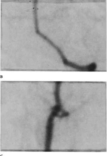
A C
Figure 1. (A) Angiographr in a control animal. (B) Magnified t,ieu' of the intracranial vertebral and basilar artery ~reproduced x 10). (C) Magni- fied riew of the extracranial vertebral artery (reproduced × 10).
dure was repeated. Following this, the animals were monitored clinically at least twice daily.
The time of the second hemorrhage was considered day zero (day 0). The first 50 animals (group A) underwent angiographic examination at one of the following prefixed intervals: day 1, day 3, or day 8. Some day 1 and day 3 animals also underwent computed tomography (CT) scanning examination. In three animals, saline instead of blood was injected into the cisterna magna. In this group, angiography was per- formed 3 days after the second and last injection.
In the last 19 animals (group B), a control angiogram was obtained just prior to the first intracisternal injec- tion of blood. A final angiogram was then performed 3 days after the second experimental SAH.
For the purpose of angiography, the animals were
again anesthetized as previously, and the femoral artery was catheterized with a No. 4.0 French (Flexo Pulmo- cath Selon orandjean) catheter. Positioning of the tip of the catheter into the left subclavian artery close to the origin of the vertebral artery was checked with fluoro- scopy. Digital angiography (Angiotron-Siemens Elec- tronics) was performed using standardized conditions (i.e., distance between the head of the animal and the source of radiation, angiographic mask) via manual injection of 5 mL of water-soluble contrast agent (Con- ray 60). In the group B animals, the catheter was removed following the control angiographic study and a temporary clip was placed on the artery proximal to the puncture site, the latter in order to allow a later, second catheterization. The animals were sacrificed following the final angiogram, and their brains were removed for gross inspection.
The diameter of the basilar artery was measured at the junction between its lower and middle third, using a 10-fold magnification of the subtraction films. Appro- priate calipers were used for at least two different
410 Surg Neurol Spallone and Pastore 1989;32:408-17
B
A
evaluations. We also considered the ratio between the mean diameter o f the extracranial vertebral artery (mea- sured at two different levels) and that of the basilar artery, the latter to minimize the possible influence of changes in the distance between the animal and the x-ray source, plus anatomical variability.
Statistical analysis of the data was performed using the Student's t-test and Fisher's analysis of variance.
R e s u l t s
Group A (50 Animals)
General observations.
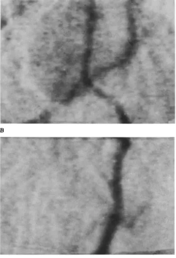
We recorded 10 deaths (20%) in this group, including 2 deaths that it is likely were related to the angiographic procedure. The incidence of drowsiness was high, being present in 24 rabbits. Hemi- paresis was noted in two cases. Apnea after SAH was observed in 50% o f animals but in none of the saline- injected rabbits, with intervals between 8 and 30 sec- onds. As a rule, the animals presenting with apnea didFigure 2. (A) Angiography in an animal I day after SAH. (B)
Magnified view of the intracranial vertebral and basilar artery (reproduced
× l 0). (C) Magnified view of the extracranial vertebral artery (reproduced ×10).
not show a substantially different clinical course in comparison with nonapneic animals. Mechanical venti- lation was required in four cases where apnea exceeded a duration of 30 seconds. Nevertheless, two of these animals did not recover from acute respiratory distress occurring after the subarachnoidal injection of blood in spite of adequate ventilation and resuscitative maneu- vers. In these two animals, arterial blood gas monitoring showed values compatible with the clinical syndrome of irreversible acute respiratory insufficiency (PO2 < 60 mm Hg, PCO2 > 40 mm Hg, p H about 7.25).
Blood pressure monitoring consistently showed a rise in the mean values immediately after the experi- mental hemorrhage, and this subsided in about 10 seconds.
Cerebral Vasospasm in Rabbit Surg Neurol 4 l 1 1989;32:408-17
A C
Figure 3. (A) Angiography in a rabbit 3 days following SAH. (B) Magnified view of the intracranial vertebral and basilar artery (reproduced xlOL (C) Magnified view of extracranial vertebral artery (reproduced x I0).
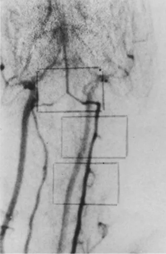
A r t e r i o g r a p h i c results. The subtraction films of the vertebrobasilar system were of satisfactory quality in 38 of the 40 cases (Figures 1-5). The numerical data obtained from the experimental animals are summa- rized in Table 1.
Values obtained from the "saline" group were similar to the mean values o f control group. Figure 6 is a graphic representation o f the observed angiographic values. As expected, there was an inverse relationship between the measured diameters of the basilar arteries and the ratio mean extracranial vertebral artery/basilar artery (MV/BA). The increase in the ratio suggested a decreasing diameter o f the basilar without substantial changes in that of extracranial vertebral artery. There was a trend showing a reduction in the diameter of the
basilar artery in SAH animals, with a peak on day 3 after the last hemorrhage. This trend appears to be more evident when the ratio MV/BA was used for the measurements.
Statistical comparison of the changes in the diameters of the basilar arteries showed significant difference between controls and day 1 animals (p < 0.05) and between controls and day 3 animals (p < 0.005). A statistically significant difference existed between day 1 animals and day 3 animals (p < 0.05).
N o difference was detected between control animals, day 8 animals, and the saline-injected animals.
On the other hand, statistical comparison of the MV/BA ratio values between controls and day 3 rabbits showed a highly significant difference (p < 0.001). The same p value (< 0.001) was obtained comparing MV/BA ratios of day 1 and day 3 animals.
N o significant difference was found in control group MV/BA values when compared respectively to either those of day 8 group or day 1 group.
412 Surg Neurol Spallone and Pastore 1989;32:408-17
A
B
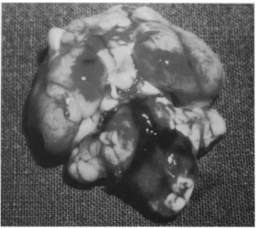
Gross pathologicalfindings. Autopsy findings demon-
strated the presence of blood clots on the basal surface o f the brain in the day 1 animal group (Figure 7). This finding was in agreement with the CT scan results (Figure 8). N o clots were found in the day 3 group specimens.
G r o u p B (19 A n i m a l s )
General observations. Mortality related to the procedure
was very high. In fact, 19 animals were required to obtain 10 complete angiographic studies. In particular, five animals died during the second angiography, three after the second hemorrhage, and one between the first and second hemorrhage. In those animals, monitoring of vital signs showed hypotension and respiratory fail- ure as a cause o f death. Four o f the survivors showed a milder form o f the same syndrome during the second angiography, but these animals recovered.
Figure 4. (A) Angiography in an animal 8 days following SAH. (B)
Magnified view of the intracranial vertebral and basilar artery (reproduced
× I 0). (C) Magnified view of the extracranial vertebral artery (reproduced
xlO).
Arteriographic results. We found the same pattern
with a vessel caliber reduction of 1 5 % - 2 0 % for the basilar artery 3 days after the SAH (Figures 9-12). The data are summarized in Table 2. Statistical analysis using the Student's t-test again showed statistical significance (p < 0.05).
Gross pathological findings. As expected, no clots
were found macroscopically in this group of animals.
D i s c u s s i o n
The occurrence of ischemic complications presumably owing to vasospasm following SAH is a grave prognos-
Cerebral Vasospasm in Rabbit Surg Neurol 413 1989;32:408-17
Figure 5. Angiography in a saline-injected animal
tic factor and a major cause of death in patients with ruptured intracranial aneurysms [2,12]. The pathogene- sis of cerebral arterial spasm has still to be clarified in most of its aspects and the treatment results are gener- ally unrewarding [ 1,11,16,23,27,29].
Carefully designed experimental studies, as well as animal models reproducing the pathological events leading to the development of cerebral arterial spasm in humans, would be a useful research model. Information from this model could be used for better clarification of the pathophysiology of this pathological condition and, it is hoped, improvement in the clinical outcome. Several experimental models of SAH have been re- ported in the literature, with the aim of studying the development of vasospasm [3,4,9,10,22,27,28].
The rabbit is a relatively inexpensive, easily available experimental animal; however, it has been used infre- quently in experimental studies of SAH [5,8,13,15,20]. Its size allows angiography of the cerebral vessels to be performed adequately without too much sophistication needed in the technique.
Because of its proximity in philogeny to primates [21], rabbits possess comparable cerebrovascular mor- phological features that might also be involved in the pathogenesis of ischemic complications of SAH [6,7,
17,25].
The double-injection model of SAH, recently de- scribed by Varsos et al [27] in dogs, appears to re- produce with good approximation the occurrence of delayed cerebral vasoconstriction following SAH. Ow- ing to the different size of the animals, an open surgical
m y / ~
Figure 6. Graphic representation of the mean angiographic values. Top: ratio M V / B A (see text). Bottom: diameters of t-Ee-basilar arteries.
i i n
0 () 1 2 ~1 4 5
d~yu
414 Surg Neurol SpaUone and Pastore 1989;32:408-17
F i g u r e 7. Pathological specimen of a day 1 group animal A diffuse SAH is evident, mainly at the level of the prepontine cistern and around the basilar artery.
F i g u r e 8. Computed tomography scan of a day 1 group animal," there is evidence of blood surrounding the basilar artery.
Table 1. Measurement Values in Individual Animals
Group BA (ram) Ratio MV/BA
C 1.3 2.85 C 1.3 3.85 C 1.4 3.57 C 1.5 3.47 C 1.4 2.93 C 1.3 3.46 C 1 3.20 C 1.3 3.31 C 1.4 2.93 C 1.2 2.78 D. 1 1 3.60 D.1 1.2 3.68 D.1 1.3 3.47 D.1 1.0 3.51 D.1 0.9 3.56 D. 1 0.9 3.88 D.1 1.2 3.04 D.1 1.1 3.37 D.1 1.3 3.25 D.3 1 4.32 D.3 1.1 3.98 D.3 0.9 4.67 D.3 0.8 5.25 D.3 0.8 5.02 D.3 0.7 5.17 D.3 0.8 4.52 D.3 1.1 3.65 D.8 1.3 2.90 D.8 1.5 2.98 D . 8 1.1 3.05 D . 8 1.2 2.82 D . 8 1.4 2.87 D . 8 1.3 3.32 D.8 1.3 3.34 D.8 1.3 3.52 S 1.5 2.63 S 1.25 3.24 S 1.30 3.15
See text for statistical analysis.
Abbreviations: BA = basilar artery diameter; ratio MV/BA (see text for explanation); C = control group; D.1 = day 1 group; D.3 = day 3 group; D.8 = day 8 group; S = saline-injected animals.
technique was used in the present study to avoid accidental contamination of cerebrospinal fluid (CSF) and to obtain visual control of injection. We are not aware of other studies using a double-injection model of SAH in rabbits. Conflicting results regarding the occur- rence of vasospasm and related changes have been reported by other studies using either a single-injection SAH model [5] or a triple-injection SAH model [13] in rabbits. The present closed skull model does not re- quire manipulations o f brain structures and allows an experimental reproduction of the effects of sudden
Cerebral Vasospasm in Rabbit Surg Neurol 415 1989;32:408-17
ography in rabbits. Accurate measurements of angiogra- phic data were essential for the purpose of the present study. Digital subtraction angiography demonstrated satisfactory angiographic images and allowed us to keep constant the possible parameters that might influence the comparison of films obtained from different animals. The possible influence of even small variations in the distance between the x-ray source and the animal was overcome by using the ratio MV/BA in the mea- surements.
In a large number of animals in which a single angiogram was performed, we observed a considerable interanimal variation in the size of the basilar artery. However, the diameter of the basilar artery appeared to be reduced in SAH animals, with a definite peak at about the third day after the last SAH. This applies well to the hypothetical timing of release of spasmogenic substances from clots accumulated in the subarachnoid spaces and their possible mechanism of action [5,9,11,
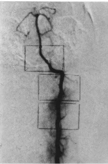
Figure 10. Group B. Angiography of number two animal3 days after the second SAH.
Figure 9. Group B. Angiography of number two animal before the SAH.
increased intracranial pressure (ICP) that occurs in hu- man SAH. Injection of I mL of blood over 20 seconds was well tolerated by the animals. Computed tomogra- phy scanning examinations and autopsy showed clots located anteriorly around the basilar artery 1 day after the last hemorrhage, thus supporting the effectiveness of the described procedure.
Furthermore, the chosen 24-hour interval between hemorrhages allowed the animals to recover from the effect of both anesthesia and the experimental proce- dure. However, mortality was by no means negligible. Several factors might play a role regarding mortality. Excluding technical errors, these factors included respi- ratory distress, general stress owing to surgical manipu- lations, and sudden increased ICP. Perhaps the effects owing to raised ICP in this experimental model might be minimized by removing an equal amount of cerebrospinal fluid prior to blood injection. No particu- lar problems were encountered when performing angi-
416 Surg Neurol Spallone and Pastore 1989;32:408-17
Figure 11. Group B. Angiography of number seven animal before the SAH.
13,14,16,19,20,24,26]. M o r e o v e r , the reduction in the d i a m e t e r o f "spastic" basilar artery was statistically significant.
R e p e a t e d serial angiographic studies, in the same animal, are the o p t i m a l way to assess angiographic vasospasm, particularly in m o d e l s that are being phar- macologically manipulated for b e t t e r clinical results. In this study, we p e r f o r m e d r e p e a t e d angiographic exami- nations 3 days after the second h e m o r r h a g e in the last 19 animals, in which a control angiogram had b e e n p e r f o r m e d p r i o r to the e x p e r i m e n t a l SAHs. Although the occurrence o f a significant vasospasm o f the basilar artery was d e m o n s t r a t e d , an unacceptably high mortal- ity rate was also recorded. W e believe that multiple surgical p r o c e d u r e s and anesthesia were mainly respon- sible for the disappointing death rate in the g r o u p B animals. Using a longer interval b e t w e e n control angio- g r a m and e x p e r i m e n t a l S A H could be a possible way to reduce the mortality in this model.
T h e p r e s e n t results raise the question as to w h e t h e r the validity o f this m o d e l regarding the production o f vasospasm is, in fact, affected by the high surgical mortality rate. In addition, there are recent suggestions [13,18,27] that the double intracisternal injection o f blood does not a p p e a r to enhance cerebral vasospasm (N.F. Kassel, personal communication, 1987). These studies, as well as o u r results, suggest a consistent limitation to the use o f the p r e s e n t S A H model. Future studies using a single-injection m o d e l are being contem- plated.
The authors are deeply indebted to the management and technical staff of "Clinica Nuova Latina," Rome, Italy, for their valued support and assistance in the angiographic studies. The authors would also like to express their thanks to James T. Goodrich, M.D., Ph.D., Depart- ment of Neurological Surgery, Albert Einstein College of Medicine, New York, for his editorial comments during the preparation of this manuscript.
Figure 12. Group B. Angiography of number seven animal 3 days after the second SAH.
C e r e b r a l V a s o s p a s m in R a b b i t Surg N e u r o l 417 1 9 8 9 ; 3 2 : 4 0 8 - 1 7
T a b l e 2. Group B-Measurement Values in Individual
Animals
Control Three days
1. 2. 3. 4. 5. 6. 7. 8. 9. 10. BA (mm) ratio MV/BA BA (mm) ratio MV/BA BA (mm) ratio MV/BA BA (mm) ratio MV/BA BA, mm) ratio MV/BA B A mm) ratio M V / B A B A mm) ratio M V / B A BA mm) ratio M V / B A BA mm) ratio MV/BA BA mm) ratio MV/BA 1.3 O.9 3.41 4.94 1.3 0.8 3.77 4.57 1.3 0.9 3.85 4.48 1.4 0.9 3.08 4.85 1.3 0.8 3.34 4.86 1.4 1.0 3.35 4.38 1.5 1.0 3.38 4.72 1.4 1.1 3.80 4.50 1.3 0.9 3.43 5.07 1.3 1.0 3.30 4.50 Student's t-test: p ~ 0.05.
Abbreviations: BA = basilar artery diameter; ratio MV/BA (see text for explanation); control = control angiography values; 3 days = third day angiography values.
References
1. Alksne JF, Branson PJ. Pathogenesis of cerebral vasospasm. Neurol Res 1980;2:273-82.
2. Allcock SM, Drake CG. Ruptured intracranial aneurysms: the role of arterial spasm. J Neurosurg 1965;22:21-9.
3. Allen GS, Gold LHA, Chou SN, French LA. Cerebral arterial spasm. Part 3. In vivo introcisternal production of spasm by serotonin and blood and its reversal by phenoxybenzamine. J Neurosurg 1974;40:451-8.
4. Barry KJ. Small animal model for investigation of SAH and cerebral vasospasm. Stroke 1979;10:538-41.
5. Chan RC, Durity FA, Thompson GB, Nugent RA, Kendall M. The role of the prostacyclin-thromboxane system in cerebral vasospasm following induced subarachnoid hemorrhage in the rabbit. J Neurosurg 1984;61:1120-8.
6. Conway LM, McDonald LW. Structural changes of the intradural arteries following subarachnoid hemorrhage. J Neurosurg 1972; 37:715-23.
7. Crompton MR. The pathogenesis of cerebral infarction following the rupture of cerebral berry aneurysms. Brain 1964;87: 491-510.
8. Duckies SP, Bevan R, Bevan JA. An in vitro study of prolonged vasospasm of a rabbit cerebral artery. Stroke 1976;7:174-8. 9. Echlin FA. Experimental vasospasm, acute and chronic, due to
blood in the subarachnoid space. J Neurosurg 1971;35:646-56. 10. Echlin FA. Spasm of the basilar and vertebral arteries caused by
experimental SAH. J Neurosurg 1965;23:1-11.
11. Kapp J, Mahaley MS, Odom GA. Cerebral arterial spasm. Part 2.
Experimental evaluation of mechanical and humoral factors in pathogenesis. J Neurosurg 1968;29:339-49.
12. Kassel NF. Timing of aneurysm surgery--Report of Interna- tional Study. Presented at the 8th International Congress of Neurological Surgery, Toronto, Canada, July 1985.
13. Liszczak TM, Black PM, Tzouras A, Foley L, Zervas NT. Morphological changes of the basilar artery, ventricles, and choroid plexus after experimental SAH. J Neurosurg 1984; 61:486-93.
14. Liszczak TM, Varsos VG, Black PM, Kistler JP, Zervas NT. Cerebral arterial constriction after experimental subarachnoid hemorrhage is associated with blood components within the arterial wall. J Neurosurg 1983;58:18-26.
15. Logothetis J, Karacostas D, Karoutas G, Artemis N, Mansouri A, Milonas I. A new model of subarachnoid hemorrhage in experi- mental animals with the purpose to examine cerebral vasospasm. Exp Neurol 1983;81:257-78.
16. Lye RH, Paul KS, Forster CM, Whalley ET, Dutton J. Effect of fibrin-fibrinogen degradation products on human basilar artery preparations. Possible role in the etiology of cerebral arterial spasm. J Neurosurg 1982;56:339-43.
17. Mayberg MR, Houser OW, Sundt TM Jr. Ultrastructural changes in feline arterial endothelium following subarachnoid hemorrhage. J Neurosurg 1978;48:49-57.
18. Norwood CW, Poole GJ, Moody D. Treatment of experimental delayed cerebral arterial spasm with a beta2-adrenergic stimulator and a phosphodiesterase inhibitor. J Neurosurg 1976;45:491-7. 19. Ozaki N, Mullan S. Possible role of the erythrocyte in causing
prolonged cerebral vasospasm. J Neurosurg 1981;55:869-76. 20. Peerless SJ, Yasargil MG. Adrenergic innervation of cerebral
blood vessels in the rabbit. J Neurosurg 1979;35:773-8. 21. Petter EO, Kristiansen K, Torvik A. Subarachnoid hemorrhage
and cerebrovascular spasm: morphological study of intracranial arteries based on animal experiments and human autopsies. J Neurosurg 1981;55:869-876.
22. Ritchie WL, Weir B, Overton TR. Experimental subarachnoid hemorrhage in the cynomolgus monkey: evaluation of treatment with hypertension, volume expansion, and ventilation. Neurosur- gery 1980;6:57-62.
23. Sasaki T, Wakai S, Asano T, Takakura K, Sano K. Prevention of cerebral vasospasm after SAH with a thromboxane synthetase inhibitor, O K Y - 1 5 8 1 . J Neurosurg 1982;57:74-82.
24. Schumacher MA, AlksneJF. Mechanism of whole blood induced cerebral arterial contraction. Neurosurgery 1981;9:275-82. 25. Tanabe Y, Sakata K, Ito T, Takada M. Cerebral vasospasm and
ultrastructural changes in cerebral arterial wall. J Neurosurg 1978;49:229-38.
26. Tanishima T. Cerebral vasospasm: contractile activity of hemo- globin in isolated canine basilar arteries. J Neurosurg 1980; 53:312-22.
27. Varsos VG, Liszczak TM, Han DH, Kistler JP, VielmaJ, Black PM, Heros RC, Zervas NT. Delayed cerebral vasospasm is not reversible by aminophylline, nifedipine, or papaverine in a "two hemorrhage" canine model. J Neurosurg 1983;58:11-7. 28. Weir B, Erasmo R, Miller J, McIntyre J, Secord D, Mielke B.
Vasospasm in response to repeated subarachnoid hemorrhages in the monkey. J Neurosurg 1970;33:395-406.
29. Zabramsky J, Spetzler RF, Bonstelle C. Chronic cerebral vaso- spasm: effect of calcium antagonists. Neurosurgery 1986;18: 129-35.