UNIVERSITÀ DEGLI STUDI DEL PIEMONTE ORIENTALE
“AMEDEO AVOGADRO”
Tesi di Dottorato di Ricerca in MEDICINA MOLECOLARE
XXVI CICLO
CHARACTERIZATION OF MOLECULAR MECHANISMS
INVOLVED IN MESOTHELIOMA CARCINOGENESIS
AND IDENTIFICATION OF NEW BIOMARKERS FOR
TREATMENT SELECTION.
Coordinatore:
Prof. Emanuele Albano
Supervisore:
Prof. Renzo Boldorini
Dottorando:
Rosanna Mezzapelle
Table of Contents
1 INTRODUCTION ... 1
1.1 Malignant Mesothelioma ... 1
1.2 Etiology and Pathogenesis... 2
1.2.1 Asbestos and Mesothelioma ... 2
1.2.2 SV40 and mesothelioma ... 3 1.2.3 Genetic predisposition... 4 1.2.4 Radiation ... 4 1.3 Clinical features ... 5 1.4 Diagnosis ... 5 1.5 Therapy ... 7 1.5.1 Surgery ... 7 1.5.2 Radiation therapy ... 8 1.5.3 Chemotherapy ... 8 1.5.4 Immunotherapy ... 9
1.5.5 New agent under study ... 10
1.6 Epidermal growth factor receptor (EGFR). ... 11
1.6.1 EGFR mutations and inhibitors ... 13
1.7 v-Ki-ras2 Kirsten rat sarcoma viral oncogene homolog (KRAS) ... 14
1.8 v-raf murine sarcoma viral oncogene homolog B1 (BRAF) ... 14
1.9 Phosphoinositide 3- kinase (PI3K) ... 15
1.10 Prognostic and predictive biomarkers. ... 15
1.10.1 Excision repair cross-complementing group-1 (ERCC1) ... 16
1.10.2 Thymidylate synthase (TS) ... 17
1.11 Clonality analysis ... 18
1.11.1 X Chromosome inactivation ... 19
1.11.2 Mechanisms of X-chromosome inactivation ... 20
2 AIMS OF THE STUDY ... 22
3 MATERIALS AND METHODS ... 23
3.1 Novara samples ... 23
3.1.1 Samples collection and preparation ... 23
3.1.2 DNA extraction ... 24
3.1.3 Mutational analysis... 24
3.1.4 Protein and gene expression analysis... 27
3.1.5 Statistical analysis. ... 28
3.2 US samples ... 29
3.2.1 HUMARA Assay ... 29
3.2.2 Data Analysis... 31
4 RESULTS ... 32
4.1.1 Clinical Features ... 32
4.1.2 Mutational Analysis ... 32
4.1.3 Statistical analysis. ... 33
4.2 Predictive and Prognostic Biomarkers ... 36
4.2.1 Clinical features ... 36
4.2.2 Survival analysis ... 36
4.2.3 ERCC1 Protein expression ... 38
4.2.4 ERCC1 Gene Expression ... 42
4.2.5 Thymidylate Synthase protein expression. ... 43
4.2.6 TS Gene Expression ... 45
4.3 Clonality Assessment ... 49
4.3.1 Sensitivity assay ... 49
4.3.2 Clonality analysis. ... 51
5 DISCUSSION ... 56
5.1 Mutational analysis of EGFR and downstream pathway in pleural malignant mesothelioma samples. ... 56
5.2 Prognostic and predictive biomarkers in malignant pleural mesothelioma. ... 58
5.3 Evaluation of clonal origin of malignant mesothelioma. ... 60
1
1 INTRODUCTION
1.1 Malignant Mesothelioma
Mesothelial cells form the serosal lining of the pleural, pericardial and peritoneal cavities. Among the most undifferentiated cells of our body mesothelial cells are able to differentiate morphologically into epithelial-like cells or fibroblast-like cells (Carbone et al, 2002). Malignant mesothelioma (MM) is a rare but very aggressive tumour which arises from the mesothelial cells; the pleural subtype is the most frequent (80%) (Boutin et al,1998). Malignant pleural mesothelioma (MPM) is strongly related to asbestos and/or asbestos-like fibers exposure, moreover MPM etiology is linked to Simian virus 40 infection, radiation and genetic susceptibility. MPM is characterized by a long latency (interval between first exposure to risk factors and the development of the pathology) that ranges from 10 to 45 years. The incidence of MPM in Italy is 2.94/100.000 for men and 1.06/100.000 for women. In those areas in which there were asbestos production factories, like for example Casale Monferrato in Piedmont region, the incidence rises to 43.7/100.000 for men and 27/100.000 for woman (Centro di Riferimento per l’Epidemiologia e la Prevenzione Oncologica in Piemonte). According to epidemiologic studies, it is estimated that MPM mortality rates will continue to increase by 5-10% per year in most industrialized countries for the next 2-3 decades, despite asbestos abatement efforts. In Italy the peak will be reach in 2015 (Peto J. et al, 1999) (Figure 1).
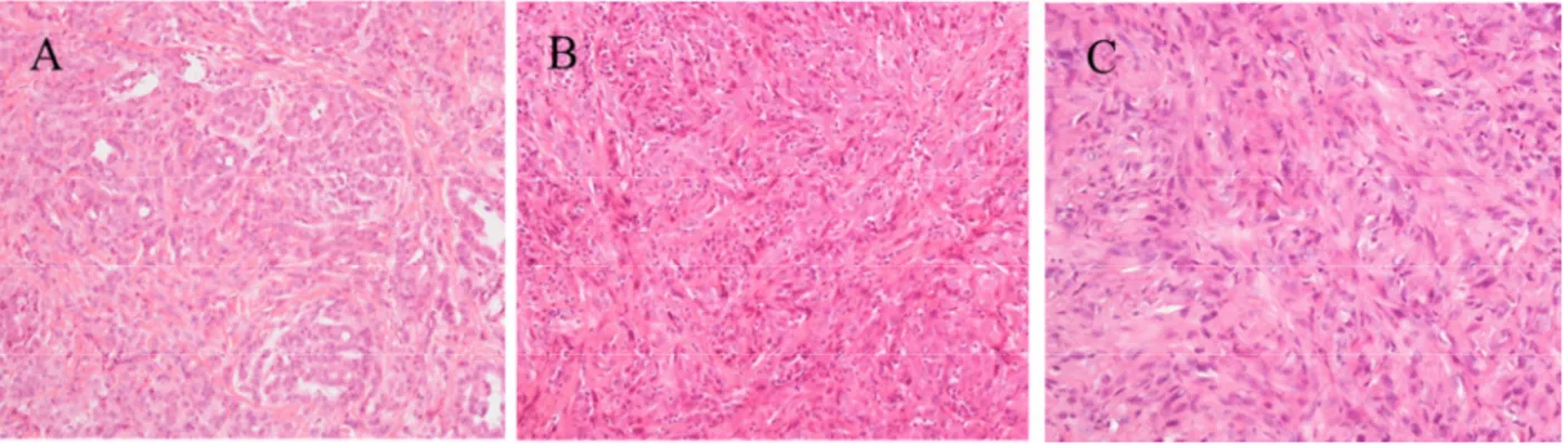
Figure 1. Observed (to 1989) and predicted (1990–2029) annual numbers of pleural cancer deaths in men in Italy. Prognosis of MPM is poor, the overall survival in no treated patients ranges from 4 to 12 months. (Pass et al, 2001). According to the amount of epithelial and spindle cells we can distinguish three histological subtypes: epithelioid, sarcomatoid and biphasic (Figure 2); they are associated with a different prognosis. The epithelioid subtype is considered the less aggressive and most responsive to treatments, with the best prognosis (Boutin et al, 1998; Robinson et al,2005).
2
Figure 2 Example of histological subtypes of MPM: A) Epithelioid B) Biphasic C) Sarcomatoid. Original magnification 20X
1.2 Etiology and Pathogenesis
1.2.1
Asbestos and Mesothelioma
Prior to the 1950s, malignant mesotheliomas (MM) were extremely rare. The first documented case of mesothelioma, according to current diagnostic criteria, was published in 1947 (King et al, 1947). Asbestos is a generic name for a family of naturally silicate minerals with different carcinogenic potential (Mossman et al, 1990). The various types of asbestos are divided into two major groups: serpentine represented by crysotile, the most common and economically important form of asbestos in the Western World; and the amphiboles, which include crocidolite, the most oncogenic type of asbestos, amosite, anthophyllite, and tremolite. The link between asbestos fibers and MPM development is well established (Boutin et al, 1996; Bocchetta et al, 2007), moreover Qi et al. recently showed how chrysotile can cause transformation in human mesothelial cells via HMGB1 and TNF-α signaling (Qi et al, 2013). Amphibole are very thin fibers (diameter 3µm) which have the capacity to reach the pleura either through the lymphatic, or by direct penetration and to cause fibrosis, pleural plaques, and eventually mesothelioma. Moreover they can damage the mitotic spindle of the cell leading to aneuploid and DNA damage. (Ault et al, 1995; Kamp et al, 1995). A key mechanism by which asbestos causes the transformation of mesothelial cells has recently been elucidated: working with primary human mesothelial (HM) cells, Yang et al discovered that asbestos induces necrotic cell death with resultant release of HMGB-1 in the extra cellular space. HMGB-1 release causes a chronic inflammatory response, macrophage accumulation and the secretion of TNF-α, which in turn activates NF-kB, leading to the survival of HM cells that have accumulated genetic damage because of asbestos exposure (Figure 3) (Yang et al, 2010).
3
Figure 3 Mechanism of asbestos-induced pathogenesis.
Erionite is an asbestos-like mineral more carcinogenic than asbestos to induce mesothelioma (Hill et al, 1990); Wagner and colleagues showed that mice injected with erionite develop MM in almost all cases, instead mice injected with asbestos fibers has MM in a lower percentage of cases (48%) (Wagner et al, 1985). Carbone linked erionite with endemic cases of mesothelioma in some Turkish villages of Cappadocia (Emri et al, 2002) (erionite is natural component of the stones of this region) and in North and South Dakota, showing that it is a serious cause of environmental pollution (Carbone et al, 2011). Some studies have shown that asbestos exposure causes activation of MAPK, phosphatidylinositol-3-kinase (PI3K)-AKT and the downstream mTOR (the target for rapamacyin) in MM (Altomare et al, 2005; Wilson et al, 2008).
1.2.2
SV40 and mesothelioma
Simian Virus 40 is a normal guest of macaque species of monkeys, it is a double circle DNA virus with two coding regions: early and late according to which is first coded. Early region codes T antigen (large T antigen) and t antigen (small t antigen). The ability to induce tumor transformation in the host cells is linked to the large T antigen, indeed it binds and inactivates essential tumor suppressor genes, like p53 and pRb, stimulates Met, Notch-1 and telomerase activity. (Carbone et al, 1997; De Luca et al, 1997; Cacciotti et al, 2001; Bocchetta et al., 2003; Foddis et al, 2002).
4
Hamsters intracardially injected with SV40 develop MM in the 60% of cases (6-9 months), as well as intrapleurally injected mice show MM in the 100% of cases (4-6months) (Cicala et al, 1993). In the 60’-70’, millions of people worldwide were injected with the inactivated (Salk) and early live attenuated (Sabin) forms of polio vaccines that were contaminated with SV40, however its ability to cause tumour in human is not very clear since several conflicting data have been reported.
1.2.3
Genetic predisposition
Some individuals develop mesothelioma following exposure to small amounts of asbestos, whereas others exposed to heavy amounts do not. Carbone et al. have reported mesothelioma clustering in several Turkish families in which up to 50% of members developed mesothelioma (Roushdy-Hammady et al, 2001; Carbone et al, 2007). This incidence far exceeds that observed in cohorts exposed to high levels of asbestos (4.6%), suggesting a genetic predisposition. Afterwards Carbone et al focused on two American families with high incidence of mesothelioma to identify putative mesothelioma susceptibility genes. The members of these families were neither exposed to erionite nor had occupational exposure to asbestos, thus removing the confounding factor of heavy exposure to carcinogens known to cause a high incidence of mesothelioma. Family members developed various malignancies, although mesothelioma predominated (Testa et al, 2011). Array-comparative genomic hybridization (CGH) analysis of two tumors (one per family) uncovered alterations encompassing or adjacent to the BAP1 (BRCA-1 associated protein 1) locus at 3p21.1. BAP1 germline mutations have been also liked to uveal melanoma and to a type of benign melanocytic tumors that called mBAITS (melanocytic BAP1-mutated atypical intradermal tumours (Carbone et al, 2013) suggesting that BAP1 may rather represent gene predisposing to a new cancer syndrome.
1.2.4
Radiation
Radiation exposure has also been linked to MM, even though these cases are rarely observed (Goodman et al, 2009). Patients who received radiation treatments, specifically in the thoracic or abdominal regions, or who received Thorotrast intravascularly have shown increased risks in developing MM (Amin et al, 2001). Moreover, studies in rats demonstrate that radiation is a causative co-factor of MM in combination with asbestos exposure (Lafuma et al, 1980).
In summary, the association between asbestos, erionite, SV40 infection, genetic predisposition and radiation exposure suggests a multifactorial origin for malignant mesothelioma and each factor plays a crucial role in necrosis, inflammation and genetic damage.
5
1.3 Clinical features
The mean age at presentation is 60 years, because of the long latency from the time of first exposure to asbestos to the development of clinically evident disease (Britton M., 2002). The incidence is higher in men, presumably because more men have worked in asbestos-related trades. Symptoms and physical findings are generally not specific for the disease. Most patients present with non-pleuritic chest pain or dyspnea. Compared to that of metastatic pleural diseases, the pain from mesothelioma can be severe, aching, and often very difficult to control. Less common complaints are cough, fevers, chills, sweats, and fatigue. Fatigue, cachexia and pain are common in advanced disease. Physical examination is usually only remarkable for signs related to the presence of a pleural effusion or mass. Later in the course of disease one can often appreciate volume loss and decreased mobility of the chest wall on the side of the primary tumor. Occasionally, the tumor may extend directly into the chest wall, and be detected as a tender or non-tender chest wall mass.
1.4 Diagnosis
Diagnosis of malignant mesothelioma requires a careful evaluation of the clinical and radiological features, and it must be confirmed by a pleural biopsy.
The main radiologic techniques used to diagnose mesothelioma are:
• Computer Tomography (TAC)TAC is able to detect pleural effusion, pleural thickening, calcification, intralobular thickening and the potential thoracic invasion. However TAC cannot distinguish between benign tumour, adenocarcinoma and mesothelioma. TAC scanning may help fine needle aspiration/biopsy of pleural mass.
• Magnetic resonance Imaging (MRI)MRI scanning allows to determine tumour size and to better detect the tumor area and distinguish the normal part. MRI is more accurate than TAC to evaluate the mediastinic lymphonodal enlargement.
• Positron emission tomography (PET) PET imaging is a nuclear technique that produces three-dimensional image, it is currently the better way to locate the onset tumor sites. Fluorodeoxyglucose PET and particularly PET/CT shows promise as a tool to differentiate benign from malignant disease and as an adjunctive tool for staging. A combination of the
6
imaging techniques may be necessary for determining the best approach to the patient (Wang et al, 2004).
Histology
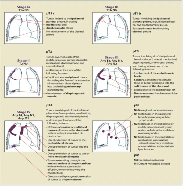
In order to obtain a definitive diagnosis of MPM the tissue biopsy and/or pleural effusion exams are essential. The pleural effusion often shows high level of bloody cells, high protein concentration, low level of white cells and low pH. The high content of hyaluronic acid is suggestive of mesothelioma, but it is poorly specific, so the cytologic analysis, as well as the trans-needle aspiration rarely leads to a definite diagnosis. The histological evaluation of pleural biopsy is therefore of crucial support to the diagnosis. However, in many cases, to confirm the MPM diagnosis it is necessary to investigate a panel of tumoral markers by the mean of immunohistochemistry. According to the embryologic histogenesis of mesothelial tissue, MPM shows epithelial and mesothelial markers such as cytocheratin 5/6, carletinin, thrombomodulin, mesothelin and the Wilms Tumor 1 (WT-1). The presence of at least two of positive markers in the context of a clinical and histological suspicion, is sufficient to confirm the diagnosis of MPM (Chierieac et al, 2009). Very often the diagnosis of MPM occurs in its late stage. TNM-based staging system by the International Mesothelioma Interest is the most widely used staging system for MPM (Rusch, 1995), it can be summarized as in Figure 4.
7
Figure 4 IMIG Staging System for Malignant Pleural Mesothelioma.
1.5 Therapy
There are no therapeutic standards for malignant mesothelioma and the treatment options depend on performance status, pulmonary function, stage, and age of the patient.
1.5.1
Surgery
The two potential goals of surgical therapy for pleural mesothelioma are palliation of symptoms and debulking of tumor with therapeutic intent.For surgical debulking of mesotheliomas, two surgical approaches are commonly employed, pleurectomy with decortication or extrapleural
8
pneumonectomy (EPP). Pleurectomy with decortication removes all gross disease from all pleural surfaces and preserves the underlying lung. EPP entails en bloc removal of the lung along with surrounding parietal pleura, pericardium, and diaphragm, with the pericardium and diaphragm then replaced by synthetic grafts. These are both technically challenging procedures and should be performed only by surgeons with extensive experience. EPP is especially difficult, and was originally associated with an unacceptably high morbidity of 30%. However, with advances in surgical, anesthetic, and critical care techniques, and more exacting patient selection, experienced centers now report mortality rates of < 4%, a rate comparable to standard pneumonectomy (Sugarbaker et al, 1999).
1.5.2
Radiation therapy
Although in vitro studies suggest that mesothelioma is more sensitive to radiation than non-small cell lung cancer (Charmichael et al, 1989), the clinical experience reported by radiation oncologists suggests that it is an especially radio-resistant tumour. In addition, radiation of the involved chest is limited by the presence of radiosensitive organs and the extensive nature of the tumour. As a consequence, its use appears limited to adjunctive therapy for patients who have undergone EPP, and to palliative treatment of painful chest wall lesions. Prophylactic chest wall irradiation may reduce the incidence of chest wall recurrences at incision sites but there is no consensus on its use and randomized controlled trials are needed (Lee et al, 2009). An area of active ongoing research is the role of high-dose hemithorax irradiation after EPP for early stage disease. In carefully staged patients, this approach has resulted in a marked reduction in local tumor recurrences, although nearly one half of patients subsequently developed isolated distant metastases (Senan et al, 2003).
1.5.3
Chemotherapy
Most patients with mesothelioma are not candidates for surgical or radiotherapy treatment and chemotherapy is their main option. The most commonly regimen used now includes the multitargeted antifolate drug pemetrexed with a platinum drug such as cisplatinum. The use of this combination has been compared to cisplatin alone in a large Phase III study of 456 patients (Vogelzang et al, 2003). Response rates were significantly better in the pemetrexed/cisplatin arm than in the cisplatin alone arm (41.3% Vs 16.7%), and survival was significantly better as well (median survival 12.1 months Vs 9.3 months). Addition of folic acid and vitamin B12 significantly reduced toxicity without altering survival benefit. The other regimen used commonly is the false nucleotide gemcitabine with a platinum agent. Nearly half of the patients on this doublet regimen noted symptom improvement, 33% had a partial response, and 60% had stable disease; no survival
9
benefit was demonstrated compared to historical controls (Novak et al, 2002). Similarly, treatment with the combination of gemcitabine and oxaliplatin has been reported to improve symptoms, but not significantly improve survival (median survival of 13 months) (Schutte et al, 2003). Platinum compounds act through the formation of platinum-DNA adducts. Removal of these adducts, which leads to chemoresistance, is mainly carried out by the nucleotide excision repair (NER) system that consists of at least 30 identified polypeptides, including the pivotal protein excision repair cross-complementing group-1 (ERCC1) (Sancar A., 1995). It is hypothesized that low expression of ERCC1 might predict increased sensitivity to platinum-based chemotherapy, possibly due to the saturation of the enzyme complex; conversely, high levels of ERCC1 may predict a resistance to platinum-based chemotherapy. Pemetrexed (commercial name Alimta
), is a multitargetedantifolate agent that inhibits dihydrofolate reductase (DHFR), thymidylate synthase (TS), and glycinamide ribonucleotide formyltransferase (GARFT), enzymes involved in purine and pyrimidine synthesis. However, pemetrexed is a weak inhibitor of GARFT, and when TS is inhibited, tetrahydrofolate oxidation stops and there is no longer a need for DHFR activity (Chattopadhyay et al, 2007). Therefore, most studies have focused on pemetrexed effects on TS. TS mRNA levels were inversely correlated with pemetrexed activity in different tumor cells (Hanauske et al, 2007; Giovannetti et al,2008), whereas other studies suggested a correlation between high levels of TS protein expression and reduced sensitivity to pemetrexed in colon and lung cancer cells (Sigmond et al, 2003).
1.5.4
Immunotherapy
It is known that an immune response is induced by mesothelioma, but it is weak (Robinson et al, 2000). This knowledge has prompted a number of investigators to study different ways to consolidate that response. The intrapleural instillation of cytokines is limited by the short half-life of most cytokines, necessitating repeated injections or continuous infusion via a pleural catheter. Intrapleural interferon-gamma twice weekly for 2 months was reported to induce response rate of 56% in early stage disease (Boutin et al, 1991). A continuous intrapleural infusion of interleukin-2 induced a partial response in four of 21 patients and an overall survival of 16 months (Goey et al, 1995). In both cases, side effects were minimal and consisted primarily of fever and constitutional symptoms. Studies in animals suggest that interferons have an antiproliferative effect on mesothelioma cells and enhance the cytotoxic effect of cisplatin. The results from these studies led to the development of a Phase II trial of cisplatin-doxorubicin and interferon alpha-2 in advanced malignant mesothelioma. The overall response rate was 29% and the median survival was 9.3
10
months with a one year survival of 45% and two year of 34% (Parra et al, 2001). However, severe myelosuppression was seen in 60% of patients limiting the application of this treatment.
1.5.5
New agent under study
Studies of the molecular biology of mesothelioma and the cellular mechanisms leading to a malignant phenotype have led to the identification of several possible therapeutic targets for treatment of this disease. Some of these are already under investigation in clinical trials, for example, several receptor tyrosine kinases are aberrantly expressed in these tumors, including the epidermal growth factor receptor (EGFR) (Janne et al, 2002). Other novel agents targeting growth factors found to be overexpressed in mesothelioma, e.g. vascular endothelial growth factor and its receptor, are under investigation. Other agents under study include anti-angiogenic agents, e.g. AZD2171, thalidomide and PTK/ZK787, inhibitors of histone deacetylase superoylanilide and hydroxamic acid (SAHA), proteasome inhibitors, and histone deacetylase inhibitors (PXD101). Furthermore, two classes of EGFR antagonists, small molecule tyrosine kinase inhibitors (TKIs) and monoclonal antibodies (mABs), have been approved by the Food and Drug administration (FDA) and the European Medicines Evaluation Agency (EMEA) for the treatment of metastatic NSCLC, colorectal cancer (mCRC), squamous-cell carcinoma of the head and neck and pancreatic cancer (Gridelli et al, 2007; Sridhar et al, 2003). Gefitinib and erlotinib, two reversible TKIs, inhibit the EGFR phosphorylation and its downstream cascade by blocking the ATP pocket located in the intracellular catalytic domain of the receptor. Cetuximab and panitumumab, two anti-EGFR mABs, target the extracellular domain of the receptor and upon the receptor binding they inhibit its dimerization and subsequent phosphorylation and signal transduction (Figure 5). The introduction of cetuximab and panitumumab in clinical practice, either in combination with chemotherapy or as single agent, has shown to improve the outcome of metastatic CRC and NSCLC patients (Saltz et al, 2004). Preclinical studies have shown that EGFR TKIs are highly efficacious in mesothelioma cell cultures (Barbieri et al, 2011), but two phase II studies of gefitinib and erlotinib used alone to treat malignant pleural and peritoneal mesotheliomas failed to demonstrate their clinical efficacy. However it needs to be pointed out that the patients in both trials were not selected on the basis of any molecular criteria (Govindan et al, 2005; Garland et al, 2007). One recent study has shown that cetuximab effectively blocks the growth of MPM cells in cell cultures and mouse models (Kurai et al, 2012) and, as in the case of colorectal cancer and lung adenocarcinomas, the potential efficacy of these TKIs in MPM may depend on the mutation status of EGFR gene and its downstream effectors (Lie`vre et al, 2006).To the best of our knowledge, only a few low-powered studies have investigated the presence and frequency of EGFR gene mutations in MPM (Cortese et al, 2006;
11
Enomoto et al, 2012), and none has searched for mutations in the KRAS, BRAF, and PI3KCA downstream effectors.
Figure 5 EGFR inhibitors, mABs and TKIs.
1.6 Epidermal growth factor receptor (EGFR).
Epidermal growth factor (EGF) was first discovered by Stanley Cohen in 1960 during his study of nerve growth factor in mouse sub-maxillary glands and subsequently, in 1975, he confirmed the presence of plasma membrane receptors in human fibroblasts (Cohen et al, 1960, Carpenter et al, 1975). EGFR was isolated in 1982 as a 170 kDa transmembrane glycoprotein with an EGF binding site on the extracellular surface (Cohen et al, 1982). The structure of EGFR was found to be the human equivalent of the mammalian v-erb-B oncogene protein from the avian erythroblastosis virus. Unlike the human EGFR, the v-erb-B oncogene protein did not have the extracellular EGF binding domain thereby demonstrating that the intracellular domain may play an important role in tumourigenesis. EGFR belongs to the human epidermal growth factor receptor (HER) family, which has four structurally related receptor tyrosine kinases. The EGFR gene localized on chromosome 7 p11-13, the protein consists of 1186 amino acids. The receptor structure consists of extracellular, transmembrane and intracellular domains. The extracellular domain consists of cysteine-rich clusters, which form the ligand-binding domain. Upon binding with ligands such as EGF or transforming growth factor alpha (TGF-α), the EGFR monomers form homodimers with another
12
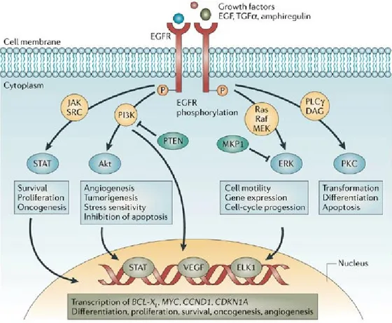
EGFR or heterodimers with another receptor of the HER family. The intracellular domain has tyrosine kinase activity. Dimerisation of the EGFR results in structural rearrangement of the intracellular tyrosine kinase domain, adenosine triphosphate (ATP) is then recruited into the catalytic domain, resulting in its auto-phosphorylation. This leads to the activation of a cascade of intracellular signal transduction pathways resulting in cell proliferation, anti-apoptosis, invasion and metastasis (Citri et al, 2006; Hynes et al, 2005; Bogdan et al, 2011). Among a host of various intracellular signalling pathways stimulated by EGFR, the major pathways activated are the RAS/RAF/MAPK pathway resulting in cell proliferation, metastasis and invasion, and the PI3K/AKT/mTOR pathway resulting in inhibition of apoptosis (Ciardello et al, 2008) (Figure 6). The first signaling cascade shown to be downstream of the EGFR was the Ras–mitogen-activated protein kinase or MAP kinase pathway. When the pathway is activated, the SOS guanine nucleotide exchange factor is recruited to the plasma membrane via the Grb2/Drk/Sem5 adapter protein. SOS stimulates the exchange of GTP for GDP on the small G-protein Ras. Subsequently activated Ras stimulates the MAP kinase pathway to promote cell proliferation. Phosphoinositide 3-kinase (PI3K) plays a crucial role in effecting alterations in a broad range of cellular functions in response to extracellular signals. A key downstream effector of PI3K is the serine-threonine kinase Akt which in response to PI3K activation, phosphorylates and regulates the activity of a number of targets including kinases, transcription factors and other regulatory molecules (Paez et al, 2000). The complexity of signaling is further increased by cross-talk between individual pathways. Since EGFR is associated with an oncogenic phenotype, its inhibition may result in an anti-neoplastic effect. As mentioned before, in the last decade several inhibitors of EGFR have been developed, including monoclonal antibodies (cetuximab) and small molecule inhibitors (gefitinib, erlotinib), which have been shown to be effective in animal models, in preclinical and clinical studies (Mendelsohn et al, 2003). A correlation between EGFR expression and response to therapy has been reported in some human cancers (breast, lung and prostate) (Santoro et al, 2004; Cappuzzo et al, 2003).
13
Figure 6 EGFR downstream signaling pathways
1.6.1
EGFR mutations and inhibitors
Activating EGFR mutations have been reported in cancers such as non-small-cell lung cancer (NSCLC) and head and neck cancers, and are predictive of the response to gefitinib or erlotinib therapy (Lynch et al, 2004; Paez et al, 2004; Lee et al, 2005). Approximately 90% of EGFR mutations affect small regions of the gene within exons (18 to 24) that code for the EGFR tyrosine kinase domain. The most common mutations are an in-frame deletion in exon 19 around codons 746 to 750 (accounting for 45 to 50% of EGFR mutations) and a missense mutation leading to a substitution of arginine for leucine at codon 858 (L858R) in exon 21 (35 to 45% of EGFR mutations) (Sharma et al, 2007). Somatic EGFR mutations are found in approximately 5 to 15% of unselected white patients and in 25 to 35% of unselected Asian patients with NSCLC. These mutations seem to be limited to NSCLC, since they have rarely been detected in other types of human cancer.
14
1.7 v-Ki-ras2 Kirsten rat sarcoma viral oncogene homolog (KRAS)
KRAS is a small GTPases that regulate cell growth, proliferation and differentiation, it is normally activated in response to the binding of extracellular signals, such as growth factors, RTKs (Receptor Tyrosine Kinases) and TCR (T-Cell Receptors). In the resting cell, KRAS is tightly bound to GDP (Guanosine Diphosphate); as a result of extracellular stimuli to cell membrane receptors the guanine nucleotide exchange factors (GEFs) release GDP and allow GTP (Guanosine Triphosphate) binding. In the GTP-bound form, KRAS interacts specifically with effector proteins, thereby initiating cascades of protein-protein interactions that may finally lead to cell proliferation.Active GTP-bound KRAS interacts with several effector proteins: among the best characterized are the Raf kinases and phosphatidylinositol 3-kinase (PI3K) (Hancock JF., 2003). KRAS gene is localized on chromosome 12. More than 95% of KRAS activating gene mutations occurs at codon 12 and 13 of exon 2. Less frequent mutations occurs at codon 61. KRAS activating mutations cause resistance to anti-EGFR mABs target therapies.
1.8 v-raf murine sarcoma viral oncogene homolog B1 (BRAF)
BRAF gene is localized on chromosome 7q34, it encodes the protein BRAF belonging to the raf/mil family of serine/threonine protein kinases. This protein plays a role in regulating the MAP kinase/ERKs signaling pathway, which affects cell division, differentiation, and secretion, in fact activated BRAF triggers mitogen-activated protein kinase (MAPK) and extracellular-signal regulated kinase (ERK, MEK1 and MEK2) by serine phosphorylation. Mutations in BRAF gene are associated with cardiofaciocutaneous syndrome, a disease characterized by heart defects, mental retardation and a distinctive facial appearance. Mutations in this gene have also been associated with various cancers, including non-Hodgkin lymphoma, colorectal cancer, malignant melanoma, thyroid carcinoma, non-small cell lung carcinoma, and adenocarcinoma of lung. The highest frequency of BRAF mutations is in malignant melanoma. The most common mutation is a T to A nucleotide transversion leading to a V600E amino acid substitution within the activation segment of the Raf serine/threonine kinase gene product, increases the catalytic activity of B-Raf and leads to subsequent activation of MEK and ERK MAPKs (Davies et al, 2002: Pollock et al, 2003). Since constitutively active BRAF mutants commonly cause cancer by excessively signaling to cell growth, inhibitors of BRAF have been developed for both the inactive and active conformations of the kinase domain as cancer therapeutic candidates (Bollag et al, 2010; Wan et al, 2004). Sorafenib
15
and Vemurafenib are currently the two BRAF molecular inhibitors approved by the FDA for the treatment of primary liver and kidney cancer and for late stage melanoma.
1.9 Phosphoinositide 3- kinase (PI3K)
The class I PI3Ks catalyse the conversion of phosphatidylinositol-3,4-bisphosphate (PtdIns-3,4-P2)
to phosphatidylinositol-3,4,5- trisphosphate (PtdIns-3,4,5-P3). These specialized lipids serve to
recruit pleckstrin homology (PH) domain-containing proteins such as the serine-threonine kinase Akt and PDK1 (phosphoinositide-dependent kinase 1) to the plasma membrane. After recruitment to the membrane, Akt is phosphorylated and consequently activated, by PDK. In turn, Akt phosphorylates multiple proteins on serine and threonine residues. Through phosphorylation of these targets, Akt carries out its role as a key regulator of a variety of critical cell functions including glucose metabolism, cell proliferation and survival. The PI3K family comprises eight members divided into three classes according to their sequence homology and substrate preference. PI3K enzymatic structure shows a catalytic subunit (p110) associated with a regulatory one (p85). The catalytic subunit PI3KCA is encoded by a gene localized at chromosome 3p26.32. Mutations in the PIK3CA gene are not frequent in colon rectal cancer, occurring in about 15% of these tumours. PIK3CA mutations mainly occur in exons 9 and 20, with exon 9 showing the highest incidence (68.5% approximately). These mutations can be found in the same tumour together with KRAS and BRAF mutations, and this makes difficult to evaluate their own role in defining the sensitivity to anti-EGFR mAbs. (Benvenuti et al, 2008; Samuel et al, 2004; Di Nicolantonio et al, 2010).
1.10 Prognostic and predictive biomarkers.
In the majority of patients MPM is diagnosed in stage III/IV, and systemic therapy represents the only potential treatment option for most cases. The combination of platinum or cisplatin and pemetrexed represents the standard of care in the first-line treatment of MPM. Several studies carried out on NSCLC showed that protein and mRNA ERCC1 expression have a consistent prognostic and predictive value in patient treated with cipslatin (Olaussen et al, 2006; Zheng et al, 2007). Similarly, in NSCLC cell lines, high baseline TS gene expression levels confer resistance to pemetrexed and TS protein levels are correlated to pemetrexed efficacy in a variety of solid tumours (Gomez et al, 2006; Rose et al, 2002; Zucali et al, 2011; Righi et al, 2010). Due to the epithelioid phenotype of the most part of MPM it could be interesting to investigate the ERCC1 and TS
16
gene/protein expression in order to determine whether they can have a prognostic and/or predictive value in mesothelioma patients.
1.10.1
Excision repair cross-complementing group-1 (ERCC1)
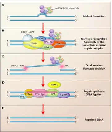
Platinum compounds function by binding to DNA resulting in intrastrand or interstrand crosslinks, which disrupt the DNA structure. These lesions may interfere with base pairing and generally obstruct transcription and normal replication processes, ultimately leading to apoptosis. The nucleotide excision repair (NER) pathway is 1 of 5 recognized DNA repair pathways (mismatch repair, double-strand break repair, base excision repair and direct repair) that maintain DNA integrity and defend DNA against environmental damage. It is generally well accepted that each of the repair pathways identifies distinct lesion types (Hoeijmakers, 2001). NER has been identified to repair bulky, helix-distorting DNA lesions caused by UV light or chemicals, including platinum compounds. NER pathway acts through the recognition of DNA repair, followed by the formation of a complex to unwind the damage portion and excise it. Finally, the excised area is resynthesized and bound to the undamaged DNA, restoring the double helix. After the repair process is complete, the entire complex is disassembled. Excision repair cross-complementation group 1(ERCC1) protein functions within the repair complex as it heterodimers with the Xeroderma pigmentosum complementation group F (XPF) protein and functions as an endonuclease producing a 5’ single strand cut of 20 nucleotides from the lesion (Figure 7). Early reports suggested that ERCC1 mRNA levels, and not XPF, were correlated with DNA repair capacity when exposed to UV light, suggesting that ERCC1 might be rate-limiting (Bohanes et al, 2011; Vogel et al, 2000). Those data suggest the hypothesis that ERCC1 gene expression might be used as a predictive marker for DNA repair capacity, and thus predict platinum compounds cytotoxicity. ERCC1 gene localized on chromosome 19q13.32 and encodes a 32 kDa protein located in the nucleus.
17
Figure 7 Simplified model of genome Nucleotide Excision Repair (NER) system.
1.10.2
Thymidylate synthase (TS)
Thymidylate synthase (TS) is a key enzyme in the de novo synthesis of DNA. The reaction catalyzed by TS is the methylation of dUMP, through the transfer of the methyl group provided by the cofactor methylenetetrahydrofolate (CH2THF) dUMP is converted into
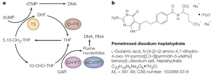
deoxythymidine-5′-monophosphate (dTMP). Subsequently, dTMP is phosphorylate by two successive steps to 2’-deoxythymidine-5’-triphosphate (dTTP) an essential precursor for DNA synthesis. TS is target for chemotherapeutic agents because of its central role in DNA synthesis, and it is also of interest because of its rich mechanistic features. Pemetrexed and 5-fluorouracil are the main antitumour agents targeted to the TS (Figure 8). The gene encoding TS is localized on chromosome 18p11.32, the protein consists of 313 amino acids and has a ubiquitous localisation in the cell, i.e. in the nucleus, in the cytoplasm and in the mitochondrion inner membrane and matrix.
18
Figure 8 a)Simplified illustration of some key enzymatic reactions of folate metabolism, showing enzymes affected by pemetrexed, or its polyglutamates. b) Structure of pemetrexed disodium. AICARFT: aminoimidazole carboxamide
ribonucleotide formyltransferase; DHFR: dihydrofolate reductase; GARFT: glycinamide ribonucleotide formyltransferase; THF: tetrahydrofolate, TS: thymidylate synthase.
1.11 Clonality analysis
In spite of a number of different approaches that have been shortly described above, malignant mesothelioma is not yet to be cured. Understanding more about the initiation of the tumor development and the factors that trigger it is crucial to have the best way to treat or even prevent this cancer. Given the fact that a cell population could be a mixture of slightly different cells, any single method for the treatment may include the chance of the failure, because of the differences of drug sensitivity of the single cells. The most common method to investigate whether a tumour population is homogenous or not is to determine its clonal origin. This approach explores the nature of tumour initiation and categorizes the tumour as mono- or poly-clonal. A clonal population of tumour cells is defined as those cells arising from the mitotic division of a single somatic cell (Seeker-Walker et al, 1985), while a polyclonal tumor is known to be initiated by division of multiple differentiated cells. Categorization of a cell population as mono- or polyclonal is made possible by the determination of the inactivated X chromosome of the cells in a given population. The natural event of X chromosome inactivation occurs in all female cells during the early embryogenesis and provides a sufficient tool for tracking a population to their ancestral stage, because once it is determined, the same X chromosome is kept inactivated during the mitosis of the same cell.
19
1.11.1
X Chromosome inactivation
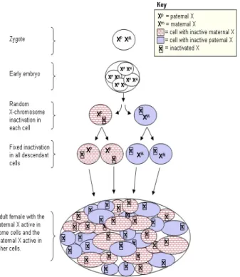
In females inactivation of one X chromosome occurs in each somatic cell in early embryonic development and is passed onto the progeny of the cell in a stable fashion(Lyonization) (Martin et al, 1978; Lyon et al, 1988, Lyon, 1961). Females heterozygous for polymorphic X-chromosome genes are therefore mosaics with respect to X-chromosome activity (Figure 9). In 1961, Mary Lyon proposed that the Barr body, a unique cytological entity situated near the nucleolus that distinguishes female from male cells, was the condensed, inactive female X chromosome. She proposed random X‐chromosome inactivation (XCI) as an explanation for this cytological entity. The Lyon hypothesis suggested that one of the two X chromosomes is entirely silenced or inactivated at random in the soma to balance the X‐linked gene dosage between XX females and XY males. Her hypothesis was based on the observations of X-linked coat color mutations in heterozygous female mice. In these mice, the phenotype was always a mosaic, consisting of patches of normal or mutant color, rather than a homogenous blending, suggesting that early in development, in the pigmented cells either one or the other X chromosome was inactivated. Thus, if the X chromosome carrying the mutant allele was inactivated, the patch was of normal color, whereas if the X chromosome carrying the normal allele was inactivated, the patch was of mutant color. Beutler and colleagues formulated the XCI hypothesis using studies of the human X chromosome glucose 6-phosphate dehydrogenase (G6PD) gene (Beutler et al, 1962). They found that, in females, G6PD activity was not twice as much that of males, as expected by the presence of two X chromosomes, and postulated a dosage compensation mechanism. In females heterozygous for G6PD deficiency, dosage compensation results in G6PD expression at half the rate of normal hemizygous males. This could be attributable to either half-level activity in all cells or normal expression in some cells and low expression in other cells, resulting in overall half-level expression. Using a mixture of male cells with deficient G6PD activity and normal G6PD activity, Beutler and colleagues measured G6PD activity (by glutathione stability) and compared it with the response of female erythrocytes. They found that the response curves of the 2 samples were similar in shape and concluded that intermediate activity in females was probably attributable to the same mechanism as in the mixture of male normal and G6PD activity-deficient erythrocytes. There is evidence that chromosome inactivation is related to differential methylation of cytosine in the DNA of X-chromosome genes (Holliday R, 1987).
20
Figure 9 X chromosome inactivation Xp: clonal cell with active paternal X chromosome Xm: clonal cell with active maternal X chromosome
1.11.2
Mechanisms of X-chromosome inactivation
The exact molecular mechanisms underlying XCI are still not fully clarified, but involve several steps, including the determination of the number of X chromosomes per cell, selection of either the paternal or maternal X chromosome for subsequent inactivation, and initiation of the actual inactivating process. It has been demonstrated in mice that there are 3 non coding loci, located near the center of inactivation of X chromosome that play a pivotal role in the mechanism of X-chromosome inactivation. These loci are: non coding RNA X (inactive)-specific transcript (Xist), its antisense partner Tsix, and the intergenic locus Xite. Xist is necessary for cis inactivation of the X chromosome. In vitro, Xist is able to silence also the autosomal surrounding chromatin in case of X: autosome translocation, but in an incomplete manner, due to instability of autosome inactivation (Lee J.T et al, 1999). Tsix and Xite work in parallel to Xist by maintaining X-chromosome transcriptional competence (Ogawa et al, 2003). Although the functions of these 3 loci have been deduced using complementary cell lines, the actual physical interactions of these components are less well known. Xist is proposed to achieve cis-inactivation of the X chromosome through close interactions between its RNA transcript and the segment of X chromosome to be inactivated (Penny G.D. et al, 1996). The putative trans-interactions, based on the need to determine one X to be exclusively activated and the other X to be exclusively inactivated, remained elusive, until the
21
recent demonstration that the 2 X chromosomes undergo inter-chromosomal pairing (Heard E., 2004). It is remarkable that inter-chromosomal pairings typically occur in germ cells undergoing meiosis, rather than in somatic cells undergoing mitosis. X-chromosome inactivation timing is crucial to the interpretation of X chromosome inactivation pattern (XCIP)-based clonality assays. It has been assumed that pre-blastocyst embryos express both X chromosomes and that inactivation did not occur until after implantation and the embryonic stem cells began to differentiate into separate cell lineages (Okamoto et al, 2004). Recent experiments, however, demonstrate that XCI occurs as early as the 4-cell stage of the embryo, but is variable and leaky and does not become stabilized until after implantation, but before differentiation of embryonic stem cells into the various cell lineages (Huynh et al, 2003). XCI before cell lineage differentiation is crucial for the interpretation of XCIP clonality studies. Hematopoietic cell lines derive not from a single embryonic stem cell but from several progenitors, allowing for the mosaic expression of genes from both X chromosomes (Prchal et al, 1996).
22
2 AIMS OF THE STUDY
1)
Given that only a few low-powered studies have investigated the presence and frequency of EGFR gene mutations in malignant pleural mesothelioma (Cortese et al, 2006; Enomoto et al, 2012), and none has searched for mutations in the KRAS, BRAF and PI3KCA downstream effectors, in this study we searched a large series of histological MPM samples for mutations in EGFR gene and its main downstream signaling effectors in order to evaluate their frequency and possible prognostic significance, and their possible use as predictors of the response of MPM to targeted therapies.2)
The golden standard in the treatment of patients with malignant pleural mesothelioma is the multimodal administration of platinum compounds and pemetrexed. Currently there are no biomarkers predicting the clinical outcome and the prognosis for this tumour. The second aim of this work was to retrospectively investigate in a series of MPM patients, randomly treated with platinum and pemetrexed (alone and in combination), ERCC1 and TS gene and protein expression to determine whether they can provide information about the clinical outcome and/or can have a prognostic value.3)
Identifying whether a tumour is monoclonal or polyclonal at start have critical implications in terms of early therapeutic intervention. The third aim of the study was to evaluate the clonality pattern of malignant mesothelioma.23
3 MATERIALS AND METHODS
In this study we used two different clusters of MPM tumour samples: in the first case we collected the samples from the Thoracic Unit of the University Hospital of Novara on which we performed the sequencing analysis of EGFR and downstream pathways and the evaluation of ERCC1 and TS protein and gene expression (Novara samples).
In the second case the tumour samples we used to evaluate the clonality assessment of malignant mesothelioma were obtained from Dr. H. I. Pass (NYU, New York) and from Dr.Paul Sugarbaker (WCI, Washington, DC) in accordance with protocols approved by the Institutional Review Board of each center and upon patients informed consent (US samples).
3.1 Novara samples
3.1.1
Samples collection and preparation
In this study we involved a large number of MPM patients admitted to the Thoracic Unit of the University Hospital of Novara between January 2008 and March 2013, all of whom were diagnosed as MPM on the basis of multiple pleural biopsies taken by means of video-assisted thoracoscopy. The tumour samples were immediately fixed in formalin for 24 h, embedded in paraffin, and routinely processed for histology and immunohistochemistry. The diagnosis of MPM was based on standard histological and immunohistochemical criteria, including positivity to calretinin, vimentin, and cytokeratins 5 and 6, and negativity to carcinoembryonic antigen, thyroid transcription factor 1, and Ber Epy 4. From the analysis of the clinical records we obtained the following data:
• Personal data of the patient • MPM diagnosis date • Surgery or biopsy date • Treatment
• Status (Dead/Alive)
• Follow up: from the date of diagnosis to June 2013 for patients alive, and from the date of diagnosis to the date of death for dead patients.
The MPMs were classified on the basis of the WHO classification of pleural tumours (Travis et al, 2004), and clinically and pathologically staged on the basis of the TNM staging system (Sobin et al,
24
2009). Looking through the WINANA database of the Pathology Department of “Maggiore della Carità” Hospital we got these data (snomed # M-90523):
• haematoxylin/eosin-stained slides of the pleural biopsies and corresponding formalin-fixed, paraffin-embedded blocks
• histotype: epithelioid, sarcomatoid and biphasic.
An expert pathologist reviewed the haematoxylin/eosin-stained slides of each case to:
• confirm the diagnosis and the histotype;
• select the area with 70% of tumour cells (minimum required for the sequencing analysis and gene expression evaluation);
• identify the best sample, in term of cellularity, in case we have more than one biopsy or surgical specimen.
The tumoral areas of the formalin-fixed paraffin-embedded sections were macro-dissected manually, and then five 5 µm thick sections were prepared and collected in a 1.5 mL tubein order to perform the DNA and RNA extraction. 3 µm thick sections were cutted to perform immunohistochemistry staining.
3.1.2
DNA extraction
Genomic DNA was extracted using 450 µl of EDTA-SDS/proteinase K (SDS 1%, EDTA 20mM, Tris HCl pH 7,5 20mM) followed by phenol-chloroform, and resuspended with 30 µL of DEPC-treated and RNAse free water (Promega, Madison, USA). We include a negative control of extraction every 5 samples. The DNA concentration was evaluated by reading the absorbance at 260nm by mean of a spectrophotometer (Eppendorf, Amburgo, Germania). The purity of DNA preparation were measured by evaluating 260/280 and 260/230 ratios.
3.1.3
Mutational analysis
EGFR gene
All of the samples were analysed using the TheraScreen EGFR29 Mutation Kit (QIAGEN, Manchester, UK), which combines the two technologies of ARMS and Scorpion chemistry in order
25
to detect mutations in a real-time polymerase chain reaction (PCR). This kit allows the detection of in-frame deletions on exon 19, insertions on exon 20, and G719X, S768I, T790M, L858R and L861Q mutations against a background of wild-type genomic DNA with a sensitivity of 1%. PCR plate and reaction mix were performed according to the manufacturer’s instructions, with 50 ng of DNA for each well. DNA amplification was performed with the following cycles: denaturation at 95°C for 4 min, 40 cycles of 95°C for 30 sec and 60°C for 1 min. Negative and positive controls were used in every reaction plate, moreover the kit contains a control reaction mix that amplify exon 2 of EGFR to evaluate amplifiable DNA in the tested samples. Results interpretation was done following the datasheet instruction: sample quantification cycle must be within the range of 21.92 and 37.00. Sample was classified as positive when both control and mutation curves were positive according to the above limits. In order to determine the presence of other less common mutations, the samples underwent further PCRs in order to amplify the whole sequence of exons 18-21 of the EGFR gene. PCR conditions primers and are shown in Table 1.
KRAS gene
KRAS gene was analysed by means of a mutant-enriched PCR (ME-PCR) in order to detect the hotspots in codons 12 and 13 of exon 2 that include more than 95% of the known gene mutations. The ME-PCR consisted of two amplification steps (semi-nested PCR) in which artificial restriction sites were introduced into the wild-type amplicon using mismatched primers. The restriction sites (BstNI for codon 12 and BglI for codon 13) introduced during the first PCR step were localised immediately next to the KRAS codon in the analysis in order to distinguish wild-type and mutant sequences. The wild-type amplicons were then digested by restriction enzymes and the mutant products were enriched for a second round of amplification. Each round of amplification was followed by overnight digestion with BstNI and BglI restriction enzymes(10 U/ml); respectively for codon 12 we digested with BstNI at 60°C and for codon 13 with BglI at 37°C in a 20 µl reaction volume. Products of PCR amplification were analyzed by gel electrophoresis on 3% agarose gel containing Ethidium Bromide (10ug/ml), and resolved DNA bands were visualized on a UV transilluminator (MarcoVe UV-20, Hoefer). ME-PCR has a sensitivity of up to 0.01%. All of the samples were underwent automated sequencing by using an ABI PRISM 3130 (Applied Biosystems, Foster City; CA, USA) and reverse primers.
BRAF gene
Exon 15 of the BRAF gene (which contains the hotspot codon 600, where more than 90% of gene mutations occur) was analysed by means of direct sequencing after PCR reaction starting from 50 ng of genomic DNA. The primers and PCR conditions are shown in Table 1.
26
PI3KCA gene
The analysis of the PIK3CA gene was concentrated on exons 9 and 20, which include all of the hotspot codons, the primers and PCR conditions are shown in Table 1. The mutational status of PIK3CA was then investigated by means of direct sequencing.
Sequence analysis
All of the PCR products and KRAS second enzymatic digestions were analysed by means of 3% agarose gel electrophoresis, and then purified using NucleoSpin Gel and the PCR clean-up kit (Macherey-Nagel, Düren, Germany). The sequence of each gene was analysed using an ABIPrism 3130 Genetic Analyzer (Applied Biosystems, Foster City, CA, USA), and all of the mutated cases were confirmed twice starting from independent PCR reactions.
Table 1 Forward and reverse primers, PCR conditions and amplicon length of KRAS, EGFR, BRAF and PI3KCA exons investigated.
27
3.1.4
Protein and gene expression analysis.
mRNA extraction and reverse transcription.
RNAs were isolated from paraffin-embedded MPM tumor samples verified by an expert pathologist to contain at least 50% of tumor cells. After deparaffinization with xylene, RNA was isolated by the RecoverAll Total Nucleic Acid Isolation Kit (Ambion, Applied Biosystems) following the datasheet instruction and resuspended in 60 µl of elution solution. RNA yields were checked by reading the absorbance at 260nm by mean of a spectrophotometer (Eppendorf, Amburgo, Germania). 500 ng sample of total mRNA was reverse transcribed to cDNA using RevertAid First Strand cDNA synthesis kit (Fermentas, St. Leon-Rot, Germany) using 0,2 µg/µL of random examers.
Quantitative Real-time PCR.
Quantitative real-time polymerase chain reaction (qRT-PCR) was performed in triplicate with 3µl of cDNA, 1X of TaqMAn Universal PCR Master Mix no AmpErase UNG, 1X of premade TaqMan Gene Expression Assay (Assay ID: ERCC1: Hs01012161_m1; TS:Hs00426586_m1, Applied Biosystems) in a final reaction volume of 20 µl. Samples were amplified bythe ABI 7500 real-time PCR machine (Applied Biosystems) under the following thermal profile: an initial incubation at 95°C for 20 seconds, 40 cycles of denaturation at 95°C for 15 seconds followed by annealing and extension at 60°C for 30 seconds. Assay results were normalized to 18S rRNA (Eukaryotic 18S rRNA Endogenous Control; Applied Biosystems) and gene expression quantification was performed by ∆∆CT methods using Sequenze Detector System 7500 software v 2.0.4. We used as a calibrator a pool of normal tissues including lung, liver, colon and pleura.
Immunohistochemistry
Immunohistochemistry was performed on 3-µm thick tissue sections by using anti-ERCC1 (clone 8F1, dilution 1:100; ThermoScientific, Erembodegem, Belgium) and anti-TS (clone TS106, dilution 1:50; Dako, Glostrup, Denmark) monoclonal antibodies. ERCC1 immuno reaction was performed on Ventana BENCHMARK® XT instrument using UltraView DAB kit (Ventana Medical Systems, Tucson, USA), whereas DAKO Autostainer (Dako, Glostrup, Denmark) was used for TS immunostaining. For epitope retrieval, slides were exposed on heat EDTA, then, endogenous peroxidase activity was blocked by incubation with H2O2 3% (ERCC1: 30 min EDTA and 4 min
H2O2– TS: 14 min EDTA and 10 min H2O2). ERCC1 primary antibody incubation was carried out
for 32 minutes at 37°C while anti-TS incubation was performed for 60 minutes at room temperature. The reaction was revealed with EnVision HRP Rabbit/mouse detection system (DakoCytomation, Denmark), using 3’ 3-diaminobenzidine (Dako) as chromogen. Negative
28
controls were obtained omitting the primary antibody. Proliferating germinal center lymphocytes of a reactive lymph node and normal mesothelial cells adiacent the tumor served as positive controls for TS and ERCC1, respectively. (Olaussen KA et al. 2006; Zucali AP et al., 2011). The slides were counterstained with hematoxylin, dehydrated, and mounted.
Immunohistochemistry evaluation.
The sections were reviewed and scored by a pathologist that was blinded to patient identity and clinical outcome. In agreement with previous studies (Olaussen et al, 2006), the results were interpreted by a system on the basis of staining intensity and the number of stained cells. Staining for ERCC1 was considered positive when tumor cells showed nuclear reactivity, while TS positivity was on the basis of both nuclear and cytoplasmic reactivity. The percentage of positive tumor cells and the staining intensity were analyzed by a semiquantitative histological score (H-score). In particular, the staining intensity of tumor—ranging from low (score 1) to moderate and high scores (2 and 3)—was multiplied by the percentage of positive neoplastic cells, in detail: 0 if 0%, 0,1 if 1% to 9%, 0,5 if 10% to 49%, and 1 if 50% or more, thus obtaining values from 0 to 3.
3.1.5
Statistical analysis.
Mutational data analysis.
The associations between categorical variables were determined using the chi-squared or Fisher’s exact test. The statistical differences of the average values were tested using a Student’s t test and analysis of variance followed by Bonferroni’s test. The impact of the different variables on long-term outcomes was analysed using the Kaplan-Meier method of analysing disease specific survival (DSS); the survival data were compared using the log-rank test. P values of <0.05, with a 95% confidence interval, were considered statistically significant
Biomarkers data analysis.
Patient characteristics were described in terms of number and percentage, median and range. DSS was calculated from the time of diagnosis to the time of death (caused by the specific disease). DSS was evaluated with the Kaplan–Meier method and groups were compared with the Log-rank. The association between mRNA or H-score and the clinical-pathological features of the patients was analised respectively by the mean of Kruskal Wallis test and Fisher’s Exact test. The correlation between TS and ERCC1 gene expression was analised by the Pearson’s test, whereas the correlative analysis between TS and ERCC1 protein expression was carried out by the Kendall test. All the statistical analyses were performed using the R software. The level of significance was set at P=.05.
29
3.2 US samples
US tumour samples derived from MM female patients who underwent surgery; they were collected at the following institutions: Department of Cardiothoracic Surgery, New York University, New York, NY; MedStar Washington Hospital Center, Washington, DC; University of Wisconsin School of Medicine and Public Health Department of Surgery, Madison, WI, and at the Department of Surgery, Penn Presbyterian Medical Center, Philadelphia, PA, in accordance with protocols approved by the Institutional Review Board of each center and upon patients informed consent. Human specimens tissues were collected during surgical tumour resection, immediately frozen and processed for laser microdissection and DNA extraction. The identification of tumour and normal tissues in each sample was performed by hematoxylin-eosin (H&E) staining.
3.2.1
HUMARA Assay
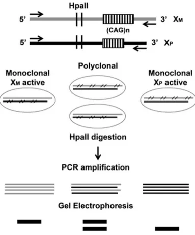
Tumors and normal tissues were dissected by Laser Capture Microdissection using a MMI CellCut Plus (Molecular Machines & Industries, MI, USA). LCM tubes were incubated for 48 hours at 37°C, centrifuged, and subjected to protein digestion for two additional days at 55°C, by adding fresh Proteinase K daily. DNA was extracted by using DNeasy Blood&Tissue Kit (Qiagen, Valencine, CA) (Figure 10). DNAs were then digested with HpaII enzyme: 100 ng of either tumor or normal DNA were digested with 10 U Hpa II restriction enzyme (New England Biolabs, Ipswich, MA, USA) at 37ºC overnight in a 20 µl reaction volume. Separate aliquots of DNA were subjected to mock digestion without the enzyme. After incubation, the restriction enzyme was inactivated at 65ºC for 20 min. HpaII-digested or mock-digested DNA was then subjected to PCR reaction, using the following primers: 5 FAM-labeled forward primer, 5’ACC GAG GAG CTT TCC AGA AT3’; reverse primer, 5’TGG GGA GAA CCA TCC TCA C3’. Thermal cycling conditions included the following steps: denaturation at 95°C for 10 minutes; 30 cycles at 95°C for 30 seconds, 55°C for 30 seconds, and 72°C for 30 seconds; and a final extension at 72°C for 10 minutes. Products of PCR amplification were analyzed by gel and capillary electrophoresis. Gel electrophoresis was performed on 3% agarose gel containing ethidium bromide (10ug/ml), and resolved DNA bands were visualized on a UV transilluminator (Biorad). For capillary electrophoresis, PCR products were mixed with 95% formamide and loading buffer (5% blue dextran, 25 mM EDTA) containing Rox-500. The mixture was then loaded on a 5% Long Ranger–6 M urea gel in TBE buffer. Electrophoresis was performed at 200 W for 2.25 hours, and the data were analyzed by an on a ABI 3100 Genetic Analyzer (Applied Biosystems, Foster City, CA) and quantified by Genescan 3.1 software (Applied Biosystems) (Figure 11). We used DNA extracted from female melanoma cell
30
line (labelled #1290) as a monoclonal control and DNA obtained from a healthy female blood sample (labelled L-IV-II) as a polyclonal control.
Figure 10 Laser capture microdissection of MM. H&E of a representative MM tumor section is shown before A) and after B) tumor tissue was collected by laser capture microdissection.
Figure 11 Schematic diagram of the HUMARA assay. Maternal and paternal X chromosomes carry different numbers of CAG repeats at the Humara locus. HpaII methylation sensitive sites are located at the polymorphic CAG region. During embriogenesis, random X chromosome inactivation occurs in female individuals, resulting in methylation of either the paternal or maternal X chromosome in different cells. Therefore, monoclonal cell population, derived from the division of a single ancestor cell, shares the same inactivated X chromosome, while a polyclonal population, derived from more than one ancestor cell, contain both cells with either inactive maternal or paternal X chromosome. HpaII digestion removes the unmethylated alleles, allowing amplification of the methylated HUMARA locus. Electrophoresis of the PCR products will resolve respectively in a single band or two bands of different size. Arrows indicate the primer sites, HpaII denotes the methylation sensitive endonuclease sites; arrows indicate primer annealing regions. Cross bars indicate the methylated chromosome.
31
3.2.2
Data Analysis
For each sample, the allele intensities were measured as the peak areas of both alleles, which is proportional to the molar amount of DNA. The allele ratios were first calculated by dividing the ratio (RU=A1U/A2U) of the non-HpaII digested sample, by the ratio (RD=A1D/A2D). The AR calculation (AR=RU/RD) corrects for any preferential amplification of one allele that might occur if the alleles are different in length. The clonality ratio is then calculated by dividing the AR of the tumor DNA by the AR calculated for the normal tissue (CR=ART÷ARN). This final calculation corrects for a potential skewed lyonization. A CR ≥ 3.0 or ≤ 0.3, representing a preferential loss of intensity in the digested sample of one of the two alleles present in the tumor sample, was scored as a monoclonal pattern.
32
4 RESULTS
4.1 Mutational analysis of EGFR and downstream pathways
4.1.1
Clinical Features
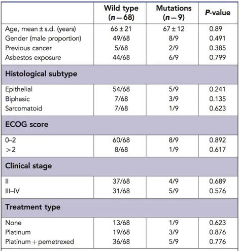
Out of 77 MPM patients studied, 57 were male (74%) and 20 were female (26%); their average age at the time of diagnosis was 68 years (range 43–90, median 64.5 years). Of these, 50 patients (64.9%) had previously been exposed to asbestos at work. Histological examination showed that 59 MPMs (77%) were epithelioid, 10 (13%) biphasic, and 8 (10.4%) sarcomatoid. In total, 41 patients had stage II tumours, 30 stage III tumours, and 6 stage IV tumours. Eastern Cooperative Oncology Group PS was 0–2 in 68 patients, and >2 in nine patients. In all, 41 patients were treated with platinum plus pemetrexed (Alimta) and 22 with platinum alone; 14 received no treatment because their performance status (PS) was >2 or because they refused. For this work we stopped the up (FU) at June 2012, at this date we collected FU data from 74 patients (three were lost to follow-up). In all, 15 patients were still alive at June 2012 with a median FU of 24.5 months (range 14–39 months). The median disease specific survival (DSS) of the cohort as a whole was 12.5 months (range 1–39 months) (Table 2).
4.1.2
Mutational Analysis
EGFR gene mutational profiling.
No mutations were detected in the EGFR gene by direct sequencing or the Scorpions-ARMS assay, even though the latter has a sensitivity of 1% (vs the10–20% of direct sequencing).
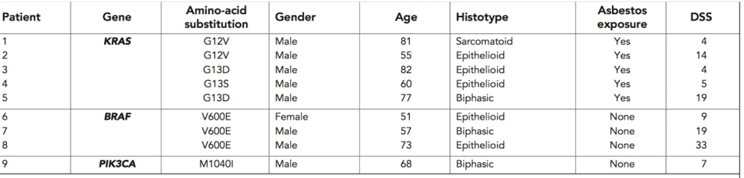
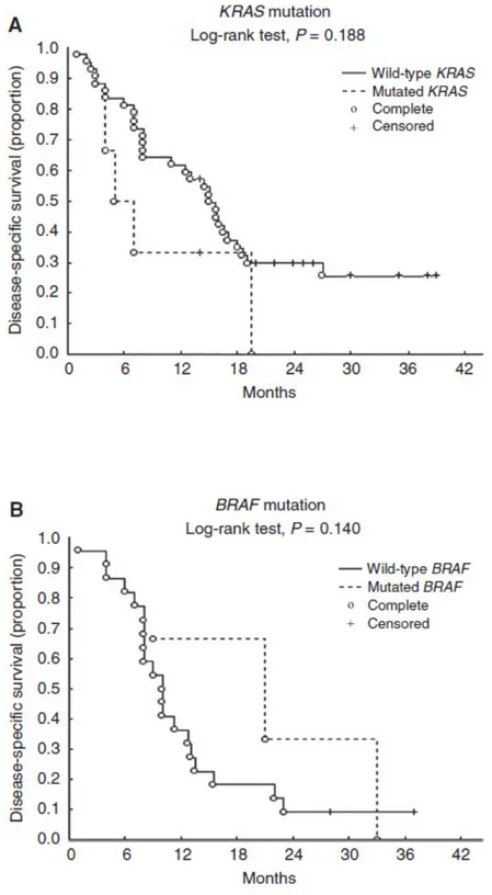
KRAS and BRAF gene mutational profiling.
KRAS gene was successfully amplified in all of the samples, five of which showed mutations: two patients had the GGT-GtT point mutation in codon 12 leading to a glycine-to-valine amino-acid substitution (G12V); two had the GGC-GaC point mutation in codon 13 leading to a glycine-to aspartic acid substitution (G13D); and one had the rare GGC-aGC mutation in codon 13 leading to a glycine-to-serine substitution (G13S). Three of the five mutations occurred in patients with epithelioid MPMs (G12V, G13D, and G13S), one in a patient with a biphasic MPM (G13D), and one in a patient with a sarcomatoid subtype (G12V) (Table 2). All five patients with KRAS mutations reported previous occupational asbestos exposure. The BRAF gene mutational analysis