UNIVERSITY OF PIEMONTE ORIENTALE
“Department of Health Science”
Doctoral course in MEDICAL SCIENCES AND BIOTECHNOLOGY
Cycle XXXI
Three-dimensional oral mucosa models:
development and applications
Candidate: Rita Sorrentino
Coordinator: Marisa Gariglio
Supervisor: Lia Rimondini
Index
Abstract……….………...7
Graphical Abstract……….………...9
Chapter 1: Background……….………...10
I. Introduction……….10
1.1. Tissue engineering (TE)……….10
1.1.1. In-vivo models for biocompatibility assessment of implants……….10
1.1.1.1. Legislature………...10
1.1.1.1.1.
Reduction……….…12
1.1.1.1.2.
Refinement……….12
1.1.1.1.3.
Replacement……….12
1.1.1.1.3.1.
In vitro models – 2-Dimensional VS 3-Dimensional.13
1.1.1.2. Animal models for oral implantology: An overview………...16
1.2. Oral Cavity………..…..……19
1.2.1. Anatomy………..…….19
1.2.2. Oral mucosa………...21
1.2.2.1. Oral epithelium……….22
1.2.2.2. Basement membrane……….23
1.2.2.3. Lamina Propria……….24
1.2.3. Periodontium………..……25
1.3. Oral mucosa model…………..………..……27
1.3.1. Monolayered Keratinocytes cultures……….…..27
1.3.2. Histotypic oral epithelial models……….……28
1.3.2.1. Bilayer cultures………..…..28
1.3.2.2. Commercial models………28
1.3.2.3. Organotypic oral mucosa model………..29
1.3.2.4. Scaffolds……….……….…..30
1.3.2.4.1.
Naturally Derived Scaffolds……….………..……30
1.3.2.4.2.
Collagen-based Scaffolds………..……….…………..…….30
1.3.2.4.3.
Gelatin-based Scaffolds……….….……….31
1.3.2.4.4.
Synthetic and hybrid Scaffolds……….….……….31
1.3.3. Cell Source and culture medium………..……31
1.3.4. Applications and development of engineered oral mucosa………..…. 32
1.3.4.2. Mucosa model implementation………32
1.3.4.3. Infected model development……….33
1.4. Aims and Thesis structures……….34
II. Bibliography……….…..…..36
Chapter 2: Models setting and optimizations………..………42
III. Introduction……….……… 42
3.1. Human mesenchymal stem cells (hMSC) and the osteogenic differentiation.42
3.1.1. Osteoblast and bone engineering………45
3.2. Human gingival fibroblast……….……46
3.3. Human oral keratinocytes……….……..…..……….……47
3.4. Cross-talk between mesenchyme and keratinocytes..……….47
IV. Materials and methods………..………..………49
4.1. Standard Cells culture condition…..……….……….……….……49
4.1.1. Primary cells culture condition………..…………..………..….…49
4.1.2. Cell lines culture condition………..………..…….49
4.1.3. Optimization of culture media for every cell type……….…….50
4.1.4. Viability assay………...……….……..50
4.1.5. Migration assay………...….….……….51
4.2. Osteogenic differentiation protocol………..………..…….51
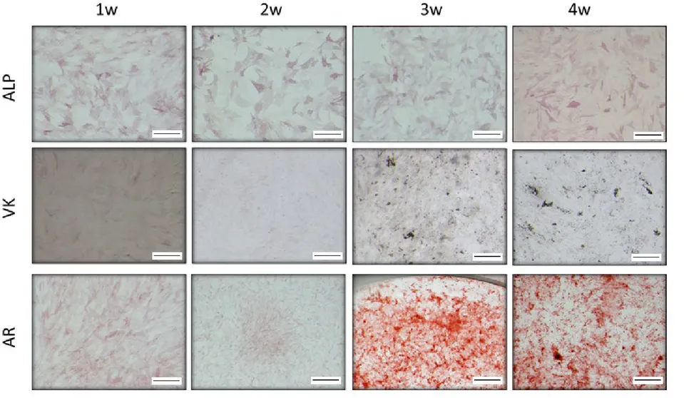
4.2.1. Osteogenic differentiation protocol evaluation: Alkaline Phosphatase
(ALP), Von Kossa and Alizarin Red staining………51
4.3. Bone substitute optimization……… 52
4.3.1. Scaffold preparation ………..……….52
4.3.2. Scaffold repopulation………..……...52
4.3.3. Dynamical Mechanical Properties (DMA) analysis………..………52
4.3.4. Transmission electron microscopy (TEM) imaging………..…..53
4.3.5. Confocal imaging………..…..53
4.4. Established a custom made keratinized oral mucosa model……….….53
4.4.1. Haematoxylin and eosin stain………..……….54
V. Results………..55
5. Establishment of the common media for co-culture………..55
5.1.1. Monolayer growth condition establishment: Viability Assay……….55
5.1.2. Evaluation of hMSCs secretory capability into EpiLife medium: Migration
assay……….……57
5.3. Scaffold development………..………60
5.3.1. Preliminary data……….………..………..………60
5.3.2. Mechanical properties evaluation…………..……….………..61
5.3.3. hMSC viability evaluation……….……….62
5.4. Establishment of the 3D parakeratinized (masticatory) epithelium model…..65
VI. Discussion……….…………..………68
VII.
Bibliography………..………...70
Chapter 3: Epithelial -Mesenchymal cross-talk.………..73
VIII. Introduction………73
8.1. Oral epithelium……….………73
8.1.1. Keratinized epithelia……….73
8.1.2. Non-Keratinized epithelium……….……….……….76
8.1.3. Cytokeratin………...76
8.2. Gingival mucoperiosteum ………78
8.3. Oral mucosa models………..………..79
IX. Materials and methods……….…………..…….81
9.1. Evaluation of hMSC on Oral Mucosa model……….………..….81
9.1.1. Paracrine effect of hMSC on Oral Mucosa model…………..……….81
9.1.1.1. hMSC and Oral Mucosa Co-Cultures………..…..81
9.2. Evaluation of hMSC derived Osteoblast effect on Oral Mucosa……….…82
9.2.1. Paracrine effect of hMSC-derived Osteoblast onto keratinocytes
stratification and differentiation...82
9.2.2. Osteogenic induced hMSC and Oral Mucosa Co-Cultures……….….82
9.3. Haematoxylin and eosin stain……….…82
9.4. Immunohistochemistry analysis………..………...83
9.5. Proteomic Array. ………..………..……….83
9.6. Elisa...……….……..………..………83
9.7. Mucoperiosteum model……….84
9.7.1. Masson Trichrome assay……….84
X. Results………85
10.1.
Paracrine effect evaluation….………85
10.1.1.
hMSCs paracrine effect on keratinocytes stratification and
differentiation………85
10.1.2.
Paracrine effect of hMSCs -derived osteoblast effect on
keratinocytes stratification and differentiation……….………85
10.1.3.
Proteomic array and ELISA ….……….………89
10.2.
Co-Culture of the oral mucosa and hMSCs with or without differentiation
factors: cross-talk effects….……….………..……….…93
10.2.1.
hMSCs affects keratinocytes stratification and differentiation……93
10.2.2.
Mucosa model affects hMSC osteogenic differentiation ……….95
10.3.
Connective tissue development: fibroblast-hMSCs interactions effect
onto keratinocytes stratification and differentiation……….96
10.4.
3D composite model……….96
10.4.1.
Histological evaluation………96
10.4.2.
Mechanical properties evaluation………96
XI. Discussion………..……….99
XII. Bibliography………...103
Chapter 4: Innervated Mucoperiosteal model development……….…………...106
XIII. Introduction…..……….106
13.1. Periodontium innervation……….106
13.2. Innervated epithelial model………...….………..107
13.3. Aims………..……….……..107
XIV. Materials and methods….………..………..108
14.1.
Mucosa model effect on nervous system….……….……….108
14.1.1.
Axonal outgrowth evaluation….….………108
14.1.2.
DRG Isolation….………..….108
14.1.3.
Quantification of axonal growth………..……….108
14.2.
Mucoperiosteum model……….………..……….…….109
14.2.1.
Masson Trichrome assay………..………109
14.2.2.
Immunofluorescence analysis………109
XV. Results……….……….110
15.1.
Effect of oral mucosa on the neuronal compart……….………110
15.1.1.
ND7/23………..……….…………110
15.1.2.
Dorsal Root Ganglia………..………..…………..113
15.2.
3D composite model………..………..……….115
15.2.1.
Scaffold suitability………..……….115
15.2.1.1. Histological analysis………...115
XVI. Discussion………....………117
XIX. Conclusion and future prospective.……….………..……….121
XX. Abbreviation list……….……….123
XXI. Figure index………..……….125
XXII. Table index………..………..……….127
Abstract
INTRODUCTION: Animal experimentation has been extensively and for a long time applied in several research fields, but since 2011 it has been substantially limited by the Commission of the European Parliament to ensure people/animals safety and reduce research costs. To respond to these directives, many attempts have been focused on the development and validation of new in vitro 3D systems, bypassing the traditional 2D cell cultures. In this regard, diverse approaches to tissue-engineered bone and oral mucosa have been developed. Despite the promising premises and the cutting-edge results, the used 3D in vitro bone-oral mucosal models still lack interaction between the mucosal and the bone components. Therefore, this project aimed to create 3D models, entirely made with primary human cells (keratinocytes, fibroblasts, and osteoblasts), able to mimic the natural structure and interaction of bone and oral mucosa. A direct future application will be the multi-tissue periodontal regeneration, needing synchronized restoration of the gingival and bone compartments, besides cementum and periodontal ligament.
EXPERIMENTAL METHODS: For the in vitro 3D oral mucosa assessment, a collagen-based lamina propria was enriched with a pool of primary human fibroblasts (HGF), freshly obtained from the normal gingiva of young healthy and informed consent donors, put into a culture insert and submerged into defined culture media. A pool of primary human oral keratinocytes (HOK) were seeded upon the gel. When keratinocytes reached confluence, they were grown at the air-liquid interface, stratifying in about two weeks. The oral mucosa model, obtained as above described, was used to evaluate the effect of two mesenchyme cell types, mesenchymal stem cells (hMSC) and osteoblast (hFOB) on keratinocytes stratification. Mesenchyme effect was assessed in 3 different set-ups: paracrine effect (conditioned media, CM), indirect co-culture (transwell system), and direct co-culture (hMSC embedded in the connective tissue substitute). Oral mucosae were, after that, histologically examined. The conditioned media from those experiments were used to growth murine nervous system cells (immortalized ND7/23 and primary dorsal root ganglia DRGs) and quantified the effect on axonal outgrowth.
Regarding the bone compartment, a bovine tendon collagen (BTC) and nano-hydroxyapatite (nHA) sponges developed by Salgado et al., (2015) was synthesized by lyophilization process. The sponge was subsequently implemented with a fibrin coating to increase the stiffness and mechanically characterized by the dynamic mechanical analysis machine. Primary human osteoblasts (hFOB) were integrated into the BTC/nHA sponge diluted directly in the fibrin gel. Oral mucosae, produced as above described, were air-lifted onto the repopulated bone substitute and co-cultivated for others 12 days to allow cell-interaction and keratinocytes stratification. This latter innovative model was histologically and mechanically characterized.
RESULTS AND DISCUSSION: the histological analysis showed an unexpected effect of osteogenically induced hMSC (hMSC-OB) onto keratinocyte stratification. Under the hMSC-OB stimulation, the ratio between the spinosum and the corneum strata resulted impaired due to the increase of the keratinization in treated 3D models. This impairment results correlated to a higher expression of the cytokeratin 10 (typical of keratinized epithelia) with a slight reduction of the cytokeratin 13 (typical on non-keratinized epithelia). These results suggest that hMSC and, in particular, one of their differentiated forms, the osteoblasts (hOB), play a crucial role in mucosal differentiation fate. The proteomic analysis
induces keratinocytes proliferation and differentiation. The KGF production (amount increased to 208±36 pg/ml after 14 days of differentiation and to 347±15 pg/ml after 28 days of differentiation), confirmed in ELISA, may explain why, despite the increase in keratin production, most of the keratinocytes retain the nuclei producing a para-keratinized phenotype. At the same time, two 3D hMSC-OM co-cultures was developed. In the first one, hMSC and oral mucosae were kept in contact with a transwell porous system. The histological analysis of 3D culture shows that the crosstalk between oral mucosa and hMSCs improve the keratinocytes behaviour ensuring a complete stratification process. The differentiation analysis of co-cultured hMSC showed that, in the presence of osteogenic factors, oral mucosae halve the time required for hMSC differentiation. In the second model, hMSC were seeded with HGF within the collagen matrix used as lamina propria substitute. The histological analysis showed that hMSC speeded up the keratinocyte proliferation and stratification, obtaining the fully epithelium resembling the native one in only 7 days. Once again, the proliferative state induced by hMSCs resulted uncontrolled. Indeed, after another 1 week of direct co-culture, keratinocytes lose their organized structure. The obtained results were used to set-up a mucoperiosteal model composed of an engineered oral mucosa air-lifted and let stratified onto an osteoblast repopulated hard-sponge mimicking the bone counterpart. The mechanical evaluation of this model suggested that mesenchymal cells (hMSC and HGF) and epithelial cells collaborate to remodel the synthetic bone matrix. Moreover, the effect of those models on the nervous system was evaluated by calculating the axonal outgrowth of both immortalized neuronal cells and murine dorsal root ganglia (DRGs). Although the treaded models induce a slight increase of axonal elongation on immortalized neuronal cells, an adverse impact is registered on the ganglia of the back. These recent results suggest that oral mucosae produce pro-innervation factors (i.e., NGF observed by IF analysis) but also specific molecules that inhibit the migration/elongation of unsuitable axons. Finally, the obtained results were used to set-up an innervated mucoperiosteal model composed of an engineered oral mucosa enriched with ND7/23 let stratified at the air/liquid interface directly onto an osteoblast repopulated hard-sponge mimicking the bone counterpart enriched with ND7/23. In the innervated model, it is possible to observe several cells migrating within the bone counterpart; these cells were characterized by immunofluorescence analysis and, at the interface between bone and oral mucosa, several TBR1/β-tubulin type III positive cells identified as ND7/23. However, any axonal prolongations were found despite the presence of secreted NGF within the model.
CONCLUSION: In the present work, the regulatory role of the mesenchymal tissue onto epithelia was evaluated. The main results showed that that during the differentiation hMSC produce and secrete factors that induce the keratinization and the expression of the marker of differentiation CK10; in particular in the middle stage of differentiation (OB14). The proteomic analysis revealed that this effect can be ascribable to KGF secretion. This finding may impact the design of new implantable devices able to induce, alone, the epithelial growth and keratinization to improve implant graft avoiding epithelial graft linked to the morbidity of another zone. Moreover, we also showed that OM might have a pro-innervation effect, at least during the last stages of keratinocytes stratification. Finally, we characterized an innervated mucoperiosteal model that could open new in vitro frontiers for oral biomaterials validation as well as improve knowledge regarding the mesenchymal stem cells roles onto oral mucosa development.
Graphical Abstract
Figure 1 Graphical Abstract. Assessment of stromal-epithelial crosstalk and mucoperiosteal development. JE: junctional
Chapter 1
Background
I. Introduction
1.1. Tissue engineering (TE)
In 1993, Langer and Vacanti described the tissue engineering (TE) as “an interdisciplinary field that applies the principles of engineering and life sciences toward the development of biological substitutes that restore, maintain, or improve tissue function or a whole organ”. This definition still well represents many TE applications and nowadays, even comprises the development of biological substitutes aimed to mimicking the tissue environment and function in vitro (as a tissue model) for being used in the cell biology and embryology fields as a tool to study the cell and tissue response to exterior influences (i.e., drugs, biomaterials, etc.) (Olson et al., 2011; Ikada et al., 2006)
Starting from this background, in vitro models have been recently developed to mimic not only the single tissue function but also the complexity of whole organs, including tissue interfaces (Atala et al., 2012). These multi-tissue models have been mainly proposed as tools for analysing the cells-cells interaction within a physiological microenvironment(Gothard et al., 2014) and the fundamental mechanisms involved in cell signaling during tissue repair.
These in vivo models represent valuable tools to speed-up the biocompatibility assessment of implantable materials by reducing the recourse to in vivo experimentation.
1.1.1.
In vivo models for biocompatibility assessment of implants
The term “in vivo” is referred to all the experiments conducted in living organisms. Currently, they still represent a necessary tool to evaluate both the safety and the efficacy issues of new drugs or implantable devices. Indeed, despite several drawbacks mainly related to i) ethical dilemmas, ii) model handlings complexity, iii) long experiments duration, iv) limited accessibility, v) expensive management of animal facilities and vi) inflexible limiting laws (Knight et al., 2011) in vivo models use remain irreplaceable.
1.1.1.1.
Legislature
In 2015, nine European countries presented a petition to the European Commission (EC) to ban the use of animals in research. The EC reject the petition but stated that ethical justification and adoption of protocol which follow the 3Rs (Replacement, Reduction, and Refinement) must be approved before proceed with each experimental studies (http://ec.europa.eu/environment/chemicals/lab_animals/pdf/vivisection/en.pdf), not only for ethical purposes but also because the use of healthy animals growth in adequate animal facility (accordingly to actual European directive and 2Rs statements) allow the production of more robust and reliable results, underlying valid scientific outputs (Hurst
and West, 2010; Singhal et al., 2014). Globally, legislation differs between countries and geographical regions, but in general, the well-being of the animal used in research is protected.
The 3Rs concept was first developed by Russell and Burch (1959) and has become rooted in legislation and guidelines concerning animal experimentation in many Countries (fig. I-1). The 3 Rs stay for “Replacement” which stated that, when applicable, alternative methods must be applied to avoid or reduce animal use in research, “Reduction” which encourage the use of strategies that enable the obtainment of reliable data starting from as few animals as possible, or, at least, to obtain more information from the same number of animals and “Refinement” which impose the use of methods that alleviate or minimize potential pain, suffering or distress, and enhance animal welfare for the animals used.
The directive 2010/63/EU follow the 3D principles and states that "every project proposal in EU member states involving procedures on living non-human vertebrates and cephalopods has to be approved in a review process, including a harm-benefit-analysis (HBA), to assess whether the harm to the animals in terms of suffering, pain, and distress is justified by the expected outcome taking into account ethical consideration and may ultimately benefit human beings, animals or the environment"(Eggel et al., 2018).
Figure I-1 Graphical representation of the significant ethical concepts and key questions that scientists must address under the
traditional view of the 3Rs – Replacement, Reduction, and Refinement – to justify the use of animals in experimentation, from planning the program of work through to publication. (http://jeb.biologists.org/content/220/17/3007#ref-85)
1.1.1.1.1.
Reduction
‘Reduction’ requires the limitation in the number of animals used for experimentation in "just enough data and no more". The better strategies to overcome this limitation involve the improvement of the experimental design and the precision of measurement as well as the addition of reliable concomitant measurements (that can be used as internal control) to reduce the variability in the pilot studies (i.e., sex or age). At the same time, it is essential to include, for each experimentation, adequate control groups and to choose the correct animal model (i.e. use aged animal if in the investigation must be considered the senescence) to obtain reliable translational results (McClelland et al., 2000; Eng et al., 2003; de Boo and Hendriksen, 2005). Moreover, the guidelines state that the
calculation of statistical
power should be included to optimize the sample size.
1.1.1.1.2.
Refinement
Refinement is an "integral component of improving laboratory animal welfare, which is vital for healthy biological functioning and a normal behaviour repertoire". This part allows more reliable results since the scarce animal condition can interfere with the measurement. However, for most species, the adequate tools or protocols to assess their health and welfare (i.e., pain assessment is highly developed for mammals compared with other animal groups) were upgraded only in the last few years (Sneddon et al., 2014; Sneddon, 2015). The EC Directive (2010; http://eur-lex.europa.eu/legal-content/EN/TXT/?uri=CELEX:32010L0063) proposes that “all protected animals should have enriched environments in which to live”. Indeed, an adequate environment, improve with social housing and apparatus to allow exercise or sensory stimulation reduce the stress in animals reducing the effect of this factor on the experiment outcomes (Singhal et al., 2014).
The term "refinements" is also referred on the experimental procedures; handle animals with proper procedure reduce the stress related to invasive procedures. For instance, to minimize mice anxiety is suggested to avoid collection them by their tail. In general, when applicable, it is recommended the use of painkillers after surgery and non-invasive imaging techniques to collect data (O'Farrell et al., 2013).
1.1.1.1.3.
Replacement
According to several regulatory organs, the youngest forms of many species do not suffer. For instance, the UK Animals (Scientific Procedures) Act 1986 (https://www.gov.uk/government/publications/consolidated-version-of-aspa-1986) and European Directive 2010/63/EU (http://eur-lex.europa.eu/legal-content/EN/TXT/?uri=CELEX:32010L0063) allow the use of fish until they acquire the capability of independent feeding and allows the use of embryos (i.e., from chickens) (Tazawa et al., 2002). However, this approach is not valid in several research fields and in particular, it is not valid for biomaterials studies. Due to EU's decision to ban animal testing for cosmetics (EU1223/2009) and the restriction in animal use occurs after 2015, several in vitro models platform including 3D cell cultures have been developed to bridge conventional 2D tissue cultures and animal experimentation Those platforms are often aimed to identify potentially dangerous chemicals and recently, extensive research has gone into i) mammalian tissue studies, used to develop ex vivo tissue techniques such as the precision-cut tissue slice (Fisher RL and Vickers AE, 2013) and ii) stem
cell-derived organoid 3D cultures to improve the translational potency of the studies (Liu et al., 2016; Muthuswamy, 2017). A precision-cut tissue slice is a technique in which an organ, or a part of it, is sectioned in identical portions (usually 0,3 mm thick) and kept in cultures for a limited time. These latter represent an in vitro model able to closely mimics the organ complexity since the “slice” maintain the structural and functional features of the whole organ while organoids are in vitro derived 3D cell aggregates with organ functionality which offer a comparable structure to primary tissue and a stable system for prolonged cultivation, and that can be easily cryopreserved.
One of the main advantages of 3D cultures is to facilitate the adherence to the 3Rs ethical concepts.
1.1.1.1.3.1.
In vitro models – 2-Dimensional vs 3-Dimensional
The term in vitro refers to experiments carried out on segments derived from living organisms and cultivated on external support. In vitro models are mainly divided in 2 categories, the models growth in 2-"dimension"-D (length and width) that are represented by isolated cells growth as monolayer on synthetic supports (such as coated tissue culture plastic, TCP) and 3-"dimension"-D (length, width, and thickness) that can be referred both to the same cells grown as a multi-layered system on or within heterogenic supports or to tissue sections ex-vivo cultivation (Duval et al., 2017).
Two-dimensional culture techniques have been one of the foremost breakthroughs in the biomedical field. They’ve been represented, since the early ‘900, the possibility to carry out experiments with living biological specimens in highly standardized and controlled conditions (Vanderburgh et al., 2017). However, there are several differences between monolayered cells and the natural tissues, and they include tissue-specific substrate stiffness, the spatial cues (such as cell-matrix and cell-cell interaction or receptor topography), and, consequently, the concentration gradients of nutrients, secreted factors and gas (i.e., oxygen). Since the early '90, several researchers focused on the mechanical stimulation of different cell types, and, nowadays, it is well-known that the mechanical stimulus has the same importance of chemical stimuli. Accordingly, the substrate stiffness has a significant effect on cell behaviours such as differentiation and migration process, and substrate morphology severely affects cell appearance via cytoskeleton regulation (Pedersen et al, 2005).
Moreover, the presence of the ECM influences the gene expression; for instance, Mishra et al. (2012) compared metalloproteinase (MMPs) expression in 2D and 3D culture and showed a low or absent expression of some MMPs, indispensable marker for evaluating the pro-metastatic capability of cancer cells, in monolayer condition. Finally, the absence of the third dimension imposes several changes in cellular morphology and receptors topography, and this induces several complications in translate dose/response curve in more complex models (Bradbury et al., 2012).
To overcome these limitations, several 3D models have been developed. Indeed, 3D models have the potential to overcome not only 2D cultures unable to mimic the physiological tissue behaviour but also some animal models because of the lower financial disbursement. The main advantages and disadvantages of 2D and 3D models are summarized in table I-1.
As previously described, the organ complexity influences the difficultness in obtaining reproducible and affordable models. The modern methods in the production of functional tissue and/or organ models entail a multi-disciplinary approach in which engineering, chemical, physical, mathematics, biological, and physiological knowledge are exploited in a trial-and-error tactic (Sharifikia et al., 2017). The development of biological 3D multi-tissue models, intended to mimic physiological tissues, is a complex but promising way to improve the power of biological studies. With these models, the understanding and prediction of human organ response to external stimuli (i.e., xenobiotic molecules roles, mechanical stress, paracrine cross-talk, biomaterials compatibility, drugs safety, etc.) or the cytofunctionality analysis [i.e., the study of the de novo production of extracellular matrix (ECM)] could be assessed in a physiological context. This study approach can provide reliable information on cell interaction useful in implant substrate design.
This methodology has so far become a pivotal tool for evaluating necessary processes such as matrix remodelling, cell crosstalk, growth factor secretion, gene expression, regulation, etc. of human cells in their microenvironment. Moreover, as previously described, these models represent a high 3Rs-friendly approach by reducing the use of small animal models (and by lowering the animal-related cost for researchers).
Table I-1 Principal differences between 2D and 3D cultures
2D Cell Culture Systems
3D Cell Culture Systems
Advantages
Disadvantages
Advantages
Issue and Future
Prospective
Low-cost The absence of real cell environments
More relevant cell models
Throughput (the initial study is more
expensive and time-consuming; however, results
are trustable) Well established Lack of predictivity
(with increasing cost and failure rate of further
studies)
Direct interaction between different types
of cells (organotypic models)
Guarantee the right oxygenation to the inner part
of the model A lot of comparative
literature
False results caused by the growth media and
expansion of cells
Presence of connective/stromal tissue or barrier tissues
Standardize protocols
Easier cell observation and measurement
Better simulation of conditions in a living
organism
Increase the complexity
Reduces the use of animal models Represent a more realistic way to grow and
treat cancer cells
The general approach to design engineering materials is peculiar when the main goal is to obtain implantable tissue instead of tissue models. Implantable tissue required to be safe, fully biomimetic and biocompatible, and the ideal mechanical properties and morphology of the implanted scaffold are studied to guarantee the first phases of the graft and ensure the host tissue migration within both acellular and repopulated implant. On the contrary, tissue models need to resemble, as close as possible, all the features of the mimicked tissue.
Indeed, it is expected that healthy resident cells can invade the free space of the scaffold and interact with implant structure and resident cells to secrete new ECM proteins and structures to re-activate the biological function of the tissue (Antoni et al., 2015). On the contrary, tissue models are designed to mimic, ex vivo, all the features and the three-dimensional topography of the single (histotypic model) or the composite (organotypic model) tissue of interest. When histotypic and organotypic models are considered, the priority is to obtain an easy-handling model suitable for a wide range of tests. These models are mainly developed as platforms for evaluating different biological responses to external stimuli, such as microbial infections, drugs, materials application, or mechanical stress. Above all, these models are addressed to give information regarding gene regulation and expression, macromolecules or vesicles secretion, receptor polarity, and tissue morphology changes in response to certain stimuli. To reach the targets, histotypic and organotypic models must be realized with highly standardized and reproducible protocols and with fully characterized cells (Antoni et al., 2015). This enables to easily and separately identify each variable or cell components offering a considerable advantage in comparison with animal models, where the contributions of some tissue type or cells are often overlapped and hidden by other biological parts.
Besides, another essential advantage of 3D models in comparison with in vivo models is found in the study cost, as shown in table I-2.
In both 3D and in vivo models, a key role is played by connective tissue. Indeed, in both pathological and healthy condition, the connective tissue support and regulate the behaviour of all the other tissues. For instance, in tumours, the surrounding fibroblast (nowadays known as tumour-associated fibroblast, TAF) interacts and regulates cancer behaviour. In healthy condition, the connective tissue secretes crucial molecules which regulate and drive the fate of other cells such as the ones which form the lining epithelia. Thus, several studies have been focused on elucidating the interaction between the connective or stromal tissue and the covering epithelia, and in reproducing this interaction in refined 3D models. To pursue this research term, several types of 3D cell culture techniques have been designed to replicate lining epithelia such as gastric epithelium, alveolar epithelium, corneal, skin, or oral mucosa.
Pluristratified squamous epithelia may be resembled by using tissue-specific basal-like keratinocytes on natural or synthetic connective tissue substitutes repopulated by tissue-specific fibroblasts. In this case, keratinocytes stratify at the air-liquid interface, retaining the specific epithelial belongings. Among them, one of the most engineered specialized tissues is the keratinized pluristratified squamous epithelium of the oral cavity (Gibbs et al., 2000).
Table I-2 Toxicity test costs, in animals and in vitro, as reported by Human Society International
(http://www.hsi.org/issues/chemical_product_testing/facts/time_and_cost.html)
TYPE OF TOXICITY TEST TYPE STUDY COST ($ US)
GENE TOXICITY
CHROMOSOME ABERRATION In Vivo 30000
In Vitro 20000
SISTER CHROMATID EXCHANGE In Vivo 22000
In Vitro 8000
UNSCHEDULED DNA SYNTHESIS In Vivo 32000
In Vitro 11000
EYE IRRITATION/CORROSION
DRAIZE RABBIT EYE TEST In Vivo 1800
BOVINE CORNEAL OPACITY In Vitro 1400
SKIN CORROSION
DRAIZE RABBIT SKIN TEST In Vivo 1800
EPIDERM™ In Vitro 1600
CORROSITEX® In Vitro 500
1.1.1.2.
Animal models for oral implantology: An overview
In the last three decades, several researchers have been focused on their osteointegration and soft tissue integration study of dental implants using several animal models.
Literature reports several papers focused on histological responses to implants in primates (forbidden in Europe) or in dogs with all expected related ethical concerns since no life-saver devices are involved.
Rodents (mice, rats, and rabbits), ovine, and swine have been also used with less restriction; choosing the appropriate animal model for each experimentation allows the production of reliable and reproducible data.
The mouse is the most used animal model in research, and, through the years, several syngeneic and transgenic strains have been established to meet experimental protocols requirements. Mice are moderately inexpensive, easy to handle and reproduce quickly in laboratory settings. The mouse genome has high homology with human one and can be easily modified to mimic human disease, included bone defects and age-related disease. However, mice models are not often used to test oral implantable materials or devices mainly because of their reduced size, which requires to scaled-down the devices (Rahal et al., 2000; Beppu et al., 2011) and makes the surgical procedures almost tricky.
Rats, in particular, Wistar and Sprague-Dawley strain, have been extensively used in the implantology field. Wistar rats are variants of the congenital albino rat, whereas Sprague-Dawley rats are of an outbred origin. Due to their bigger size, male rats are more often used than females, and as for mice, extra-oral bones are preferred for dental
material implantation. Some authors, such as Haga et al. (2009), preferred to use mini implants for teeth replacement and evaluating the maxillary bone formation and maturation processes around implants. Following similar strategies, Rinaldi and Arana-Chavez (2010) were able to examine implants in contact with the periodontal ligament of adjacent teeth in the mandible while Hou et al. (2009) shown that the mechanical force of oral environment improves titanium integration. Torricelli et al. (2002) suggested the use of aged rats, instead of young rats, for bone implants evaluation since they showed a reduction in bone formation in aged rats. In the same work, the authors suggest the use of a demineralized bone matrix to improve bone healing and re-mineralization in osteoporotic condition. Genetically-modified rat strains or diseased animals have also been used to mimic human diseases such as diabetes (Biobreeding diabetes-prone rats) or hypertension (Zucker rat); for instance, Hasegawa et al. (2008) showed that the osteointegration and bone volume are reduced in diabetic mice in comparison with non-diabetic rats. Similarly, ovariectomized rats were used to assess the effect of implanted materials onto osteopenic bones. In 2002, Fini et al. compared the osteointegration of titanium (Ti6AI4V) implants in ovariectomized and sham-aged rats and sheep. In their experiments, the author demonstrated that the titanium implants were less osteointegrated in both trabecular and cortical osteopenic bone in comparison with those in normal bone; the authors also observed a decrease of bone microhardness in both trabecular and cortical bone, but this decrease resulted widespread in the osteopenic bone suggesting implant-associated delay in both bone formation and maturation.
Despite the positive results, the bone structure of rodents is poorly representative of human ones and lacks
Haversian-type remodelling. Due to this factor, data obtained by those models are not always confirmed in pre-clinical or human models (Li et al., 2015).
Rabbit is one of the most used models in musculoskeletal studies. It is often used in implant dentistry (Neyt et al., 1998) mainly because its knees are large enough to host dental implants designed for humans.
Rabbit has faster bone turnover than human with significant intracortical Haversian remodelling, which corresponds to a quicker osteointegration and to a reduction of time requires for the experimentation (Pearce et al., 2007). For this reason, it is considered as a pre-translational animal model useful to screen new oral implant technologies.
Among all rabbit strains, the New Zealand White with a bodyweight between 2 and 5 kg is the most (Vidigal et al., 2009).
Nowadays, one of the elective models for implant dentistry is represented by sheep and/or goat since their bone dimension is comparable with the human one despite the higher bone density (around 2-fold) (Rasmusson et al., 1999). Domestic sheep are easy-handling animals that are widely used to study numerous musculoskeletal pathologic conditions or to test different kinds of implants. The use of adult sheep is strongly suggested since the complete bone maturation occurs a long time after puberty (average age: nine months within different breed). The sheep trabecular bone well resembles the human one while the cortical bone presents slight differences in the bone mineral composition and Haversian canals content, lower in sheep than in humans (Ravaglioli et al.,1996; Aerssens et al., 1998). Although, the sheep bone healing and a bone remodeling rate are comparable with that of humans (Pastoureau et al., 1989;
sheep under hormone treatments are a suitable model for osteoporosis (Dvorak et al. 2011). Due to those features, sheep is a standard model in the implantology field even if more expensive and time-consuming (the healing and recovering time is higher) then rodents.
Swine is often used for translational purposes in pharmaceutical research. However, for the dental implantology field have been created specific "mini-pig" since the domestic adult one (suitable for dental implants) are too complicated to handle (difficulties in housing because of size and in surgically widening oral tissues) (Pearce et al., 2007). Different strains of mini-pig were developed since the sixties: Yucatan (in its two forms of minipig and micropig), Hanford, Sinclair Hormel (also called Minnesota), Pitman-Moore, Kangaroo Island, Ohmini, Lee Sung, Morini, and Göttingen. Despite small differences in plaque formation and bone structures between minipig strains and in comparison, with human, minipig bone is considered one of the most representative of human bone in terms of bone remodelling processes, density and mineralization (Mosekilde et al., 1987 and 1993; Aerssens et al., 1998; Ma et al., 2009).
Animal model studies enabled a considerable improvement in several research fields. However, the time and financial disbursement required for experimentation with adult and/or diseased animals are higher in comparison with 3D models, and also the reliability for getting reliable results is not 100% (Shanks et al.; 2009). Moreover, these models require highly specialized personnel to be correctly used, and besides, they are subjected to ethical issues and the strict control of national and international regulatory authorities. The regulation regarding in vivo model is described in paragraph 1.1.1
1.2. Oral Cavity
1.2.1. Anatomy
The oral cavity is the first segment of the digestive system, bounded externally by the lips and internally by the pharynges. It is formed by different structures composed of hard and soft tissue. The first and outer portion of the oral cavity, named vestibule or vestibulum oris, includes the lips and the cheek externally while the inner part is formed by gums and teeth (fig. I-2).
The lips, or vermilion zone, are lined by a thin keratinized layer balanced out by a prominent stratum lucidum and its basal layer results full of melanocytes. The upper lips are innervated by the maxillary (V2) and the mandibular (V3) branches of the trigeminal nerve (V) and the infraorbital branch of the V2 while the lower lips are innervated by V2, V3 and the mental nerve branch of V3, the latter also innerve the oral mucosa (Sadrameli and Mupparapu, 2018).
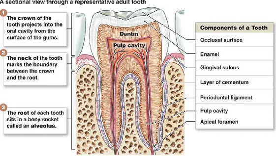
The teeth are composed of a crown that projects into the mouth, and roots which are connected with the alveolar bone by a flexible but reliable joint called periodontal ligaments. The enamel and the dentin compose the hard and external part of the teeth. In contrast, the soft and inner part, the pulp, is mainly composed of stromal connective tissue supplied by nerve, lymphatics vessel, and blood capillaries. The dental pulp contains fibroblasts (PF) and oral mesenchymal stem cells (O-MSCs) (fig. I-3).
Figure I-3 Diagram illustrating the anatomy of the tooth and the and foremost function of its components
(https://www.printablediagram.com/diagrams-of-teeth-printable/diagram-of-teeth-anatomy/)
Figure I- 2Diagram illustrating the anatomy and central structures of the oral cavity (https://pocketdentistry.com/1-oral-structures-and-tissues/
The gingival margin has a scalloped-like course across the dentition due to interdental papillae that fill the interdental spaces beneath the tooth contacts. Moreover, the gingiva is histologically divided into two portions:
• The free gingiva, which is characterized by two non-keratinized epithelia in its inner part, known as sulcular epithelium (in the free part) and the junctional epithelium (in direct contact with the teeth). The sulcular epithelium forms the wall of the sulcus and, together with the junctional epithelium, form the dento-gingival junction. The junctional epithelium has two basal lamina, the internal one that faces the tooth and the external one that meets connective tissue; due to this characteristic, it results in the most permeable epithelium and the lamina propria underlying the junctional epithelium is characterized by a
chronic inflammatory state derived by the filtration of several microbial antigens. On the contrary, the outer surface which is mainly keratinized.
• The attached gingiva, which is covered by a keratinized epithelium and extends apically from the free gingiva towards the alveolar mucosa. The latter is defined "attached" because it is firmly adherent to the alveolar bone and in case of bone fenestration to the root cementum.
(Hall and Lundergan, 1993; Garnick and Ringle, 1988).
The lamina propria of both free and attached gingiva is rich in nerve, blood, and lymphatic vessels and resident innate immune system cells. Gingival tissues structures are summarised in figure I-4
The second and inner part of the mouth, known as the oral cavity proper, start with the alveolar process of the mandibular and maxillary bone, where teeth are located fixed and enclosed by the periodontium, covered by a soft tissue called mucosa. In the superior zone, the hard and soft palate separates the oral cavity by the nasal cavity while the lower part of the oral cavity is filled by the tongue and the floor of the mouth.
The hard palate is composed of the upper mouth bones lined by a keratinized epithelium while the soft palate is located behind the hard palate and doesn't contain bone but is mainly composed of muscle lined by a non-keratinized epithelium and is innervated by the V3 and the vagus nerve.
The tongue surface is covered by 4 types of lingual papillae named circumvallate, foliate, filiform, and fungiform positioned in different parts of the tongue and assign to taste different savours. Fifth cranial nerve supplies general sensory innervation (not the gustative one) to the anterior 2/3 of the tongue. Finally, the flour of the mouth borders the lower limit of the oral cavity and connect, with the lingual frenum, the gingival tissue with the tongue.
Figure I-4 Structures of gingival tissues. https://sites.google.com/site/dentalhygieneportfoliofelicia/hom e)
1.2.2. Oral mucosa
The oral mucosa is the linin organ of the oral cavity; from a histological point of view, it shares several characteristics with the skin, the dry lining organ of the human body, and other covering membranes such as the oesophagus. Structurally, both skin and lining membranes are composed of two interconnected tissue: a connective tissue covered by a lining epithelium, mostly a squamous pluristratified epithelium. These two components are strictly interconnected with the epithelial cells that infiltrate within the connective tissue forming the rete ridge. The lining epithelia and the connective tissue are attached by a thin but complex structure known as basement membrane.
The principal function of the oral mucosa is to protect the surrounded tissue and glands against external stimuli such as microbial pathogens as well as resident bacteria to avoid the infection and consequent damage of underlying tissue. Physiologically, the oral cavity is daily subject to different mechanical stimuli, such as compression, stretching, and shearing force, towards which the oral mucosa developed a high withstand.
As above mentioned, the oral mucosa can be distinguished in three different types of stratified squamous epithelia underlined by a connective
tissue named lamina propria, accordingly to their structures and function. The lining mucosa is the most broaden type of the oral mucosa and covers the lips, the cheeks, the soft palate, and the floor of the mouth. On the contrary, the specialized mucosa which coats the tongue and the masticatory mucosa that include the hard palate and the gingiva covers less than the 40% or the total area (fig. I-5).
The oral mucosa and the skin shared several features. However, albeit they are composed of the same type of cells, mainly keratinocytes and fibroblast, skin and mucosal membranes are considerably different between each other. The first and microscopical difference notable is the colour. Indeed, the coloration depends on several factors, such as the thickness of the epithelium, the degree of keratinization, the dilatation of underlining capillary, and the quantity of melanin pigment. Moreover, accordingly to the anatomic position, also the structure present in the connective tissue diverges. The connective tissue of the skin, named dermis, contains numerous hair follicles and sweat and sebaceous glands, while the lamina propria contains mainly salivary glands that release the salivary trough the duct and within the oral cavity. Within the membranes, the differences depend primarily on their firmness; the masticatory mucosa is a fixed and immobile structure while the lining mucosa is softer and bendable. This difference has an essential application in clinical applications; indeed, injections are easy in loose structures such as the one of lining mucosa, so it is preferable
Figure I-5 Anatomic location of masticatory mucosa (deep gray), lining mucosa
(white), and specialized mucosa (light gray). (Squier and Brogden, Human Oral Mucosa: Development, structure and function, 2011)
sensible to the inflammation process since the inflammatory fluid can be easily resorbed while in the masticatory mucosa, the provoke pain and swallowing. However, the firm masticatory mucosa is most suitable for biopsy since the lining mucosa requires suturing when surgically incise while masticatory mucosa may not.
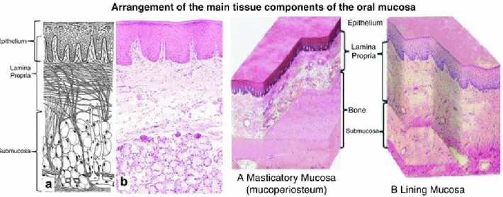
Finally, differently by other body structures, oral mucosa misses the musculature structure that bounded the mucosa by underlying tissue. To separate the mucosa lining cheek, lips and a part of the hard palate from the underlying bone or muscle there is a loose stromal tissue full of glands and fat named submucosa while the oral mucosa covering the gingiva and the more significant part of the hard palate are directly attached to the bone. This latter arrangement is called mucoperiosteum and provide a stable and rigid attachment (fig. I-6).
Figure I-6 Arrangement of the leading tissue components of the oral mucosa. (Squier and Brogden, Human Oral Mucosa:
Development, structure and function, 2011)
1.2.2.1.
Oral epithelium
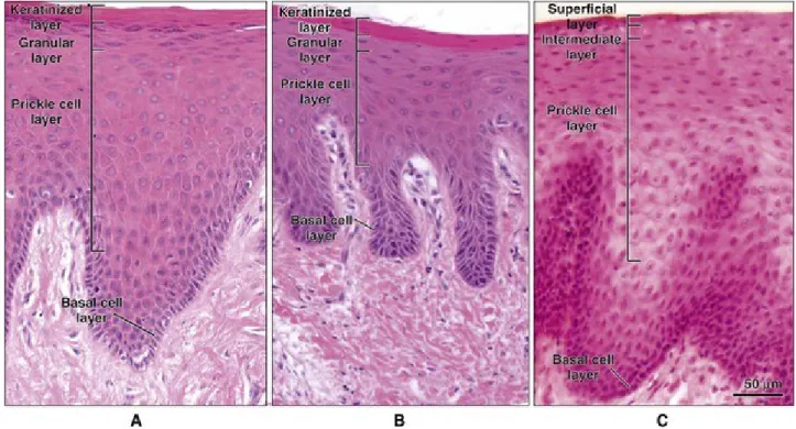
Oral epithelium is a covering and lining epithelium composed of different squamous pluristratified epithelia consisting of cells tightly attached to each other and to the basement membrane which are well organized in distinct layers. Each layer, or stratum, is easily recognized in cross-sectional histological analysis and have a specific role. The main histological difference between the epithelia is the keratinization degree.
Epithelia are classified in keratinized and non-keratinized epithelium; the first is characteristic of the masticatory and the second of the lining mucosa (fig. I-7). The features and differences between oral epithelia are described in Chapter 3.
Figure I-7 Histological features of the leading oral epithelial type. (Squier and Brogden, Human Oral Mucosa: Development, structure
and function, 2011)
1.2.2.2.
Basement membrane
The basement membrane is a thin and fibrous layer that connects the epithelial tissue with the underlying connective tissue. It is mainly composed of ECM protein, in direct contact with the epithelial tissue are more prominent the laminin, the type IV collagen and the type XVII collagen (present within the hemidesmosome) while in contact with the connective tissue are mainly present type VII collagen, anchoring fibrils and microfibrils (Kamaguchi et al., 2019). The upper layer is a thin electron-dense membrane in which thick collagen IV fibrils are strongly interconnected and supported by the heparan sulphate-rich proteoglycan perlecan, laminin, integrins, entactin, and dystroglycan. On the contrary, the lower part, which strongly binds the stromal tissue, is rich in substrate adhesion molecules (SAMs). Those protein have also been evaluated as a coating for 2D or 3D cultures. For instance, perlecan is necessary for epithelial formation since it not only supports the collagen structures but also regulates survival and terminal differentiation of 3D keratinocytes (Sher et al., 2006). Laminin, and in particular the sub-unit α-5, is involved in epithelial-mesenchymal signaling and hallow keratinocytes adhesion similarly as a feeder layer (Wegner et al., 2016; Tjin et al., 2018). Glycosaminoglycans improve fibroblast shaping in 3D and increase the number of seeded fibroblasts in S or G2 phases; in this way, an enhanced re-epithelialization process was guaranteed (Belvedere et al., 2018).
The basement membrane is also necessary for several other processes such as the angiogenesis, since basement membrane proteins stimuli endothelial differentiation, or barrier function towards malignant cells invasion.
1.2.2.3.
Lamina Propria
The connective tissue which underly the oral epithelia is named "lamina propria" and it is characterized by a huge heterogenicity in terms of cell types, fibres density, and organization. Generally, it is histologically divided into two principal types: the superficial papillary layer (which interval the epithelial ridges) and the deeper reticular layer (characterized by a "net-like" collagen-based structures). The two layers are characterized by an abundant and dense type I collagen fibres that in the papillary layer are thin and loosely arranged while in the reticular layer has collagen fibres arranged in thick bundles that tend to lie parallel to the surface plane.
The lamina propria can be in direct contact with bone, the mucoperiosteal structures (attached gingiva and hard palate) or a looser connective structure of the reticular layer characterized by the presence of type III collagen and others elastic fibres. The lamina propria is also rich in cell types (macrophages, plasma cells, mast cells, and lymphocytes, endothelial cells, and mesenchymal stem cells), blood vessels, and neural elements.
The lamina propria is made of three-dimensional fibres network (mainly collagen, elastin, and fibronectin) and a ground substance composed of water, glycoproteins, and proteoglycans and serum-derived proteins (Mohd at al., 2017).
Currently, at least 28 different collagen subtypes, composed of 46 distinct polypeptide chains have been identified (Shoulders and Raines, 2009). Collagen is typically organized as three parallel α-polypeptide strands coiled in a helical conformation around each other; each triple helix auto-assemble in a complex structure that can be easily observed under the macroscopic. The helix conformation leads the classification of different collagen subtypes; for instance, collagen type I, II, III, and V can be identified with an electron microscope by a characteristic helix arrangement that arises by fibrils with a banding pattern of 64 nm under SEM analysis
The elastic fibres are composed of elastin, which guarantees the elasticity and the support of mature fibres, and microfibrils, which is composed of fibrillin, a glycoprotein, microfibril associated glycoproteins (MAGPs), fibulins and elastin microfibril Interface Located Protein (EMILIN) (Wagenseil and Mecham, 2007). The elastic fibres can be stained by aldehyde fuchsin, orcein, or Weigert's elastic stain and are abundant in the lining mucosa, which needs more flexibility.
The ground substance consists of two heterogeneous protein/ carbohydrate complexes groups: proteoglycans (the polypeptide core with attached glycosaminoglycans chains) and glycoproteins (branched polypeptide chains to which only a few simple hexoses are attached).
The nerve supplying oral mucous membrane is predominantly sensory and arises mainly from the second and third divisions of the trigeminal nerve; afferent fibres of the facial (VII), glossopharyngeal (IX), and vagus (X) nerves are also involved. The myelin sheaths are lost in the reticular layer, where the bare fibres form network terminating in a subepithelial plexus between the epithelial ridges (Basbaum et al., 2009).
Finally, lymphatic and blood vessels are in the lamina propria while ducts, arising from different glands, are located in the deeper submucosa.
The main cell type found in lamina propria is fibroblast. Fibroblasts play an essential role in several processes, including epithelial morphogenesis (keratinocytes adhesion or integrin expression for the cellular junction formation). Moreover, they can be easily isolated and maintain their ECM secretion capability when cultivated in 3D, and they are essential for keratinocytes proliferation in vivo (see chapter 2).
1.2.3. Periodontium
The periodontium is the specialized multi-tissues structures composed by gingiva, periodontal ligament (PDL), cementum, and alveolar bone. The main role of the periodontium is protecting the bordered teeth (fig. I-8). The periodontal ligament, formed by
specialized fibroblasts, provides a firm but flexible connection for the teeth, necessary to give the proper withstand towards the mechanical stress. The alveolar bone, the thickened ridge of bone that contains the tooth sockets, forms the alveolar arch and confers stiffness. Like other bones, alveolar bone regularly undergoes a remodelling process regulated by mechanical stimulation. In particular, the tooth pocket is highly subject to this physiologic
phenomenon. The bone-forming cells, osteoblast and osteocytes, act on the areas of tension while the bone resorption cells, the osteoclast, line the areas of compression. This process guarantees the "high-quality" of the bone and, consequently, the adequate support to teeth. Periodontium development seems to be regulated mainly by the negative regulation of Sfrp3/Frzb derived from the Wnt pathway (de Jong et al., 2017).
Recently, human mesenchymal stem cells (hMSCs) have been found in the periodontium, especially close to the periodontal ligaments and in the gingiva and in dental pulp. That specific dental pulp hMSC (DPMSCs) subpopulation showed an enhanced differentiation potential when compared with bone-marrow-derived hMSC since they can reproduce the entire periodontium but also easily trans-differentiate in neuron-like cells. In the last decades, the secretome of MSCs, isolated from different sources, has acquired interest by researchers involved in tissue regeneration since it has shown impressive potential both in vivo and in vitro studies.
MSCs secrete several types of soluble factors, such as cytokines, chemokines, or growth factors, which mediate diverse functions and regulates the crosstalk between different cell types.
MSCs are well-known to act a pivotal role in tissue regeneration both locally, with the differentiation in specific Figure I-8 Histology and morphology of the periodontium.
signalling potential. For instance, after injuries, MSCs are reduce the cellular damage by inhibiting the fibrotic tissue formation, promoting angiogenesis to increase the nutrients available to the damaged site, modulating the immune response, and recruiting progenitors and other stem cells to the injured site to start the tissue renewal. The principal involved factors in tissue regeneration are summarized in figure I-9.
MSC features are better described in chapter 2
1.3. Oral mucosa model
1.3.1. Monolayered Keratinocytes cultures
The first successful monolayer culture of keratinocytes was performed in 1975 by Rheinwald and Green; to improve the viability and proliferation of keratinocytes in vitro they coated the plastic support with a feeder layer composed of irradiated 3T3 mouse fibroblasts. Keratinocytes were then cultivated in a home-made enriched culture medium named Green’s medium.
This method has been lately development to obtain a single-layer epithelial sheet; however, the derived models were often described as challenging to handle due to their fragility. Thus, these monolayer cultures have been improved and used for decades to study the basic biology of oral keratinocytes. For example, several studies showed that oral and skin keratinocytes have different behaviour in terms of differentiation, mitotic ratio, gene expression, and stimuli response. However, those monolayer cultures miss the typical structures and gene expression change to whom keratinocytes undergo in vivo during the terminal differentiation process. Thus, the development of a three-dimensional multilayer culture system was a vital innovation in epithelial biology studies, and this innovation will be fully analysed in chapter 3.2.
Nowadays, oral and skin keratinocytes are cultivated in 2D using polarized plastic support coated with collagen and serum and calcium-free media (such as the KGM from Gibco or EpiLife from Thermofisher). Thus, several 2D-based co-culture systems, mainly based on transwell systems, have been used to evaluate keratinocyte interaction with other cell types. As previously described, epithelial cells growth on the top of a stromal tissue mainly repopulated by fibroblast and, although in lower numbers, by mesenchymal stem cells. Due to hMSCs potential in tissue regeneration, several authors tried to establish the relation which intercourse between SCs and keratinocytes. For instance, Sivamani et all. (2015) showed that keratinocytes induce the epithelial trans-differentiation of hMSCs in direct co-culture and myofibroblast differentiation of hMSCs when they are co-cultivated with a transwell model.
Keratinocytes behaviour is severely affected by the growth condition. Alike for other cell types, keratinocytes gene expression, behaviour, proliferation potential, and morphology are deeply related to the three-dimensional stratification. However, differently by other cells, in the 3D histological structure are represented all differentiative stages of keratinocytes. For instance, in the basal layer keratinocytes are characterized by stem capability, which results lost in the spinosum layer. This peculiarity is crucial in the toxicological and pharmaceutical test since the same molecules can have different effects on the fourth layer, which characterizes the epithelium. Despite this intrinsic ability of keratinocytes to differentiate during the stratification, the particularities of each stratum are under the regulation of the closest mesenchyme in both healthy and damaged condition.
3D cell culture endowments the possibility to grow simultaneously different cellular populations in their proper microenvironment; thus, the resulting co-cultures can accurately mimic the cellular functions and the effect of crosstalk in terms of paracrine and autocrine regulation, and cell-cell interaction can be effectively studied. On the contrary, when cells are grown in 2D-based co-cultures, several phenomena cannot be appropriately evaluated. During the years, this statement resulted true in particular for mesenchyme-epithelial interaction.
1.3.2.
Histotypic oral epithelial models
In the last three decades, several advances in tissue engineering have provided an alternative approach to in vivo studies. Indeed, several three-dimensional models of oral epithelia (keratinocytes alone) or oral mucosa (keratinocytes growth on a fibroblast repopulated scaffold) were established as in vitro model aimed to study the developmental or wound healing processes and to evaluate the muco-toxicity and the biocompatibility of new biomaterials and drugs to be used for clinical application Scaffolds and ECM substitutes for connective tissue engineering, and optimization of technique for keratinocytes cultivations are technological key points for three-dimensional model developing.
1.3.2.1.
Bilayer cultures
Rosdy et al. (1990), in the first nineties, started to cultivate the keratinocytes from a different source on a permeable cell culture membranes support at the air/liquid interface. This cultivation method allows keratinocytes to arrange multilayer epithelia with different cytokeratin expression accordingly to the layer and the presence of keratin for the keratinized mucosa.
1.3.2.2.
Commercial models
Some companies, such as SkinEthic Laboratories (Nice, France) MatTek Corp. (Ashland, MA, USA), developed their own 3D models. The SkinEthic Laboratories model consists of a polycarbonate cell culture inserts on which human TR146 keratinocyte cells or gingival-derived cells are growth at air/liquid interface. However, TR146 are cancer cells derived by a squamous cell carcinoma and miss the capability to differentiate and form the keratin layer.
MatTek Corp developed two specific tools, called EpiOralTM and EpiGingivalTM, respectively mimicking the lining non-keratinized epithelium and the gingival keratinized epithelium arising, respectively, by buccal and gingival keratinocytes. Both models express the CK13 and produce cytokines, growth factors, and antimicrobial peptide as IL-10, VEGF, and the β-defensins and are made by primary single-donor cells. Both companies also provide models modification such as co-culture with fibroblast or pathological models. However, the media provided are under patent, and models cannot be furtherly developed. Indeed, as shown in table I-3, those models are mainly used for cytocompatibility and genotoxicity tests.
Table I-3 In this table are summarised the principal application of commercially available oral mucosa
MODEL
COMPANY
APPLICATION
AUTHOR
JOURNAL
DATE
EPIGINGIVAL MatTek Cytomegalovirus infection Hai et al Virology Journal 2006 EPIORAL, EPIGINGIVAL
MatTek Toxicological tests Klausner et al Toxicol in
vitro
2007
EPIORAL MatTek Irritancy tests Delves et al Toxicology 2008
SKINETHIC RHO Episkin Microbial tests and genotoxicity
Challacombe Stephen at al
Microbiology 2008
EPIORAL MatTek Mucoadhesive test Hu et al Ph.D. Thesis 2010
SKINETHIC RHO Episkin Microbial tests and genotoxicity
Challacombe at al
Cell Host & Microbe
2010
EPIORAL MatTek Permeability study Koschier et al Food Chem Toxicol
2011
EPIGINGIVAL MatTek Oral care tests Yang et al International dental Journal
2011
SKINETHIC RHE, SKINETHIC RHO
Episkin Oral care tests Alonso at al AAPS Pharm Sci Tech
2011
EPIORAL, EPIGINGIVAL
MatTek DNA repair evaluation
Mitchell et al Photochem Photobiol
2012
EPIGINGIVAL MatTek UV-radiation tests Agrawal et al Photochemist ry and Photobiology
2013
EPIORAL MatTek Xerogels insulin delivery tests
Boateng et al Protein pept Lett
2014
EPIORAL, EPIGINGIVAL
MatTek Toxicology test Schalge et al Toxicol Mech Methods
2014
EPIORAL MatTek γ-irradiation study Lambros et al Evid Based Complement Alternat Med
2015
EPIORAL MatTek Mucoahesive test MucoLox PCCA 2015
EPIORAL MatTek Tobacco tests Zanetti et al Chem Res Toxicol
2016
EPIGINGIVAL MatTek Tobacco tests Sundar et al Virology Journal 2016 EPIORAL, EPIGINGIVAL MatTek Ag nanoparticles tests
Pinďáková et al Int J Pharm. 2017
EPIORAL MatTek Mucoahesive test Song at al AAPS Pharm Sci Tech
2017
1.3.2.3.
Organotypic oral mucosa model
The models named “full thickness engineered oral mucosa” are characterized by a lamina propria substitute and an overlying stratified epithelium. The lamina propria is represented by a biocompatible scaffold fully repopulated