P
LANT
R
ESPONSE
TO
M
ODIFIED
C
ONDITIONS OF
L
IGHT AND
N
UTRIENTS
Polzella Antonella
TUTOR:
Prof. Scippa Gabriella Stefania
CO-TUTORS:
Prof. Chiatante Donato
Dr. Montagnoli Antonio
T
HESISS
UBMITTED FOR THED
EGREE OFD
OCTOR OF
P
HILOSOPHY
AT THE
U
NIVERSITY OFM
OLISED
EPARTMENT OFB
IOSCIENCES ANDT
ERRITORYCURRICULUM BIO-ENVIRONMENTAL
CYCLE XXXI
i
D
ECLARATIONExcept where reference is made to other sources, I declare that the contents in this thesis are my own work and have not been previously submitted, in part or in full, for the award of a higher degree elsewhere.
P
UBLICATIONS ARISING FROM THIS WORK“Toward an understanding of mechanisms regulating plant response to biochar application”
Antonella Polzella, Elena De Zio, Simona Arena, Gabriella Stefania Scippa, Andrea Scaloni, Antonio Montagnoli, Donato Chiatante, Dalila Trupiano
ii
D
EDICATIONTo myself, to my courage and stubbornness. To pillars of my life: my beloved family and Giovanni.
iii
A
CKNOWLEDGEMENTSI am very grateful for having this opportunity and experience to do my PhD in Pesche, Varese and Lancaster. I really learned a lot during the past three years and I enriched my scientific knowledge and my personality. Today, looking back I know to thank many people, who helped and accompanied me in this long and nice journey.
Firstly, I would like to express my deepest gratitude to my tutor and co-tutors. Prof. Scippa Gabriella Stefania, for her scientific guidance and many constructive suggestions, Prof. Chiatante Donato, it is a big shame he is retiring, only who has met him can say to have been lucky, and Dr. Montagnoli Antonello, for me also a big and dear friend who showed me the solution each time I couldn’t find it.
I am very grateful to my Lancaster supervisor, Prof. Toledo-Ortiz Gabriela, for her lovely, infinite and kind support. Without her, it would not have been possible for me to know, study and manipulate Arabidopsis mutants.
I am indebted to several people, which I have collaborated with or have helped me in various ways during all PhD period. My colleagues but mainly “academic friends” in University of Pesche: Dalila, Elena, Melissa, Antonio and Pamela. They have been always ready to give me a scientific comment, a personal suggestion or simply a hug. The great people I met in University of Varese: Mattia, Barbara, Oriana, Federica, Rosy, Daniela and Cristiano, whit whom I shared so many beautiful and formative experiences. For one year, they have been my second family and I will bring them in my heart forever. The lovely people in University of Lancaster: Jon, Dennis, Brittany and Phoebe for supporting work in every moment I needed.
Obviously, it cannot miss my infinite thank you to all my big and beautiful family, without them it would have been difficult! Particularly, a special thank you to Giovanni, my lifetime love, my future husband, he always believes in myself, that is why I can do everything.
iv
In natural environments plant growth, development, productivity, and distribution are highly dependent on a wide number of different biotic and abiotic factors. Among all water, temperature, light, and nutrients are the most important ones. Understanding mechanisms and adaptive responses of plant growth to changes in the availability of these environmental components is of the fundamental importance. In this framework, the present thesis aimed at widen the knowledge on plant response to modifications of soil nutrient availability and to the alteration in quality and quantity of light spectrum.
To accomplish this aim, the effects of changes in nutrient composition have been investigated by adding biochar amendment to the soil, whereas alterations in quality and quantity of light spectrum have been obtained by using different artificial lighting systems. The response to biochar soil amendment has been analyzed at morpho-physiological and molecular levels in different plant species (i.e. tomato, pea and Arabidopsis), alone and in combination with light spectra alterations. Results obtained in this thesis show that although biochar addition misbalances the photosynthetic machinery in tomato plants, it might improve Pisum and Arabidopsis growth, even at higher magnitude when the light spectrum is characterized by a specific composition. In addition, morpho-physiological plant response leads to hypothesize that photoreceptors such as phyA, phyB, and light signaling components such as pifs, could be involved in processes of growth stimulation in nitrogen and light stress conditions.
As part of the Ph.D project, the effects of a new artificial lighting system named CoeLux®, on morpho-physiology of several different plant species (i.e. Anthurium, Basilicum, Q. ilex) have been investigated. Experiments with CoeLux® lighting system showed a species-specific plant response
mechanism and a high plant efficiency to receive and use CoeLux® lighting system by performing
v
S
OMMARIOIn natura la crescita, lo sviluppo, la produttività e la distribuzione delle piante sono altamente influenzati da un ampio numero di diversi fattori biotici e abiotici. Tra tutti i fattori biotici, l’acqua, la temperatura, la luce e i nutrienti restano quelli di maggiore rilevanza. Così come resta di fondamentale importanza lo studio dei meccanismi della crescita e delle risposte di adattamento delle piante ai cambiamenti della disponibilità di queste stesse componenti ambientali. In tale contesto, il presente lavoro di tesi mira ad ampliare la conoscenza sulla risposta delle piante alla disponibilità modificata di nutrienti nel suolo e alla manipolazione qualitativa e quantitativa dello spettro di luce.
Per conseguire questo obiettivo sono stati studiati gli effetti dei cambiamenti nella disponibilità di nutrienti nel terreno attraverso l’aggiunta di un ammendante organico quale il biochar, invece le alterazioni sia qualitative che quantitative dello spettro di luce sono state ottenute usando diversi sistemi di illuminazione artificiale. Il biochar è stato utilizzato solo ed in combinazione con diversi spettri di luce, di cui gli effetti sono stati analizzati sia al livello morfo-fisiologico che molecolare in diverse piante (ad es. pomodoro, pisello e Arabidopsis).
I risultati ottenuti in questa tesi dimostrano che sebbene il biochar aggiunto nel terreno porta ad uno squilibrio dell’apparato fotosintetico nelle piante di pomodoro, esso potrebbe migliorare la crescita delle piante di Pisum e Arabidopsis, in maggior misura se si utilizza in combinazione con una luce caratterizzata da una specifica composizione spettrale. Inoltre, la risposta morfo-fisiologica delle piante porta ad ipotizzare che i fotorecettori, come phyA, phyB e fattori coinvolti nella segnalazione luminosa come pifs potrebbero essere convolti in processi di stimolazione della crescita in condizioni di stress di luce e di azoto.
Come parte del progetto di dottorato, sono stati studiati gli effetti di un nuovo sistema di illuminazione artificiale chiamato CoeLux® sulla morfo-fisiologia di diverse specie di piante (ad es. Anthurium, Basilicum, Q. ilex). Gli esperimenti con il sistema di illuminazione CoeLux® hanno dimostrato un meccanismo di risposta specie-specifico ed un’alta efficienza della pianta nel ricevere ed usare la luce CoeLux® attraverso lo svolgimento di una buona attività sia fotosintetica che stomatica.
vi
DECLARATION AND PUBLICATIONS ARISING FROM THIS WORK i
DEDICATION ii
ACKNOWLEDGEMENTS iii
ABSTRACT iv
SOMMARIO v
CHAPTER I-General Introduction 1
1.1 The Influence of Endogenous and Exogenous Factors on Plant Growth 2 1.2 The Plant Response to Nutrients and Light Supply Changes 3
1.3 The Aim and Structure of the Thesis 6
1.4 References 8
CHAPTER II - Toward an Understanding of Mechanisms Regulating Plant Response to
Biochar Application 15
2.1 Introduction 16
2.2 Material and Methods 18
2.2.1 Experimental Set Up and Biochar Treatment 18
2.2.2 Soil Analysis 19
2.2.3 Plant Growth Analysis 19
2.2.4 Protein Extraction and Two-Dimensional Electrophoresis 19 2.2.5 In-Gel Digestion, Mass Spectrometry Analysis and Identification 20
vii
2.2.6 ACO Expression Measurements 21
2.2.7 PCR-Based Detection of P. infestans in Leaves 21
2.3 Results 22
2.3.1 Biochar Effect on Soil Characteristics 22 2.3.3 Effects of Biochar on Leaf Proteome 24 2.3.4 Analysis of LeACO1 Gene Expression Patterns 29 2.3.5 Detection of the Tomato Late Blight Pathogen: Phytophthora infestans 29
2.4 Discussion and Conclusions 30
2.5 Acknowledgments 34
2.6 References 35
CHAPTER III-The Interplay of LED Spectra and Biochar in Arabidopsis columbia and Pisum
sativum L. Morpho-Physiological Traits 41
3.1 Introduction 42
3.2 Materials and Methods 45
3.2.1 Plant Material and Experimental Setup 45
3.2.2 Growth Room Characteristics 46
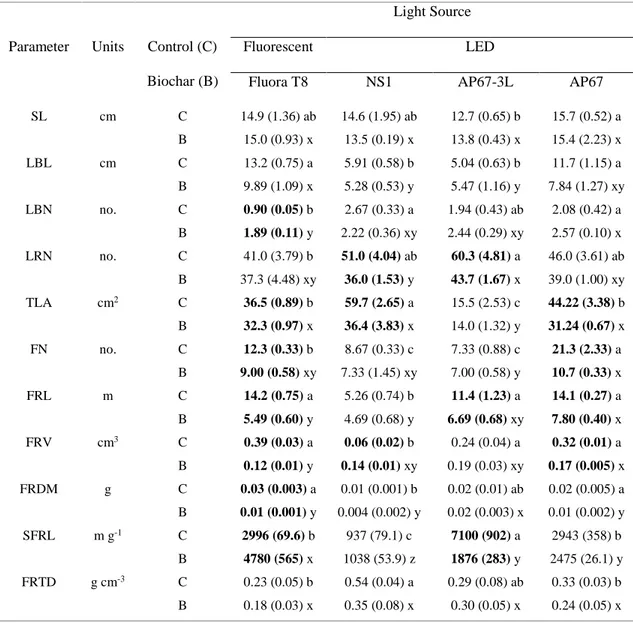
3.2.3 Biochar Characterization 47 3.2.4 Soil Characterization 48 3.2.5 Morphological Measurements 48 3.2.6 Physiological Measurements 49 3.2.7 Statistical Analysis 50 3.3 Results 50
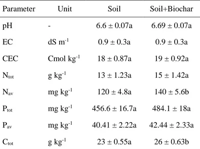
3.3.1 Biochar and Soil Characteristics 50
viii 3.3.4 Physiological Traits 56 3.4 Discussion 59 3.5 Conclusions 64 3.6 Acknowledgments 64 3.7 References 65
CHAPTER IV-The Possible Photoreceptor Role in Response to Modified Light and Nitrogen
Availability 75
4.1 Introduction 76
4.2 Materials and Methods 78
4.2.1 Hydroponic Growing System 78
4.2.2 Plant Material, Solution Preparation and Growing Conditions 78
4.2.3 N Starvation Treatment 79
4.2.4 Morphological Measurements and Determination of Photosynthetic Pigment
Contents 80
4.2.5 Statistical Analysis 80
4.3 Results 81
4.3.1 Morphological Traits 81
4.3.2 Photosynthetic Pigment Contents 84
4.4 Discussion and Conclusions 86
4.5 References 89
CHAPTER V-The Plant Response to CoeLux® Lighting System 95
5.1 Introduction 96
ix
5.2.1 The CoeLux® Lighting System 98
5.2.2 Plant material and Experimental Design 99
5.2.3 Morpho-physiological measurements 102
5.3 Results 103
5.3.1 Short-Term Experiments 103
5.3.2 Long-Term Experiments 105
5.4 Discussion and Conclusions 111
5.5 References 113
CHAPTER VI-Synthesis and Outlook 119
ORAL/POSTER PRESENTATIONS FROM THIS WORK 123
LIST OF ABBREVIATIONS 125
LIST OF TABLES 127
1
Chapter I
2
I
1.1 The Influence of Endogenous and Exogenous Factors on Plant
Growth
Plant growth and development are finely regulated by the integration of many environmental and endogenous signals. Because they dictate plant characteristics from within the cell, the genetic factors are considered internal factors (i.e. endogenous) and they represent the overall plant genetic material, including genes, chromosomes, genomes and all those that represent gene expression (Johnson and Stinchcombe, 2007; Bailey et al., 2009; Schweitzer et al., 2012b). On the other hand, the environmental factors, external non-genetic factors (i.e. exogenous), are generally divided into two groups: biotic and abiotic. Biotic factors include all living components, such as animals, plants, fungi, and bacteria; abiotic factors are all non-living components (Buchmann, 2000) comprising (i) topography (intended as all earth physical features such as land elevation, slope, terrain, etc.), (ii) soil physical and chemical properties (e.g. soil nutrient availability, texture, structure, pH, and so on) and (iii) climate factors comprising light, temperature, water, aeration, etc. (Dunson and Travis, 1991).
To ensure their living and surviving in a natural environment, plants have evolved mechanisms to rapidly respond to modify conditions produced by the interactions between the above-mentioned biotic and abiotic factors. For instance, excess or deficit of water availability affects plant growth and yield in terms of tissue development, transpiration, stomatal activity, CO2
assimilation, photosynthetic activity, and flowering (Osakabe et al., 2014; Abdallah et al., 2018). Similarly, plants are able to perform vital activities in certain and optimal temperature range and it is known that high and low temperatures mostly alter the flowering and photosynthetic processes (Allakhverdiev et al., 2008; Ashraf and Harris, 2013; Prasad and Djanaguiraman, 2014). Likewise, gases, pollutants, and wind strongly affect plant biomass, productivity, and evapotranspiration (Zengin and Munzuroglu, 2005). Several studies have shown that low light causes the reduction of plant growth and photosynthetic pigment accumulation (Adelusi and Aileme, 1977), whereas others have reported that the plant root structure is affected by soil texture, structure and nutrient availability (Oke, 1985). Furthermore, it is well established that the influence of the interaction of these factors on plant growth, for example, the interaction between temperature, water and salt stress may be associated with photo-inhibition (Osmond et al., 1987). On the other hand, the close relation between light and temperature can positively affect the plant developmental stages, for instance the optimal plant growth and photosynthesis proved at medium irradiance and high
General Introduction
3
I
nutrient levels in Thompson et al.’s report (1988). However, plants are able to counteract the changes of the biotic and biotic environmental components. Indeed plants respond to modified water availability by activating a series of signaling pathways (Zhu, 2002) and respond to temperature variations through production of secondary metabolites (Mathur et al., 2014). Furthermore, plants are capable to activate tolerance mechanisms for pollutant detoxification as for instance metal immobilization, sequestration and compartmentalization (Patra et al., 2004; John et al., 2009; Dalvi and Bhalerao, 2013). Finally, plants are able to acclimatize certain irradiance values and nutrient availability by physiological adjustment promoting carbon gains (Thompson et al. 1988).
1.2 The Plant Response to Nutrients and Light Supply Changes
Plant growth is highly dependent on mineral nutrient uptake (Clarkson, 1980; Sinclair, 1992). In seeds, roots and leaves, the nutrient content in the growth media affects several activities, including organ function, rate of organ growth and turnover, and plant life-history strategies (Kerkhoff et
al., 2006). Generally, the nutrients required for plant growth are classified into three groups. The
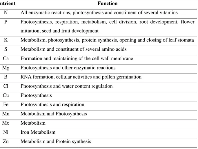
first group is composed by three basic elements that plants can obtain from water and atmosphere such as carbon (C), hydrogen (H), and oxygen (O). The other two groups comprises the so-called soil-derived nutrients that are the (i) macronutrients such as nitrogen (N), phosphorus (P), potassium (K), sulfur (S), calcium (Ca), and magnesium (Mg) and the (ii) micronutrients like boron (B), chloride (Cl), copper (Cu), iron (Fe), manganese (Mn), molybdenum (Mo), nickel (Ni) and zinc (Zn) (Mahler, 2004). In Table 1.1 these elements are summarized in relation to their specific function in plant biological processes affecting the growth and development. Although plants mainly take up C from the air, they acquire the rest of nutrients almost exclusively from the soil through the root systems. Thus, the nutrient concentrations in plant tissues are dependent on resource availability in the soil which in turn depends on the rate of their uptake as well as on the rate of replacement due to bacterial N fixation, organic matter mineralization, atmospheric deposition or weathering (Lukac et al., 2010). Among all the above-listed elements, N and P remain the most limiting ones in many ecosystems and for many plant biological processes (Wassen et al., 2005; Chapman et al., 2006; Lambers et al., 2008).
4
I
Different plant species respond to changes in soil nutrient availability by modulating organ development. Different levels of nutrient availability in the soil may induce an alteration in leaf dry matter content (Hodgson et al., 2011). The root system morphological and architectural characteristics, tightly associated with nutrient uptake efficiency, as well as the root/shoot biomass allocation might be also modulated in the case of nutrient shortage conditions (Hill et al., 2006). Generally, the P deficiency causes the inhibition of plant primary root growth. On the contrary, plants increase both the growth and density of lateral (i.e. secondary) and hair root growth in response to N, P, Fe and S deficiency (López-Bucio et al., 2003; Britto and Kronzucker, 2008). Furthermore, also high nutrient levels might be toxic for plants causing the production of reactive
Nutrient Function
N All enzymatic reactions, photosynthesis and constituent of several vitamins
P Photosynthesis, respiration, metabolism, cell division, root development, flower initiation, seed and fruit development
K Metabolism, photosynthesis, protein synthesis, opening and closing of leaf stomata S Metabolism and constituent of several amino acids
Ca Formation and maintaining of the cell wall membrane Mg Photosynthesis and other enzymatic reactions
B RNA formation, cellular activities and pollen germination Cl Photosynthesis and water content regulation
Cu Photosynthesis
Fe Photosynthesis and respiration Mn Metabolism and Photosynthesis Mo Metabolism
Ni Iron Metabolism
Zn Metabolism and Protein synthesis
Table 1.1. Essential macro- and micro- nutrients for plant growth and their functions within plant tissue (Baligar et al., 2007).
Table 1.0.2. Essential macro- and micro- nutrients for plant growth and their functions within plant tissue (Baligar et al., 2007).
Table 1.0.3. Essential macro- and micro- nutrients for plant growth and their functions within plant tissue (Baligar et al., 2007).
Table 1.0.4. Essential macro- and micro- nutrients for plant growth and their functions within plant tissue (Baligar et al., 2007).
Table 1.0.5. Essential macro- and micro- nutrients for plant growth and their functions within plant tissue (Baligar et al., 2007).
Table 1.0.6. Essential macro- and micro- nutrients for plant growth and their functions within plant tissue (Baligar et al., 2007).
General Introduction
5
I
oxygen species (ROS), which in turn cause a plant cellular damage (Connolly and Walker, 2008; Morgan and Connolly, 2013).
Light is an essential element for plant life. Plants, as sessile organisms, developed specialized structures to receive, modulate and convert different quality and quantity of light that can reach plant tissue in different directions. The electromagnetic spectrum is composed of different types of radiations such as radio waves, microwaves, infrared, visible light, ultraviolet, X-rays and gamma rays. Among them, only the visible light can be perceived by the human eye, characterized by wavelengths between about 400 nm and 700 nm (DeVany et al., 1969; Fig. 1.1a). Photosynthetic organisms such as plants, algae, and cyanobacteria are able to perform the photosynthetic process in which light is converted to chemical energy. Plants receive and absorb light through light-absorbing molecules called pigments, mainly Chlorophyll a, Chlorophyll b and carotenoids that are three key pigments absorbing light energy in a specific wavelength range (Fig. 1.1b). Beside pigment molecules, in plant tissue, there are also photoreceptor proteins that detect and use the light for important biological processes.
a
b
c
Figure 1.1. (a) Spectrum of visible light detected with Handheld Spectrometer (UPRtek). (b) Absorption spectra of the two key pigments and β-carotene, the representative of the carotenoid group (OpenStax College, Biology). (c) Absorption regions of main photoreceptors (Modified from Parihar et al., 2016).
6
I
There are three classes of plant signal-transducing photoreceptors, namely Phytochromes (Phys), Cryptochromes (Crys) and Phototropins (Phots). Each photoreceptor operates in a specific range of the visible spectrum as shown in the detail in Fig. 1.1c.
The Phys are involved in many important photomorphogenic responses in plant growth and development, e.g. germination, stem elongation, leaf expansion, formation of some pigments, chloroplast development and flowering (Franklin and Quail, 2010). They exist in two photoconvertible forms absorbing in the light spectrum region between 600 and 750 nm, in the red (Pr) and far-red (Pfr) light, which represent the biologically active and inactive form, respectively (Quail, 1997; Reed, 1999; Shinomura et al., 2000). The Crys are blue-light and UV-A sensitive photoreceptors absorbing in the 320-520 nm range of visible spectrum functioning mainly as entrainment of the circadian clock, and in the flowering and photomorphogenic activities. Similarly, the Phots operate in blue, UV-A and green light (320-520 nm range) playing a key role in phototropism, chloroplast movement and stomatal opening (Briggs et al., 2001; Möglich et al., 2010; De Wit et al., 2016; Parihar et al., 2016).
Optimal light irradiance is required for a normal plant growth, however, the possible high or low level of irradiance might affect photosynthetic process and in turn plant yield (Ma et al., 2015). For example, Powles and Critchley (1980) showed that bean plants under a low level of light had a reduced growth due to a lower rate of photosynthetic electron transport and carbon dioxide (CO2) assimilation. Nevertheless, Guo et al. (2006) reported that in the bayberry tree the
high irradiance caused the depression of photosynthesis and photosystem II (PSII) efficiency and activated the protective mechanism of photoinhibition. Additionally, other responses to variation in light availability include changes of morphological and physiological features of organs involved in the light acquisition and carbon assimilation processes, such as leaf and shoot (Valladares and Niinemets, 2008).
1.3 The Aim and Structure of the Thesis
How plant responds to the alteration of environmental components affecting their growth and development is of great importance for widening the understanding of biological mechanisms that may allow plants to adapt to important modifications such as “climate change”. Nutrient and light availabilities meaningfully affect plant growth and development, however, the majority of the
General Introduction
7
I
studies carried so far on plant response to change of these two environmental components, have manipulated a single parameter. For instance, several studies have investigated the influence of nutrient contents on plant growth (da Silveira Pontes et al., 2010; Kazakou et al., 2014), as well as other reports have studied the effect of light spectra on numerous plant growth parameters (Smirnakou et al., 2017; Montagnoli et al., 2018). Whereas the interplay of these two factors have been rarely investigated (Siebenkäs et al., 2015). In this framework, the present thesis focused on investigating plant response to the combining manipulation of nutrient supply and light irradiation both at qualitative and quantitative levels. In particular, plant morpho-physiological traits were measured in response to the application of biochar amendment, a source of soil nutrient supply, and the modification of light spectra emitted by various LED lighting systems. Particularly, since it is well known that biochar amendment increases soil nitrogen content and availability, the present work aimed to cover the possible role of nitrogen and different lighting conditions on plant growth.
In detail, proteomic and molecular analysis on leaves of tomato plants were carried to investigate the response to changes in soil quality by using biochar amendment (see Chapter II). In chapter III the morpho-physiological traits of Pisum sativum and Arabidopsis thaliana (Columbia ecotype) were analyzed in relation to biochar application and the alteration of light spectra sourced by the LED. Furthermore, the possible role of photoreceptors was investigated in
Arabidopsis mutants grown in a hydroponic system and in manipulated availability of nitrogen
and artificial lighting (see Chapter IV). Finally, part of the study was focused on the morpho-physiological changes of ornamental, aromatic, forestry and agronomic plant species grown under CoeLux® lighting system (see Chapter V).
8
I
1.4 References
Abdallah M. B., Trupiano D., Polzella A., De Zio E., Sassi M., Scaloni A., Zarrouk M., Youssef N. B., Scippa G. S. (2018). Unraveling physiological, biochemical and molecular mechanisms involved in olive (Olea europaea L. cv. Chétoui) tolerance to drought and salt stresses. Journal of Plant Physiology, 220, 83-95.
Adelusi A. A., Aileme J. D. (1997). Effect of light and mineral nutrient stress on the yield and yield components of cowpea (Vigna unguiculata). Nigerian Journal of Botany, 10, 1-6. Allakhverdiev S. I., Kreslavski V. D., Klimov V. V., Los D. A., Carpentier R., Mohanty P. (2008).
Heat stress: an overview of molecular responses in photosynthesis. Photosynthesis
Research, 98, 541–550.
Ashraf M., Harris P.J.C. (2013). Photosynthesis under stressful environments: an overview.
Photosynthetica, 51(2), 163–190.
Bailey J. K., Schweitzer J. A., Ubeda F., Koricheva J., LeRoy C. J., Madritch M. D., Rehill B. J., Bangert R. K., Fischer D. G., Allan G. J., Whitham T. G. (2009). From genes to ecosystems: a synthesis of the effects of plant genetic factors across levels of organization.
Philosophical Transactions of the Royal Society of London B: Biological Sciences, 364(1523), 1607-16.
Baligar V. C., Fageria N. K. (2007). Agronomy and Physiology of Tropical Cover Crops. Journal
of Plant Nutrition, 30, 1287–1339.
Briggs W. R., Olney M. A. (2001). Photoreceptors in Plant Photomorphogenesis to Date. Five Phytochromes, Two Cryptochromes, One Phototropin, and One Superchrome. Plant
Physiology, 125(1), 85-88.
Britto D. T., Kronzucker H. J. (2008). Cellular mechanisms of potassium transport in plants.
Physiologia Plantarum, 133, 637-650.
Buchmann N. (2000). Biotic and abiotic factors controlling soil respiration rate in Picea abies stands. Soil Biology and Biochemistry, 32, 1625-1635.
General Introduction
9
I
Chapin F. S., Bloom A. J., Field C. B., Waring R. H. (1987). Plant responses to multiple environmental factors. BioScience, 37, 49–57.
Chapman S.K., Langley J. A., Hart S. C., Koch G. W. (2006). Plants actively control nitrogen cycling: uncorking the microbial bottleneck. The New Phytologist, 169, 27-34.
Cheng L., Tang X., Vance C. P., White P. J., Zhang F., Shen J. (2014). Interactions between light intensity and phosphotus nutrition affect the phosphate-mining capacity of white lupin (Lupinus albus L.). Journal of Experimental Botany, 65(12), 2995-3003.
Connolly E. L., Walker E. L. (2008). Time to pump iron: iron-deficiency-signaling mechanisms of higher plants. Current Opinion in Plant Biology, 11, 530-535.
da Silveira Pontes L., Louault F., Carrère P., Maire V., Andueza D., Soussana J.-F. (2010). The role of plant traits and their plasticity in the response of pasture grasses to nutrients and cutting frequency. Annals of Botany, 105, 957–965.
Dalvi A. A., Bhalerao S. A. (2013). Response of plants towards heavy metal toxicity: an overview of avoidance, tolerance and uptake mechanism. Annals of Plant Sciences, 2(9), 362–368. De Vany A. S., Eckert R. D., Meyers C. J., O'Hara D. J., Scott R. C. (1969). A Property System
for Market Allocation of the Electromagnetic Spectrum: A Legal-Economic-Engineering Study. Stanford Law Review, 21, 1499.
De Wit M., Galvão V. C., Fankhauser C. (2016). Light-Mediated Hormonal Regulation of Plant Growth and Development. The Annual Review of Plant Biology, 67, 513-37. http://doi.org/10.1146/annurev-arplant-043015-112252.
Dunson W. A., Travis J. (1991). The role of abiotic factors in community organization. The
American Naturalist, 138(5), 1067-1091.
Franklin K. A., Quail P. H. (2010). Phytochrome fuonctions in Arabidopsis development. Journal
of Experimental Botany, 61(1), 11-24.
Guo Y. P., Guo D. P., Zhou H. F., Hu M. J., Shen Y. G. (2006). Photoinhibition and xanthophyll cycle activity in bayberry (Myrica rubra) leaves induced by high irradiance.
10
I
Hill J. O., Simpson R. J., Moore A. D., Chapman D. F. (2006). Morphology and response of roots of pasture species to phosphorus and nitrogen nutrition. Plant and Soil, 286, 7–19.
Hodgson J. G., Montserrat-Martí G., Charles M., Jones G., Wilson P., Shipley B., Sharafi M., Cerabolini B. E. L., Cornelissen J. H. C., Band S. R., Bogard A., Castro-Diez P., Guerrero-Campo J., Palmer C., Perez-Rontome M. C., Carter G., Hynd A., Romo-Diez A., de Torres Espuny L., Royo Pla F. (2011). Is leaf dry matter content a better predictor of soil fertility than specific leaf area? Annals of Botany, 108, 1337-1345.
John R., Ahmad P., Gadgil K., Sharma S. (2009). Heavy metal toxicity: effect on plant growth, biochemical parameters and metal accumulation by Brassica juncea L. International
Journal of Plant Production, 3(3), 65–76.
Johnson M. T., Stinchcombe J. R. (2007). An emerging synthesis between community ecology and evolutionary biology. Trends in Ecology & Evolution, 22(5), 250-7.
Kazakou E., Violle C., Roumet C., Navas M.-L., Vile D., Kattge J., Garnier E. (2014). Are trait-based species rankings consistent across data sets and spatial scales? Journal of Vegetation
Science, 25, 235–247.
Kerkhoff A. J., Fagan W. F., Elser J. J. Enquist B. J. (2006) Phylogenetic and growth form variation in the scaling of nitrogen and phosphorus in the seed plants. The American
Naturalist, 168, 4.
Lambers H., Chapin III F. S., Pons T. L. (2008) Plant physiological ecology, Springer Verlag. López-Bucio J., Cruz-Ramírez A., Herrera-Estrella L. (2003). The role of nutrient availability in
regulating root architecture. Current Opinion of Plant Biology, 6(3), 280-7.
MA X., Song L., Yu W., Hu Y., Liu Y., Wu J., Ying Y. (2015). Growth, physiological, and biochemical responses of Camptotheca acuminata seedlings to different light environments. Frontiers in Plant Science, 6, 321.
Mathur S., Agrawal D., Jajoo A. (2014). Photosynthesis: response to high temperature stress.
General Introduction
11
I
Möglich A., Yang X., Ayers R. A., Moffat K. (2010). Structure and Function of Plant Photoreceptors. The Annual Review of Plant Biology, 61, 21-47. http://doi.org/ 10.1146/annurev-arplant-042809-112259.
Montagnoli A., Dumroese R. K., Terzaghi M., Pinto J. R., Fulgaro N., Scippa G. S., Chiatante D. (2018). Tree seedling response to LED spectra: implications for forest restoration. Plant
Biosystems, 152, 515-523.
Morgan J. B., Connolly E. L. (2013). Plant-soil interactions: nutrient uptake. Nature Education
Knowledge, 4(8), 2.
Oke S. O. (1985). Effect of soil texture nutrient stress and water stress on yield of Andropogon gayanus. knuth and Schizchyrium sanguineum (Rertz) alston. M.Sc. Thesis, University of Ife (Now Obafemi Awolowo University), Nigeria.
Osakabe Y., Osakabe K., Shinozaki K., Tran L.-S. P. (2014). Response of plants to water stress.
Frontiers in Plant Science, 5, 86.
Osmond C. B., Austin M. P., Berry J. A., Billings W. D., Winner W. E. (1987). Stress physiology and distribution of plants. Bioscience, 37, 33-47.
Parihar P., Singh R., Singh S., Tripathi D. K., Chauhan D. K., Singh V. P. and Prasad S. M. (2016). Photoreceptors mapping from past history till date. Journal of Photochemistry and
Photobiology, B: Biology, 162, 223-231.
Patra M., Bhowmik N., Bandopadhyay B., Sharma A. (2004). Comparison of mercury, lead and arsenic with respect to genotoxic effects on plant systems and the development of genetic tolerance. Environmental and Experimental Botany, 52(3), 199–223.
Poorter H., Niklas K. J., Reich P. B., Oleksyn J., Poot P., Mommer L. (2011). Biomass allocation to leaves, stems and roots: meta‐analyses of interspecific variation and environmental control. New Phytologist, 193(1), 30-50.
Powles S. B., Critchley C. (1980). Effect of light intensity during growth on photoinhibition of intact attached bean leaflets. Plant Physiology, 65, 1181–1187.
12
I
Prasad P. V. V., Djanaguiraman M. (2014). Response of floret fertility and individual grain weight of wheat to high temperature stress: sensitive stages and thresholds for temperature and duration. Functional Plant Biology, 41, 1261-1269.
Quail P. (1997). An emerging molecular map of the phytochromes. Plant Cell and Environment,
20, 657-666.
Reed J.W. (1999). Phytochromes are Pr-ipatetic kinases. Current Opinion in Plant Biology, 5, 393-397.
Schweitzer J. A., Madritch M. D., Felker-Quinn E., Bailey J. K. (2012b). From genes to ecosystems: how plant genetics links above- and below-ground processes. In: Wall D, editor. Soil ecology and ecosystem services. Oxford, U.K: Oxford University Press, pp. 82–98.
Shinomura T., Uchida K., Furuya M. (2000). Elementary processes of photoperception by Phytochrome A for high-irradiance response of hypocotyls elongation in Arabidopsis.
Plant Physiology, 122, 147-156.
Siebenkäs A., Schumacher J., Roscjer C. (2015). Phenotypic plasticity to light and nutrient availability alters functional trait ranking across eight perennial grassland species. AoB
Plants, 7, plv029.
Smirnakou S., Ouzounis T., Radoglou K. (2017). Continuous spectrum LEDs promote seedling quality traits and performance of Quercus ithaburensis var. macrolepis. Frontiers in Plant
Science, 8, 188.
Stuefer J. F., Huber H. (1998). Differential effects of light quantity and spectral light quality on growth, morphology and development of two stoloniferous Potentilla species. Oecologia,
117, 1-8.
Thompson W. A., Stocker G. C., Kriedemann P. E. (1988). Growth and photosynthetic response to light and nutrients of Flindersia brayleyana F. Muell., a rainforest tree with broad tolerance to sun and stade. Australian Journal of Plant Physiology, 15, 299-315.
General Introduction
13
I
Uchida R. (2000). Essential Nutrients for Plant Growth: Nutrient Functions and Deficiency Symptoms. Plant Nutrient Management in Hawaii’s Soils, Approaches for Tropical and
Subtropical Agriculture, (eds. College of Tropical Agriculture and Human Resources).
Valladares F., Niinemets Ü. (2008). Shade tolerance, a key plant feature of complex nature and consequences. Annual Review of Ecology, Evolution, and Systematics, 39, 237-257. Wassen M.J., Venterink H. O., Lapshina E. D., Tanneberger F. (2005) Endangered plants persist
under phosphorus limitation. Nature, 437, 547-550.
Zengin F. K., Munzuroglu O. (2005). Effects of some heavy metals on content of chlorophyll, proline and some antioxidant chemicals in bean (Phaseolus vulgaris L.) seedlings. Acta
Biologica Cracoviensia Series Botanica, 47(2), 157–164.
Zhu J. K. (2002). Salt and drought stress signal transduction in plants. Annual Review of Plant
15
Chapter II
Toward an Understanding of Mechanisms
Regulating Plant Response to Biochar
Application
Polzella Antonella
a, De Zio Elena
a, Arena Simona
b, Scippa Gabriella Stefania
a,
Scaloni Andrea
b, Montagnoli Antonio
c, Chiatante Donato
c, Trupiano Dalila
aa
Department of Biosciences and Territory, University of Molise, Pesche, Italy.
bProteomics and Mass Spectrometry Laboratory, ISPAAM, National Research
Council, Napoli, Italy.
c
Department of Biotechnology and Life Science, University of Insubria, Varese,
Italy.
16
II
Abstract
Plant growth and development are affected by several environmental factors, among which soil nutrient availability. Biochar addition to soil is recognized to exert beneficial effects on soil fertility and thus plant growth; furthermore, it is a promising option for climate change mitigation. However, multi-species studies and meta-analyses have indicated considerable variations in biochar responses among plant species. To date, information on the biochar effect on plants, especially at molecular level, are still scarce.
Using a multi-target approach with a model plant such as tomato, we demonstrate that biochar has a negligible effect on soil nutrient content and plant growth, even if it misbalances the plant photosynthetic machinery, as well as mechanisms recognizing pathogen-derived molecules. Ethylene could be one of the signal-molecule driving the alteration of tomato-pathogen recognition signaling by inactivation of vesicle trafficking. All these modifications could be at the basis of the increased susceptibility of biochar treated plants to pathogen attack.
Further organ and tissue specific multi-level studies, from resolution internal processes towards high-throughput external phenotyping, coupled with powerful biostatistic and informatic analysis, will help to decipher, in a network-type fashion, all the factors and signaling mechanisms related to the complex interaction between different plant, soil and biochar types.
Keywords: leaves, Lycopersicon esculentum, ethylene, morphology, Phytophthora infestans, proteome, soil
amendment.
2.1 Introduction
Biochar application to soil is considered as a promising strategy to sustain soil fertility, thus to promote plant growth, simultaneously sequestering atmospheric CO2 and reducing greenhouse
gases emissions, such as CO2, CH4, N2O (Agegnehu et al. 2017). It is a carbonaceous product
obtained from the pyrolysis of plant and waste feedstocks occurring at high temperatures (between 350-700 °C) in oxygen-limiting conditions (Lehmann and Joseph, 2009).
The wide variety of both biochar and soil with overall complex physical, chemical and biological interactions between them, induce different effects on plant growth and response (Mukherjee and Lal, 2013). In detail, several reports prove biochar benefits on many soil parameters, including nutrient retention, cation-exchange and water-holding capacity, electric conductivity, pH, microbial and mycorrhizal activity (Glaser et al., 2002), which improve soil
Toward an Understanding of Mechanisms Regulating Plant Response to Biochar Application
17
II
fertility and thereby plant growth (Major et al., 2010; Jeffery et al., 2011; Trupiano et al., 2017). Nevertheless, other reports show conflicting effects. For instance, biochar negatively influences plant NH4+ adsorption and uptake, due to its surface properties, determining a lower nitrogen (N)
release, and thus negatively affecting leaf N and chlorophyll content in tomato plants (Akhtar et
al., 2014). As well as, in saline sodic soil, there is an antagonist interaction between biochar and
phosphorus (P), producing a detrimental effect on the P availability and Suaeda salsa growth (Xu
et al., 2016). Furthermore, in species with indeterminate habit, such as tomato, the growth
enhancement using biochar is unlikely to occur (Vaccari et al., 2015). However, multi-species studies and meta-analyses proved considerable variations in biochar responses among plant species (Thomas and Gale, 2015), although molecular mechanisms sustaining these different responses are almost unknown.
Recently, Viger et al. (2015) have defined a first model to explain the early response, signaling and altered gene expression in Arabidopsis thaliana grown in biochar-amended soil. Although in their work, they have noted a biochar-induced increase of Arabidopsis plants rate of growth, contrarily to a previous study (Meller Harel et al., 2012), a down-regulation of a large suite of plant defense genes and related-stress signaling response was also observed.Mechanisms related to plant defense signaling are still not understood, but phytohormones, such as ethylene, jasmonic and salycilic acid, seem to have a key role in the regulation of stress-related genes. This highlights the complex plant-soil-biochar interaction and suggests the importance of future investigations on different biochar types and plant species, determining whenever expression changes in genes related to defense may result in an increased organism susceptibility to pathogen attack.
Tomato plant (Lycopersicon esculentum, Mill.) is one of most widely grown vegetable (Tranchida-Lombardo et al., 2018) and it is susceptible to certain fungal diseases, such as the late blight caused by Phytophthora infestans, which lead often to localized cell death through a hypersensitive plant response (Rigano et al., 2014).
Thus, tomato is used often as a model plant system, although, to date, information on the effects of biochar on tomato growth and pathogen-resistance are still scarce or completely absent. In the present work, we provide a complete picture of the tomato plant response to biochar application, describing the changes in plant morphological parameters in combination with
18
II
corresponding changes in proteome profiles and susceptibility to P. infestans attack. Results obtained contribute elucidating molecular mechanisms regulating plant growth-defense response.
2.2 Material and Methods
2.2.1 Experimental Set Up and Biochar Treatment
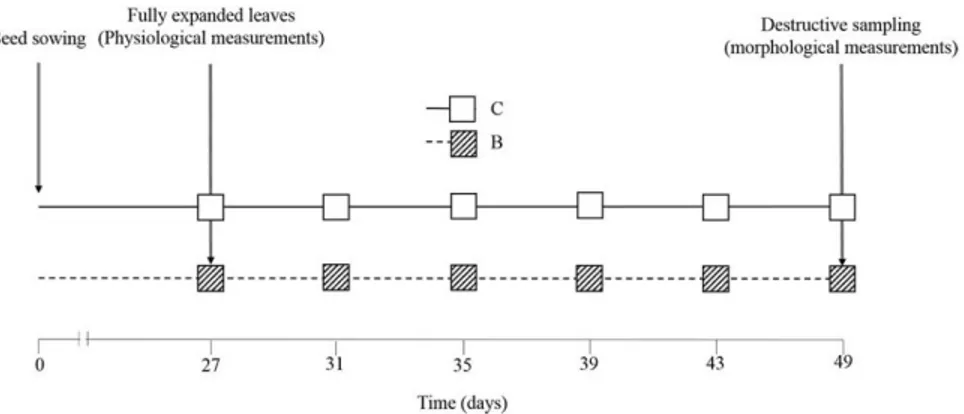
One-month-old tomato seedlings (Lycopersicon esculentum, Mill.) (var. San Marzano; Vivaio Mignogna, Ripalimosani, CB - Italy) were transplanted in plastic pots (10 L) prepared with non-amended (control=C) and biochar-non-amended (treatment=B) soils. Soil was collected from an uncultivated pasture area, located in Pesche, at a depth of 0–20 cm. It is unlikely that these soils contain charcoal already, since there has not been a tradition of crop residue or other burning on the land. The soil was found to be neutral, relatively low in organic matter and to have a clay soil texture (%clay: 52.7±3.4, %sand: 15.7±0.6, %silt: 31.6±2.7). For the experiment, soil was air dried for 72 h, weighed and finely crushed then mixed thoroughly before packing lightly in the pots on top of 100 g of pebbles placed on the base to improve drainage. The weight of each filled pot was 10000 g. The biochar used was a commercial charcoal provided by “Romagna Carbone s.n.c.” (Italy), which was obtained from orchard pruning biomass through a slow pyrolysis process at 500 °C in a transportable ring kiln of 2.2 m in diameter and holding around 2 ton of feedstock. The biochar doses were equivalent to application rates of 65 g for Kg of dry soil. Resulting biochar was crushed into particles smaller than 5 cm of diameter before the soil application. Biochar chemical characteristics are described in Trupiano et al. (2017). After mixing, the pots were filled in order to ensure the same soil bulk density.There were ten pots (one plant per pot) for each treatment arranged in a complete randomized block design and rotated each day to a different position within the block for the duration of the trial. The pots were fully irrigated to prevent water stress (twice a day, as required), and a suspended shade cover net was used to reduce exposure to sunlight.
Toward an Understanding of Mechanisms Regulating Plant Response to Biochar Application
19
II
2.2.2 Soil Analysis
Soils were sampled at the end of the experiment and air dried for 72 h. Methods for the characterization of moisture, electrical conductivity (EC), Cation exchange capacity (CEC), total nitrogen (Ntot), total phosphorus (Ptot), available phosphorus (Pav) pH and particle size distribution
were determinate following standard procedure, described in Trupiano et al. (2017). Organic carbon (Corg) was assessed according to Walkley-Black test method (1934).
2.2.3 Plant Growth Analysis
After plants treatment, the main morphometric parameters were measured weekly: LN=leaflets number; CLN=compound leaves; SB=stem branching; SH=stem height. The Image J 1.41 (Wayne Rasbanb, National Institute of Health, Bethesda, MD; http://rsb.info.nih.gov/ij/) software was used for analysis. At the end of the experiment, leaf and root biomass allocation was determined before (leaf and root fresh weight=LFW and RFW) and after (leaf and root dry weight=LDW and RDW) two days of drying in an oven at 60 °C.
Chlorophyll (Chl a and b) and carotenoid (Car) contents were determined spectrophotometrically by using N,N’-dimethylformamide (DMF) method, detailed reported in Trupiano et al. (2017). The following equations were used: Chla = 12.70A664.5 - 2.79A647; Chlb =
20.70A647 - 4.62A664.5; total Chl = 17.90A647 + 8.08A664.5;Carotenoids = [(1000 * A480 ) – (1,12 *
Chla) – (34,07 * Chlb)] / 245 V/W, where A = absorbance in 1.00 centimeter cuvettes, V= DMF volume used to dissolves samples and W=mg of fresh tissue. All the measurements were performed on six plants.
2.2.4 Protein Extraction and Two-Dimensional Electrophoresis
Total proteins were extracted from leaves samples following the phenol protocol with minor modifications, as reported in Trupianoet al. (2014). For isoelectrofocusing (IEF), 17 cm, linear
pH 4–7 isoelectrofocusing pH gradient (IPG) ReadyStrip strips (Bio-Rad, Hercules, CA, USA) were rehydrated and focused as reported in Ialicicco et al. (2012). After focusing, proteins were reduced by incubating the IPG strips with 1% w/v dithiothreitol in 2,5 ml of 50 mM Tris–HCl (pH 8.8), 6 M urea, 30% w/v glycerol, 2% w/v SDS for 20 min, and then alkylated with 2.5% w/v
20
II
iodoacetamide in 2,5 ml of 50 mM Tris–HCl (pH 8.8), 6 M urea, 30% w/v glycerol, 2% w/v SDS for 20 min.
Electrophoresis in the second dimension was carried out on 12% polyacrylamide gels (17×24cm×1 mm) with a Protean apparatus (Bio-Rad) in 25 mM Tris–HCl, pH 8.3, 1.92 M glycine and 1% w/v SDS, with 90 V applied for 19 h, until the dye front reached the bottom of the gel. 2-DE gels were stained with colloidal Coomassie G250. Each sample was run in biological triplicate. Gel scanning, densitometric and statistical analysis 2-DE gels were scanned using a GS-800 calibrated densitometer (Bio-Rad). Image analysis was performed using the PDQUEST software (Bio-Rad) to identify differentially expressed proteins. Spot detection and matching between gels were performed automatically, followed by manual verification. After normalization of the spot densities against the whole-gel densities, statistical Student’s t-test analysis at significance level (P < 0.01) was chosen to find out significant changes between samples. An absolute two-fold change in normalized spot densities was then considered indicative of a differentially modified protein; values > 2 or < 0.5 were associated with increased or decreased protein amounts after treatment, respectively.
2.2.5 In-Gel Digestion, Mass Spectrometry Analysis and Identification
Protein spots of interest were excised from gels and triturated. After a washing step with water, proteins were reduced, S-alkylated and digested with trypsin as previously reported (Trupiano et
al., 2012a). Briefly, digest aliquots were removed and subjected to a desalting/concentration step
on mZipTipC18 (Millipore Corp., Bedford, MA, USA) using 5% formic acid/50% acetonitrile as eluent. Then, peptide mixtures were analyzed by nano-liquid chromatography-electrospray ionization-linear ion trap-tandem mass spectrometry (nanoLC-ESI-LIT-MS/MS) using an LTQ XL mass spectrometer (Thermo Finnigan, San Jose, CA, USA), which was equipped with a Proxeon nanospray source connected to a nanoEasy chromatographer (Proxeon, Odense, Denmark). The Mascot software package (Matrix Science, UK) was used to identify spots unambiguously (Trupiano et al., 2012a).
Toward an Understanding of Mechanisms Regulating Plant Response to Biochar Application
21
II
2.2.6 ACO Expression Measurements
RNA was extracted from 0.1 g of a pooled sample becoming form the fourthly expanded leaves of 10 plants by using the RNeasy Plant Mini kit (Qiagen, Valencia, CA), according to manufacturer’s suggestions. RNA concentration, integrity and quality was checked as detailed reported in (Trupiano et al., 2012b). cDNA was synthesized by using 1.0 μg of total RNA, the poly(A) oligonucleotide primer and Superscript III reverse transcriptase (Invitrogen Co., Carlsbad, CA, USA). LeACO1 specific primers (F5’-ATGGAGAACTTCCCAAT-3’; R
5’-CTAAGCACTTGCAATTG-3’) were used for PCR amplification (Barry et al., 1996). To account for small differences in RNA loadings, data were normalized to α-tubulin gene expression (α-Tub; F5’-TGACGAAGTCAGGACAGGAA-3’; R5’-CTGCATCTTCTTTGCCACTG-3’; Solyc04g077020.2; Chen et al., 2013).
Conditions for RT-PCR reactions (25 μl of vol) were as follows: 95 °C for 1 min, followed by 35 cycles of 94 °C for 45 s, 55 °C (α-Tub) or 52 °C (LeACO1) for 1 min, 72 °C for 1 min, then followed by 1 cycle of 72 °C for 5 min. Three independent extractions were performed, each with two technical replications. Images of gels were acquired by Chemidoc (Quantity One software; Bio-rad) and analyzed using Image J 1.41 software. Negative controls devoid of template were used in each experiment to check for contaminated reagents.
2.2.7 PCR-Based Detection of P. infestans in Leaves
PCR was applied to determine the growth of P. infestans on plants grown on non-amended and biochar-amended soils. DNA was isolated from a pooled sample becoming form the fourthly expanded leaves of 10 plants using the DNeasy Plant Mini Kit (Qiagen). The above reported
α-Tub primers were used to quantify L. esculentum DNA (Chen et al., 2013), while the primers
PiO8-3-3F (5’-CAATTCGCCACCTTCTTCGA-3’) and PiO8-3-3R (5’-GCCTTCCTGCCCTCAAGAAC-3’), which were designed based on highly repetitive sequences from the P. infestans genome (Judelson and Tooley, 2000), were used to quantify P. infestans DNA.
Conditions for PCR reactions (25 μl of vol) were as follows: 95 °C for 1 min, followed by 40 cycles of 94 °C for 45 s, 55 °C (α-Tub) or 58 °C (PiO8-3-3) for 1 min, 72 °C for 1 min, then followed by 1 cycle of 72 °C for 5 min. Three independent extractions were performed, each with
22
II
two technical replications. Images of gels were acquired and analyzed as reported above. Negative controls devoid of template were used in each experiment to check for contaminated reagents.
2.3 Results
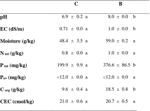
2.3.1 Biochar Effect on Soil Characteristics
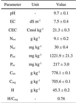
Soil chemical analysis showed that biochar addition resulted in an increase of pH, EC, Ptot and Corg
values, while moisture, Ntot, Pav and CEC were unchanged (Table 2.1).
Table 2.1. Substrates chemical properties. Data represent the mean (n=4) ± standard error. Mean values marked with
the same letter are not statistically different. One-way ANOVA was applied to weigh the effects biochar treatments (𝑝 ≤ 0.05).
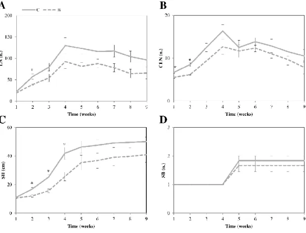
2.3.2 Biochar Effect on Plant Growth
No difference in growth parameters were recorded between non-amended and biochar-amended plants. In detail, LN, CLN, SB and SH values were unchanged (Fig. 2.1) producing no variation
C B pH 6.9 ± 0.2 a 8.0 ± 0.0 b EC (dS/m) 0.71 ± 0.0 a 1.0 ± 0.0 b Moisture (g/kg) 48.4 ± 3.5 a 59.0 ± 0.2 a N tot (g/kg) 0.8 ± 0.0 a 1.0 ± 0.0 a P tot (mg/kg) 199.9 ± 9.9 a 376.6 ± 86.5 b Pav (mg/kg) <12.0 ± 0.0 a <12.0 ± 0.0 a C org (g/kg) 9.6 ± 0.4 a 18.5 ± 0.8 b CEC (cmol/kg) 21.0 ± 0.6 a 20.7 ± 0.5 a
Toward an Understanding of Mechanisms Regulating Plant Response to Biochar Application
23
II
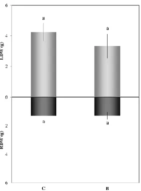
in total biomass accumulation, both as LDW and RDW (Fig. 2.2); chlorophyll and carotenoid contents were also unchanged (data not shown).
Figure 2.1. Morphological analysis. The main plant parameters were analyzed, i.e. LN=leaflets number;
CLN=compound leaves; SB=stem branching; SH=stem height. Data represent the mean (𝑛 = 6) ± standard error. Mean values marked with asterisks are statistically different at ∗𝑝 ≤ 0.05. C= tomato plants grown in non-amended soils (control); B= tomato plants grown in biochar-amended soils.
24
II
Figure 2.2. Leaf and root biomass (g of dry tissue weight). Data represent the mean (𝑛 = 6) ± standard error. Mean values marked with the same letter are not statistically different (𝑝 ≤ 0.05). C= tomato plants grown in non-amended soils (control); B= tomato plants grown in biochar-amended soils.
2.3.3 Effects of Biochar on Leaf Proteome
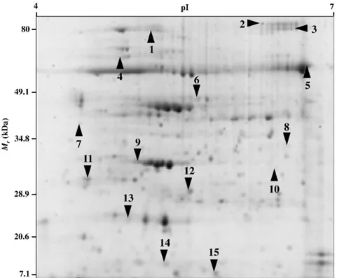
Leaf proteomic maps of tomato plants grown on non-amended and biochar-amended soils contained an average of 300 well-resolved spots, ranging in Mr from about 76 to 12 kDa. We here provide the first proteomic profile of tomato leaves influenced by presence/absence of biochar. These maps were highly reproducible, most spots detected in 2-DE gels showed analogous positions and intensities, as indicated by the degree of gel similarity between the various samples and the reference map.Computer-assisted comparison of 2-DE maps revealed 15 protein spots as differentially represented (P<0.01) among samples, which were subjected to trypsinolysis and further nanoLC-ESI-LIT-MS/MS analysis for protein identification (Fig. 2.3). The list of all identified proteins together with their information and representation profiles are shown in Table 2.
When compared to plants grown on non-amended soil, those grown on biochar-amended soil presented an over-representation of 7 components, namely two isoforms of ribulose
Toward an Understanding of Mechanisms Regulating Plant Response to Biochar Application
25
II
bisphosphate carboxylase/oxygenase activase (RCA; spots 1 and 2), remorin (spot 3), 2-cysteine peroxiredoxin B (spot 7), soluble inorganic pyrophosphatase (PPA6; spot 9), chitinase (spot 12) and a class I heat shock (spot 15), together with a down-representation of 8 components, namely ATP synthase CF1 alpha subunit (spot 4), ribulose bisphosphate carboxylase large chain (rbcL; spot 5), RAB GTPase homolog E1b (spot 6), two isoforms of subtilisin-like protease (spots 8 and 10), oxygen-evolving enhancer protein 2-1 (OEE2; spot 11), embryo defective protein 1241 (fragment; spot 13), and a photosystem I reaction center subunit II-2 (PSAD2; spot 14) (Table 2.2).
Figure 2.3. Reproducible 2-DE maps of tomato leaves (control) showing 15 differentially represented proteins.
26 Spot Acces-sion Gene name Protein description Mascot Score Theor. Mr/pI Exp. Mr/pI Peptides matched Sequences matched Coverage % Functional classification Details classification Protein representation levels 1 K4D489 RCA Ribulose bisphosphate carboxylase/ oxygenase activase 1621 49.2/8.62 77.0/5.16 188 25 51.8 Photosynthesis 'PS.calvin cycle.rubisco interacting' 2 K4D489 RCA Ribulose bisphosphate carboxylase oxygenase activase 1218 49.2/8.62 81.0/6.40 69 23 50.3 Photosynthesis 'PS.calvin cycle.rubisco interacting' 3 Q9XEX 8
rem-1 Remorin family 280 21.8/5.64 79.0/6.68 12 7 28.7 Transcription
'RNA. regulation of transcription.un-classified' 4 A0A0C 5CHA6 atpA ATP synthase CF1 alpha subunit 189 55.4/5.14 67.0/4.84 4 4 10.1 Photosynthesis 'PS.lightreaction. ATPsynthase. alpha subunit' 5 A0A0C 5CHE6 rbcL Ribulose bisphosphate carboxylase large chain
573 52.9/6.55 57.5/6.70 16 11 24.7 Photosynthesis 'PS.calvin cycle.rubisco large subunit' 6 K4C8Q 1 TUFA
RAB GTPase homolog E1b 510 48.9/6.17 44.0/5.60 11 10 24.9 Protein synthesis 'protein.synthesis. elongation' 7 K4D389 BAS1 2-cysteine peroxiredoxin B 157 29.5/6.00 37.0/4.46 5 4 20 Disease/Defense 'redox.peroxire-doxin.BAS1' 0 1000 2000 3000 0 200 400 600 0 1000 2000 3000 0 300 600 900 0 20000 40000 60000 0 1000 2000 3000 0 100 200 300 C B
Toward an Understanding of Mechanisms Regulating Plant Response to Biochar Application
27 8 O04678 SBT1.7 Subtilisin-like protease (fragment) 351 76.4/6.17 34.6/6.60 8 7 12.7 Protein destination and storage 'protein.degrada-tion.subtilases' 9 K4B2L1 PPA6 Soluble inorganic pyrophosphatase 1 271 32.4/6.01 33.0/4.99 11 5 18.6 Metabolism 'nucleotide metabolism.phos-photransfer and pyrophosphata-ses.misc' 10 O04678 SBT1.7 Subtilisin-like protease (fragment) 646 76.4/6.17 33.8/6.51 22 12 24.1 Protein destination and storage 'protein.degrada-tion.subtilases' 11 P29795 PSBP1 Oxygen-evolving enhancer protein 2-1 542 20.2/5.32 30.0/4.52 42 10 43.3 Photosynthesis 'PS.lightreaction.p hotosystem II.PSII polypeptide subunits' 12 Q05539 T15B16. 5
Chitinase family 352 25.0/5.63 27.5/5.56 32 6 38.5 Disease/Defense 'stress.biotic'
13 K4D311 - Embryo defective 1241 (fragment) 297 42.1/4.77 23.6/4.92 6 5 14.3 Protein destination and storage 'protein.folding' 14 P12372 PSAD2 Photosystem I reaction center subunit II-2
107 17.5/9.14 14.8/5.28 4 2 12 Photosynthesis 'PS.lightreaction.p hotosystem I.PSI polypeptide subunits' 0 100 200 0 4000 8000 12000 0 50 100 150 0 1000 2000 3000 0 4000 8000 12000 0 150 300 450 0 100 200 300
28
Table 2.2. Differentially-represented proteins in leaves from tomato plants grown in non-amended soils (C) and tomato plants grown in biochar-amended soils (B).
The list includes: spot number on the reference gel (see Fig. 2.3), hit and accession number, protein description, Mascot Score, theoretical and experimental protein Mr and pI values, peptides and sequences matched, sequence coverage (%), functional and detail classification, and protein representation levels.
15
Q9SYU 8
HSP17. 8
Class I heat shock 363 17.7/5.84 10.0/5.86 17 8 55.2 Disease/Defense 'stress.abiotic.heat'
0 1000 2000 3000
Toward an Understanding of Mechanisms Regulating Plant Response to Biochar Application
29
II II
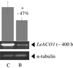
2.3.4 Analysis of LeACO1 Gene Expression Patterns
Results of LeACO1 gene expression analysis showed a lower expression level of this gene and, an indirect lesser ethylene hormone amount, in the leaves of plants grown on biochar-treated soil compared to those grown on non-amended soil (Fig. 2.4).
Figure 2.4. LeACO1 gene expression level. The LeACO1 expression level in leaves of tomato plant grown on
non-amended (C) and biochar-non-amended (B) soil. Three independent biological replicates were run for each tissue, each with two technical replications. Data were normalized to α-tubulin as a loading control. Bars represent the standard error of mean values. Asterisk indicates significant differences at (𝑝 ≤ 0.05).
2.3.5 Detection of the Tomato Late Blight Pathogen: Phytophthora
infestans
Genomic DNA was extracted from tomato leaves to assess the presence/absence of P. infestans pathogen. Molecular analysis showed the presence of highly conserved genomic region PiO8-3-3 that highlighted the presence of P. infestans pathogen in plants grown on biochar-treated soil (Fig. 2.5).
30
II Figure 2.5. Detection of P. infestans. Photos in the upper panel shows representative picture of P. infestans infected
leaves of tomato plant grown on biochar-amended soil. The lower panel shows the detection of P. infestans PiO8-3-3 DNA region in leaves of tomato plant grown on non-amended (C) and biochar-amended (B) soil. Three independent biological replicates were run for each tissue, each with two technical replications. Data were normalized to α-tubulin as a loading control. Bars represent the standard error of mean values.
2.4 Discussion and Conclusions
In the present study, we investigated the almost-unknown effects of biochar amendment on tomato plants by using a multi-target approach. Data showed that the application of biochar to nutrient-poor and neutral soil induced an increase of pH, EC, Ptot and Corg values, leaving unchanged the
CEC, Ntot and Pav counterparts. At the same time, it determined a slight decrease of tomato leaflets
number, compound leaves and stem height only in the first phases of plant growth after the transplant, and not over-time.
It is widely known that plant biomass accumulation is highly dependent on N and P concentration, largely due to the key role of these two macronutrients in a variety of biochemical processes, including photosynthesis, energy metabolism, signal transduction, biosynthesis of macromolecules and protein regulation (Wright et al., 2004; Abbasi and Yousra, 2012; Tavarini
et al., 2015). Under an adequate N amount, it is expected an increased N allocation toward
chloroplast thylakoid membrane proteins and pigment-protein complexes, thus increasing the light-saturated photosynthesis rate. On the other hand, P is essential for plant development and growth, being a major component of nucleic acids, sugar phosphates and phospholipids.
Biochar usually contains N, P and basic cations like Ca2+, Mg2+ and K+ (Major et al., 2010);
thus, directly or indirectly it improves soil nutrients availability and use efficiency (Lehmann and Joseph, 2009). However, depending on feedstock properties, pyrolysis conditions and soil characteristics, biochar has been noted for its ability to retain NH4+-N (Gai et al., 2014), and/or
Toward an Understanding of Mechanisms Regulating Plant Response to Biochar Application
31
II II
NO3-N (Gale et al., 2017). Both biochar and N, as a limiting factor, might be detrimental on P and
K soil content (Chan et al., 2008). The effects of biochar on P availability have been positively proved in acidic soils, whereas its impact on alkaline soils is generally negative due to P sorption or precipitation (Parvage et al., 2013). In fact, alkaline pH and large amount of P sorption sites, such as Ca2+,Mg2+, Al3+ and Fe3+ oxides, which are contained in biochar, may be responsible for P precipitation and insolubilization (Marks et al., 2014).
A limited availability of P generally activates the plant metabolic and developmental processes to maximize inorganic phosphate (Pi) acquisition transport and use (Yang and Finnegan, 2010). In tomato plants grown on biochar-treated soil, proteomic data, reported in our work, reveled an over-representation of PPA6, a member of a class of enzymes that catalyzes the hydrolysis of PPi to produce two orthophosphates (2Pi) and release energy (Heinonen, 2001). Thus, PPA6, through recycling of the pyrophosphate (PPi) deriving from many biosynthetic reactions, may play a role in the plant adaptation to phosphorus deficiency condition. Hernández-Domíguez et al. (2012) demonstrated that, in common bean, Pi deficiency induces an ATP reduction, thereby the PPi hydrolysis rates must increase to compensate the P limitation. Furthermore, transgenic tobacco and potato plants expressing the E. coli ppa1 gene showed a stunted growth mainly due to a decrease in hexose phosphates and PPi content (Sonnewald, 1992). Authors hypothesized that the decrease of cytosolic PPi may reduce the energy gain and, thereby, sugar produced by photosynthesis can be accumulated in source leaves, inhibiting long-distance sucrose transport, and mobile energy source for all plant cells (Gaxiola et al., 2012). Sugar accumulation in leaves determines a reduction in the photosynthetic process probably due to a metabolite feedback regulation, which in turn induces electron transfer and RubisCO amount and activity decrease (Lemoine et al., 2013). This effect was confirmed in tomato biochar-treated plants by down-representation of rbcL and ATP synthase CF1 alpha subunit, as well as by the reduction of the embryo defective protein 1241, chloroplast GrpE protein, important forchloroplast protein import and functionality maintenance (Flexas et al., 2006).In these plants, although the chlorophyll and carotenoid contents resulted unchanged, the reduction of PSAD2 and OEE2 may limit the assembly/functionality of the photosystems I and II, altering the electron transport chain and the photosynthetic machinery.
In biochar amended tomato plants, photosynthesis misbalance may dramatically determine ROS production, as demonstrated by the over-representation of 2-cysteine peroxiredoxin B and a