Research Article
Aerobic Training Prevents Heatstrokes in Calsequestrin-1
Knockout Mice by Reducing Oxidative Stress
Flávia Alessandra Guarnier
,
1,2,3Antonio Michelucci,
1,2Matteo Serano,
1Laura Pietrangelo,
1,2Claudia Pecorai,
1,4Simona Boncompagni
,
1,2and Feliciano Protasi
1,41Center for Research on Ageing and Translational Medicine (CeSI-MeT), University G. d’Annunzio, 66100 Chieti, Italy 2Department of Neuroscience, Imaging, and Clinical Sciences (DNICS), University G. d’Annunzio, 66100 Chieti, Italy 3Department of General Pathology, Londrina State University, 86057-970 Londrina, PR, Brazil
4Department of Medicine and Aging Science (DMSI), University G. d’Annunzio, 66100 Chieti, Italy
Correspondence should be addressed to Flávia Alessandra Guarnier; [email protected]
Received 24 November 2017; Revised 1 February 2018; Accepted 21 February 2018; Published 3 April 2018 Academic Editor: Yong Zhang
Copyright © 2018 Flávia Alessandra Guarnier et al. This is an open access article distributed under the Creative Commons Attribution License, which permits unrestricted use, distribution, and reproduction in any medium, provided the original work is properly cited.
Calsequestrin-1 knockout (CASQ1-null) mice suffer lethal episodes when exposed to strenuous exercise and environmental heat, crises known as exertional/environmental heatstroke (EHS). We previously demonstrated that administration of exogenous antioxidants (N-acetylcysteine and trolox) reduces CASQ1-null mortality during exposure to heat. As aerobic training is known to boost endogenous antioxidant protection, we subjected CASQ1-null mice to treadmill running for 2 months at 60% of their maximal speed for 1 h, 5 times/week. When exposed to heat stress protocol (41°C/1 h), the mortality rate of CASQ1-null mice was significantly reduced compared to untrained animals (86% versus 16%). Protection from heatstrokes was accompanied by a reduced increase in core temperature during the stress protocol and by an increased threshold of response to caffeine of isolated extensor digitorum longus muscles during in vitro contracture test. At cellular and molecular levels, aerobic training (i) improved mitochondrial function while reducing their damage and (ii) lowered calpain activity and lipid peroxidation in membranes isolated from sarcoplasmic reticulum and mitochondria. Based on this evidence, we hypothesize that the protective effect of aerobic training is essentially mediated by a reduction in oxidative stress during exposure of CASQ1-null mice to adverse environmental conditions.
1. Introduction
Hyperthermia is an abnormal rise in body temperature above the hypothalamic set point caused by excessive accumulation of external (environmental) or internal (metabolic) heat. When the core body temperature rises above 40°C, hyper-thermia may result in heatstroke, a life-threatening episode characterized by dysfunction of central nervous system and peripheral organs [1].
Malignant hyperthermia (MH), identified and described for thefirst time in 1960 [2], is an inherited pharmacogenetic disorder that manifests as a life-threatening hypermetabolic response to the administration of volatile anesthetics such as halothane or isofluorane [2, 3]. The main clinical features
of MH crises include uncontrolled muscle contracture, rup-ture of musclefibers (i.e., rhabdomyolysis), increased circu-lating levels of creatine kinase (CK) and K+, and increased oxygen consumption [3]. Interestingly, hyperthermic crises known as exertional/environmental heatstroke (EHS), but virtually identical to anesthetic-induced MH episodes, have also been reported in humans exposed to elevated environ-mental temperatures or strenuous exercise performed in challenging conditions [1, 4–6].
Most of families (70–80%) affected by MH susceptibility present mutations in the RYR1 gene [7], which encodes for a 2200 kDa protein forming the sarcoplasmic reticulum (SR) Ca2+-release channel of skeletal muscle, the ryanodine receptor type-1 (RyR1) [8, 9]. An association between RYR1
Volume 2018, Article ID 4652480, 14 pages https://doi.org/10.1155/2018/4652480
variants and exertional- or heat-induced rhabdomyolysis and sudden death has been reported [10–13]. The correlation between MH and EHS is also supported by evidence col-lected in animal models: (a) in porcine stress syndrome (PSS), swine carrying a point mutation in RYR1 trigger MH episodes in response to halothane administration but also following exposure to either heat or emotional/ physical stress [14, 15]; (b) knock-in mice carrying gain-of-function point mutations in RYR1 linked to MH in humans exhibit heat- and anesthetic-induced MH episodes [16, 17]. In addition, we discovered that male mice lacking calsequestrin-1 (CASQ1-null), the main Ca2+-binding pro-tein located in the lumen of SR terminal cisternae that modulates RyR1 opening probability [18–20], exhibit lethal hyperthermic episodes when exposed to anesthetics, heat, and strenuous exercise [18, 21–23].
The molecular mechanisms underlying rhabdomyolysis of skeletal muscle fibers during MH/EHS crises appear to be complex cascade of events revolving around an increased leak of Ca2+from the mutated RyR1 [24–28] and an excessive production of reactive oxygen/nitrogen species (ROS/RNS) [24]. Michelucci et al. [23] demonstrated that administration of antioxidants (N-acetylcysteine and trolox) protects CASQ1-null mice from anesthetic- and heat-induced lethal crises by reducing mitochondrial production of superoxide anion and global oxidative stress.
Oxidative stress levels in a cell are the net result of pro-duction and removal of oxidative species since the early 80s, it has been demonstrated that aerobic training promotes mitochondrial biogenesis in skeletal muscle while boosting endogenous antioxidant levels [29]. It seems that, although free radical production increases during exercise [30], their rise may act as a signal leading to both upregulation and increased activity of antioxidant enzymes [31–33].
In the present study, we hypothesized that aerobic training, by boosting antioxidant defenses [31–33], could reduce mortality of CASQ1-null mice during hyperthermic crisis. The results collected in this study indicate that training effectively protects CASQ1-null mice from EHS, an effect essentially mediated by a significant reduction in oxidative stress.
2. Materials and Methods
2.1. CASQ1-Null Male Mice. CASQ1-null mice were gener-ated as previously described [27]. All animals used in this study were males, as this gender is more susceptible to MH/ EHS-like crises when exposed to halothane and heat [22]. Mice were housed in microisolator cages at 20°C in a 12 h light/dark cycle, provided free access to water and food. Age-matched C57BL/6 (WT) mice were used as controls. All experiments were conducted according to the Directive of the European Union 2010/63/UE and were approved by the Italian Ministry of Health (1199/2015-PR).
2.2. In Vivo Experiments
2.2.1. Incremental Test. This protocol consisted of a single session of exercise on a treadmill with no incline, according
to the protocol described by Cunha et al. [34] and Gladden and Hogan [35], performed at room temperature of 20 ± 2°C. A mild electrical stimulus (0.5 mA) was applied to
mice that stepped off the treadmill to keep them exercising. The test started with a warm-up of 10 min at a speed of 6 m/min. The speed of the treadmill was then increased by 3 m/min (from 6 to 39 m/min) every 3 min until exhaustion, defined as the time when the mice were no longer able to maintain regular gait. Workloads corresponding to 85 and 60% of peak workload were determined for each mouse. 2.2.2. Constant Load Test and Lactate Measurements. This protocol consisted of a warm-up period of 10 min at 6 m/ min followed by a 28 min constant load running on a tread-mill with no incline (at 85% of maximal speed reached in the incremental test) [33], performed at room temperature of 20± 2°C. Blood samples (~50 μL) were collected from the tail vein while mice were kept running, every 7 min. Blood was then transferred to 1.5 mL microtubes, centrifuged at 3000×g for 15 min at 4°C for plasma separation, and stored in 200μL microtubes at −20°C. The lactate concentration in the blood was analyzed in plasma using a colorimetric enzy-matic assay kit (Lactate Assay Kit II; Sigma-Aldrich®, St. Louis, MO, USA), following the manufacturer’s instructions. The lactate concentration in the colorimetric reaction was measured spectrophotometrically at 450 nm (Spectra MAX 190; Molecular Devices, Sunnyvale, CA, USA) and expressed as mmol of lactate/L.
2.2.3. Grip Strength Test. The strength developed by mice during instinctive grasp was measured as previously described [23, 36, 37]. Briefly, mice were held by the tail and allowed to grasp a metal grating connected to a Shimpo Fgy 0.5X transducer (Metrotec Group, Spain). Once the mouse had firmly grasped the grating, a steady and gentle pull was exerted on the tail. Measurements of the peak force generated by each mouse using forelimbs were repeated three times with appropriate intervals (about 30 s) to avoid fatigue. Average peak force values were then normalized to the total body mass.
2.2.4. Aerobic Training Protocol. WT (n = 7) and CASQ1-null (n = 21) mice were enrolled in the study at 2 months of age after weight and grip strength were measured. Each mouse was then (a) accustomed for 5 days to treadmill run-ning (Columbus Instruments, Columbus, OH, USA), (b) subjected to the incremental test (see above), and (c) ran-domly assigned to the CASQ1-null training (n = 7) or to the control groups (n = 14). The training group of CASQ1-null mice was subjected to aerobic training [33, 34] on the treadmill at 60% of their individual maximal speed reached during the incremental test. After 4 weeks, trained CASQ1-null mice performed a new incremental test to readjust the training load and guarantee the 60% of workload in the fol-lowing 4 weeks. At the end of 8 weeks of training, body weight and grip strength were reassessed for all mice (WT and CASQ1-null control and trained), which were also reevaluated with the incremental test. After 2 days of rest, all animals additionally performed a constant load test (see
above) with blood collection, to measure the lactate accumu-lation/removal ratio. A detailed overview of experimental protocol is shown in Figure 1.
2.2.5. Heat Stress Protocol and Core Temperature Recording. Three days after the constant load test (see above), all mice were subjected to a heat stress protocol. Animals were placed in an environmental chamber in which temperature was maintained at 41°C, 1 h [21]. During exposure to heat, core body temperature was measured using a rectal thermometer taped to the tail of the animals and recorded every 5 min throughout the duration of heat challenge. Breathing and spasmodic contractions were visually monitored, while mus-cle rigidity was manually confirmed by limb resistance imme-diately after animal death. Surviving animals were returned to normal housing conditions and monitored for 24 h to assess possible delayed deaths. After 3 days, the mice that survived from the heat stress protocol were sacrificed, and muscle sam-ples collected and processed for further analysis.
2.3. Ex Vivo and In Vitro Experiments
2.3.1. In Vitro Contracture Test (IVCT) in (EDL) Muscles. Intact EDL muscles were dissected from hind limbs of mice, placed in a dish containing Krebs-Henseleit solution, and pinned and tied withfine silk sutures at each end. Muscles were then mounted vertically between two platinum elec-trodes immersed in an organ chamber filled with Krebs-Henseleit solution and attached to a servomotor and force transducer (model 1200A; Aurora Scientific, Aurora, ON, Canada). Before starting the experimental protocol, stimula-tion level and optimal muscle length (L0) were determined using a series of 80 Hz train stimulus in order to adjust the muscle to the length that generated maximal force (F0). Dur-ing the experiments, temperature was kept constant at 25°C. To determine caffeine sensitivity of resting tension, EDL muscles were subjected to an in vitro contracture test (IVCT) as previously described [23]. Briefly, isolated EDL muscles were continuously stimulated at 0.2 Hz at 23–25°C, caffeine
concentration in the bath was changed every 3 minutes (no wash between applications) as follows: 2, 4, 6, 8, 10, 14, 18, and 22 mM. Muscle basal tension was measured at the end of each step of caffeine application and reported both as specific and relative force. Specific force was calculated by normalizing the absolute force to the cross-sectional area of the muscle.
2.3.2. Preparation of Total Homogenates and Isolation of Mitochondria and SR Membranes. Total homogenates were prepared by placing muscles tibialis anterior (TA) and gas-trocnemius on ice in an Ultra-Turrax homogenizer (2× 30 s at 14,500 rpm) containing 50 mg/mL of tissue in 10 mM KH2PO4/K2HPO4 buffer and 120 mM KCl at pH 7.4 [38].
Total homogenate preparations were used for assessment of total diene conjugates production, protein carbonylation, and calpain activity assays (see below).
Mitochondria and SR membranes were isolated from gastrocnemius to assay-specific organelle lipid peroxidation. Samples were placed in an Ultra-Turrax homogenizer (2× 30 s at 14,500 rpm) in 5 vol of 30 mM KH2PO4, 5 mM EDTA, 3.0 M sucrose, 0.5 mM dithiothreitol, 0.3 mM phenyl-sulfonylfluoride, and 1% (v/v), 1 μM leupeptin, 1 μM pepsta-tin (pH 6.8). All steps for isolation were performed at 4°C. Mitochondrial fraction was prepared from the total homoge-nates by differential centrifugation as previously described [39]. The supernatant from afirst homogenate centrifugation (1000×g for 10 min) was centrifuged at 14,000 ×g for 35 min. The pellet was then suspended in 30 mM imidazole, 60 mM KCl, and 2 mM MgCl2 (pH 7.0) and stored at −80°C until
use. This resuspension was then used to assay cytochrome c oxidase activity and lipid peroxidation in mitochondrial membranes. Microsomes vesicles were prepared by isoelec-tric precipitation from the supernatant of mitochondrial iso-lation as previously described [40, 41]. Sodium acetate (1 mM) was added to the samples until the pH was 4.0 and then centrifuged at 10,000×g for 10 min. The supernatants were discharged, and pellets were resuspended in the same volume that was initially used, of KCl 1.15% glycerol (4 : 1 v/v). The pellets were mechanically broken using a vor-tex and then centrifuged again in 10,000×g for 10 min. The final pellet was resuspended in 100 mM KH2PO4and glycerol
(4 : 1 v/v), mixed using a vortex, and stored in microtubes at −80°C. For Ca2+
-dependent ATPase activity, the pellets were resuspended in 3% polyethylene glycol, 5 mM azide, 80 mM KCl, and 0.1 mM ouabain (pH 7.5). Total proteins were quantified by the method of Bradford [42], using bovine serum as a standard.
2.3.3. Determination of Ca2+ ATPase Activity. Activity of sarco/endoplasmic Ca2+ ATPase (SERCA), the main SR Ca2+ pump of skeletal muscle, was estimated in SR iso-lated membranes from gastrocnemius by using a colori-metric assay that quantifies the amount of inorganic phosphate (Pi) that complexes with ammonium molybdate
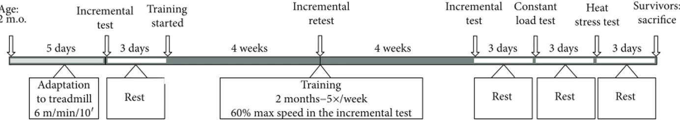
Age: 2 m.o. 5 days 3 days Incremental test Incremental test Constant load test Heat stress test Survivors: sacrifice Incremental retest Training started
4 weeks 4 weeks 3 days 3 days 3 days
Adaptation to treadmill 6 m/min/10′ Rest Training 2 months−5×/week 60% max speed in the incremental test
Rest Rest Rest
Figure 1: Schematic view of the experimental protocol. Two months old CASQ1-null male mice were randomly selected to perform 2 months of aerobic training. Control WT and untrained (control) CASQ1-null mice were always subjected to identical procedures, without the aerobic training. m.o.: months old.
and malachite green following release from SERCA-mediated ATP hydrolysis [43, 44]. Briefly, the reagent to quantify Pi was prepared by mixing 1 vol of 10% (w/v)
ammonium molybdate in 4 M HCl with 3 vol 0.2% (w/v) malachite green in 4 M HCl. The reaction medium, com-posed by 2 mM EDTA, 10 mM CaCl2, 2 mM MgCl2, and 2 mM ATP, was mixed to malachite green/ammonium molybdate dye reagent, and then the reaction was started with the addition of 300μg protein/mL of membranes resus-pension. When Piwas complexed with ammonium
molyb-date and malachite green in 4 M HCl, it creates a green color which was quantified by reading the absorbance spec-trophotometrically at 660 nm and compared to a standard curve of known Piconcentrations (0–15 nmols of PO4−2from
NaPO4). The color formation was monitored for 5 min, the Pi concentration at every minute was calculated using a stan-dard curve, and the differences are considered the activity of Ca2+ATPase pump [44]. All the reactions were repeated in the presence of thapsigargin (100 nM), an inhibitor of the SERCA family of Ca2+ pumps [45], to differentiate SERCA activity from any other ATP-dependent activity that could interfere with the measurement.
2.3.4. Determination of Calpain Activity. The activity of cal-pain [46, 47] was measured in total homogenates from gas-trocnemius muscle, by a chemiluminescence assay using a calpain protease assay kit (Calpain-Glo Protease Assay®; Promega, Madison, WI, USA). The assay provides a prolumi-nescent calpain substrate, in a buffer system optimized for calpain and luciferase activities. During the assay, calpain cleavage of the substrate generates a glow-type luminescent signal produced by the luciferase reaction. In this homoge-neous, coupled-enzyme format, the signal is proportional to the amount of calpain activity present in the sample [48]. Muscle homogenates were prepared as described above, diluted to a concentration of 6.25 mg/mL in 10 mM KH2PO4 buffer, pH 7.4 in 0.9% NaCl, and finally processed according to the manufacturer’s instructions. Results were expressed as calpain activity/mg of muscle tissue.
2.3.5. Preparation of Samples for Electron Microscopy (EM). EDL muscles were dissected from sacrificed animals, pinned on a Sylgard dish, fixed at room temperature with 3.5% glutaraldehyde in 0.1 M sodium cacodylate (NaCaCo) buffer (pH 7.2), and then stored in the fixative at 4°C. Small bundles of fixed tissue were then postfixed, embed-ded, stained en bloc, and sectioned for EM as described previously [49]. Ultrathin sections (~50 nm) were then cut in a Leica Ultracut R microtome (Leica Microsystem, Austria) using a Diatome diamond knife (Diatome Ltd. CH-2501 Biel, Switzerland). Sections were examined at 60 kV (after double staining with uranyl acetate and lead citrate) with a FP 505 Morgagni Series 268D electron
microscope (FEI Company, Brno, Czech Republic),
equipped with a Megaview III digital camera (Munster, Germany) and Soft Imaging System (Munster, Germany). 2.3.6. Quantitative EM Analysis of Mitochondrial Volume and Damage. (A) Mitochondrial volume was determined
using the well-established stereology point-counting tech-nique [50, 51] in EM micrographs collected from transverse sections of samples at 8900× magnification. Briefly, after superimposing an orthogonal array of dots at a spacing of 0.20μm to the electron micrographs, the ratio between num-bers of dots falling within mitochondrial profiles and total number of dots covering the whole image was used to calcu-late the relativefiber volume occupied by mitochondria. (B) In the same set of micrographs, the number of severely dam-aged mitochondria was evaluated and reported as percentage of the total number. Mitochondria with one of the following ultrastructural alterations were classified as severely dam-aged: (a) presenting disruption of the external membrane, (b) presence of internal vacuolization and/or disrupted inter-nal cristae, and (c) containing myelinfigures.
2.3.7. Cytochrome c Oxidase Activity. Cytochrome c oxidase activity was measured in isolated mitochondria from gastroc-nemius muscles, by a colorimetric assay kit (Cytochrome c Oxidase Assay Kit; Sigma-Aldrich, St. Louis, MO, USA) based on the observation of the decrease in absorbance at 550 nm of ferrocytochrome c caused by its oxidation to ferri-cytochrome c by ferri-cytochrome c oxidase [52]. All samples used in the assay had the same amount of protein (300μg protein/ mL), and the results were expressed as cytochrome c oxidase activity (U/mL× 10−3).
2.3.8. Oxidative Stress Measurements. Protein carbonyl group formation is a classic and immediate biomarker of oxidative modification to proteins [53, 54]. Here, carbonyl protein content was measured with a modified version of a protocol previously described [53]. Briefly, TA (50 mg/mL) was homogenized in 50 mM of phosphate buffer, 1 mM ethylene-diaminetetraacetic acid, pH 7.4. Samples were then centri-fuged at 600×g for 10 min at 4°C. A volume of 200μL of 2,4-dinitrophenylhydrazine (DNPH) was added to 200μL of supernatant and incubated at room temperature. After 30 min of incubation, 100% trichloroacetic acid (TCA) was added and samples were placed on ice for 5 min and then spinned at maximal speed for 2 min. Supernatants were dis-carded, while pellets were washed in cold acetone and placed at−20°C for 5 min. Then, acetone was carefully removed, and pellets were resuspended in 0.5 mL 6 M guanidine hydrochlo-ride to be spectrophotometrically read at 375 nm. To calcu-late the protein carbonyl content, the following formula was used: C = [(OD 375 nm)/6.364× 100] nmol/well, where 6.364 is the extinction coefficient using the enclosed 96-well plates in mM (=22 mM−1 cm−1× 0.2893 cm path length in well). Results were expressed as nmol carbonyl/mg of total protein, which were quantified in each sample at 280 nm.
Oxidation of fatty acids forms conjugated dienes that absorb UV light at 230 to 235 nm. Measurement of dienes is a useful index of peroxidation in pure lipids or isolated lipoproteins and has the advantage that it measures early stages in peroxidation [55]. Muscle homogenate and SR ves-icles were dispensed in concentration of 20μg/mL protein in solution with 10 mM phosphate buffer containing 1% Lubrol [56]. The absorption spectrum was then recorded. The rate of conjugated diene formation was estimated according to the
lipid oxidation index, A233nm/A215nm, which provides a sensi-tive method for determination of lipid peroxidation [57]. 2.4. Statistical Analysis. The statistical analysis is reported in the legend of eachfigure. Mean ± SD was used when variabil-ity representative values are needed (higher number of sam-ples/animals), while mean± SEM was used to represent precision of a measurement (lower number of samples/ animals).
3. Results
3.1. Aerobic Training Increased Functional Output and Aerobic Capacity of CASQ1-Null Mice. Before the 2 months training protocol, and two days after the last training session, body weight and grip strength were evaluated in each mouse (Table 1). Body weight between WT and CASQ1-null mice, although slightly lower in the latter at both 2 and 4 months of age, was not significantly different. Significant differences were observed for the functional output, as the grip strength test revealed that untrained CASQ1-null mice were about 60% weaker than age-matched WT at both 2 and 4 months of age. On the other hand, the maximum speed reached dur-ing the incremental test was surprisdur-ingly higher in CASQ1-null mice: this last result is likely due to the increased number and volume of mitochondria in CASQ1-null muscles (see below; see also [27]). Two months of aerobic training (2 to 4 months of age; see Figure 1), while did not change the aver-age body weight of CASQ1-null mice, significantly improved both functional parameters: (a) the grip strength output nor-malized by weight increased from 3.0± 0.8 g/g of body weight in untrained CASQ1-null to 4.8± 1.1 g/g of body weight in trained CASQ1-null and (b) the maximum speed reached during the incremental test was slightly higher in trained than in untrained CASQ1-null mice, respectively, 32.5± 3.9 versus 28.1± 2.7 m/min.
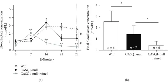
As lactate accumulation/removal ratio is considered a good marker of aerobic capacity [58, 59], we subjected all mice at 4 months of age to a 28-minute constant load test (at 85% of the maximal speed reached during the incremental test) and measured the lactate accumulation in the blood-stream every 7 minutes (Figure 2(a)). WT mice displayed a typical lactate accumulation curve, increasing from baseline (0.09± 0.30 mmol/L) to a peak at the 14th minute of 2.87
± 0.90 mmol/L, followed by a plateau. On the other hand, the lactate levels in the bloodstream in untrained CASQ1-null mice (1.11± 0.46 at baseline) reached a peak of 3.38 ± 1.10 mmol/L at the 14th minute, but then declined in the second part of the experiment. This decay was even greater in trained CASQ1-null mice, with a peak of 2.10 ± 0.73 mmol/L at 7th minute followed by a constant decline. Indeed, the blood lactate concentration at the end of the con-stant load test (28th minute; dashed line in Figure 2(a)) exhibited a reduction compared to WT mice of, respectively, ~39% and ~70% in untrained and trained CASQ1-null animals (Figure 2(b)).
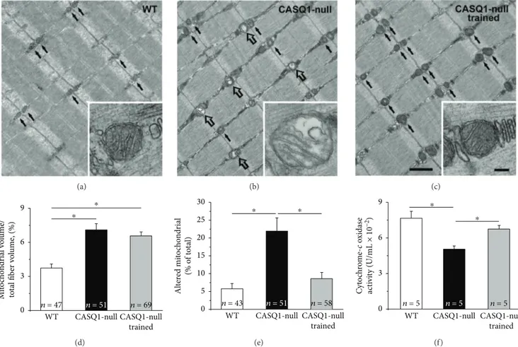
3.2. Aerobic Training Decreased Mitochondrial Damage and Improved Mitochondrial Function in CASQ1-Null Mice. In EDLfibers of WT mice, mitochondria are usually positioned at the I band in proximity of Z lines (Figures 3(a)–3(c) black arrows; see [60]). Healthy mitochondria usually exhibit an electron dense dark matrix (inset in Figure 3(a)), while when damaged, they would appear swollen and with a clear matrix [61]. Afirst qualitative assessment suggested that damaged mitochondria were more numerous in untrained CASQ1-null than in WT mice (Figure 3(b), empty arrows), but again rare in trained CASQ1-null mice (Figure 3(c)). The visual observations were supported by the quantitative analysis: (i) the relative volume occupied by mitochondria and (ii) the percentage of mitochondria presenting structural alterations were significantly higher in untrained CASQ1-null (6.6 ± 0.6% and 21.6 ± 3.8%) than in WT mice (3.9 ± 0.4% and 5.8± 1.4%) (Figures 3(d) and 3(e)). On the other hand, in trained CASQ1-null mice, while the relative volume occupied by mitochondria did not change significantly compared to untrained CASQ1-null mice (6.2± 0.6% versus 6.6 ± 0.6%), the percentage of damaged mitochondria was significantly reduced to a value close to that observed in WT muscles (8.6± 1.4% versus 5.8 ± 1.4%; see above) (Figures 3(d) and 3(e)). The structural improvement of mitochondrial struc-ture would also suggest improved functional properties of mitochondria. Indeed, when we measured cytochrome c oxi-dase activity, we found that it was decreased in untrained CASQ1-null mice (0.05± 0.006 U/mL) compared to WT (0.076± 0.01 U/mL), but rescued to values closer to those of WT following training (0.067± 0.006 U/mL; Figure 3(f)). The data collected in the analysis of mitochondria could have Table 1: Body weight and functional output of mice before and after training.
Pretraining (2 months of age) Posttraining (4 months of age) WT (n = 7) CASQ1-null (n = 21) Untrained Trained WT (n = 7) CASQ1-null (n = 14) CASQ1-null (n = 7) Body weight (g) 24.6± 2.9 23.6± 2.5 29.9± 2.8 27.0± 2.9 25.7± 3.0
Grip strength normalized (peak force g/g weight) 9.7± 1.5 3.9± 1.6∗ 7.9± 1.7 3.0± 0.8† 4.8± 1.1# Max. speed in the incremental test (m/min) 24.8± 3.1 27.8± 3.0∗ 24.0± 2.1 28.1± 2.7† 32.5± 3.9#
Data are shown as mean± SD.∗p < 0 05 compared to age-matched WT, as evaluated by two-tailed unpaired Student’s t-test for the 95% confidence intervals; †p < 0 05 when compared to WT at 4 months, and#p < 0 05 compared to untrained CASQ1-null, as evaluated by one-way ANOVA followed by Tukey’s post
implications for (i) the improved functional output (Table 1) and (ii) the higher capability of trained CASQ1-null mice to remove lactate (Figure 2).
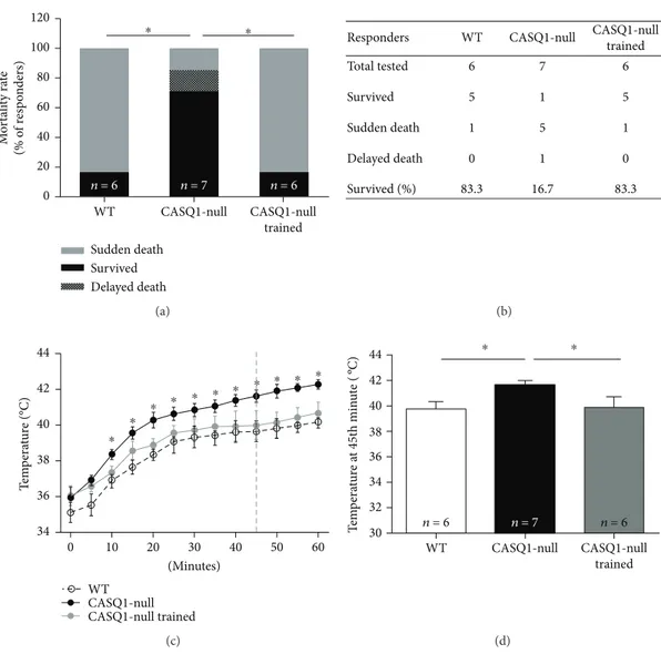
3.3. Aerobic Training Protects Male CASQ1-Null Mice from Heat-Induced Sudden Death by Reducing Hyperthermia. Three days after the constant load test, all mice were subjected to a heat challenge in an environmental chamber (41°C for 1 h). Consistent with previous publications [21, 23], mortality rate during the heat stress protocol was signi fi-cantly higher in CASQ1-null than in WT mice: 86% versus 18%, respectively (Figures 4(a) and 4(b)). Though, the mortal-ity rate of CASQ1-null mice was reduced to only 16% after two months of aerobic training (Figures 4(a) and 4(b)). As a typical MH crisis is characterized by an abnormal rise in body tem-perature, namely, hyperthermia [1], we also monitored the core temperature throughout the entire duration of the heat challenge. Whereas core temperature raised in all animals (including WT) during the experiment, this increase was sig-nificantly greater in untrained CASQ1-null mice than in the other two groups of animals tested (Figure 4(c)). Specifically, the temperature calculated at 45th minute of the protocol, that is the average time-to-onset of hyperthermic crises in CASQ1-null mice [18, 23], was (i) on average 1.85°C higher in untrained CASQ1-null than in WT mice (Figure 4(d)) and (ii) on average 1.79°C lower in trained CASQ1-null than in untrained CASQ1-null mice, close to the temperature observed in WT animals (Figure 4(d)).
3.4. Aerobic Training Normalizes IVCT in CASQ1-Null EDL Muscles. Intact EDL muscles were dissected from all mice and subjected to IVCT, the gold standard for the diagnosis of MH susceptibility in humans [62, 63]. Basal force of EDL
muscles was measured during exposure to increasing con-centrations of caffeine, a potent agonist of RyR1 that triggers release of Ca2+from the SR. Force is displayed in Figures 5(a) and 5(b) as absolute and relative basal tension, respectively. While no differences in the specific basal tension were recorded among the three groups of animals in the absence of caffeine (7.9 ± 0.4 mN/mm2, 7.8± 0.2 mN/mm2, and 8.0
± 0.4 mN/mm2 for WT untrained and trained CASQ1-null
mice, resp.), when exposed to the IVCT protocol, EDL muscles from untrained CASQ1-null mice exhibited a greater caffeine sensitivity compared to those observed from WT (Figure 5). Specifically, EDL muscles from untrained CASQ1-null mice started to develop tension already at 10 mM and reached a final tension value of 12.5 ± 0.4 mN/ mm2at 22 mM of caffeine. Two months of aerobic training normalized the responsiveness to caffeine of CASQ1-null EDL muscles to values similar to that observed in WT: at 22 mM of caffeine, the specific basal tension reached by EDL muscles from WT and trained CASQ1-null mice was 9.7± 0.3 mN/mm2 and 10.4± 0.3 mN/mm2, respectively (Figure 5(a)). In Figure 5(b), force is displayed as relative basal tension normalized to control conditions (no caffeine) during the exposure to increasing concentrations of caffeine. 3.5. Calpain Activity Was Reduced in Muscles of Trained CASQ1-Null Mice. Cytosolic Ca2+ levels have been shown to be slightly elevated in CASQ1-null fibers [18, 21, 23] and may lead to an increased need of Ca2+ removal from the cytosol [37, 64]. For this reason, we measured SERCA activity in microsomal membranes of gastrocnemius mus-cles, estimated as production of Pi in the presence of ATP (Figure 6(a)). Our results showed that the rate of Pi
gener-ated in untrained CASQ1-null samples was significantly WT CASQ1-null CASQ1-null trained ⁎⁎ ⁎⁎ ⁎⁎ ⁎⁎ ⁎ ⁎ # # 7 14 21 28 0 (Minutes) 0 1 2 3 4 5 B lo o d lac ta te co ncen tra tio n (mmo l/L) (a) n = 6 n = 7 n = 6 ⁎ ⁎ 0 1 2 3 4 Final b lo o d lac ta te co ncen tra tio n (mmo l/L) WT CASQ1-null trained CASQ1-null (b)
Figure 2: Accumulation of lactate in the bloodstream during constant load test. (a) Lactate accumulation in the blood during a 28-minute constant load test (at 85% of maximal speed reached during the incremental test). The vertical dashed line represents the time point in whichfinal blood lactate concentration (plotted in panel (b)) was evaluated. #p < 0 05, as evaluated by two-way ANOVA (differences among paired curves); ∗p < 0 05, CASQ1-null versus WT, and ∗∗p < 0 05, CASQ1-null versus CASQ1-null trained, as evaluated by Bonferroni’s post hoc test (indicating differences among the same time point). (b) Average blood lactate concentration at the end of incremental test (28th minute; dashed line in panel (a)). Data are given as mean± SD; ∗p < 0 05, as evaluated by one-way ANOVA followed by Tukey’s post hoc test. n = number of mice.
higher (32%) compared to that generated in WT. To demon-strate that this Pi generation was specifically dependent on
the activity of SERCA, the same microsomes were treated with thapsigargin (TG, 100 nM), a noncompetitive inhibitor of SERCA [45]: after TG treatment, all samples exhibited a signif-icant decay to~1.0 nmol Pi∙mg protein−1∙min−1, supporting
the view of a different rate of SERCA activity in WT versus CASQ1-null fibers (Figure 6(a)). Though, SERCA activity was not reduced by aerobic training in CASQ1-null muscle. However, as increased levels of cytosolic Ca2+may also result in an enhanced activation of proteolysis pathways [46], we also measured the total calpain activity, one of the most important nonlysosomal classes of proteases in skeletal muscle fibers, which is activated by chronic high Ca2+levels [46, 65]. In this case, calpain activity, measured in total homogenates of gas-trocnemius muscles, that was 2.7-fold higher in untrained CASQ1-null than WT specimens, was significantly lowered in trained CASQ1-null samples (Figure 6(b)).
3.6. Oxidative Stress Was Reduced by Aerobic Training in Muscles of CASQ1-Null Mice. Oxidative stress has been
shown to be elevated in CASQ1-null muscles [23]. To determine if aerobic exercise improved the capabilities of skeletal muscle to reduce oxidative damage, we evaluated (i) levels of carbonyl proteins (carbonyl protein content is an important biomarker of oxidative modification of proteins [53, 54]) and (ii) diene conjugates in total homogenates of TA muscle and in SR-and-mitochondria membranes of gastrocnemius muscles (diene conjugates are a by-product of lipid peroxidation chain, which is an indicator of structural oxidative modification to mem-branes [55]). The results obtained from these experiments revealed that (a) total carbonyl protein content was abnor-mally elevated in total muscle homogenates of untrained CASQ1-null compared to that of WT mice, while lowered of about 39% in muscles of trained CASQ1-null mice (Figure 7(a)); (b) levels of diene conjugates were signifi-cantly increased either in total homogenates or in isolated mitochondria and SR membranes obtained from untrained CASQ1-null compared to WT muscles but reduced of about 40% in all three preparations obtained from trained CASQ1-null muscles (Figures 7(b)–7(d)).
(a) (b) (c) WT CASQ1-null trained CASQ1-null ⁎ ⁎ n = 51 n = 69 n = 47 0 3 6 9 M it o ch o n dr ial v o lu me/ to ta l fib er v o lu me , (%) (d) WT CASQ1-null trained CASQ1-null ⁎ ⁎ n = 51 n = 58 n = 43 0 5 10 15 20 25 30 Al te re d mi to ch o n dr ial (% o f t o ta l) (e) WT CASQ1-null trained CASQ1-null ⁎ ⁎ n = 5 n = 5 n = 5 0 3 6 9 Cy to ch rom e-c ox id as e ac ti vi ty (U/mL × 10 −2 ) (f)
Figure 3: Structural and quantitative analysis of mitochondria. (a–c) Representative electron micrographs (longitudinal sections) of EDL fibers where small black arrows point to healthy mitochondria (see enlargements in (a) and (c)), while empty arrows point to damaged mitochondria (see enlargement in (b)). Scale bars in (a–c), 1 μm; insets, 0.1 μm. (d) Quantitative analysis of the relative fiber volume occupied by mitochondria. (e) Quantitative analysis of damaged mitochondria calculated as percentage of total. (f) Cytochrome c oxidase activity assayed in TA muscles. Data in (d) and (e) are shown as mean± SEM;∗p < 0 05, as evaluated by two-tailed unpaired Student’s t-test for the 95% confidence intervals. Data in (f) are shown as mean ± SD; ∗p < 0 05, as evaluated by one-way ANOVA followed by Tukey’s post hoc test. n = number of fibers analyzed (in panels (d) and (e)) and number of mice (in panel (f)).
4. Discussion
In the last ten years, compelling evidence has been collected in animal models to demonstrate that EHS shares common molecular mechanisms with classic MH susceptibility, the reaction caused by exposure to halogenated anesthetics. Indeed, we and others have shown that knock-in mice carry-ing mutations in RYR1 linked to MH in humans (RYR1Y522S/
WT mice) and CASQ1-null mice trigger lethal crises when
exposed to anesthetics, heat, and also physical exertion [16– 18, 21]. The molecular events leading to rhabdomyolysis of skeletal fibers during hyperthermic crises in both mouse models involve Ca2+leak from RyR1 and excessive produc-tion of ROS/RNS, which then feeds a feedforward cycle that promotes additional SR Ca2+release [24]. We demonstrated that this cycle can be interrupted by administration of
antioxidants (N-acetylcysteine and trolox) [23, 24]. Here, we tested if aerobic training (Figure 1) can break the vicious cycle triggered by overproduction of ROS/RNS by boosting endogenous antioxidant defenses and, hence, prevent heat-strokes in CASQ1-null mice exposed to high environmental temperatures.
4.1. Main Findings of the Study. Our results show that aero-bic training (a) protects mice from hyperthermic episodes, lowering both the increase in core temperature in vivo and the responsiveness of intact EDL muscles during IVCT (Figures 4 and 5), and (b) reduces oxidative stress, hence reducing mitochondrial damage (Figures 3, 6, and 7). Additionally, we also show that aerobic training had improved in vivo muscle performance, as shown by higher
grip strength and maximal speed reached in the
Sudden death Survived Delayed death CASQ1-null trained CASQ1-null WT n = 6 n = 7 n = 6 0 20 40 60 80 100 120 M o rt ali ty ra te (% o f r esp o n der s) ⁎ ⁎ (a)
Responders WT CASQ1-null CASQ1-null trained 6 5 1 0 83.3 7 1 5 1 16.7 6 5 1 0 83.3 Total tested Survived Sudden death Delayed death Survived (%) (b) WT CASQ1-null CASQ1-null trained ⁎ ⁎ ⁎ ⁎ ⁎ ⁎ ⁎ ⁎ ⁎ ⁎ ⁎ 34 36 38 40 42 44 T em p era tur e (°C) 10 20 30 40 50 60 0 (Minutes) (c) 44 42 40 38 36 34 32 30 WT n = 6 n = 7 n = 6 CASQ1-null CASQ1-null trained T em p era tur e a t 45t h min u te ( ºC) ⁎ ⁎ (d)
Figure 4: Mortality rate and measurements of core temperature in mice exposed to heat stress. (a) Incidence of sudden and delayed deaths (i.e., within 24 h after challenge) following exposure to the heat stress protocol (41°C for 1 h);∗p < 0 05, as evaluated by two-tailed Fisher’s exact test. (b) Table showing (i) the number of mice tested during the heat challenge and (ii) the experimental outcome. (c) Increase in absolute core temperature, recorded every 5 minutes during exposure to the heat stress protocol: dashed vertical line represents the mean time-to-onset of lethal crises in CASQ1-null mice; ∗p < 0 05, trained CASQ1-null versus both WT and trained CASQ1-null mice, as evaluated by two-way ANOVA followed by Bonferroni’s post hoc test. (d) Average core temperature in mice at 45th minute of the heat stress protocol (dashed line in panel (c)). Data in (c) and (d) are given as mean± SD; ∗p < 0 05, as evaluated by one-way ANOVA followed by Tukey’s post hoc test. n = number of mice (in panel (d)).
incremental test and by the reduced lactate accumulation in the bloodstream during the constant load test (Table 1 and Figure 2). These main points are discussed below in the same order.
4.1.1. Protection from Lethal EHS Episodes. The training program in CASQ1-null mice resulted in a striking protec-tion from heat-induced lethal episodes, with the survival rate raising from 17% in untrained to 83% in trained CASQ1-null
mice (Figure 4). The protective effect of aerobic training is likely the direct result of (i) a reduced rise in core tempera-ture (i.e., hyperthermia) in trained versus untrained CASQ1-null animals (Figure 4) and (ii) a normalized IVCT (Figure 5), which is an indirect, but quite reliable, measure-ment of Ca2+handling, as muscle tension directly correlates with intracellular Ca2+concentration. The fact that intracel-lular Ca2+ levels are slightly elevated in CASQ1-null mice has been demonstrated and discussed in depth in several WT (n = 5) CASQ1-null (n = 5) CASQ1-null trained (n = 5) ⁎ ⁎ ⁎ 4 8 12 16 20 24 0 Caffeine (mM) 8 10 12 14 Sp ecific bas al t en sio n (mN/mm 2) (a) WT (n = 5) CASQ1-null (n = 5) CASQ1-null trained (n = 5) ⁎ ⁎ ⁎ 4 8 12 16 20 24 0 Caffeine (mM) 1.0 1.2 1.4 1.6 1.8 Rela ti ve bas al t en sio n (no rmalized t o 0 ca ff eine) (b)
Figure 5: Caffeine dependence of basal tension in isolated EDL muscles during IVCT. (a) Specific basal tension of intact EDL muscles, recorded during exposure to increasing concentrations of caffeine. (b) Relative basal tension normalized to control conditions (no caffeine), during exposure to increasing concentrations of caffeine (same EDL muscles shown in panel (a)). Data are given as means ± SEM;∗p < 0 05, as evaluated by ANOVA repeated measures, followed by Tukey’s post hoc test for the pairwise comparisons. n = number
of mice tested. 6 5 4 3 2 1 0 n = 6 n = 6 n = 7 n = 7 n = 6 n = 6 TG WT TG CASQ1-null TG CASQ1-null trained nmo l P i. m g p ro tein −1. min −1 ⁎ ⁎ ⁎ ⁎ (a) n = 6 n = 7 n = 6 WT CASQ1-null CASQ1-null trained ⁎ ⁎ 20,000 15,000 10,000 5,000 0 C al p ain ac ti vi ty/m g tissue (b)
Figure 6: Measurements of SERCA and calpain activities. (a) SERCA activity assayed in microsomes isolated from gastrocnemius muscles in the absence and presence of 100 nM of thapsigargin (TG). (b) Calpain activity assayed in gastrocnemius muscles total homogenates. Data are given as mean± SD;∗p < 0 05, as evaluated by one-way ANOVA followed by Tukey’s post hoc test. n = number of mice.
previous publications [18, 21, 23, 61]. The elevated intracel-lular Ca2+levels in untrained CASQ1-null muscles are here underlined both by IVCT (see above) and by increased SERCA and calpain activities (Figure 6). Indeed, both SERCA and calpain have been demonstrated to have their activities increased under conditions in which myoplasmic Ca2+ concentration is elevated [46, 47, 66]. Although aer-obic training did not reduce the activity of SERCA, it did lower that of calpain, an effect that could contribute to several aspects presented in this work (see below). 4.1.2. Protection from Oxidative Stress and Damage. Aerobic training resulted in a striking protection from heat-induced lethal episodes in CASQ1-null mice (Figure 4). Noticeable, a similar protection against EHS was also achieved treating CASQ1-null animals with antioxidants [23], or with estro-gens [67], the primary female sex steroid hormones which have been shown to exhibit antioxidant properties [68–71]. Here we show that aerobic training, similarly to antioxidants and estrogens, reduces oxidative stress in CASQ1-null mice.
Specifically, we demonstrated that protein carbonylation and diene conjugate formation, well-established markers of oxidative damage to proteins [53] and of lipid peroxidation [55], are greatly reduced in CASQ1-null mice subjected to aerobic training (Figure 7). This effect is possibly achieved, thanks to the increased endogenous antioxidant protection, as lipid peroxidation is strongly influenced by endogenous antioxidant defenses [55, 56]. The fact that aerobic train-ing reduces oxidative stress is not surpristrain-ing: the traintrain-ing protocol used in the present study (Figure 1) was indeed demonstrated to increase aerobic capacity and to counter-act protein and lipid oxidative modifications in skeletal muscles of heart failure-bearing animals [34]. In addition, many investigators have reported that exercise training boosts antioxidant defenses [33, 34, 72–74] while decreas-ing lipid peroxidation [29] and oxidative modifications to proteins and RNA [55].
4.1.3. Adaptations of CASQ1-Null Mice to Exercise. Muscle function and lactate production were evaluated in all three 10 8 6 4 N m o l ca rb o n yl /m g p ro tein 2 0 n = 4 n = 5 n = 5 WT CASQ1-null CASQ1-null trained ⁎ ⁎ Total homogenates (a) 1.0 0.8 0.6 0.4 Diene co n juga tes co n ten t (A 233 /A 215 ) 0.2 0.0 n = 4 n = 5 n = 5 WT CASQ1-null CASQ1-null trained ⁎ Total homogenates⁎ (b) 0.5 0.4 0.3 0.2 Diene co n juga tes co n ten t (A 233 /A 215 ) 0.1 0.0 n = 4 n = 5 n = 5 WT CASQ1-null CASQ1-null trained ⁎ ⁎ Sarcoplasmic reticulum (c) 0.5 0.4 0.3 0.2 Diene co n juga tes co n ten t (A 233 /A 215 ) 0.1 0.0 n = 4 n = 5 n = 5 WT CASQ1-null CASQ1-null trained ⁎ Mitochondria ⁎ (d)
Figure 7: Oxidative modifications in proteins and membranes. Protein oxidation levels assayed by carbonyl protein content (a) and lipid peroxidation levels assayed by diene conjugate levels (b) in total homogenates from TA muscles. (c, d) Oxidative modifications assayed by diene conjugate levels in SR microsomes and in mitochondria isolated from gastrocnemius muscles. Data are given as mean± SD;∗p < 0 05, as evaluated by one-way ANOVA followed by Tukey’s post hoc test. n = number of mice.
groups of mice (Table 1) and allowed to determine differ-ences caused by adaptation of muscle to lack of CASQ1 (control CASQ1-null versus WT) and to training (control versus trained CASQ1-null). While grip strength test indi-cated that control CASQ1-null mice are weaker than age-matched WT (Table 1), a result in accordance with data previously published [23, 61], the maximal speed reached during the incremental test was greater in knockout ani-mals. While surprising, this finding is possibly explained by the much greater content of mitochondria in muscle fibers from CASQ1-null mice (Figure 3 and [27]) and by the higher capability of knockout animals to remove lactate from their bloodstream (Figure 2). On the other hand, not surprising was the fact that aerobic training in CASQ1-null mice improved both the grip strength and the performance in the incremental test (Table 1). The improved capabilities of CASQ1-null following training may be the direct result of (i) the reduced blood lactate accumulation (Figure 2) and of (ii) the improved mito-chondrial structure and function (Figure 3).
4.2. Altered Mitochondria Are a Potential Source of Excessive Oxidative Stress. Generation of free radicals inside the cells may arise from multiple sources, including mitochondria [75]. The enzyme cytochrome c oxidase is a transmembrane protein found in the inner mitochondrial membrane, crucial for ATP synthesis [52], a process that can produce superox-ide anion (O2•−) [76]. Production of oxidative species may increase once mitochondria are damaged [76, 77]. Indeed, in untrained CASQ1-null muscle fibers, mitochondria are more frequently damaged than in WT while activity of cyto-chrome c is reduced and oxidative stress is elevated (Figures 3 and 7). In previous publications, we already showed elevated mitochondrial damage and increased levels of superoxide dismutase 1 and 2, respectively, the cytosolic and the mito-chondrial isoforms [23, 67]. Present data also show that aer-obic training in CASQ1-null mice reduces mitochondrial damage while increasing cytochrome c oxidase activity, likely as the consequence of reduced calpain activity (Figure 6) and oxidative stress (Figure 7). In turn, improved mitochondrial structure could also contribute to the improved muscle per-formance (Table 1).
4.3. Closing Remarks. In latest years, global warming has become reason of health concern [78, 79] because of (a) the increased frequency and severity of unusual heat waves (i.e., period of abnormally high temperatures and humidity) [78] and (b) a dramatic rise in the mortality rate due to heat-related illnesses during these heat waves [80, 81]. The impact of heat waves is especially severe in urban areas and well doc-umented in literature (more than 1000 scientific reports). Surprisingly, these reports indicate that 95% of human deaths due to natural hazards are caused by hot and humid weather [82, 83]. The 2003 heat wave in France, one of the better described in literature, was accompanied by an excess mortality of exceptional magnitude: 14,947 excess deaths for the period of August 4–18. Interestingly, mortality rate of the population returned to its normal level starting on August 19 [84]. Also striking was the rate of mortality in
the 1995 heat wave in Chicago [81]. A recent report indicates that the effects of high temperatures over consecutive days on human health are quite similar to those caused by sin-gle unusually hot days [85]. Even if several factors may contribute to the death caused by high environmental temperatures (age, preexisting disease, urban residence, isolation, poverty, and air pollution), the most common cause of death attributable to heat is dehydration, heat cramps and exhaustion, and hyperthermia, that is, in one word EHS [85]. EHS is life-threatening mainly because skeletal muscle represents a high percentage of the human body mass, and even rhabdomyolysis of a small percentage offibers will cause a significant modification of the blood parameters, which in turn may challenge heart and kidney functions [1]. Understanding the molecular mechanisms underlying EHS, and dissect which molecular pathways must be interrupted to prevent/revert crises, is crucial. Also urgent is the development of (a) drugs to be used in emergency situations and (b) life habits that may pre-vent the triggering of EHS in hot weather. Here we have shown that in CASQ1-null mice, which are susceptible to both exertional and environmental heatstroke [18, 21, 23], aerobic training significantly reduced the mortality rate during exposure to heat by lowering oxidative stress. This knowledge may help in the future to develop guide-lines for those populations on earth that are frequently exposed to high environmental temperatures, hence to the risk of EHS.
Abbreviations
CASQ1: Calsequestrin type-1
CASQ1-null: CASQ1 knockout mice
CK: Creatine kinase
EDL: Extensor digitorum longus
EHS: Exertional/environmental heatstroke
EM: Electron microscopy
IVCT: In vitro contracture test
MH: Malignant hyperthermia
RyR1: Ryanodine receptor type-1
SERCA: Sarcoplasmic/endoplasmic reticulum Ca2+ ATPase
SR: Sarcoplasmic reticulum
TA: Tibialis anterior
WT: Wild type.
Conflicts of Interest
The authors declare that they have no conflicts of interest.
Authors
’ Contributions
Feliciano Protasi directed the study. Flávia Alessandra Guar-nier conceived and designed the experimental protocol of Figure 1. Flávia Alessandra Guarnier, Antonio Michelucci, Matteo Serano, Laura Pietrangelo, Claudia Pecorai, and Simona Boncompagni performed the experimental work and data analysis. Flávia Alessandra Guarnier performed (a) training, blood lactate collection, and measurements
(Figure 2); (b) in vivo experiments of mortality rate and inter-nal temperature measurements (Figure 4); (c) Ca2+ATPase and calpain activities (Figure 6); and (d) measurements of oxi-dative stress (Figure 7). Matteo Serano contributed to training and blood collection. Simona Boncompagni, Laura Pie-trangelo, and Claudia Pecorai performed the EM analysis (Figure 3). Antonio Michelucci performed IVCT experiments in isolated EDL muscles (Figure 5). Finally, Flávia Alessandra Guarnier, Antonio Michelucci, Simona Boncompagni, and Feliciano Protasi wrote and edited the manuscript.
Acknowledgments
This study was supported by the following grants: (a) Ital-ian Telethon ONLUS Foundation (Rome, Italy) no. GGP13213 to Feliciano Protasi; (b) Brazilian Ciências sem Fronteiras no. 233898/2014-1 to Flávia Alessandra Guarnier; (c) National Institute of Health (Bethesda, MD, USA) no. AR059646-06 (subcontract to Feliciano Protasi); (d) Italian Ministry of Education, University and Research no. PRIN 2015ZZR4W3 to Feliciano Protasi; and (e) Italian Ministry of Health (Rome, Italy) no. GR-2011-02352681 to Simona Boncompagni.
References
[1] A. Bouchama and J. P. Knochel, “Heat stroke,” The New England Journal of Medicine, vol. 346, no. 25, pp. 1978–1988, 2002.
[2] M. A. Denborough, J. F. Forster, R. R. Lovell, P. A. Maplestone, and J. D. Villiers,“Anaesthetic deaths in a family,” British Jour-nal of Anaesthesiology, vol. 34, no. 6, pp. 395-396, 1962. [3] H. Rosenberg, M. Davis, D. James, N. Pollock, and K. Stowell,
“Malignant hyperthermia,” Orphanet Journal of Rare Diseases, vol. 2, no. 1, pp. 21–34, 2007.
[4] P. M. Hopkins, F. R. Ellis, and P. J. Halsall, “Evidence for related myopathies in exertional heat stroke and malignant hyperthermia,” The Lancet, vol. 338, no. 8781, pp. 1491-1492, 1991.
[5] F. Lehmann-Horn, W. Klinger, and K. Jurkat-Rott, “Nonanes-thetic malignant hyperthermia,” Anesthesiology, vol. 115, no. 5, pp. 915–917, 2011.
[6] T. Pamukcoglu, “Sudden death due to malignant hyperther-mia,” The American Journal of Forensic Medicine and Pathol-ogy, vol. 9, no. 2, pp. 161-162, 1998.
[7] R. Robinson, D. Carpenter, M. A. Shaw, J. Halsall, and P. Hopkins,“Mutations in RYR1 in malignant hyperthermia and central core disease,” Human Mutations, vol. 27, no. 10, pp. 977–989, 2006.
[8] C. Franzini-Armstrong and F. Protasi,“Ryanodine receptors of striated muscles: a complex channel capable of multiple interactions,” Physiological Reviews, vol. 77, no. 3, pp. 699– 729, 1997.
[9] M. F. Schneider, “Control of calcium release in functioning skeletal musclefibers,” Annual Review of Physiology, vol. 56, no. 1, pp. 463–484, 1994.
[10] M. Davis, R. Brown, A. Dickson et al., “Malignant hyper-thermia associated with exercise-induced rhabdomyolysis or congenital abnormalities and a novel RYR1 mutation in
New Zealand and Australian pedigrees,” British Journal of Anaesthesiology, vol. 88, no. 4, pp. 508–515, 2002.
[11] J. R. Tobin, D. Jason, V. R. Challa, T. E. Nelson, and N. Sambuughin,“Malignant hyperthermia and apparent heat stroke,” JAMA, vol. 286, no. 2, pp. 168-169, 2001.
[12] J. F. Capacchione, N. Sambuughin, S. Bina, L. P. Mulligan, T. D. Lawson, and S. M. Muldoon,“Exertional rhabdomyolysis and malignant hyperthermia in a patient with ryanodine receptor type 1 gene, L-type calcium channelα-1 subunit gene, and calsequestrin-1 gene polymorphisms,” Anesthesiology, vol. 112, no. 1, pp. 239–244, 2010.
[13] L. Groom, S. M. Muldoon, Z. Z. Tang et al., “Identical de novo mutation in the type 1 ryanodine receptor gene asso-ciated with fatal, stress-induced malignant hyperthermia in two unrelated families,” Anesthesiology, vol. 115, no. 5, pp. 938–945, 2011.
[14] T. E. Nelson, E. W. Jones, J. H. Venable, and D. D. Kerr, “Malignant hyperthermia of Poland China swine: studies of a myogenic etiology,” Anesthesiology, vol. 36, no. 1, pp. 52–56, 1972.
[15] E. W. Jones, T. E. Nelson, I. L. Anderson, D. D. Kerr, and T. K. Burnap, “Malignant hyperthermia of swine,” Anesthesiology, vol. 36, no. 1, pp. 42–51, 1972.
[16] M. G. Chelu, S. A. Goonasekera, W. J. Durham et al., “Heat-and anesthesia-induced malignant hyperthermia in an RyR1 knock-in mouse,” The FASEB Journal, vol. 20, no. 2, pp. 329-330, 2006.
[17] T. Yang, J. Riehl, E. Esteve et al.,“Pharmacologic and func-tional characterization of malignant hyperthermia in the R163C RyR1 knock-in mouse,” Anesthesiology, vol. 105, no. 6, pp. 1164–1175, 2006.
[18] A. Michelucci, C. Paolini, S. Boncompagni, M. Canato, C. Reggiani, and F. Protasi, “Strenuous exercise triggers a life-threatening response in mice susceptible to malignant hyperther-mia,” The FASEB Journal, vol. 31, no. 8, pp. 3649–3662, 2017. [19] D. H. MacLennan and P. T. Wong, “Isolation of a
calcium-sequestering protein from sarcoplasmic reticulum,” Proceed-ings of the National Academy of Sciences of the United States of America, vol. 68, no. 6, pp. 1231–1235, 1971.
[20] N. A. Beard, M. M. Sakowska, A. F. Dulhunty, and D. R. Laver, “Calsequestrin is an inhibitor of skeletal muscle ryanodine receptor calcium release channels,” Biophyscal Journal, vol. 82, no. 1, pp. 310–320, 2002.
[21] M. Dainese, M. Quarta, A. D. Lyfenko et al., “Anesthetic-and heat-induced sudden death in calsequestrin-1-knockout mice,” The FASEB Journal, vol. 23, no. 6, pp. 1710–1720, 2009.
[22] F. Protasi, C. Paolini, and M. Dainese,“Calsequestrin-1: a new candidate gene for malignant hyperthermia and exertional/ environmental heat stroke,” The Journal of Physiology, vol. 587, no. 13, pp. 3095–3100, 2009.
[23] A. Michelucci, C. Paolini, M. Canato et al., “Antioxidants protect calsequestrin-1 knockout mice from halothane- and heat-induced sudden death,” Anesthesiology, vol. 123, no. 3, pp. 603–617, 2015.
[24] W. J. Durham, P. Aracena-Parks, C. Long et al., “RyR1 S-nitrosylation underlies environmental heat stroke and sudden death in Y522S RyR1 knockin mice,” Cell, vol. 133, no. 1, pp. 53–65, 2008.
[25] A. Carsana, “Exercise-induced rhabdomyolysis and stress-induced malignant hyperthermia events, association with
malignant hyperthermia susceptibility, and RYR1 gene sequence variations,” The Scientific World Journal, vol. 2013, Article ID 531465, 6 pages, 2013.
[26] P. Aracena-Parks, S. A. Goonasekera, C. P. Gilman, R. T. Dirksen, C. Hidalgo, and S. L. Hamilton,“Identification of cysteines involved in S-nitrosylation, S-glutathionylation, and oxidation to disulfides in ryanodine receptor type 1,” Journal of Biological Chemistry, vol. 281, no. 52, pp. 40354–40368, 2006.
[27] C. Paolini, M. Quarta, A. Nori et al.,“Reorganized stores and impaired calcium handling in skeletal muscle of mice lacking calsequestrin-1,” The Journal of Physiology, vol. 583, no. 2, pp. 767–784, 2007.
[28] F. Protasi, C. Paolini, M. Canato, C. Reggiani, and M. Quarta, “Lessons from calsequestrin-1 ablation in vivo: much more than a Ca2+buffer after all,” Journal of Muscle Research and Cell Motility, vol. 32, no. 4-5, pp. 257–270, 2011.
[29] H. M. Alessio and A. H. Goldfarb, “Lipid peroxidation and scavenger enzymes during exercise: adaptive response to train-ing,” Journal of Applied Physiology, vol. 64, no. 4, pp. 1333– 1336, 1988.
[30] K. Davies, A. Quintanilha, G. Brooks, and L. Packer,“Free rad-icals and tissue damage produced by exercise,” Biochemical and Biophysical Research Communications, vol. 107, no. 4, pp. 1198–1205, 1982.
[31] L. L. Ji, F. W. Stratman, and H. A. Lardy, “Antioxidant enzyme systems in rat liver and skeletal muscle: influences of selenium deficiency, chronic training, and acute exercise,” Archives of Biochemistry and Biophysics, vol. 263, no. 1, pp. 150–160, 1988.
[32] D. Criswell, S. Powers, S. Dodd et al.,“High intensity training-induced changes in skeletal muscle antioxidant enzyme activ-ity,” Medicine & Science in Sports & Exercise, vol. 25, no. 10, pp. 1135–1140, 1993.
[33] J. C. Ferreira, A. V. Bacurau, C. R. Bueno Jr. et al.,“Aerobic exercise training improves Ca2+handling and redox status of skeletal muscle in mice,” Experimental Biology and Medicine, vol. 235, no. 4, pp. 497–505, 2010.
[34] T. F. Cunha, A. V. Bacurau, J. B. Moreira et al., “Exercise training prevents oxidative stress and ubiquitin-proteasome system overactivity and reverse skeletal muscle atrophy in heart failure,” PLoS One, vol. 7, no. 8, p. e41701, 2012. [35] L. B. Gladden and M. C. Hogan,“Lactic acid accumulation is
an advantage/disadvantage during muscle activity,” Journal of Applied Physiology, vol. 100, no. 6, pp. 2100-2101, 2006. [36] A. M. Connolly, R. M. Keeling, S. Mehta, A. Pestronk, and J. R.
Sanes,“Three mouse models of muscular dystrophy: the natu-ral history of strength and fatigue in dystrophin-, dystrophin/ utrophin-, and laminin α2-deficient mice,” Neuromuscular Disorders, vol. 11, no. 8, pp. 703–712, 2001.
[37] M. Munkvik, P. K. Lunde, and O. M. Sejersted, “Causes of fatigue in slow-twitch rat skeletal muscle during dynamic activity,” American Journal of Physiology Regulatory, Integra-tive and ComparaIntegra-tive Physiology, vol. 297, no. 3, pp. R900– R910, 2009.
[38] F. A. Guarnier, A. L. Cecchini, A. A. Suzukawa et al.,“Time course of skeletal muscle loss and oxidative stress in rats with Walker 256 solid tumor,” Muscle & Nerve, vol. 42, no. 6, pp. 950–958, 2010.
[39] E. Babušíková, P. Kaplán, J. Lehotský, M. Jesenák, and D. Dobrota, “Oxidative modification of rat cardiac
mitochondrial membranes and myofibrils by hydroxyl radi-cals,” General Physiology and Biophysics, vol. 23, no. 3, pp. 327–335, 2004.
[40] R. Cecchini, O. I. Aruoma, and B. Halliwell, “The action of hydrogen peroxide on the formation of thiobarbituric acid-reactive material from microsomes, liposomes or from DNA damaged by bleomycin or phenanthroline. Artefacts in the thiobarbituric acid test,” Free Radical Research Communica-tions, vol. 10, no. 4-5, pp. 245–258, 1990.
[41] B. A. Eason, Purification and Properties of Skeletal Muscle Microsomes, Retrospective Theses and Dissertations Iowa State University Digital Repository, Ames, IA, USA, 1969. [42] M. M. Bradford, “A rapid and sensitive method for the
quantitation of microgram quantities of protein utilizing the principle of protein-dye binding,” Analytical Biochemis-try, vol. 72, no. 1-2, pp. 248–254, 1976.
[43] H. H. Taussky and E. Shorr,“A microcolorimetric method for the determination of inorganic phosphorus,” Journal of Biolog-ical Chemistry, vol. 202, no. 2, pp. 675–685, 1953.
[44] D. C. Mc Mullen, W. S. Kean, A. Verma, J. T. Cole, and W. D. Watson,“A microplate technique to simultaneously assay cal-cium accumulation in endoplasmic reticulum and SERCA release of inorganic phosphate,” Biological Procedures Online, vol. 14, no. 1, pp. 4–11, 2012.
[45] J. Lytton, M. Westlin, and M. R. Hanley, “Thapsigargin inhibits the sarcoplasmic or endoplasmic reticulum Ca-ATPase family of calcium pumps,” Journal of Biological Chem-istry, vol. 266, no. 26, pp. 17067–17071, 1991.
[46] P. Costelli, P. Reffo, F. Penna, R. Autelli, G. Bonelli, and F. M. Baccino,“Ca2+-dependent proteolysis in muscle wasting,” The International Journal of Biochemistry & Cell Biology, vol. 37, no. 10, pp. 2134–2146, 2005.
[47] D. E. Goll, V. F. Thompson, H. Li, W. Wei, and J. Cong,“The calpain system,” Physiological Reviews, vol. 83, no. 3, pp. 731– 801, 2003.
[48] F. H. Borges, P. C. Marinello, A. L. Cecchini, F. P. Blegniski, F. A. Guarnier, and R. Cecchini, “Oxidative and proteolytic profiles of the right and left heart in a model of cancer-induced cardiac cachexia,” Pathophysiology, vol. 21, no. 4, pp. 257–265, 2017.
[49] L. Pietrangelo, A. D’Incecco, A. Ainbinder et al., “Age-depen-dent uncoupling of mitochondria from Ca2+release units in skeletal muscle,” Oncotarget, vol. 6, no. 34, pp. 35358–35371, 2015.
[50] A. V. Loud,“A method for the quantitative estimation of cyto-plasmic structures,” Journal of Cell Biology, vol. 15, no. 3, pp. 481–487, 1962.
[51] B. A. Mobley and B. R. Eisenberg,“Sizes of components in frog skeletal muscle measured by methods of stereology,” The Journal of General Physiology, vol. 66, no. 1, pp. 31–45, 1975. [52] M. V. Gilmour, M. R. Lemberg, and B. Chance,“Cytochrome
oxidase and its derivatives. IX. Spectrophotometric studies on the rapid reaction of ferrous cytochrome c oxidase with molecular oxygen under conditions of complete and partial oxygenation,” Biochimica et Biophysica Acta (BBA) - Bioener-getics, vol. 172, no. 1, pp. 37–51, 1969.
[53] A. Z. Reznick and L. Packer,“Oxidative damage to proteins: spectrophotometric method for carbonyl assay,” Methods in Enzymology, vol. 233, pp. 357–363, 1994.
[54] D. Weber, M. J. Davies, and T. Grune,“Determination of pro-tein carbonyls in plasma, cell extracts, tissue homogenates,
isolated proteins: focus on sample preparation and derivatiza-tion condiderivatiza-tions,” Redox Biology, vol. 5, pp. 367–380, 2015. [55] B. Halliwell and J. M. C. Gutteridge, Free Radicals in Biology
and Medicine, Oxford University Press Inc., New York, NY, USA, 4th edition, 2007.
[56] J. M. Brauhgler, L. A. Duncan, and R. L. Chase,“The involve-ment of iron in lipid peroxidation. Importance of ferric to fer-rous ratios in initiation,” Journal of Biological Chemistry, vol. 261, no. 22, pp. 10282–10289, 1986.
[57] R. A. Klein,“The detection of oxidation in liposome prepara-tions,” Biochimica et Biophysica Acta (BBA) - Lipids and Lipid Metabolism, vol. 210, no. 3, pp. 486–489, 1970.
[58] T. Yoshida, M. Chida, M. Ichioka, and Y. Suda,“Blood lactate parameters related to aerobic capacity and endurance perfor-mance,” European Journal of Applied Physiology and Occupa-tional Physiology, vol. 56, no. 1, pp. 7–11, 1987.
[59] J. C. Ferreira, N. P. Rolim, J. B. Bartholomeu, C. A. Gobatto, E. Kokubun, and P. C. Brum,“Maximal lactate steady state in running mice: effect of exercise training,” Clinical and Experimental Pharmacology and Physiology, vol. 34, no. 8, pp. 760–765, 2007.
[60] A. E. R. Boncompagni, M. Micaroni, G. V. Beznoussenko, R. S. Polishchuk, R. T. Dirksen, and F. Protasi,“Mitochondria are linked to calcium stores in striated muscle by developmentally regulated tethering structures,” Molecular Biology of the Cell, vol. 20, no. 3, pp. 1058–1067, 2009.
[61] C. Paolini, M. Quarta, L. Wei-LaPierre et al.,“Oxidative stress, mitochondrial damage, and cores in muscle from calsequestrin-1 knockout mice,” Skeletal Muscle, vol. 5, no. 1, pp. 10–17, 2015.
[62] European Malignant Hyperthermia Group,“A protocol for the investigation of malignant hyperpyrexia (MH) suscep-tibility. The European Malignant Hyperpyrexia Group,” British Journal of Anaesthesiology, vol. 56, no. 11, pp. 1267– 1269, 1984.
[63] M. G. Larach,“Standardization of the caffeine halothanemus-cle contracture test,” Anesthesia & Analgesia, vol. 69, no. 4, pp. 511–515, 1989.
[64] A. R. Tupling, H. J. Green, B. D. Roy, S. Grant, and J. Ouyang, “Paradoxical effects of prior activity on human sarcoplasmic reticulum Ca2+ -ATPase response to exercise,” Journal of Applied Physiology, vol. 95, no. 1, pp. 138–144, 2003. [65] D. E. Croall and G. N. DeMartino,“Calcium-activated neutral
protease (calpain) system: structure, function, and regulation,” Physiological Reviews, vol. 71, no. 3, pp. 813–847, 1991. [66] J. C. Calderón, P. Bolaños, and C. Caputo,“The excitation–
contraction coupling mechanism in skeletal muscle,” Biophys-ical Reviews, vol. 6, no. 1, pp. 133–160, 2014.
[67] A. Michelucci, S. Boncompagni, M. Canato, C. Reggiani, and F. Protasi,“Estrogens protect calsequestrin-1 knockout mice from lethal hyperthermic episodes by reducing oxidative stress in muscle,” Oxidative Medicine and Cellular Longevity, vol. 2017, Article ID 6936897, 15 pages, 2017.
[68] K. Sugioka, Y. Shimosegawa, and M. Nakano,“Estrogens as natural antioxidants of membrane phospholipid peroxida-tion,” FEBS Letters, vol. 210, no. 1, pp. 37–39, 1987.
[69] A. D. Mooradian, “Antioxidant properties of steroids,” The Journal of Steroid Biochemistry and Molecular Biology, vol. 45, no. 6, pp. 509–511, 1993.
[70] M. A. Gomez-Zubeldia, J. J. Arbues, G. Hinchado, A. G. Nogales, and J. C. Millán,“Influence of estrogen replacement
therapy on plasma lipid peroxidation,” Menopause, vol. 8, no. 4, pp. 274–280, 2001.
[71] C. Borras, J. Gambini, M. C. Gómez-Cabrera et al., “17β-oes-tradiol up-regulates longevity-related, antioxidant enzyme expression via the ERK1 and RK2MAPK/NFκB cascade,” Aging Cell, vol. 4, no. 3, pp. 113–118, 2005.
[72] R. R. Jenkins, “Exercise, oxidative stress and antioxidants: a review,” International Journal of Sports and Nutrition, vol. 3, no. 4, pp. 356–375, 1993.
[73] A. T. Quintanilha,“Effects of physical exercise and/or vitamin E tissue oxidation metabolism,” Biochemical Society Transac-tions, vol. 12, no. 3, pp. 403-404, 1984.
[74] A. Done and T. Traustadóttir,“Nrf2 mediates redox adapta-tions to exercise,” Redox Biology, vol. 10, pp. 191–199, 2016. [75] D. Munro and J. R. Treberg, “A radical shift in perspective:
mitochondria as regulators of reactive oxygen species,” Journal of Experimental Biology, vol. 220, no. 7, pp. 1170–1180, 2017. [76] G. A. Loschen, A. Azzi, C. Richter, and L. Flohé,“Superoxide radicals as precursors of mitochondrial hydrogen peroxide,” FEBS Letters, vol. 42, no. 1, pp. 68–72, 1974.
[77] C. Saporito-Magriñá, R. Musacco-Sebio, J. Acosta et al., “Copper(II) and iron(III) ions inhibit respiration and increase free radical-mediated phospholipid peroxidation in rat liver mitochondria: effect of antioxidants,” Journal of Inorganic Biochemistry, vol. 172, pp. 94–99, 2017.
[78] J. T. Houghton, Y. Ding, D. J. Griggs et al.,“Intergovernmental Panel on Climate Change,” in IPCC Third Assessment Report, Cambridge University Press, Cambridge, NY, USA, 2001. [79] R. Samu and J. M. Samet,“Relation between elevated ambient
temperature and mortality: a review of the epidemiologic evi-dence,” Epidemiologic Reviews, vol. 24, no. 2, pp. 190–202, 2002.
[80] D. J. Gaffen and R. J. Ross, “Increased summertime heat stress in the US,” Nature, vol. 396, no. 6711, pp. 529-530, 1998. [81] J. C. Semenza, C. H. Rubin, K. H. Falter et al.,“Heat-related
deaths during the July 1995 heat wave in Chicago,” The New England Journal of Medicine, vol. 335, no. 2, pp. 84–90, 1996. [82] C. Posey,“Heat wave,” Weatherwise, vol. 33, no. 3, pp. 112–
116, 1980.
[83] A. Blong, Centre Issues Paper No. 4, University College, London, London, 2005.
[84] M. Poumadère, C. Mays, S. Le Mer, and R. Blong,“The 2003 heat wave in France: dangerous climate change here and now,” Risk Analysis, vol. 25, no. 6, pp. 1483–1494, 2005. [85] T. P. Yeo, “Heat stroke: a comprehensive review,” AACN
Clinical Issues: Advanced Practice in Acute and Critical Care, vol. 15, no. 2, pp. 280–293, 2004.
Stem Cells
International
Hindawi www.hindawi.com Volume 2018 Hindawi www.hindawi.com Volume 2018 INFLAMMATIONEndocrinology
International Journal ofHindawi www.hindawi.com Volume 2018 Hindawi www.hindawi.com Volume 2018
Disease Markers
Hindawi www.hindawi.com Volume 2018 BioMed Research InternationalOncology
Journal of Hindawi www.hindawi.com Volume 2013 Hindawi www.hindawi.com Volume 2018 Oxidative Medicine and Cellular Longevity Hindawiwww.hindawi.com Volume 2018
PPAR Research
Hindawi Publishing Corporation
http://www.hindawi.com Volume 2013 Hindawi www.hindawi.com
The Scientific
World Journal
Volume 2018 Immunology Research Hindawi www.hindawi.com Volume 2018 Journal ofObesity
Journal of Hindawi www.hindawi.com Volume 2018 Hindawi www.hindawi.com Volume 2018 Computational and Mathematical Methods in Medicine Hindawi www.hindawi.com Volume 2018Behavioural
Neurology
Ophthalmology
Journal of Hindawi www.hindawi.com Volume 2018Diabetes Research
Journal ofHindawi
www.hindawi.com Volume 2018
Hindawi
www.hindawi.com Volume 2018 Research and Treatment
AIDS
Hindawiwww.hindawi.com Volume 2018 Gastroenterology Research and Practice
Hindawi www.hindawi.com Volume 2018