Università degli Studi del Piemonte Orientale
“Amedeo Avogadro”
Dipartimento di Scienze Chimiche, Alimentari, Farmaceutiche e
Farmacologiche
Dottorato di Ricerca in Biotecnologie Farmaceutiche ed Alimentari
XXVII ciclo a.a.2011-2014
DISSECTING CHANGES IN THE B CELL
REPERTOIRE IN RESPONSE TO VACCINE
ANTIGENS
Liliana Alleri
Supervised by Oretta Finco and Grazia Galli
Contents
Chapter 1 1
Introduction
Chapter 2
41
Outline of the thesis
Chapter 3
43
Ex vivo analysis of human memory B lymphocytes specific for A and B influenza hemagglutinin by polychromatic flow cytometry
Chapter 4
76
Dissecting the Immunoglobulin repertoire of human fHbp-specific B cells and Plasmablasts in response to Meninogococcus B vaccination
Chapter 5
106
Conclusions
1
1.1. Immunological memory is long-lived after infection
or vaccination
The generation and the maintenance of a serological memory are fundamental for the immune system to remember the pathogens to which it was exposed. It has been so far demonstrated that a successful response remains in human sera for a lifetime after vaccination or infections (Amanna IJ1, 2006 Jun; Hammarlund E1, 2003 Sep). Different are the hypotheses suggested in the recent years to explain the mechanisms at the basis of such durable responses and it is noteworthy that all these hypotheses rely upon the establishment of a pool of Antigen-specific Plasmablasts (PBs) and Memory B cells (MBCs) (Mark K. Slifka, 1998; Traggiai E1, 2003 Jun 1) (Figure 1).
PBs are known to be the main source of antigen specific antibodies and these terminally differentiated B cells are able to maintain antibody level independently (Slifka MK1, 1998 ) or could require a refilling mediated by the continuous differentiation of MBCs into PBs (Amanna IJ1, 2006 Jun; Traggiai E1, 2003 Jun
2
1). Several are in fact the studies that supposed a requirement of MBCs for the antibody maintenance even if it seems that it is not likely to be a global mechanism for antibody maintenance (Ian J. Amanna, November 8, 2007).
Both PBs and MBCs are the result of adaptive immune responses. The adaptive capabilities of B cells strongly rely on two independent stages of development. The first one occurs in the bone marrow and is an antigen-independent process by which each B cells finally creates a unique receptor for recognition of pathogens. As a consequence, a large repertoire of antigen receptors is generated with the potential to specifically recognize many different pathogens. The naïve B cells generated in this way reach the secondary lymphoid organs where they eventually recognize eliciting agents. Following antigen recognition, the second stage of B cell development starts which is antigen-dependent and leads to the clonal expansion and receptor affinity maturation of the antigen-activated B cells. These latter events are the basis for generating effector cells that elicit strong response and long term memory in the form of MBCs and PBs (Figure 1).
1.2. Antigen-independent B cell differentiation in the
bone marrow
In humans the development of B cells occurs in the bone marrow proceeding through different stages of maturation. It has been so far demonstrated that both the development and function of B cells critically depend on the B-cell antigen receptor (BCR) (Figure 2). In its simplest form, the BCR is a heterotetramer composed by two identical heavy chains (HCs) and two identical light chains (LCs). Each chain has a variable domain at its N-terminal end a constant region at its C-terminal end. The first step of BCR generation is the biosynthesis of the Variable domain and occurs in a pre-B cell state, when a random recombination between one Variable (V), Diversity (D, only for IGH) and Joining (J) genes takes
3
place at the DNA genomic level (Tonegawa, 1983). The rearranged VDJ genes, together with one Heavy Constant gene (for the heavy chain) or the rearranged VJ and Kappa or Lambda Constant genes (for the light chain) are transcribed as a pre-messenger RNA, then spliced and translated to obtain a heavy or a light chain, respectively. This step is followed by the combinatorial pairing of the heavy and light chains. All these steps are responsible for generating the broad diversity in the repertoire of the final BCR. The random process of genes recombination introduces or subtracts nucleotides at the joints between different gene segments, while at later stages the combinatorial pairing of the different possible combinations of heavy- and light-chain V regions forming the binding site of the immunoglobulin molecules increases further the repertoire’s diversity. The variable antibody repertoire generated through these events is able to recognize both an enormous number of foreign antigens and the majority of the organism’s self-tissues molecules.
Figure 2_Maturation stages of B cell development in the bone marrow (From “The immune System” 3ed, Garland Science 2009).
4
1.3. Variations in the IG germline repertoire
Genes encoding for the Immunoglobulins (IG) are located at three primary loci in the human genome and specifically: IGH at 14q32.33, IGK at 2p11.2 and IGL 22q11.2 (Lefranc M-P, 2001). Each locus is composed by V, (D), J and C genes. The complete sequence of the human IGH locus has been reported for the first time by Matsuda et al. (Matsuda F, 1998) and comprised 44 functional/open reading frame (ORF) V genes, 85 pseudogenes, 27 D genes (whose 23 were functional) and 9 J genes (6 of which were functional). This full sequence was used by the international ImMunoGeneTics information system (IMGT) to determine the first official nomenclature of the IGH genes approved by the HUGO Nomenclature Committee (HGNC) in 1999. After this milestone and following sequencing of specific germline IGHV, IGHD and IGHJ genes and expressed IGHV gene repertoire, a comprehensive database of sequence variants has been created (IMGT).
Figure 3_The number of known alleles for each of the mapped and unmapped IGHV genes (functional/ORF) (From Genes and Immunity 2012)
5
At least 55 functional/ORF IGHV genes are known and they are grouped in seven phylogenetically related subgroups (IGHV1 to IGHV7). The repertoire size of each of the seven subgroups is variable and ranges from 1 functional gene in subgroup IGHV6 to 24 genes in subgroup IGHV3. In this context further variability is generated by the difference observed in gene allelic richness among genes, even if knowledge about inter-allelic variations is still incomplete due to the paucity of studies investigating this aspect (Figure 3). Another aspect to take into account is the segmental duplication in the IGH locus, i.e. the presence of repetitive IGHV gene-containing segments occurring multiple times across the locus, which is thought to be responsible of the occurrence of large insertion/deletion variants in the region and of the Copy Number Variation (CNV) polymorphism in the IGH locus (Matsuda F, 1998).
1.4. Central and Pheripheral human B cell development
Ig can be produced as membrane bound receptors (i.e. the BCR), or secreted as antibodies during a humoral response. The N-terminal of each heavy and light chain is involved in antigen binding and the C-terminal determines the class of antibody and mediates effector functions (Kracker, 2004) (Figure 4). The two different C-terminal domains of the transmembrane and secreted forms of IgHC are encoded by separate exons of the heavy-chain constant regions (CH) gene: the membrane-coding (MC) sequence that encodes the transmembrane region and its cytoplasmic tail, and a secretion-coding (SC) sequence that encodes the carboxy terminus of the secreted form. The production of the two forms is the result of an alternative RNA processing of the initial transcript. Different CHs, which determine the class or isotype of the antibody and thus its effector functions, are encoded by separated genes at the HC locus. The five main isotypes of immunoglobulin are IgM, IgD, IgG, IgE, and IgA. In humans, IgG antibodies can be further subdivided into four subclasses (IgG1, IgG2, IgG3, and IgG4 according
6
to their abundance in serum), whereas IgA antibodies are found as two subclasses (IgA1 and IgA2). IgM forms pentamers in serum, which accounts for its high molecular weight. Secreted IgA can occur as either a monomer or as dimers.
7
Within the variable part of both heavy and light chains, three regions are key for antigen recognition. These are termed complementarity-determining region (CDR) 1, 2 and 3. The nucleotide sequence encoding the CDRs is particularly variable compared to the surrounding sequences which are termed framework regions. CDR1 and 2 are encoded within the VH-gene segment. CDR3 comprises
the entire joint region spanning from the conserved cysteine in the 3’-end of the VH-gene
to the conserved tryptophan in 5’-end of the JH
-gene. Framework residues, that represent about 85% of the V region, define the positioning of the CDRs so that the CDRs form three loops exposed on the surface of the chain and create a dock for antigen binding (Figure 5). The rearrangements of V(D)J gene segments take place at the late pro-B cell stage. They are rearranged together by the RAG1 and RAG2 enzymes, which recognize, bind and cleave specific recombination signalling sequences (RSS) flanking each VH-, D- and JH
-gene segment (Max, 1979; Oettinger, 1990). The RSS is composed of a relatively conserved heptamer CACAGTG-3’) separated from a conserved nonamer (5’-ACAAAAACC-3’) by a non-conserved spacer region of either 12 or 23 nucleotides (Ramsden, 1994). Generally, a gene segment flanked by a 23-spacer RSS only joins with a gene segment flanked by a 12-spacer RSS. This is known as the 12/23 rule. On the IgH locus VH- and JH gene segments are flanked by 23-spacer RSS,
while D-gene segments are flanked by 12- spacer RSS. This arrangement ensures that the gene segments are associated in the correct VHDJH configuration (Early, 1980). The first rearrangement occurs between a D- and a JH-gene segment, then a
Figure 4_Figure 5. IgH variable domain structure.
Figure 5_IgH variable domain structure.
8
VH gene segment rearranges to the DJH rearrangement. If the initial VHDJH-rearrangement is functional, further VHDJH-rearrangements on the other chromosome are inhibited. This mechanism, known as allelic exclusion, prevents each single B cell from expressing more than one type of BCR (Alt, 1984). When a VHDJH-rearrangement is transcribed it will pair with a surrogate light chain (SL), leading to the assembly of a pre-BCR that will be transported to the surface membrane of large pre-B cells (Min Zhang, 2004). Pre-BCR expression is the first vital checkpoint in B cell development and marks the transition from pro- to pre-B cell. Shortly after pre-BCRs have been exposed on the cell surface, several signalling cascades (Syk- and Akt-dependent) are activated modulating the expression of rag genes (U. Grawunder, 1995) and inhibiting further RAG-mediated rearrangement in the IgH gene locus. Signalling through the pre-BCR also stimulates cell proliferation, and differentiation to small post-mitotic pre-B cells that start rearranging the IgL chain genes. The variable part of the light chain is composed of a VL- and JL-gene segment. The VL-gene segments are flanked by 23-spacer RSSs and JL-gene segment by 12- spacer RSSs. If the VLJL-rearrangement is translatable, it will replace the surrogate IgL chain in the pre-PCR. This step constitutes the second checkpoint in early B cell development and marks the transition from pre-B cell to immature B cell.
Before leaving the bone marrow, immature B cells are also tested for self-tolerance: immature B cells whose BCRs bind with high affinity to the ‘housekeeping’ molecules expressed in the bone marrow cells (and thus are potentially autoreactive) receive an intracellular signal to halt their development; those B cells that do not recognize such molecules leave the bone marrow as naïve B cells reaching the peripheral blood and secondary lymphoid tissues. Naïve B cells recirculating in the blood and co-expressing surface bound Immunoglobulin M (IgM) and D (IgD) BCRs are susceptible to face different fates based on the nature of the antigen they encounter. In fact, the exposure to eliciting agents
9
typically results in an immune response that kills, clears, or neutralizes the invader. The ability of the immune system to respond more rapidly and effectively to antigens encountered previously, also referred as immunological memory, reflects the presence of clonally expanded populations of antigen-specific B and T lymphocytes. Memory immune responses are generally different from primary responses in terms of quality and kinetics. In fact, in secondary immune responses antibodies are produced faster and have a higher affinity for the antigen in respect to previous response. Thus immune memory resides both in the antibody that continues to be made and in the B cells and T cells that can rapidly interact to reproduce successful responses.
It is well established that the affinity maturation of the antibody repertoire and the isotype switch is associated with T cell-dependent antibody responses (TD), which strongly rely on the cognate guidance of T follicular helper cells (TFH cells) and
other accessory cells following initial priming and secondary challenge with antigen. During TD responses B cell differentiation occurs through two different pathways. The initial priming of naive B cells and subsequent cognate contact with TFH cells initiates immunoglobulin class switching that results in the rapid
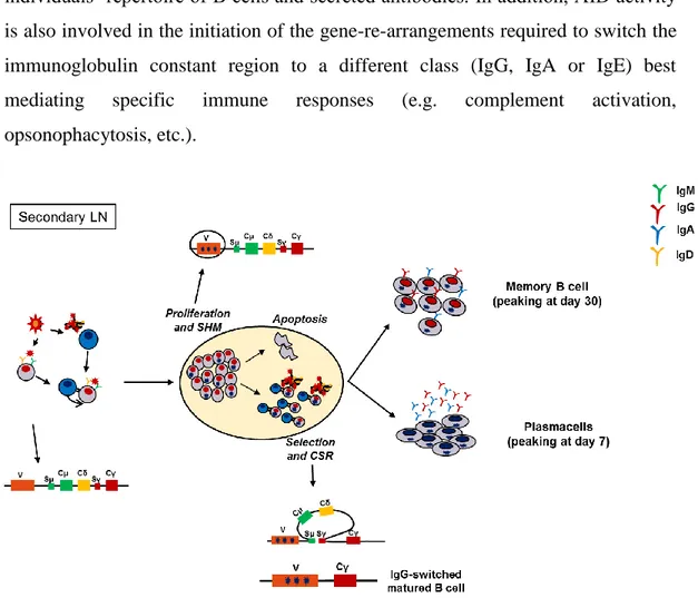
differentiation of some B cells into Antibody Secreting Cells, also called short-lived Plasma cells, outside the B cell follicles of the secondary lymphoid organs. The initial B cell–T cell contact is also required to induce the formation of specialized areas of the secondary lymphoid organs, called Germinal Centers (GCs) (IC., 1994). GCs are the unique documented sites for coupling the diversification of BCR V gene sequences, with the selective expansion of those variants with improved binding affinity for antigen. In GCs, B cells activated by antigen-recognition start to proliferate rapidly and switch on a tightly regulated nuclear machinery leading to the generation of a multitude of clones carrying modified immunoglobulin BCRs (Figure 6). In this context the Activation-induced cytidine deaminase (AID) enzyme plays a central role (Muramatsu M, 2000) (Storb U,
10
2002); by deaminating deoxycytidine residues on DNA, AID paves the way to the introduction of point mutations, deletions and duplications in the germ-line sequence of the V,D,J gene segments encoding for the variable region of Immunoglobulins (IgV). Since Immunoglobulins bind to antigens through their IgV domains, this process, named Somatic HyperMutation (SHM), modulate, and might eventually re-shape, the specificity and overall binding affinity of the individuals’ repertoire of B cells and secreted antibodies. In addition, AID activity is also involved in the initiation of the gene-re-arrangements required to switch the immunoglobulin constant region to a different class (IgG, IgA or IgE) best mediating specific immune responses (e.g. complement activation, opsonophacytosis, etc.).
Figure 6_ T cell-dependent activation of B cells leads to the formation of GC in secondary lymphoid organs. Inside the GC, BCR is modified by Somatic HyperMutation and Class Switching. Both these events drive the differentiation of activated B cells into plasma cells or memory B cells in humans
11
Inside the GC proliferating B cells compete for antigen binding and access to survival signals delivered by T cells. Ultimately, this race ends with the survival of the clones that having acquired improved antigen-binding capacity will out-compete the others, which instead will die by neglect. While clonally expanded B cells are generated and selected, some of them differentiate into plasma cells that secrete antibodies whilst others will rest and become memory B cells that recirculate in the blood and will rapidly respond to subsequent antigenic re-stimulation. Ag-specific PBs and MBCs start to populate the human blood after infection or vaccination in different timeslots and with different kinetics. PBs, representing the early component of host defence, peak in the blood around day 8 after vaccination and return quickly to undetectable levels thereafter. Otherwise, Ag-specific MBCs circulate at enriched frequency in the blood peaking at 1 month after vaccination, and contracting slightly above or equal to baseline thereafter. Even if it is still controversial, there are several emerging evidences that Ag-specific PBs and MBCs undergo affinity maturation also without GC formation in extra-follicular pathways. It seems that of the B cells remaining in the outer follicle in the early stages of the B cell response, some migrate out and establish the foci of short lived Plasma cells, whereas others may be the B cells that are the origin of GC-independent memory (Tarlinton, 2012; David Tarlinton, 2013).
1.5. Diversity of B-cell memory
A substantial fraction of B cells in humans has encountered antigen and shows hallmarks of B-cell memory. One of these memory B cell hallmark is considered the expression of CD27 (Agematsu K, 1997), but recent studies have demonstrated that also CD27- cells can present with an activated phenotype and molecular signs of antigen experience (Fecteau JF, 2006; Cagigi A, 2009), suggesting that they could be memory cells. The majority of circulating memory B cells in healthy
12
adults is derived from GC dependent reactions. The earliest GC responses generate IgM+ cells. It has been demonstrated that these cells can undergo subsequent class switching, mostly to IgG, and clonally related IgM+ and IgG+ cells can be found in human GCs and blood (Seifert M, 2009). IgA+ and IgG+ GC-derived memory B cells occur later in the course of an immune response and carry high loads of SHM. Terminal differentiation and survival of plasma cells
Plasma cells are derived from activated B cells through a different transcriptional program than memory B cells. Since plasma cells progressively loose membrane BCR expression while maturing, they depend on other mechanisms for survival. While most plasma cells are generated in lymphoid organs and long-lived plasma cells reside in bone marrow (Rozanski CH, 2011), small numbers can be found circulating in blood of healthy adults and the latter ones have high CD38, CD27 expression levels. Despite their low numbers in blood (1-5 cells/μl), plasma cells display large phenotypic heterogeneity. Interestingly, circulating IgA+ plasma cells are more frequent than IgM+ or IgG+. These IgA+ plasma cells display characteristics suggestive of a mucosal origin.
1.6. Tools to dissect the human BCR repertoire
Since the acquired immunity generates highly specific and long-lived antibody response, there is a growing interest in set up methodologies that can dissect this aspect of immune memory. It is clear that the specificity of the antibody responses for particular pathogens is achieved by the development of a vast diverse repertoire resulted by the recombination of the V-genes that finally enable antibodies recognizing an enormous numbers of potential epitopes.
To summarize, after this brief excursus on the B cell development, five are the principal mechanisms that mediate the diversity in the antigen combining site of the BCR repertoire; the first three mechanisms acting at immature B cell level whereas the last two after B cell activation:
13
1. Combinatorial diversity generated by V(D)J recombination, 2. Junctional diversity at the recombination sites of the V(D)J genes,
3. Random pairing of heavy and light chains to form the antigen binding site (Figure 7A);
4. Somatic hypermutation that introduces point mutation into the variable domain,
5. Class switch recombination that enables to change the isotypes and thus to determine a different effector function (Figure 7B).
A
B
Figure 7_Mechanisms that mediate the diversity in the antigen combining site of the BCR repertoire. A) V(D)J recombination, Juntional diversity and VH/VL pairing; B) Generation of different variants by SHM and CSR after GC reaction (from Current opinion in Immunology, 2013)
14
Due to the importance of gaining insights on the BCR nature, different progresses have been done to dissect the Ab repertoire. The earliest studies were performed by using isoelectric focusing of Antibodies on polyacrylamide gel resolving the Abs into pattern of bands based on their isoelectric pH values (al, 1968). From this study, different steps forward have been done especially with the use of PCR, electrophoresis and sequencing methods for determining the exact nucleotide sequences that encode for specific antibodies (al, 1978). These studies paved the way to a huge number of studies addressing the study of the repertoire and making use of this information to produce human monoclonal antibodies with a defined specificity. Three are the main strategies used to dissect the repertoire that have also been used to produce human monoclonal antibodies:
1. Phage display technologies_ Phage display libraries have been constructed from IgV of vaccinated or infected subjects by sorting memory B cells and total peripheral blood mononuclear cell (PBMCs) populations containing all B cell subsets. Once B cells are isolated a random RT-PCR of VH and VL follows creating a combinatorial library of random VH and VL that can be expressed as single-chain variable antibody fragments (scFvs) on phage or antigen-binding fragments (Fabs) on yeast or mammalian cells (Figure 8). In this way a multitude of monoclonal antibodies with different specificities can be obtained and in fact this strategy has been used to isolate neutralizing antibodies specific for different numbers of pathogens such as West Nile virus, rabies virus, severe acute respiratory syndrome (SARS) virus, hepatitis A virus, HIV, hantavirus, Ebola virus, yellow fever virus, hepatitis C virus, measles virus and human and avian influenza virus strains, and so on. Even if this methodology has been an high-throughput way to produce antibodies, this is not the best way to obtain information about faithful representation of the physiological antibody gene pairs due to
15
the random pairing of immunoglobulin heavy and light chain variable regions that are cloned separately. Even if this random pairing enables to create a greater diversity of antibodies and it is a helpful strategy to generate higher affinity antibodies, it is not possible to obtain information about the process of affinity maturation that a given heavy and light chain pair went through.
2. B cell immortalization_ this methodology generally involves culturing total PBMCs or sorted IgG+ memory B cells from fresh or frozen samples in the presence of the Epstain Barr Virus (EBV), that can transform all pheripheral resting B cells subsets, together with Toll Like Receptor 9 (TLR9) and/or allogeneic irradiated mononuclear cells to provide co-stimulatory signals. Under these conditions, B cells are induced to proliferate and to secrete antibodies. After several days, supernatants from cultured cells are tested for antigen binding and/or functional activity. The B cells producing the antibodies of interest are cloned by limiting dilution and further screened for the desired reactivity at the single-cell level before individual VH and VL are cloned and sequenced (Traggiai, 2012) (Figure 9). By applying this Figure 8_Scheme of the different steps to generate phage display libraries (from Nature Reviews Immunology, 2012)
16
strategy the EBV-transformed B cell clones obtained can also be fused with myeloma cells to generate hybridomas, which facilitate the stable production of high levels of antibodies. This approach offers the opportunity to immortalize and obtain information about the antibodies produced from the pool of the antigen specific memory B cells.
3. Single cell analysis of BCR repertoire_ An important advance in the study of the human BCR repertoire is represented by the single-cell reverse transcription PCR (RT-PCR) to isolate and amplify the cognate VH and VL genes from single B cells sorted by FACS (Thomas Tiller, 2008; Hua-Xin Liao, 2009) (Figure 10). This kind of approach enables to take a snapshot of the exact repertoire of the BCR expressed on the surface of particular B cells in a defined time slot. By using this approach information can be obtained also from rare, highly discrete B
Figure 9_Scheme of the different steps required for human B cell immortalization
Figure 9_Scheme of the different steps required for human B cell immortalization (form Methods in Molecular Biology, 2012)
17
cell subpopulations, such as MBCs, if those cells can be identified by FACS. This approach was firstly applied by Wardemann et al. to understand the tolerogenic selection of developing human B cell subpopulations. Thereafter, this strategy has been applied to investigate the different type and class of B cells in healthy, or immunocompromised individuals, or to study the repertoire of PBs and MBCs elicited by vaccination. There are a lot of studies focused on the repertoire of PBs isolated 7 days after vaccination because they are the first and robust source of antigen specific B cells and also because their identification by FACS basically relies upon the expression of Cluster of Differentiation (CD) surface markers. Moreover, since the time of antigen exposure is well defined, their dissection is particularly useful for examining the ongoing immune response to vaccines. For example, this method has been successful for analyzing the immune response to influenza, tetanus and anthrax vaccination. To a lesser extent also the repertoire of antigen-specific MBCs has been dissected. However the paucity of studies on this direction is ascribed to the lack of suitable tools for the identification of antigen specific Memory B cells, which circulate at very low frequencies in the blood as well as to the low efficiency in the recovery of VH and VL genes after RT-PCR. The identification of Ag-specific B cells has been so far challenging but several efforts have been done to solve this issue and one solution came from our laboratory (Bardelli M, 2013). All these studies basically rely on the use of fluorescent antigen baits or antigen tetramers to identify memory B cells engaged into BCR-specific interactions. Brightly labelled memory B cells are identified and isolated as single cells on a 96-well plate containing a lysis buffer that preserve mRNA. Thereafter, IgVH and VL genes are retro-transcribed and amplified by nested PCR .
18
Finally, various high-throughput technologies are emerging to generate a more comprehensive picture of the human B cell response. One promising approach is the use of next-generation sequencing to exhaustively sequence the entire B cell repertoire. The application of high throughput sequencing of paired VH and VL sequences to different B cell subsets isolated overtime following vaccination could be key to understand the specificity, the frequency and also the class of memory cells required to mediate protection, as well as the individual variability in the breadth of the response. Moreover, this analysis could be helpful in discriminating useless versus harming responses and thus to design better vaccine antigens able to stimulate and guide successful immune memory responses.
Figure 10_Schematic representation of scPCR approach to dissect changes in the repertoire of PBs and Ag-specific MBCs
19
2.1. Classification of influenza viruses
Influenza viruses are members of the Orthomyxoviridae family (R.A. Lamb, 2001) and are basically classified into three subtypes: A, B and C. The principal differences between the three subgroups are based on the number of the segments present in the genome and thus on the antigenic features of their internal proteins, nucleoprotein and matrix. Influenza A and C viruses infect multiple species, while influenza B almost exclusively infects humans. Influenza A and B viruses are responsible of most of the cases of human disease and cause annual epidemics. They are antigenically distinct and do not exhibit cross-immunity or gene recombination. Influenza A viruses are essentially avian viruses that occasionally infect other species including humans. Avian infection is usually asymptomatic, and viruses replicate in the intestine of aquatic birds, creating that constitute a large reservoir of potential pandemic viruses.
The eight, negative-sense, RNA segments of the influenza virus genome encode 11 different proteins, of which 8 are packaged into the infectious, enveloped, virion. On the viral surface are the two main antigenic determinants of the virus, the spike glycoproteins: hemagglutinin (HA) and neuraminidase (NA). HA mediates viral entry into cells and has receptor binding and membrane fusion activity. NA mediates enzymatic cleavage of the viral receptor at late stages of infection, allowing for the release of progeny virions (A., 1957). A third integral membrane protein, M2, is a multi-functional, proton-selective, ion channel which has roles both in virus entry as well as in assembly and budding. Inside the viral envelope, the matrix protein (M1) provides structure to the virion and bridges interactions between the viral lipid membrane and the ribonucleoprotein (RNP) core. The RNP core is composed by the RNA polymerase complex proteins, PB1, PB2 and PA, and the nucleocapsid protein (NP) which mediates binding and packaging of the viral genome. During virus replication three other proteins are expressed that are not incorporated into the mature virion. Non-structural protein 1 (NS1) is a
multi-20
functional protein with a major role in evasion of the host immune system. NS2 (NEP) plays a crucial role in mediating the export of viral RNPs from the cell nucleus during replication (García-Sastre, 2011) (Figure 11).
Influenza A viruses are grouped in subtyped based on sequence and antigenic of HA and NA. Up to now, 16 different HA and 9 NA have been described, most of which circulating in the avian reservoir. So far, cases of human infection resulting in annual epidemics have been attributed only to viruses carrying H1, H2,H3 and N1 or N2. When a virus strain with a new HA or NA subtype appears in the human population by genetic reassortment, it usually causes a pandemic because there is no preexisting immunity against the new virus. This was the case for example of the three pandemics that occurred during the last century (1918, 1957, and 1968) and also for the first pandemic of the 21st century, caused by the currently
Figure 11_schematic structure of the Influenza A virus and genomic organization
Figure 11_Schematic structure of the Influenza A virus and genomic organization
21
circulating A (H1N1) 2009 virus, which was generated by gene reassortment between a virus present in pigs of North America and a virus that circulates in the swine population of Euroasia.
2.2. Virus replication cycle
The exact mechanism for fusion of virus and cell is well established and a strong contribution to this type of knowledge has been given by crystallographic analysis. During the first steps of infection both HA and NA have crucial roles in mediating binding of the virus to the cell surface and release of the newly-formed viral particles, respectively.
HA owns its name to its capacity of causing agglutination of red blood cells in vitro and this peculiar characteristic is conferred by the capability of the glycoprotein to bind sialic acid residues exposed on cell surface. In most viral strains it is present as native un-cleaved form, also called HA0 monomer. Each HA monomer presents a host receptor binding site that allows the virus to recognize terminal sialic acid moieties on glycolipids and glycoproteins on the surface of host cells. Since there are many copies of HA on each influenza virus particle, the attachment of influenza virus on the host cell surface is multivalent and has high avidity despite low affinity. The specificity and the affinity of the viral HA for its receptor is a crucial characteristic of host transmission. Cleavage of the HA0 protein is mediated by host-produced trypsin-like proteases and produces two subunits: HA1 and HA2. The mature form of HA on infective viral particles is a homotrimeric structure where each HA molecule consists of a globular ‘head’ domain, made up exclusively by HA1, and a fibrous stem composed by the entire HA2 and part of HA1 that is inserted into the viral membrane. The HA C-terminal cytoplasmic tail interacts directly with the matrix protein layer immediately underneath the membrane envelope.
22
It is through the binding of HA to sialic acid residues that viral and host membranes fuse and thus the infective process starts by internalization of the virion inside the host cell through classical receptor mediated endocytosis in clathrin-coated vesicles. During the endocytosis process, the M2 protein allows for the influx of protons leading to an acidic environment (Matlin KS, 1981). The acidification of endosomes is critical for the next step of the replication cycle of the virus for two reasons:
6. It provokes a conformational change in the HA favoring the fusion of the viral and cellular membranes and this allows the virus to enter the cell cytoplasm
7. It promotes the uncoating of the virus and allows the RNPs to dissociate from the viral particle, such that they are released into the cytoplasm and transported to the cell nucleus.
Figure 12_Entry and replication pathway of influenza virus (from Nature Structural & Molecular Biology,2010)
23
Once in the nucleus of infected cells, the viralRNA-dependent RNA polymerase transcribes and replicates the negative sense vRNA. The positive RNA strand obtained is used as a template for the synthesis of new virion genome and for mRNA transcription and viral protein translation (Cros JF, 2003). All mRNA molecules are then transported back to the cytoplasm by the viral proteins NEP and M1 and once in the cytoplasm they are translated by the host cell machinery. The surface proteins HA, M2 and NA are synthesized in the endoplasmic reticulum (ER), glycosylated in the Golgi apparatus and finally transported to the cell membrane for virion assembly. The progeny virions assemble and bud at the plasma membrane (Figure 12).
The new progeny of virions remain initially bound to outside membrane sialic acid residues because of their interactions with HA spikes until NA sialidase activity remove them from cellular and viral glycoconjugates. For its peculiar activity, NA has also a role in preventing the aggregation of the new virions as well as in mediating their release. Once released from host cells the new virions can infect nearby cells and spread the infection.
2.3. Viral tropism and antigenic variation
Influenza A viruses circulate in a wide variety of animals, most of which are only transiently infected. The largest variety of influenza A viruses circulate in birds. Besides birds, additional animal reservoirs of influenza A viruses are found in mammalian species such as swine and horses.
The major determinants of the tropism of influenza viruses are the sialic acid molecoles because of their initial interaction with HA to start infection. Sialic acid residues are nine-carbon monosaccharides present in variable amounts on the surface of different cell types, usually bound to galactose through α2,3 or α2,6 linkages. HA from different viruses show a preferential binding avidity for the type of sialic acid linkage. Thus, viruses that infect humans bind preferentially to SA
24
with α2,6 linkages, whereas avian viruses bind mostly to SA in an α2,3 configuration (Ito T, 1998; Ito T, 2000; Ito T, 1997). The human tracheal epithelial tissue preferentially express SA with a2,6 linkages, whereas avian epithelial gastrointestinal tissue (where influenza viruses replicate in avian hosts) contain mainly SA in an a2,3 configuration. Of relevance, pig tracheal tissue has both kinds of SA linkages, and swine can thus be infected with viruses that recognize both types of receptors, hence, these animals have often been proposed as a mixing vessel for the generation of new strains from co-infections of avian and mammalian viruses.
It is noteworthy that Influenza A viruses evolve constantly using different mechanisms. Three are the best known mechanisms able to generate further variability in influenza A viruses. The most important is the lack of proofreading activity of the viral RNA polymerase during replication of the influenza genomic RNA segments, which results in a high level of point mutations, phenomenon that is also known as antigenic drift. The permanent antigenic drift in the principal targets of the immunological response, the proteins HA and NA, is the cause of the constant need to review the viruses that are included in the yearly prepared vaccines. The antigenic drift that has occurred in influenza A viruses for thousands of years has led to the current diversity of HA and NA subtypes. The second mechanism, known as antigenic shift, relies in the segmented nature of viral genome. This peculiar characteristic permits the formation of new progeny viruses with novel combination of segments when two or more different virus subtypes infect a single cell. This process is capable of introducing new proteins in circulating viral populations that can drastically change the biological properties of the virus. Antigenic shift is commonly associated with appearance of pandemic influenza viruses. A third mechanism associated with evolution of influenza A viruses is recombination by template switching. This type of recombination may involve genetic material either from more than one origin or two different
25
viralRNAsegments, and these nonhomologous recombination events could be associated with changes in viral pathogenicity.
26
3.1. Neisseria meningitidis
Neisseria meningitidis is a gram negative, encapsulated bacterium, which is generally a commensal that colonizes the mucosal epithelium of the nasopharynx of the human population. Colonizing strains can belong either to hypervirulent lineages which are generally associated with disease, or to carriage strains that provides a reservoir for meningococcal infection and can also contribute to establish host immunity (DS, 2009). For still unknown reasons, the carriage strains can invade the pharyngeal mucosal epithelium and, in the absence of bactericidal serum activity, disseminate into the bloodstream, causing septicaemia. In a subset of cases, bacteria can also cross the blood-brain barrier and infect the cerebrospinal fluid, causing meningitis.
With the exception of sporadic case reports, all known disease-causing meningococcal strains are surrounded by a capsule made up by complex polysaccharides, which confers resistance to phagocytosis and complement-mediated lysis. Even if the capsular polysaccharide (CPS) inhibits bacterial adhesion because it masks the action of meningococcal adhesins, it is known to be crucial for bacterial to survive in the blood.
Based on the immunogenicity and chemical structure of the CPS, N. meningitidis (Nm) can be classified into at least 13 serogroups A, B, C, E-29, H, I, K, L, W-135, X, Y, Z, and 29E (SE, 1953). Among them, only six serogroups (A, B, C, W-135, X, Y) have been associated with meninogococcal disease and are thus considered pathogenic. Further classification into serosubtype, serotype and immunotype is based on class 1 outer membrane proteins (PorA), class 2 or 3 (PorB) outer membrane proteins and lipopoly[oligo]saccharide structure, respectively (Rosenstein NE, 2001; Stephens DS, 2007)
27
3.2. Colonization and carriage
The first step known to establish carriage and invasive meningococcal disease is the colonization of the upper respiratory mucosal tract by Nm. The bacterium can be acquired through inhalation of respiratory droplets and secretions.
This acquisition can be either asymptomatic, or (infrequently) give rise to a local inflammation, invasion of mucosal surfaces, access to the bloodstream and fulminant sepsis, or focal infections such as meningitis (Stephens DS, 2007). Meningococcal disease usually occurs 1–14 days after acquisition of the pathogen. Acquisition may also result in upper respiratory and pharyngeal meningococcal carriage. The duration of carriage can vary from days to months. The probability of meningococcal disease after the acquisition of Nm declines very sharply, such that invasive disease becomes unlikely 10–14 days after acquisition.
3.3. Meningococcal adhesion and cell invasion
The adhesion to the respiratory epithelium is essential for Nm survival, colonization and transmission, and is also a prerequisite for invasive meningococcal disease. Upon contact with human cells, the meningococci forms microcolonies and adheres using pili. Nm has evolved numerous surface-exposed adhesive structures that facilitate interactions with human cells. Bacterial host-specificity resides in the structural specificity of the meningococcal ligands for human molecules, which can range from nutrients, to serum/secreted proteins and surface-located adhesion receptors. Mechanisms of meningococcal adhesion are multifactorial, dynamic and display temporal changes during the course of infection. After the initial colonization, there is a loss or down-regulation of the capsule, which sterically masks the outer membrane proteins. This event is key to unmask adhesins that serve to mediate subsequent adhesion to the human epithelium. Antigenic and phase variation of meningococcal outer membrane proteins (OMPs) also play an
28
important role, and the presence of multiple adhesins compensates for phase variation and may lead to an altered tissue tropism. Close adherence of meningococci to the host epithelial cells results in the appearance of cortical plaques and the recruitment of factors leading to the formation and extension of epithelial cell pseudopodia that engulf the bacteria (DS, 2009). This intracellular lifestyle can give the bacteria the opportunity to evade host immune response, finding more available nutrients, and eventually, to further cross the epithelium entering the blood stream (DS, 2009). Intracellular meningococci reside inside membranous vacuoles and are capable of translocating through basolateral epithelial tissues by transcytosis within 18-40 hours. Within the cells, meningococcus has to express again the capsule, which can prevent antibody and complement deposition (M., 1995), is anti-opsonic and anti-phagocytic and therefore aids survival in blood (M., 2009). Once in the bloodstream, meningococci may multiply rapidly to high numbers and eventually translocate across the blood-brain barrier, proliferate in the central nervous system and cause meningitis. These later steps in invasion are still poorly understood. The ability to cause invasive disease is influenced by a multiplicity of environmental and microbial factors, as well as from the absence of host’s protective antibodies.
3.4. Anti-meningococcal vaccines
Several meningococcal vaccines are available against the distinct serogroups. Vaccines against meningococcal serogroups A and C have been developed and tested in clinical trials (Costantino P, 1992) (Anderson EL, 1994); (Fairley CK, 1996). The first trials conducted in the United Kingdom with the meningococcus C conjugate showed a dramatic decline in the incidence of serogroup C disease in all age groups ( (Borrow R, 2000) (Miller E, 2001) with an efficacy of 97 and 92 per cent for teenagers and toddlers, respectively. Conjugate vaccines against
29
meningococcus are now available as monovalent (A or C) or as different tetravalent formulations (A, C, W-135 and Y).
The most critical target for vaccination is meningococcus B (MenB), which is responsible for 32 percent of all cases of meningococcal disease in the United States and for 45–80 percent of the cases in Europe. Conventional biochemical and microbiological approaches have been of little help in the development of a vaccine able to induce broad protection against menB. A polysaccharide-based vaccine approach could not be used for group B meningococcus, since the principal component of its CPS that is a polymer of Æ(2-8)-linked N-acetylneuranimic acid., which is also abundantly expressed in human tissues. Alternative approaches to develop MenB vaccines have focused on the use of surface-exposed proteins contained in outer membrane preparations (outer membrane vesicles, OMVs). The first OMV vaccines were developed in Norway and Cuba and showed efficacy in humans ranging from 50 to 80 per cent (al, 1999). However, while each vaccine was shown to induce good protection against the homologous strain, both failed to induce protection against heterologous strains (Rosenstein NE, 2001). The major protective antigen in both these vaccines is PorA, the most abundant outer membrane protein, which is known to be highly variable across different isolates of serogroup B N. meningitidis.
To overcome these limitations, a novel multicomponent recombinant protein-based vaccine, named 4CMenB or Bexsero®, has been recently developed by Novartis Vaccines and Diagnostics against group B meningococcal strains. Bexsero® is now approved in Europe and Australia.
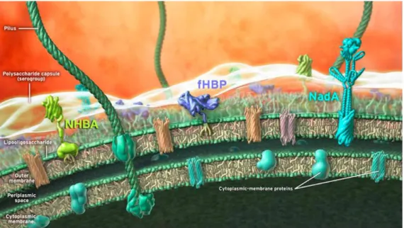
4CMenB is based on antigens identified through an innovative genetic approach known as ‘reverse vaccinology’ and combines OMVs from the New-Zealand epidemic strain (NZ98/254) with three major protein antigens: factor H-binding protein (fHbp), Neisserial Heparin-Binding Antigen (NHBA) and Neisserial adhesin A (NadA) (Giuliani MM, 2006) (Figure 13). Two of these main antigens,
30
fHpb (sub-variant 1.1) and NHBA (peptide 2), are present as fusion proteins to two minor antigens, GNA2091 and GNA1030, respectively. Another vaccine against serogroup B, developed by Pfizer, has been approved in America and contains two alleles of the recombinant fHbp.
3.5. Factor H binding protein (fHbp)
Factor H binding protein (fHbp or GNA1870) was the first discovered antigen effective in inducing bactericidal antibodies. It was identified by screening the genome of one of the most virulent strains: MC58. fHbp is known to bind specifically to human factor H (fH), an inhibitor of the alternative complement pathway. Since the evasion of the human complement system is key for Nm to cause the invasive disease, different studies have been addressed the role of fHbp
Figure 13_Schematic representation of the 4CMenB vaccine antigens on the surface of N. meningitides. The main antigens identified through reverse vaccinology approach (NHBA, fHbp and NadA) are depicted (from Vaccine, 2012).
31
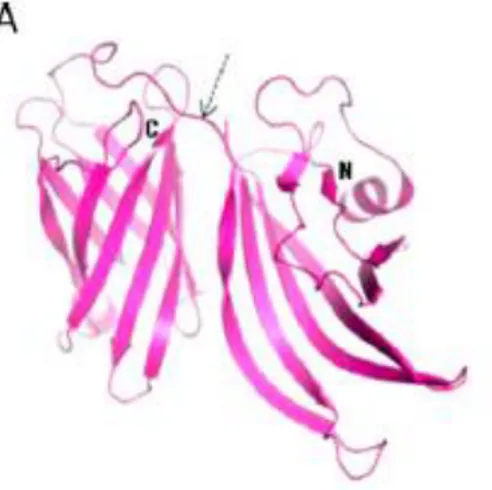
in host invasion and it has been shown that the its deletion results in increased susceptibility of most strains of Nm to killing in either serum or in whole blood. The three-dimensional (3D) solution structure of fHbp has been resolved by nuclear magnetic resonance (NMR), by spectroscopy and by X-ray crystallography.
These studies revealed that the protein is composed of two domains connected by a short linker: an N-terminal domain of 8 beta-strands, forming a highly curved anti-parallel beta-sheet, and a C-terminal domain that is a well-defined beta-barrel of 8 anti-parallel beta strands (Figure 14). Although fHbp is known to have ~300 sequence variants, multiple-sequence alignments show that the residues forming of the hydrophobic cores of both C- and N-terminal domain are well conserved predicting that the final 3D structure should be the same in all variants. The fHbp proteins can be classified into three genetic and immunogenic variants: fHbp-1, fHbp-2 and fHbp-3, which are not cross-protective, and can be further divided into subvariants. Among each variant, fHbp sequences are conserved across different strains (92 to 100%), while between the 1-3 variants sequence conservation is low (63%). This diversity has an important impact on the immunological features of fHbp, since members of each variant induce a strong protective immune response against meningococcal strains carrying homologous alleles but not against strains expressing distantly related variants (Masignani V, 2003).
Figure 14_Structure of fHbp. The currently known structure of the fHbp formed by two beta-barrel domains (one domain N-terminal and the other C-terminal) connected by a short linker (from Vaccine, 2012)
32
References
1. A. Gottschalk Neuraminidase: the specific enzyme of influenza virus and Vibrio cholera. [Revue]. - 1957. - Vol. Biochemica et biophysica acta. 2. Agematsu K Nagumo H, Yang FC, et al. B cell subpopulations separated by
CD27 and crucial collaboration of CD27+ B cells and helper T cells in immunoglobulin production. [Revue]. - 1997. - Vol. Eur J Immunol.
3. al Awdeh.Z.L. et Isoelectric focusing in polyacrilamide gel and its application to immunoglobulins [Revue]. - 1968. - Vol. Proc.Natl.Acad.Sci. U.S.A.
4. al Seidman J.G. et Multiple related Immunoglobulin Variable-region genes identified by cloning and sequence analysis [Revue]. - 1978. - Vol. Proc.Natl.Acad.Sci. U.S.A.
5. al Tappero JW et Immunogenicity of 2 serogroup B outer-membrane protein meningococcal vaccines: a randomized controlled trial in Chile [Revue]. - 1999. - Vol. JAMA.
6. Alt F.W. et al. Ordered rearrangement of immunoglobulin heavy chain variable region segments. [Revue]. - 1984. - Vol. EMBO.
7. Amanna IJ1 Slifka MK, Crotty S. Immunity and immunological memory following smallpox vaccination. [Revue] // Immunol Rev. - 2006 Jun. - pp. 211:320-37..
8. Anderson AS Jansen KU, Eiden J New frontiers in meningococcal vaccines [Revue]. - 2011. - Vol. Expert Rev Vaccines .
33
9. Anderson EL Bowers T, Mink CM, Kennedy DJ, Belshe RB, Harakeh H, Pais L, Holder P, Carlone GM. Safety and immunogenicity of meningococcal A and C polysaccharide conjugate vaccine in adults. [Revue]. - 1994. - Vol. Infect Immun..
10. Blink EJ1 Light A, Kallies A, Nutt SL, Hodgkin PD, Tarlinton DM. Early appearance of germinal center-derived memory B cells and plasma cells in blood after primary immunization. [Revue] // J Exp Med.. - 2005 Feb 21. - pp. 201(4):545-54..
11. Borrow R Fox AJ, Richmond PC, Clark S, Sadler F, Findlow J, Morris R, Begg NT, Cartwright KA. Induction of immunological memory in UK infants by a meningococcal A/C conjugate vaccine. [Revue]. - 2000. - Vol. Epidemiol Infect..
12. Cagigi A Du L, Dang LV, et al. CD27(-) B-cells produce class switched and somatically hyper- mutated antibodies during chronic HIV-1 infection. [Revue]. - 2009. - Vol. PLoS One..
13. Costantino P Viti S, Podda A, Velmonte MA, Nencioni L, Rappuoli R. Development and phase 1 clinical testing of a conjugate vaccine against meningococcus A and C. [Revue]. - 1992. - Vol. Vaccine.
14. Cros JF Palese P. Trafficking of viral genomic RNA into and out of the nucleus: influenza, thogot and Borna disease virus [Revue]. - 2003. - Vol. Virus Res.
15. David Tarlinton Kim Good-Jacobson Diversity Among Memory B Cells: Origin, Consequences, and Utility [Revue]. - 2013. - Vol. Science .
34
16. DC Stephens Biology and pathogenesis of the evolutionarily successful, obligate human bacterium Neisseria meningitidis [Revue]. - 2009. - Vol. Vaccine.
17. DS Stephens Biology and pathogenesis of the evolutionarily successful, obligate human bacterium Neisseria meningitidis. [Revue]. - 2009. - Vol. Vaccine .
18. Early P., Huang,H., Davis,M.,Calame,K., & Hood,L. An immunoglobulin heavy chain variable region gene is generated from three segments of DNA: VH, D and JH [Revue]. - 1980. - Vol. Cell.
19. Fairley CK Begg N, Borrow R, Fox AJ, Jones DM, Cartwright K. Conjugate meningococcal serogroup A and C vaccine: reactogenicity and immunogenicity in United Kingdom infants. [Revue]. - 1996. - Vol. J Infect Dis..
20. Fecteau JF Cote G, Neron S. A new memory CD27-IgG+ B cell population in peripheral blood expressing VH genes with low frequency of somatic mutation. [Revue]. - 2006. - Vol. J Immunol. .
21. Fletcher LD Bernfield L, Barniak V, Farley JE, Howell A, Knauf M, et al. Vaccine potential of the Neisseria meningitidis 2086 lipoprotein [Revue]. - 2004. - Vol. Infect Immun.
22. García-Sastre Rafael A. Medina & Adolfo Influenza A viruses: new research developments [Revue]. - 2011. - Vol. Nature.
23. Giuliani MM Adu-Bobie J, Comanducci M, Aricò B, Savino S, Santini L, Brunelli B, Bambini S, Biolchi A, Capecchi B, Cartocci E, Ciucchi L, Di
35
Marcello F, Ferlicca F, Galli B, Luzzi E, Masignani V, Serruto D, Veggi D, Contorni M, Morandi M, Bartalesi et al A universal vaccine for serogroup B meningococcus [Revue]. - 2006. - Vol. Proc Natl Acad Sci USA.
24. Good-Jacobson David Tarlinton and Kim Diversity Among Memory B Cells: Origin, Consequences, and Utility [Revue]. - 2013 . - Vol. Nature. 25. Hammarlund E1 Lewis MW, Hansen SG, Strelow LI, Nelson JA, Sexton
GJ, Hanifin JM, Slifka MK. Duration of antiviral immunity after smallpox vaccination [Revue] // Nat Med.. - 2003 Sep. - pp. 9(9):1131-7..
26. Hua-Xin Liao Marc C. Levesque, Ashleigh Nagel, Ashlyn Dixon,Ruijun Zhang, Emmanuel Walter, Robert Parks, John Whitesides, Dawn J. Marshall,Kwan-Ki Hwang, Yi Yang, Xi Chen, Feng Gao, Supriya Munshaw, Thomas B. Kepler, Thomas Denny, M. Anthony Moody,Haynes High-throughput isolation of immunoglobulin genes from single human B cells and expression as monoclonal antibodies [Revue]. - 2009. - Vol. J Virol Methods..
27. Ian J. Amanna Nichole E.Carlson and Mark K. Slifka Duration of Humoral Immunity to Common Viral and Vaccine Antigens [Revue] // N Engl J Med. - November 8, 2007. - pp. 357:1903-1915.
28. Ian J. Amanna Nichole E.Carlson, and Mark Slifka Duration of Humoral Immunity to Common Viral and Vaccine Antigens [Revue]. - 2007. - Vol. The New England Journal of Medicine.
29. Ian J.Amanna Mark K. Slifka, shane Crotty Immunity and immunological memory following smallpox vaccination [Revue]. - 2006. - Vol. Immunological reviews.
36
30. IC. MacLennan Germinal centers [Revue]. - 1994. - Vol. Annu Rev Immunol.
31. IMGT. The International ImMunoGeneTics information system: [Revue]. - http://www.imgt.org.
32. Ito T Couceiro JN, Kelm S, Baum LG, Krauss S, Castrucci MR, Donatelli I, Kida H, Paulson JC, Webster RG, Kawaoka Y. Molecular basis for the generation in pigs of influenza A viruses with pandemic potential. [Revue]. - 1998. - Vol. Journal of Virology.
33. Ito T Suzuki Y, Sazuki T, Takada A, Horimoto T, Wells K, Kida H, Otsuki K, Kiso M, Ishida H, Kawaoka Y. 2000. Recognition of N -glycolylneuraminic acid linked to galactose by the alpha2,3 linkage is associated with intestinal replication of influenza A virus in ducks [Revue]. - 2000. - Vol. Journalof Virology.
34. Ito T Suzuki Y, Takada A, Kawamoto A, Otsuki K, Masada H, Suzuki T, Kida H, Kawaoka Y. Differences in sialic acid-galactose linkages in the chicken egg amnion and allantois influence human influenza virus receptor specificity and variant selection. [Revue]. - 1997. - Vol. Journal of Virology.
35. Kracker S. & Radbruch,A. Immunoglobulin class switching: in vitro induction and analysis [Revue]. - 2004. - Vol. Methods Mol. Biol.
36. Lefranc M-P Lefranc G. The Immunoglobulin FactsBook [Revue]. - 2001. - Vol. Academic Press London.
37. M. Achtman Epidemic spread and antigenic variability of Neisseria meningitidis. [Revue]. - 1995. - Vol. Trends Microbio.
37
38. M. Virji Pathogenic neisseriae: surface modulation, pathogenesis and infection control. [Revue]. - 2009. - Vol. Nat Rev Microbiol..
39. Mark K. Slifka Rustom Antia, Jason K. Whitmire and Rafi Ahmed humoral immunity due to Long-Lived Plasma Cells [Revue]. - 1998. - Vol. Immunity.
40. Masignani V Comanducci M, Giuliani MM, Bambini S, Adu-Bobie J, Arico B, et al Vaccination against Neisseria meningitidis using three variants of the lipoprotein GNA1870 [Revue]. - 2003. - Vol. J Exp Med.
41. Matlin KS Reggio H, Helenius A, Simons K. Infectious entry pathway of influenza virus in a canine kidney cell line. [Revue]. - 1981. - Vol. Journal of Cell Biology.
42. Matsuda F Ishii K, Bourvagnet P, Kuma K, Hayashida H, Miyata T et al. The complete nucleotide sequence of the human immunoglobulin heavy chain variable region locus. [Revue]. - 1998. - Vol. J Exp Med.
43. Max E.E., Seidman,J.G., & Leder,P. Sequences of five potential recombination sites encoded close to an immunoglobulin kappa constant region gene. [Revue]. - 1979. - Vol. Proc. Natl. Acad. Sci. U. S. A.
44. Miller E Salisbury D, Ramsay M. Planning, registration, and implementation of an immunisation campaign against meningococcal serogroup C disease in the UK: a success story. [Revue]. - 2001. - Vol. Vaccine.
45. Min Zhang Gopesh Srivastava and Liwei Lu The Pre-B Cell Receptor and Its Function during B Cell [Revue]. - 2004. - Vol. Cellular & Molecular Immunology.
38
46. Muramatsu M Kinoshita K, Fagarasan S, Yamada S, Shinkai Y, Honjo T. Class switch recombination and hypermutation require activation-induced cytidine deaminase (AID), a potential RNA editing enzyme [Revue]. - 2000. - Vol. Cell.
47. Oettinger M.A., Schatz,D.G., Gorka,C.,& Baltimore,D. RAG-1 and RAG-2, adjacent genes that synergistically activate V(D)J recombination [Revue]. - 1990. - Vol. Science.
48. R.A. Lamb R.M. Krug Orthomyxoviridae: the viruses and their replication [Revue]. - 2001. - Vol. FEBS letters.
49. Ramsden D.A., Baetz,K., & Wu,G.E. Conservation of sequence in recombination signal sequence spacers [Revue]. - 1994. - Vol. Nucleic Acids Res..
50. Rosenstein NE Fischer M, Tappero JW. Meningococcal vaccines. [Revue]. - 2001. - Vol. Infect Dis Clin North Am.
51. Rosenstein NE PERKINS BA, Stephens DS, POPOVIC T, Hughes JM Meningococcal Disease [Revue]. - 2001. - Vol. N Engl J Med.
52. Rozanski CH Arens R, Carlson LM, et al. Sustained antibody responses depend on CD28 function in bone marrow-resident plasma cells. [Revue]. - 2011. - Vol. J Exp Med..
53. S. Tonegawa Somatic generation of antibody diversity [Revue] // Nature. - 1983. - pp. 575-581.
39
54. SE Branham Serological relationships among meningococci [Revue]. - 1953. - Vol. Bacteriol Rev.
55. Seifert M Kuppers R. Molecular footprints of a germinal center derivation of human IgM+(IgD+)CD27+ B cells and the dynamics of memory B cell generation [Revue]. - 2009. - Vol. J Exp Med..
56. Slifka MK1 Antia R, Whitmire JK, Ahmed R. Humoral immunity due to long-lived plasma cells. [Revue] // Immunity. . - 1998 . - pp. 8(3):363-72.. 57. Stephens DS Greenwood B, Brandtzaeg P Epidemic meningitis,
meningococcaemia, and Neisseria meningitidis. [Revue]. - 2007. - Vol. Lancet.
58. Storb U Stavnezer J. Immunoglobulin genes: generating diversity with AID and UNG [Revue]. - 2002. - Vol. Curr Biol..
59. Tarlinton Kim L.Good-Jacobson and David M. Moultiple routes to B-cell memory [Revue]. - 2012. - Vol. International Immunology.
60. Thomas Tiller Eric Meffre, Sergey Yurasov, Makoto Tsuiji, Michel C. Nussenzweig and Hedda Wardemann Efficient generation of monoclonal antibodies from single human B cells by single cell RT-PCR and expression vector cloning [Revue]. - 2008. - Vol. J Immunol Methods.
61. Traggiai E1 Puzone R, Lanzavecchia A. Antigen dependent and independent mechanisms that sustain serum antibody levels. [Revue] // Vaccine. . - 2003 Jun 1. - pp. Suppl 2:S35-7..
40
62. Traggiai Elisabetta Immortalization of Human B cells: analysis of B cell Rertoire and Production of Human Monoclonal Antibodies [Revue]. - Vol. Methods in Mol Biol .
63. U. Grawunder T.M. Leu, D.G. Schatz, A. Werner, A.G. Rolink, F. Melchers, et al. Down-regulation of RAG1 and RAG2 gene expression in preB cells after functional immunoglobulin heavy chain rearrangement [Revue]. - 1995. - Vol. Immunity, 3 (5) .
41
Outline of the thesis
B cells are crucial components of adaptive immune responses. The way by which B cells interface and sense the external ‘danger’ is through their BCR. The initial heterogeneity of B cells created by the random rearrangement of V(D)J gene segments enables recognizing a multitude of foreign antigens. In the course of human life this vast repertoire of antibody is continuously re-shaped in order to generate a pool of diversified MBCs and PBs with different antigen specificities. Different studies are underlying the importance of dissecting the BCR repertoire of different B cell subsets following vaccination to gain insights on the dynamics of the vaccine response and to predict vaccine safety. This approach could help in understanding the specificity, the frequency and also the class of memory cells required to mediate protection, as well as the individual variability in the breadth of the response. Moreover, this analysis could allow discriminating useless versus harming responses. An increased knowledge on B cells, and in particular on MBCs and PBs responding to a given vaccine antigen, could be key for designing more effective vaccines able to stimulate and guide the ‘right’ immune memory responses.
Due to the relevance of these types of studies, the principal objective of my PhD thesis was to investigate and follow the response of both MBCs and PBs over time after vaccination by sequencing the variable part of the Ig genes of single cells (through single cell PCR. scPCR of paired VH and VL) and by profiling the antibody repertoire changes
However, when I started my PhD there were different challenges in introducing this approach, mainly represented by the lack of tools to identify and isolate antigen specific MBCs and by the low efficiency of amplification of Ig scPCR obtained in MBCs compared to the pool of PBs. This has led my project to be divided in two parts. In the first part of my dissertation I will present the results published in a
42
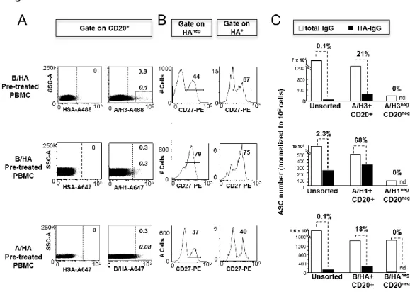
manuscript (Bardelli M, 2013) of an efficient approach developed to identify and sort HA specific MBCs as well as to amplify VH and VL genes by the use of scPCR. The robust technique developed in Novartis enabled to monitor quantitative and qualitative changes in the distribution of HA binding across different B cell subsets following vaccination, and to obtain enriched population of HA specific B cells for molecular cloning of paired VHVL Ig genes.
The overall PCR efficiency we obtained in this first study by amplifying VH-VL genes from single memory B cells isolated from frozen hPBMCs ranged between 17-35%, as described also in literature. To optimize the entire procedure of Ig-scPCR, before moving to clinical samples, a new set of primers was designed for both RT and PCR steps. The application of this strategy to different B cell subsets enables to rescue the 60-80% of PCR products with an efficiency comparable in all cell types and improved in respect with the one obtained before.
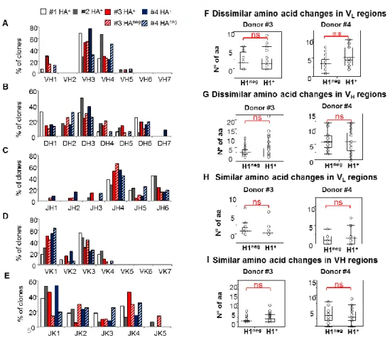
In the second part of my dissertation I will present the study that I have performed in the last year of my PhD aimed at broadly characterizing at phenotypic and molecular level the unexplored repertoire of circulating PBs and MBCs recruited in the interaction with Factor H Binding Protein (fHbp) antigen following Meningococcus B vaccination. PBs and fHbp-specific MBCs isolated ex vivo from human PBMC collected from two subjects before and at different time points after MenB vaccination have been characterized for their BCR repertoire. Moreover a comparison of the overall repertoire across the two individuals has been performed. The obtained results are described in a manuscript currently in preparation to be submitted and presented in this thesis.
43
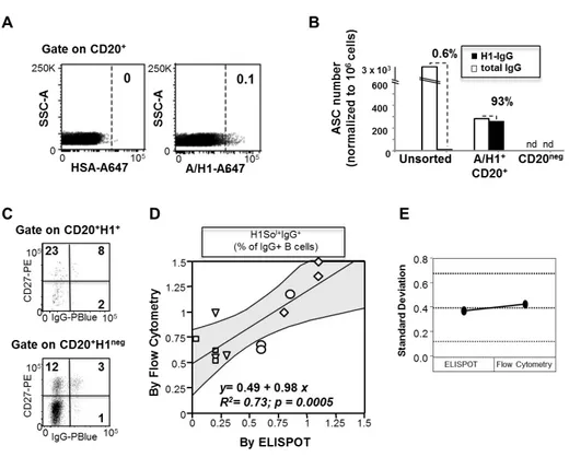
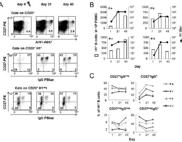
Ex vivo analysis of human memory B lymphocytes specific for A
and B influenza hemagglutinin by polychromatic flow-cytometry
Monia Bardelli1, Liliana Alleri1*, Francesca Angiolini1*§, Francesca Buricchi1, Simona Tavarini1, Chiara Sammicheli1, Sandra Nuti1, Elena Degl’Innocenti2, Isabelle Isnardi2, Elena Fragapane1, Giuseppe Del Giudice1, Flora Castellino1 and Grazia Galli1#
1
Novartis Vaccines and Diagnostics srl, 53100 Siena, Italy, 2Novartis Institutes for Biomedical Research, CH-4056 Basel, Switzerland
Published in PLoS One. 2013 Aug 15;8(8):e70620.
*these Authors gave equal contribution to this work
§ Present address: European Institute of Oncology, 20141, Milan, Italy
# Corresponding author: Grazia Galli Research Center, Novartis Vaccines and Diagnostics srl Siena, 53100, Italy. Email: [email protected] Phone: +39 055 331053 Fax: +39 055 331053