World Journal of Veterinary Science
2020 | Volume 2 | Article 1008 06
© 2020 - Medtext Publications. All Rights Reserved.
ISSN: 2689-5951
Stairstep Osteotomy for the Treatment of Radial
Shortening and Forelimb Deformity in a Dog
Case Report
Sassaroli S, Botto R*, Pennasilico L and Palumbo Piccionello A
Department of Biosciences and Veterinary Medicine, University of Camerino, Italy
Abstract
A case of radius shortening in an 11-month old female Fox Terrier referred to Veterinary Teaching Hospital of University of Camerino, Italy. Radiographic and tomographic examinations suggested a traumatic premature closure of distal and proximal physis of radius, resulting in a lateral and caudal radial shortening of 2.98 mm and moderate angular deformity in the proximal radius. Stairstep osteotomy of the radial diaphysis was performed to re-establishing the length of the bone and the elbow congruence. The distraction was obtained by temporary linear external skeletal fixation and the osteotomy was synthesized by using of a 2.4 locking compression plate applied to the cranial aspect of radius. This is the first description of sagittal sliding osteotomy used to correct both limb deformity and radial shortening.
Keywords: Radius shortening; Radioulnar incongruence; Homeroulnar subluxation; Premature closure of physis; Lengthening radius; Stairstep osteotomy
Citation: Sassaroli S, Botto R, Pennasilico L, Palumbo Piccionello
A. Stairstep Osteotomy for the Treatment of Radial Shortening and Forelimb Deformity in a Dog. World J Vet Sci. 2020; 2(1): 1008.
Copyright: © 2020 Sassaroli S
Publisher Name: Medtext Publications LLC Manuscript compiled: Jan 11th, 2020
*Corresponding author: Riccardo Botto, Department of Biosciences
and Veterinary Medicine, University of Camerino, 93/95-62024 Matelica (MC), Italy, Fax: 39 0737403402; Tel: 39 0737403403, 0737403460; E-mail: [email protected]
Introduction
The normal anatomical development of the forelimb depends on synchronous growth rates of radius and ulna. A discrepancy in the growth rate or a complete closure of one or more of the physis can result in shortened angulated limbs, elbow and carpal joints incongruity and secondary osteoarthritis [1].
Symmetric or asymmetric premature closure of the radial physis is a congenital or acquired condition that can affect the radius of young dogs [2,3]. These conditions are less common than the premature closure of the distal ulnar physis [2-4]. In a report, O’Brien et al. [1] have observed an 11% incidence of distal closure and a 6% incidence of proximal physis closure of radius among all cases of forelimb growth arrest.
The premature closure of either physis of the radius, with the premature closure of the proximal one being rarer than the distal one [1], results in radial shortening and the bone remains usually straight if the closure is symmetrical. If the premature closure of the physis is localized in an eccentric area it usually results in some degree of angular deformity [3,5,6].
When the distal radial physis is symmetrically impaired, the amount of elongation of the radius slows dramatically [4,6]. Symmetrical closure of the distal radial physis is not often associated with angular deformity although carpal varus and inward rotation of the paw may result [7].
The radius provides the major weight-bearing surface of the elbow joint (75% to 80%) [8], but the radial shortening produces an intra-articular space between the radial head and the capitulum. The humeral trochlea displaces cranially and puts pressure on the coronoid process of the ulna [4,6]. In particular, Preston and colleagues concluded that a radioulnar step may concentrate physiologic forces on the elevated lateral edge of the medial coronoid process thus resulting in fragmentation [9].
If the radioulnar angulation doesn’t concur, the rapidity of the degenerative process requires a surgical intervention in order to re-establish the congruency of the elbow joint, maintain the bone alignment and restore the limb to a functional position [4,5,10]. Rapidity and severity of osteoarthritis onset depend on patient’s age and lifestyle [11].
Early surgical intervention is recommended in dogs with physeal injuries to minimize angular limb deformities [12,13].
There are different therapeutical options for the radial shortening taking into consideration the severity of the condition and also the age of the patient, since an additional surgical correction may be needed in animals treated at a young age that haven’t still completed their growth [14].
Elbow incongruence is corrected by re-establishing the length of the radius, acutely or gradually, or by shortening the ulna [4].
Case Presentation
An 11-month-old female Fox Terrier dog breed has been referred to Veterinary Teaching Hospital of the University of Camerino, Italy. Anamnestic data report an ulnar greenstick fracture treated conservatively with a Robert-Jones bandage, 4 months prior and intermittent right forelimb lameness. Gait evaluation revealed a shortened stride in ambulatory phase and moderate head “bob” on trot and varus angulation of the elbow and valgus of the carpi during station. Orthopedic examination revealed mild generalized right forelimb muscle atrophy and pain on flexion and extension of the elbow joint with mild limitation of the elbow range of motion. Campbell’s test of the elbow was weakly positive and valgus and varus stress did not elicit elbow instability.
07
World Journal of Veterinary Science
2020 | Volume 2 | Article 1008 © 2020 - Medtext Publications. All Rights Reserved.
Radiographic and tomographic examinations were taken. Mediolateral and craniocaudal radiographic projections of the right radio-ulnar display a symmetric premature closure of the distal radial physis and asymmetric premature closure of the proximal radial physis, which were results in incongruities of the caudal and lateral radial head and a moderate valgus of the proximal anatomical radial angle (aMRPRA 90°) (Figure 1A and B) [15,16].
CT was performed on a single-slice helical CT‐scanner (CT/e General Electric single-slice computed tomography
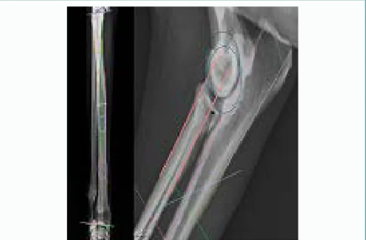
scanner) with a slide thickness of 1 mm. Images of the forelimbs were acquired in sternal recumbency with the elbow joints flexed to approximately 90 degrees. Joint incongruence was evaluated using a duplicated circle superimposition technique in sagittal CT slice (Figure 2A and B) [17-20]. The 3D reconstruction was used to obtain proximal and distal anatomical articular angle. The elbow incongruence was found to be 2.98 mm, the aMPRA and aLDRA were respectively 90° and 86° (Figure 2).
clipped from the dorsal ridge of the scapula to the level of the mid metacarpi. A 4% chlorhexidine gluconate cleanser with a minimum contact time of 5 minutes was used for the preliminary surgical site preparation. In theatre, the dog was positioned in dorsal recumbency, and the right limb was suspended from a ceiling-mounted hook. A veterinary technician performed the final skin preparation with sterile gloves, sterile gauze sponges, and a 4% chlorhexidine gluconate cleanser before application of an alcoholic chlorhexidine gluconate spray (0.5%). Cefazolin (22 mg/kg IV) was administered approximately 30 minutes before the first incision, and administration was repeated every 90 minutes throughout general anesthesia.
Stairstep frontal osteotomy was performed through craniolateral approach to the proximal-midshaft of the radius. Two pins were applied to the radius, proximally pin was inclined as joint reference angle to obtain during distraction a correction of the valgus; the two pins were connected with 2 bars, medially and laterally, building a bilateral uniplanar external fixator. The temporary linear external skeletal fixation was used to distract segments of the radius and to obtain a correction of the proximal joint angle after osteotomy. A stair-step osteotomy of the radial diaphysis was created with an oscillating saw using a narrow saw blade (Figure 3).
Figure 1: A) Craniocaudal radiographic projection. Moderate valgus of the proximal radius. B) Mediolateral radiographic projection. Incongruities of the caudal and lateral radial head.
Figure 2: A) 3D reconstruction. B) Joint incongruence evaluated by duplicated circle superimposition technique in sagittal CT slice.
The patient underwent an acute radial elongation to restore congruency of the elbow and the correct frontal proximal alignment (83° aMPRA). The anesthesia protocol consisted of premedication with
dexmedetomidine (2 µg/kg, Intramuscularly [IM]), and methadone (0.2 mg/kg Intramuscularly [IM]). General anaesthesia was induced with propofol (2 mg/kg - 4 mg/kg Intravenously [IV]) and maintained with inhalational anesthesia with isoflurane. The affected limb was
Figure 3: Intraoperative imagines. Stairstep osteotomy of the radial diaphysis and distraction by using a temporary linear external skeletal fixation.
The osteotomy was performed above the radial insertion of interosseous ligament. The radial segments were distracted for 3 mm according to preoperative measurements by using the external fixator, until the head of the radius was placed in the correct relationship with the capitulum and the coronoid process. This relationship was established by intraoperative radiographs and reproducing preoperative measurements during distraction with an orthopedic ruler. The radial proximal and distal segments were stabilized by an LCP Synthes 2.4 mm (Locking Compression Plate) and 6 locked screws used as a neutralization plate applied to the cranial aspect of the radius (Figure 4). The temporary external fixator was removed, and the soft tissues were routinely sutured.
A soft padded compressive bandage was applied for 24-48 hours to minimize swelling after surgery.
Methadone at 0.1 mg/kg was administered intravenously as needed for 24 hours postoperatively to provide analgesia. In the hospital was administered antinflammatory therapy (Carprofen 2 mg/ kg, bid), and ranitidine (2 mg/kg, BID) intravenously.
The dog was discharged from hospital after 24 hours with medical prescription of anti-inflammatory and analgesic therapy. The client was instructed to limit the motion of dog, only walking at leash was
08
World Journal of Veterinary Science
2020 | Volume 2 | Article 1008 © 2020 - Medtext Publications. All Rights Reserved.
allowed. Exercise was then gradually reintroduced, depending on the dog’s clinical progress and radiographic assessments.
Use of the internal fixation with locking plate leading less discomfort after surgery with good stability of the osteotomy [4].
Treatment of radial shortening, in skeletally mature dogs can be alternatively performed by ulnar ostectomy, which provide acute restore of mild incongruence preserving the load sharing bone [9,20]. The advantage of this technique is that proximal segment of ulna is compressed to distal segment during weight bearing and the radial head will find its own position relative to the humeral trochlea. If limb length is sufficient for reasonably good function, ulnar shortening is probably to prefer, because if is completed in a proximal to the interosseous ligament and in a dynamic fashion achieves good stability without rigid fixation [21]. However, ulnar osteotomy is not useful to manage forelimb deformity in young-adult or adult dog.
Although the incongruity was mild, preoperatory planning showed a proximal valgus of radial head due to asymmetrical physis closure and radial lengthening method was necessary to restore limb alignment. Distraction osteogenesis can be used to treat the lengthening of the antebrachium and also correcting angular deformity [22,23].
This technique allows some adjustability during continued growth, so it has advantages in the young animals [24]. Treatment of radial shortening in skeletally mature dogs can be performed with any of the gradual dynamic lengthening procedures, which would be particularly advantageous if the length discrepancy of the radius is very large and it is a feasible option for stretching the soft tissues joining the two bones [4].
Figure 4: Post-surgery mediolateral and craniocaudal radiographs.
X-ray evaluation at 60 days postoperatively revealed healing of the osteotomy of the radius and a thinning of the ulna due to the stress protection for the locking implant. The implant was subjected to a dynamization (Figures 5A and 6A) and after 90 days was completely removed (Figures 5B and 6B). On orthopedic examination, gait evaluation revealed a mild right forelimb lameness and visual inspection detected mild bilateral carpal valgus more pronounced in the right limb. The range of homolateral elbow motion was improved and pain on palpation was absent on flexion and extension. There was pain on flexion of carpal joint and Campbell’s test of homolateral carpus was positive. Radiographs showed stress protection of the bone and plate and screws were removed. X ray examination shows a better congruency of the elbow (1 mm) with a good improvement of the anatomical medial proximal radial angle (84° aMPRA), with
a mild worsening of distal varus of the distal radius (Figures 7A-D).
Figure 5: A) Patient at 60 days postoperatively. B) 90 days postoperatively.
Discussion
Acute elongation of the radius with sagittal sliding osteotomy is a good strategy technique for radial shortening in adult dogs, in particular in those with less severe radio-ulnar step. A literature doesn’t currently propose a guideline to govern the surgical choice on the need for a gradual correction with respect to the acute one. Choices are based on the surgeon experience; this case illustrates the successful use of acute one-stage radial elongation to manage radial shortening in a patient with a reduced potential growth [4].
Acute elongation technique choice was based on surgeon selection of the patient and for the slight radioulnar step incongruence detected.
Figure 6: A) 60 days postoperative mediolateral radiography: screws removal. B) 90 days postoperative mediolateral radiography: implant removal.
Figure 7: A,B) Improvement of aMPRA and mild worsening of distal varus of the radius. C,D) Improvement of congruency of the elbow.
09
World Journal of Veterinary Science
2020 | Volume 2 | Article 1008 © 2020 - Medtext Publications. All Rights Reserved.
Burton et al. [19] affirmed that acute distraction of radial segments allows a precise control over the obtained congruity, a reduced incidence of delayed healing or non-union, and a reduced morbidity associated to the use of external fixator in a gradually distraction.
To the best of our knowledge, this is the first description of sagittal sliding osteotomy used to correct both limb deformity and radial shortening. This surgery resulted in satisfactory forelimb function up to the 24 hours postoperative follow-up. No major complications were encountered. The mild lameness due to implant discomfort was completely resolved after removal. Postoperative X-ray examination displayed an improvement of proximal alignment with a mild worsening of valgus of anatomical lateral distal radial angle, without clinic significantly.
Removing the internal implants was necessary to resolve ulna resorption due to stress protection. The excessive implant’s stiffness compromised biomechanical loading of the bone results from an imbalance in bone homeostasis leading to predominant osteoclastic activity [25]. Bone resorption following plating of the radius is reported more often in small breeds dogs than in large breed dogs [26,27]. Recently, Böttcher and Eljack reported a correlation between an axially shortened radius and severe medial compartment elbow disease. The acute and accurate lengthening of the radius in dogs with a short radius may permit an immediate abaxial shift of load through the humeral trochlea from the medial coronoid process to the radial head, reducing the severity of medial compartment change that may subsequently develop [28]. Treatment of the elbow incongruence and bone lengthening were necessary to obtain a functional forelimb and prevent or slow down the progression of degenerative joint disease.
Conclusion
This report confirms that the acute re-establishment of the radial length by sagittal sliding osteotomy is an effective treatment strategy and that it is a successful technique even for the correction of angular deformity.
References
1. O’Brien TR, Morgan JP, Suter PF. Epiphyseal plate injury in the dog: a radiographic study of growth disturbance in the forelimb. J Small Anim Pract. 1971;12(1):19-36. 2. Salter RB, Harris WR. Injuries involving the epiphyseal plate. J Bone Joint Surg.
1963;3:587-62.
3. Vanderwater A, Olmstead ML. Premature Closure of the Distal Radial Physis in the Dog A Review of Eleven Cases. Vet Surg. 1983;12(1):7-12.
4. Fox DB, Radius, Ulna In, Tobias KM, Johnston SA. Veterinary surgery small animal, 2nd ed., USA: Elsevier Saunders; 2018;53:2141-92.
5. DeCamp CE, Johnston SA, Déjardin LM, Schaefer SL. Correction of Abnormal Bone Growth and Healing. In: DeCamp CE, Johnston SA, Déjardin LM, Schaefer SL, editors. Brinker, Piermattei, and Flo’s Handbook of Small Animal Orthopedics and Fracture Repair. 5th ed. USA: Elsevier Saunders; 2016;23:791-820.
6. Olson NC, Carring CB, Brinker WO. Asynchronous growth of the canine radius and ulna: effects of retardation of longitudinal growth of the radius. Am J Vet Res. 1979;40(3):351.
7. Newton CD, Nunamaker DM, Dickinson CR. Surgical management of radial physeal growth disturbance in dogs. J Am Vet Med Assoc. 1975;167(11):1011-8.
8. Noser GA, Carrig CB, Merkley DF, Brinker WO. Asynchronous growth of the canine radius and ulna: effects of cross pinning the radius to the ulna. Am J Vet Res. 1977;38(5):601-10.
9. Preston CA, Schulz KS, Taylor KT, Kass PH, Hagan CE, Stover SM. In vitro experimental study of the effect of radial shortening and ulnar ostectomy on contact patterns in the elbow joint of dogs. Am J Vet Res. 2001;62(10):1548-56.
10. Johnson AL. Treatment of growth deformities with external skeletal fixation. Vet Clin North Am Small Anim Pract. 1992:22(1):209-23.
11. Huck JL, Biery DN, Lawler DF, Gregor TP, Runge JJ, Evans RH, et al. A longitudinal study of the influence of lifetime food restriction on development of osteoarthritis in the canine elbow. Vet Surg. 2009;38(2):192-8.
12. Henney SLH, Gambardella PC. Partial ulnar ostectomy for treatment of premature closure of the proximal and distal radial physes in the dog. J Am Anim Hospl Assoc. 1990;26(2):183-8.
13. Henney SLH, Gambardella PC. Premature closure of the ulnar physis in the dog: A retrospective clinical study. J Am Anim Hospl Assoc. 1989;25(5):573-81.
14. Weigel JP. Growth deformities. Vet Clin North Am Small Anim Pract. 1987;17(14):905-22. 15. Theyse LF, Voorhout G, Hazewinkel HA. Prognostic factors in treating antebrachial
growth deformities with a lengthening procedure using a circular external skeletal fixation system in dogs. Vet Surg. 2005;34(5):424-35.
16. Burton NJ, Meakin L, Holdworth A, Parsons KJ. Reliability of CT measurement of induced radio-ulnar step in dogs using a circle superimposition technique. J Small Animl Pract. 2018;59(2):92-97.
17. Gemmill TJ, Hammond G, Mellor D, Sullivan M, Bennett D, Carmichael S. Use of reconstructed computed tomography for the assessment of joint spaces in the canine elbow. J Small Anim Pract. 2006;47(2):66-74.
18. Gemmill TJ, Mellor DJ, Clements DN, Clarke SP, Farrell M, Bennett D, et al. Evaluation of elbow incongruity using reconstructed CT in dogs suffering fragmented coronoid process. J Small Anim Pract. 2005;46(7):327-33.
19. Burton NJ, Parsons KJ, Cunliffe M, Warren-Smith CM, Ness MG, Fenton G. Canine Elbow Realignment Osteotomy (CERO): Validation of the Accuracy of Acute Radial Lengthening in a Cadaveric Incongruency Model. Vet Surg. 2016;45(5):642-50. 20. Gilson SD, Piermattei DL, Schwarz PD. Treatment of humeroulnar subluxation with a
dynamic proximal ulnar osteotomy: a review of 13 cases. Vet Surg. 1989;18(2):114-22. 21. Shields LH, Gambardella PC. Partial ulnar ostectomy for treatment of premature
closure of the proximal and distal radial physes in the dog. J Am Anim Hospl Assoc. 1990;26(2):183-8.
22. Preston CA. Distraction osteogenesis to treat premature distal radial growth plate closure in a dog. Aust Vet J. 2000;78(6):387-91.
23. Langley-Hobbs SJ, Carmichael S, Pead MJ, Torrington AM. Management of antebrachial deformity and shortening secondary to a synostosis in a dog. J Small Anim Pract. 1996;37(8):359-63.
24. Mason TA, Baker MJ. The surgical management of elbow joint deformity associate with premature growth plate closure in dogs. J Small Anim Pract. 1978;19(11):639-45. 25. Griffon D. Bone Resorption. In: Griffon D, Hamaide A. Complications in Small
Animal Surgery. Wiley Blackwell. 2016;97:658-64.
26. Gauthier CM, Conrad BP, Lewis DD, Pozzi A. In vitro comparison of stiffness of plate fixation of radii from large- and small-breed dogs. Am J Vet Res. 2011;72(8):1112-7. 27. Moens NMM. The biology of locking plate applications. In: Barnhart MD, Maritato
KC, editors. Locking Plates in Veterinary Orthopedics. USA: Wiley Blackwell; 2019;3:13-24.
28. Eljack H, Bottcher P. Relationship between axial radioulnar incongruence with cartilage damage in dogs with medial coronoid process disease. Vet Surg. 2015;44(2):174-9.