AbstrAct
Background: Carbonylation is an irreversible modifi-cation caused by the introduction into proteins of car-bonyl derivatives (aldehydes and ketones), which can alter protein structure and function and lead to cellular dysfunction and tissue damage. Chronic uremia may be associated with an increased carbonyl overload (“carbonyl stress”), though carbonyl formation has been proposed so far for major plasma proteins only. In this study we looked for evidence and for the targets of plasma protein carbonylation in patients on hemodi-alysis. We also examined the effect of in vitro carbony-lated albumin on mRNA levels of endothelial cell adhe-sion molecules involved in early atherogenesis. Methods: Carbonylated proteins in uremic plasma were detected by a covalent hydrazine bait strategy and identified by combining electrophoretic separation with mass spectrometry analysis of tryptic digests. Some plasma samples were first depleted of albumin and immunoglobulins to improve detection of lower abundance proteins. The functional impact of carbony-lation was assessed in human vein endothelial cells by studying models of modified human serum albumin. Results: Post-dialysis plasma carbonylated protein levels were significantly increased compared to pre-dialysis levels. Susceptibility to carbonyl formation was described on a open-platform investigation for a number of plasma proteins, albumin being the main scavenger of carbonyl reactive species. Incubation of endothelial cells with low doses of carbonylated
albumin caused a significant increase in intercellular adhesion molecule-1 and vascular cell adhesion mol-ecule-1 mRNA levels.
Conclusions: Chronic uremia appears as a state of “carbonyl stress” targeting several different plasma proteins. Carbonylated albumin displayed biological effects that may be relevant to uremic atherosclerosis. Key words: Albumin, Atherogenesis, Carbonylation, Hemodialysis, Proteomics, Uremia
1 Aging Research Center, Ce.S.I., G. d’Annunzio University
Foundation, Chieti-Pescara - Italy
2 Department of Biomedical Sciences, G. d’Annunzio
Uni-versity, Chieti-Pescara - Italy
3 IRCCS-Santa Lucia Foundation, Rome - Italy 4 Department of Medicine, G. d’Annunzio University,
Chieti-Pescara - Italy
5 Department of Internal Medicine, Tor Vergata University,
Rome - Italy
Barbara Pavone 1, 2, 3, Vittorio Sirolli 4, Annalisa Giardinelli 1, 2, Sonia Bucci 1, 2, Federica Forlì 1, 2, Moreno Di Cesare 4, Paolo Sacchetta 2, Natalia Di Pietro 1, 2, Assunta Pandolfi 1, 2, Andrea Urbani 3, 5, Mario Bonomini 4
Plasma protein carbonylation in chronic
uremia
IntroductIon
Protein carbonylation is an irreversible, non-enzymatic modification which is caused by the introduction into proteins of carbonyl derivatives (aldehydes and ketones) generated from direct oxidation processes or from second-ary protein reactions with reactive carbonyl compounds (RCOs). RCOs such as glyoxal and methylglyoxal, derived from both carbohydrates and lipids by oxidative and non-oxidative chemistry (1-3), are biologically active species which can react with proteins inducing structural and func-tional alterations (4, 5) and eventually form AGEs (advanced glycation end products) and ALEs (advanced lipoxidation end products) (6). A role of RCOs has been proposed in the ultrafiltration failure of patients treated with peritoneal dialysis (7, 8). Carbonylation of proteins is usually associ-ated with permanent loss of protein function, which may
be the cause of subsequent cellular dysfunction and tissue damage (9). Increased levels of protein carbonyls found in several human pathological states suggest they may have a potential causative role in disease onset and/or progres-sion (9, 10).
Chronic uremia may be characterized not only by an in-crease in oxidative stress, but also by a more generalized increase in “carbonyl stress” (carbonyl overload) resulting in chemical modifications of proteins and in accumulation of AGEs and ALEs in plasma and tissue proteins (5). The levels of both total RCOs (11) and isolated RCOs (11-13) have been found to be raised in uremic plasma, and a gen-eral increase in plasma protein carbonyls has been docu-mented in patients with end-stage renal disease (ESRD) (14-17). Moreover, in blood from uremic patients, carbonyl formation has been suggested to occur in major plasma proteins such as albumin, fibrinogen and immunoglobulins (18, 19). Whether other less abundant plasma proteins are specific targets for carbonylation in uremia is at the mo-ment unknown.
The present study was undertaken to examine via an open-platform proteomic investigation the evidence for plasma protein carbonylation and the targets of carbonylation in chronic uremia. Moreover, since in ESRD endothelium may be a key target for the action of circulating elements, such as modified plasma factors that may facilitate inflamma-tion and the vasculopathy associated with uremia (20), we investigated the in vitro effect of the main carbonylated protein species, human serum albumin, on endothelial cell mRNA levels of intercellular adhesion molecule-1 (ICAM-1) and vascular cell adhesion molecule-1 (VCAM-(ICAM-1), mem-brane proteins that play a crucial role in the early athero-sclerotic process (21).
subjects And methods
PatientsThis is an observational study examining protein carbonyla-tion in 14 hemodialysis (HD) patients (7 women and 7 men, mean age 72±10 years, dialysis vintage 50 ± 25 months). Only clinically stable patients older than 18 years were cluded in the study. Patients with diabetes (to avoid the in-fluence of diabetes per se on protein carbonyl content [16]), acute infection or blood transfusion in the past 3 months, smoking, unstabilized erythropoietin dosage, and a history of malignancy were excluded.
Patients were followed up for 3 consecutive months. Each month, blood was drawn before and after the first mid-week
HD session on the occasion of the patient’s routine monthly blood checks. After completion of the 3-month period, the plasma aliquots (1 per month) of each patient were pooled and analyzed. Patients were carefully informed about the use of drawn blood and gave written informed consent. The protocol was in conformity with the ethical guidelines of our institution, and the research was conducted in ac-cordance with the principles expressed in the Declaration of Helsinki.
Identification of carbonylated proteins
Sample preparation
Whole blood samples from uremic patients were collected in K3EDTA vials (3 mL) and centrifuged at 4000 rpm for 20 minutes at 4°C. Plasma samples were stored at -80°C un-til analysis. The Sigma-Aldrich ProteoPrep Immunoaffin-ity Albumin and IgG Depletion Kit was used for depletion of highly abundant plasma proteins, albumin and IgGs, to improve the detection of lower abundance proteins (22). Plasma depletion was carried out according to the manu-facturer’s instructions.
Protein quantification
Total protein quantification was done by the bicinchoninic acid assay (SIGMA). Samples were incubated at 60°C and using bovine serum albumin (BSA, SIGMA) standard for the calibration curve according to a protocol supplied by the vendor.
2,4-dinitrophenylhydrazine (DNPH) derivatization
Protein carbonyl groups in 10 μg of total proteins for each sample were derivatized with 10 mM DNPH in 2 N hydro-chloric acid (1:4). The mixture was incubated at room tem-perature for 30 minutes in the dark with occasional mixing. Before derivatization, proteins were precipitated using 30% TCA in ice for 30 minutes followed by a centrifugation step at 13,000 rpm for 20 minutes to remove interfering sub-stances. The pellet was then washed 3 times with an ice-cold solution containing 50% ethanol, 25% methanol, and 25% acetone and dried at room temperature (c. 15 min).
SDS-PAGE
Proteins were separated by SDS-PAGE in triplicate gels and then stained with silver nitrate (protein stain) or used for Western blot analysis (carbonyl signal).
Western blotting
After SDS-PAGE separation, proteins were electroblot-ted onto nitrocellulose membranes (0.22 μm, Protran; Whatman Schleicher & Schuell) in 25 mM Tris, 192 mM glycine and 20% v/v methanol transfer buffer according to Towbin (23). Protein transfer was carried out in an Am-ersham Bioscience transblot semidry transfer cell at 0.8 mA/cm2 constant current for 2 hours. Immunodetection
was performed as previously described (24), using anti-2,4-dinitrophenyl-keyhole limpet hemocyanin rabbit im-munoglobulin G (Invitrogen, Molecular Probes; dilution 1:1000). The immunoreactive bands were detected using goat peroxidase-conjugated antirabbit immunoglobulin G (Santa Cruz Biotechnology; dilution 1:20,000) and a chemiluminescence detection method (Santa Cruz Bio-technology). The density of each lane was analyzed with the Gel Doc 2000 system (Bio-Rad) using Quantity One software (version 4.2.1, Bio-Rad). Total carbonylated protein levels were detected as optical density normal-ized by background subtraction.
Mass spectrometry (MS) analysis
Proteins resolved by SDS-PAGE analysis, as previously described, were stained with silver nitrate, a method per-formed without glutaraldehyde (25). Protein bands were excised from the gel, in-gel reduced, thiol-alkylated, and digested with sequence-grade porcine trypsin (SIGMA) (26) in 50 mM ammonium bicarbonate (SIGMA) at 37°C for 16-18 hours; the reaction was stopped by addition of 0.1% trifluoroacetic acid (Fluka). A control piece of gel was cut from a blank region of the gel and processed in parallel with the samples.
Protein identification was carried out by matrix-assisted laser desorption/ionization time-of-flight (MALDI-TOF)/ MS mass fingerprinting. A microcrystalline matrix surface was made by spotting a saturated solution of α-cyano-4-hydroxycinnamic acid in ethanol (SIGMA) onto a ground steel MALDI plate. Tryptic peptides were extracted by ZipTip C18 (Millipore) reverse-phase material and directly eluted and crystallized in an acetonitrile/water 1:1 (v/v) saturated solution of α-cyano-4-hydroxycinnamic acid, onto the first matrix surface. The solvent was allowed to evaporate at room temperature.
MALDI mass spectra were recorded in the positive ion mode with delayed extraction on a Reflex IV time-of-flight instrument equipped with an MTP multiprobe inlet and a 337-nm nitrogen laser. A 50 pmol/μL standard peptide mix solution of angiotensin I (1296.68 Da), ACTH 18-39
(2465.19 Da), bradykinin (1060.57 Da), [Glu1]-fibrinopep-tide B (1570.68 Da) and renin substrate (1758.93 Da) was used for external calibration. Internal spectrum calibra-tion was performed by a 3-point linear fit using the au-tolysis products of trypsin at m/z 842.50, m/z 1045.56 and m/z 2211.10.
A database search with the monoisotopic peptide mass-es from mass fingerprinting peptide experiments was performed against the National Center for Biotechnol-ogy Information (NCBI) nonredundant database using the peptide search algorithm MASCOT (Matrix Science, http://www.matrixscience.com). The parameters em-ployed in the database search were: mass tolerance of 100 ppm, single miss cleavage site per peptide fragment, carboamidomethyl modification of cysteine residues and optional presence of methionine oxidation. Masses cor-responding to keratin tryptic fragments or evaluated as environmental contaminants by specific blank controls were excluded.
Protein identification by liquid chromatography (LC)-peptide fragmentation analysis was performed on ex-tracted peptide containing injected solutions (6 μL) us-ing a CapLC system (Micromass, Waters) coupled online with a nano-ESI-Q-TOF instrument (Micromass, Waters). Data were analyzed using a MASCOT MS/MS ion search engine (Matrix Science) with the NCBInr database. The query was restricted to human proteins, the maximal tol-erance for peptide masses was 50 ppm and the maximal tolerance for MS/MS data was 0.2 Da, searching peptide charges of 2+ and 3+. Peptide modifications were de-fined as previously reported for mass fingerprint analy-sis.
Endothelial cell studies
Preparation of carbonylated HSA
Human serum albumin (HSA, 1.5 mmol/L, Sigma) was car-bonylated as previously reported (27). The supernatant was removed and protein concentrated with extensive washes in a centrifugal filter with a 3 kDa cutoff (Microcon YM-3, Millipore). The HSA control (noncarbonylated HSA) was subjected to the same procedure, except for the presence of glucose in the incubating solution. To verify carbony-lation of HSA, SDS-PAGE and Western blot experiments were carried out on control HSA and carbonylated HSA af-ter incubation. Samples were derivatized with DNPH and immunodetected with anti-DNPH antibody. The concentra-tion of carbonylated HSA and control HSA was determined by Bradford’s protein measurement method.
Preparation of endothelial cells
Umbilical cords were obtained from randomly selected healthy mothers giving birth at Chieti University Hospital. Primary cultures of human umbilical vein endothelial cells (HUVECs) were obtained as previously described (28). After perfusion of umbilical cords with 0.1% collagenase at 37°C, HUVECs were grown on 0.2% gelatin-coated tissue culture plates in M199 endothelial growth medium supplemented with 20% fetal calf serum, 10 μg/mL heparin, and 50 μg/mL endothelial cell growth factor (complete medium). In all experiments cells were used between the third and sixth passage in vitro.
Experimental protocol
For real-time PCR experiments, HUVECs were plated in T75 flasks (75 cm2) (1.75 x 10⁵ cells mL-1) in complete M199
en-dothelial growth medium, and grown to confluence. After 72 hours, the HUVEC monolayer was washed twice with phos-phate-buffered saline to remove traces of serum, and incu-bated for 20 hours with DMEM/F12 with 1% fetal calf serum (basal) for starvation. Then HUVECs were stimulated for 6 hours with fresh HSA (1-25 mg/L), carbonylated HSA (1-25 mg/L), noncarbonylated HSA (1-25 mg/L), or TNF-α (1 ng/mL) as control in basal medium.
VCAM-1, ICAM-1, and endothelial nitric oxide synthase (eNOS) mRNA quantification by real-time PCR
Total RNA was extracted from the HUVECs using ABI Prism 6100 Nucleic Acid PrepStation (PE Applied Biosys-tems, Foster City, CA, USA) according to manufacturer recommendations and quantified by spectrophotometric reading at 260 nm. One microgram total RNA was used for the synthesis of first-strand cDNA (High-Capacity cDNA Archive Kit, PE Applied Biosystems). The real-time PCR reactions were optimized for VCAM-1, ICAM-1, and eNOS mRNA quantitation with specific primers and probes using TaqMan Technology (PE Applied Biosys-tems), relative to 18s rRNA level, the validated endoge-nous control gene. The real-time PCR experiments were run on Sequence Detector ABI PRISM 7900HT and data were recorded using Sequence Detector Software (SDS version 2.1, PE Applied Biosystems).
Statistical analyses
Data were normally distributed and were analyzed by t-test. P values <0.05 were considered statistically signifi-cant. The results are presented as mean ± SD.
results
Detection and identification of carbonylated pro-teins in uremic plasma
Carbonylation of plasma proteins was detected by the reaction of carbonyl groups with DNPH to form hydra-zones, which are detected by the use of antibodies against DNP (2,4-dinitrohydrazones). Immunoblotting analysis with anti-DNP was performed on plasma sam-ples obtained from 14 chronic HD patients both before and after the dialytic procedure, each patient being eval-uated independently in triplicate.
Figure 1A shows representative Western blots from an HD patient: it can be appreciated that there is consid-erably more carbonyl detected after the dialysis proce-dure. Figure 1B analyzes the extent of plasma protein carbonyl formation before and after the HD procedure in the whole group of 14 ESRD patients. In post-dialysis samples, levels of total carbonylated plasma proteins proved to be significantly increased as compared to pre-dialysis values (p<0.02).
Excised bands from silver-stained SDS-PAGE were used to identify uremic plasma protein targets of carbonyla-tion, detected by Western blot before (Fig. 2, panel A) and after (Fig. 2, panel B) depletion of albumin and im-munoglobulins. The representative Western blot image shows, in all lanes, some bands of carbonylated proteins in the range of high molecular weight between 200 kDa and 40 kDa. In particular, the intensity of 3 bands ap-pearing at about 40-80 kDa was strong and discernible in both uremic plasma samples. In the low molecular weight range (about 30 kDa) there was 1 band only. Tables I and II show the carbonylated proteins identified by MALDI-TOF/MS mass fingerprinting and nanoLC-MS/ MS analysis. The latter was coupled to an additional sep-aration strategy by nano high performance LC in order to further reduce the molecular complexity of the samples before fragmentation sequencing. The slice number cor-responds to the band number in Figure 2. The database search with the monoisotopic peptide masses was per-formed against the NCBI nonredundant database using the peptide search algorithm MASCOT (Matrix Science) with an ions score of -10*Log(P), where P is the probabil-ity that the observed match is a random event, and pro-tein scores greater than 64 were considered significant (p<0.05) for peptide mass fingerprinting analysis while individual ions scores >39 were considered significant
Fig. 1 - A) A repre-sentative Western blot analysis of DNP-modified carbonylat-ed proteins in plasma samples obtained be-fore and after hemo-dialysis in an ESRD patient. The standard protein molecular weights shown are those used in SDS-PAGE. B) Levels of total carbonylated proteins, expressed as optical density, in plasma obtained be-fore and after hemo-dialysis in the whole group of 14 ESRD patients. *p<0.02 vs. before HD.
Fig. 2 - Excised bands from a representative silver stained 1-dimensional SDS-polyacryl-amide gel used to identify car-bonylated proteins detected by Western blots in DNPH-de-rivatized whole uremic plasma A) and in albumin- and immu-noglobulin-depleted uremic plasma B).
(p<0.05) for peptide fragmentation analysis.
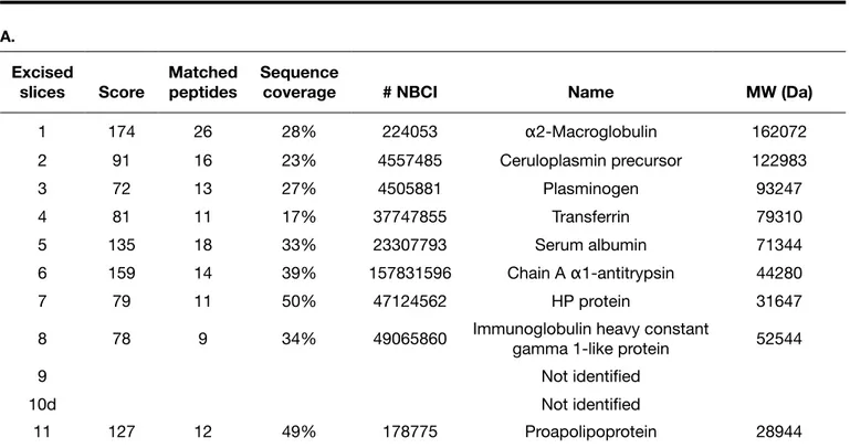
In Table I, peptide mass fingerprinting (A) and peptide fragmentation (B) analyses show that the bands pres-ent in 1-dimensional SDS-PAGE, obtained from uremic plasma without albumin and immunoglobulin depletion, contained polypeptide species from the following gene products: α2-macroglobulin; transferrin; serum albumin; 1 mixture of α1-antitrypsin-serum albumin; immunoglob-ulin gamma 1; fibrinogen gamma; 1 hypothetical albu-min fragment and proapolipoprotein. Moreover, in these experiments serum albumin was identified in multiple bands that were DNPH-positive in Western blot analysis.
Bands 2 and 3 were not identified in these experiments while they were identified, after albumin and immuno-globulin depletion, as a ceruloplasmin precursor (Tab.IIA, B) and plasminogen (Tab. IIA), respectively.
MALDI-TOF/MS mass fingerprinting and nanoLC-ESI-Q-TOF-MS/MS identification of carbonylated proteins in depleted plasma confirmed the presence of polypep-tides having an extensive sequence overlap with α2-macroglobulin, transferrin, serum albumin and proapoli-poprotein (Tab. IIA), or apoliproapoli-poprotein A-I (Tab. IIB). Band 10 shown in Figure 2, panel A, identified as a probable serum albumin fragment (Tab. IA, B), was not revealed
B. Excised slices Score Matched peptides Sequence
coverage # NBCI Name MW (Da)
1 330 10 6% 224053 α2-Macroglobulin 162072 2 Not identified 3 Not identified 4 420 11 15% 37747855 Transferrin 79310 5 130 4 5% 23307793 Serum albumin 71344 6 359 10 28% 157831596 Chain A α1-antitrypsin 44280 429 13 19% 23307793 Serum albumin 71344 7 182 5 10% 2765425 Immunoglobulin lambda heavy chain 53379 8 142 3 8% 49065860 Immunoglobulin heavy constant gamma 1-like protein 52544 9 170 6 12% 182439 Fibrinogen gamma chain 50077 10 132 4 6% 28592 Serum albumin 71316 11 317 8 30% 178775 Proapolipoprotein 28944
TABLE I
(A) MALDI-TOF/MS MASS FINGERPRINTING AND (B) nanoLC-nano-ESI-Q-TOF MS/MS IDENTIFICATION OF CARBO-NYLATED PROTEINS FROM UREMIC PLASMA
A. Excised slices Score Matched peptides Sequence
coverage # NBCI Name MW (Da)
1 159 25 29% 224053 α2-Macroglobulin 162072 2 Not identified 3 Not identified 4 78 14 23% 37747855 Transferrin 79310 5 168 18 34% 23307793 Serum albumin 71344 6 100 16 46% 157831596 Chain A α1-antitrypsin 44280 193 23 41% 23307793 Serum albumin 71344 7 71 12 25% 23307793 Serum albumin 71344 8 96 11 33% 49065860 Immunoglobulin heavy constant gamma 1-like protein 52544 9 94 12 49% 182439 Fibrinogen gamma chain 50077 10 79 13 29% 23307793 Serum albumin 71344
TABLE II
(A) MALDI-TOF/MS MASS FINGERPRINTING AND (B) nanoLC-nano-ESI-Q-TOF MS/MS IDENTIFICATION OF CARBO-NYLATED PROTEINS FROM ALBUMIN- AND IMMUNOGLOBULIN-DEPLETED UREMIC PLASMA
A.
Excised
slices Score Matched peptides Sequence coverage # NBCI Name MW (Da)
1 174 26 28% 224053 α2-Macroglobulin 162072 2 91 16 23% 4557485 Ceruloplasmin precursor 122983 3 72 13 27% 4505881 Plasminogen 93247 4 81 11 17% 37747855 Transferrin 79310 5 135 18 33% 23307793 Serum albumin 71344 6 159 14 39% 157831596 Chain A α1-antitrypsin 44280 7 79 11 50% 47124562 HP protein 31647
8 78 9 34% 49065860 Immunoglobulin heavy constant gamma 1-like protein 52544
9 Not identified 10d Not identified 11 127 12 49% 178775 Proapolipoprotein 28944 B. Excised slices Score Matched peptides Sequence
coverage # NBCI Name MW (Da)
1 937 23 17% 46812315 α2-Macroglobulin 167505 2 443 11 11% 4557485 Ceruloplasmin precursor 122983 3 Not identified 4 637 16 26% 37747855 Transferrin 79310 5 228 10 14% 23307793 Serum albumin 71344 6 44 1 2% 157831596 Chain A α1-antitrypsin 44280 7 94 2 7% 47124562 HP protein 31647 8 204 4 9% 49065860 Immunoglobulin heavy constant gamma 1-like protein 52544
9 Not identified
10d Not identified
after albumin and immunoglobulin depletion (Fig. 2, pan-el B). Furthermore, in both peptide mass fingerprinting and nanoLC-MS/MS analysis (Tab. IIA, B), α1-antitrypsin and haptoglobin (HP) were assigned without any albu-min and immunoglobulin contaalbu-mination and bands 9 and 10d were not identified, probably due to a low protein concentration. The presence of immunoglobulin chains after depletion might be due to the partial efficacy of the affinity-capturing material, as already reported (22), or to a loss of protein structure due to the high degree of car-bonyl damage present in the polypeptide structure.
Effect of carbonylated albumin on endothelial adhesion molecule mRNA levels
Figure 3 panel A shows SDS-PAGE and Western blotting of carbonylated HSA and noncarbonylated HSA sam-ples, which demonstrate carbonylation of HSA. Figure 3 panels B and C show that carbonylated HSA (1 and 25 mg/L) significantly increased VCAM-1 and ICAM-1 gene expression levels (p<0.05, respectively). By con-trast, in the same experimental conditions, eNOS gene expression was not modified (data not shown). As ex-pected, the positive control TNFα (1 ng/mL) significantly increased both VCAM-1 and ICAM-1 gene expression levels (p<0.05 for each, Fig. 3) and decreased eNOS expression (p<0.05, data not shown). No significant ef-fect on VCAM-1 and ICAM-1 mRNA levels was observed when HUVECs were cultured with noncarbonylated HSA (Fig. 3, panels B and C).
dIscussIon
The presented data provide some novel features regard-ing the process of protein carbonylation in uremia. The experimental techniques performed (SDS-PAGE, Western blotting, peptide mass fingerprinting, and peptide nano-LC-MS/MS analysis) allowed us to detect carbonyl com-pounds in plasma from stabilized nondiabetic HD patients and to identify the main protein targets of carbonyl stress. We also used a molecular reduction strategy in order to re-duce sample complexity by first applying a depletion strat-egy of the main carbonyl scavengers and then coupling an additional high performance separation by reverse-phase nanoLC. Our results are the first to show that several dif-ferent plasma proteins may be targets for carbonylation in uremia. Furthermore, the in vitro finding that even low con-centrations of carbonylated albumin may have proathero-genic properties indicates a potential role for protein car-bonylation in the complex pathophysiology of the uremic syndrome.
Carbonyl stress in chronic uremia, as documented by sev-eral studies (5, 14-19) including the present one, may be the consequence of 2 competing but not mutually exclusive mechanisms: increased generation or decreased removal (clearance or detoxification) of RCOs. Uremia-associated oxidative stress (29) could modify proteins either directly by reactive oxygen species or indirectly by RCOs generated by the autoxidation of a variety of sources (5). The buildup of RCOs in uremia might also be accounted for by a de-crease in their renal clearance (30). In addition, since RCOs react reversibly with the thiol group of glutathione and are
Fig. 3 - A) Silver-stained SDS-PAGE and Western blot of carbonylated HSA (c-HSA) and noncarbonylated HSA (nc-HSA) show-ing carbonylation of HSA. B-C) Real-time PCR evaluation of VCAM-1 and ICAM-1 gene expression. Real-time PCR analysis of VCAM-1 (B) and ICAM-1 (C) mRNA levels, relative to 18s mRNA levels, in HUVECs stimulated for 6 hours with fresh HSA (f-HSA), nc-HSA or c-HSA at 1 or 25 mg/L concentration, and expressed vs. basal (unstimulated). TNF-α (1 ng/mL) was used as a positive control. Bar plots represent the mean ± SD of 3 independent real-time PCR experiments (*p<0.05 vs. C).
subsequently detoxified, the significant decrease in both erythrocyte glutathione concentration and serum activity of glutathione-dependent enzymes found in uremia (31, 32) could result in increased levels of RCOs.
While it appears that carbonyl stress may result from ure-mia per se (17, 18), the present study supports the concept (14-16, 30, 33) that HD treatment may adversely affect the carbonyl balance and exacerbate carbonyl stress. Our data show a significant increase in plasma total carbonylated protein levels after the dialysis procedure. During hemodi-alysis there are several conditions that may all contribute, singly or in concert, to increase carbonyl stress: the ab-sence of complete correction of uremic toxicity, bioincom-patible reactions following the contact of blood with the dialyzer membrane leading, among others, to increased oxygen radical production (34, 35), reduced activities of an-tioxidants (16), and possible increased generation of RCOs (36). Identification of the exact biological mechanisms in-volved in HD-induced carbonylation of some plasma pro-teins was, however, outside the scope of the present inves-tigation and requires further studies. Such studies might help in identifying those dialysis strategies that best an-tagonize the carbonyl overload in this patient population. Previous studies in uremic patients have demonstrated the susceptibility of major plasma proteins to carbonyl forma-tion (18, 19). Our data are therefore confirmatory of those findings, providing molecular detection by peptide MS ex-periments. In particular, albumin acts as a key scavenger of carbonyl reactive species, as attested by the fact that multiple isoforms of HSA were found to be reactive to the DNPH bait. Such a buffering role of HSA has already been reported for the redox stress response in plasma from pa-tients suffering from primary nephritic syndromes (37), al-though the functional impact of the chemically injured HSA isoform has not yet been elucidated.
However, since the relative amount of a protein is not a factor determining the degree of carbonylation (38), we also examined whether less abundant plasma proteins are targets for carbonylation in uremia. To address this issue, plasma samples obtained from HD patients were depleted of albumin and immunoglobulins, and then analyzed again by the same proteomic approach. Our results indicate that several different plasma proteins may be carbonylated in chronic uremia (Tab. II), which extends previous informa-tion. Our study suggests that in uremia carbonylation could be a selective process involving also proteins present in blood in small amounts. The susceptibility of proteins to oxidation might be determined by their metal-binding sites or structural characteristics (39). Target proteins of carbo-nylation in uremia such as transferrin, albumin and
ceru-loplasmin contain 8 iron ion binding sites, and 1 and 12 copper ion binding sites, respectively (39). Different sensi-tivity to carbonylation might also depend on pathogenetic factors that make proteins more susceptible to oxidation in certain microenvironments (40).
Carbonylation can cause several different protein modifi-cations, every one of which may produce (or not) a spe-cific effect on the biological activity of different proteins (9). Carbonylation of haptoglobin and ceruloplasmin, as we de-tected in uremic blood, can impair the antioxidant protec-tive properties of those proteins (41, 42). It has been shown that carbonyl stress (AGE)-modified proteins may exhibit several biological activities initiating a range of inflamma-tory responses (reviewed in 5). Furthermore, the interac-tion of reactive carbonyl compounds (either free or protein bound) with cell surface membrane proteins may induce intracellular responses (43, 44), triggered by signaling path-ways involving P21RAS, mitogen-activated protein kinases,
and nuclear factor-κB (43).
Although the pathophysiological significance of protein car-bonylation in uremia remains to be definitively established, carbonyl stress may be relevant to various complications of chronic renal failure (6, 45). Dialysis-associated amyloido-sis, in particular, has been related to carbonyl stress (5, 6). It is thought that carbonylation of fibrinogen may contribute to the impaired clotting activity observed in HD patients (19). Accumulation of AGEs may be an important pathoge-netic factor for low bone turnover in dialysis patients (46). Moreover, several studies have demonstrated that oxidative alteration of albumin may adversely affect its vasculopro-tective effects on ESRD patients (18, 47). In addition, there is circumstantial support for the hypothesis that carbonyl stress is involved in the pathogenesis of alterations in left ventricular geometry and function in these patients (45). Atherosclerosis is another potential target of carbonylation in uremia (6). Patients suffering from ESRD experience ac-celerated atherosclerosis (48), and atherosclerotic cardio-vascular events account for a large proportion of the mor-bidity and mortality in these patients (49). It is commonly accepted that endothelial cell injury including increased expression of the endothelial adhesion molecules ICAM-1 and VCAM-1, which support firm leukocyte adhesion and transmigration into stimulated endothelium, is an initial event in atherosclerosis (21). The blood concentrations of VCAM-1 and ICAM-1, which may be shed from activated endothelial cells into the circulation, are increased in pa-tients with renal failure (50), and experimental models have demonstrated the involvement of these molecules in ure-mic atherosclerosis (51). However, just how uremia induces a proinflammatory state leading to increased endothelial
adhesion molecule expression and hence accelerated ath-erosclerosis has not been fully elucidated.
Our study supports the notion that carbonylated albumin may directly damage the endothelium, contributing to the loss of endothelial function. The results obtained in cultured HUVECs demonstrated that low doses of carbonylated al-bumin significantly increased the mRNA levels of the en-dothelial adhesion molecules in vitro, indicating that protein carbonylation processes may play a major role in the early atherogenic events of chronic uremia. On the other hand, in our cell model eNOS mRNA levels were not influenced by exposure to carbonylated albumin, indicating that the observed effect of carbonylated albumin on endothelial ad-hesion molecule expression may be independent of nitric oxide availability (20).
There are many possible applications for proteomic ap-proaches to issues related to nephrology. A recent study using proteomics revealed significant differences in the platelet expression of some proteins between dysfunc-tional uremic platelets and uremic platelets with normal functionality (52). The data presented here, obtained by means of proteomic techniques, provide further evidence of protein damage in uremia (53) by showing carbonyl for-mation in several plasma proteins, and support the view that carbonyl stress may be a contributor to uremic toxicity (6), particularly uremic accelerated atherosclerosis. Further research is required to identify carbonylating mechanisms and develop effective therapeutic strategies to reduce the carbonyl overload in uremia. Interventions targeting protein carbonylation may have the potential to prevent vascular lesions under uremic conditions, a concept which requires further investigation.
Financial support:This work was partly supported by a grant from the University of Chieti (ex MURST 60%) to M.B. The authors acknowledge the financing contribution to this work of FIRB project “Human Proteome Network” and of Fondazione Roma 2009. The funders had no role in study design, data collection and analysis, decision to publish, or preparation of the manuscript. Conflict of interest statement: None to declare.
Address for correspondence: Vittorio Sirolli, MD
Clinica Nefrologica - Emodialisi Ospedale Clinicizzato “SS. Annunziata” Via dei Vestini, 66013 Chieti, Italy [email protected]
r
eferencesBaynes JW, Thorpe SR. Role of oxidative stress in diabetic 1.
complications: a new perspective on an old paradigm. Dia-betes. 1999;48:1-9.
Baynes
2. JW, Thorpe SR. Glycoxidation and lipoxidation in atherogenesis. Free Radic Biol Med. 2000;28:1708-1716. Metz
3. TO, Alderson NL, Chachich ME, Thorpe SR, Baynes JW. Pyridoxamine traps intermediates in lipid peroxidation reactions in vivo: evidence on the role of lipids in chemical modification of protein and development of diabetic compli-cations. J Biol Chem. 2003;278:42012-42019.
Rhodes
4. J. Covalent chemical events in immune induction: Fundamental and therapeutic aspects. Immunol Today. 1996;17:436-441.
Miyata
5. T, van Ypersele de Strihou C, Kurokawa K, Baynes JW. Alterations in nonenzymatic biochemistry in uremia: Ori-gin and significance of “carbonyl stress” in long-term uremic complications. Kidney Int. 1999;55:389-399.
Miyata
6. T, Saito A, Kurokawa K, van Ypersele de Strihou C. Advanced glycation and lipoxidation end products: reactive carbonyl compounds-related uraemic toxicity. Nephrol Dial Transplant. 2001;16(Suppl 4):8-11.
Miyata T, Devuyst O, Kurokawa K, van Ypersele de Strihou 7.
C. Toward better dialysis compatibility: advances in the bio-chemistry and pathophysiology of the peritoneal membranes. Kidney Int. 2002;61:375-386.
Kakuta T, Tanaka R, Satoh Y, et al. Pyridoxamine improves 8.
functional, structural, and biochemical alterations of perito-neal membranes in uremic peritoperito-neal dialysis rats. Kidney Int. 2005;68:1326-1336.
Dalle Donne I, Aldini G, Carini M, Colombo R, Rossi R, Mil-9.
zani A. Protein carbonylation, cellular dysfunction, and dis-ease progression. J Cell Mol Med. 2006;10:389-406. Aldini
10. G, Dalle Donne I, Maffei Facino R, Milzani A, Carini M. Intervention strategies to inhibit protein carbonylation by lipoxidation-derived reactive carbonyls. Med Res Rev. 2007;27:817-868.
Miyata
11. T, Ueda Y, Yamada Y, et al. Carbonyl stress in ure-mia: Accumulation of carbonyls accelerates the formation of pentosidine, an advanced glycation end product. J Am Soc Nephrol. 1998;9:2349-2356.
Niwa
12. T, Takeda N, Miyazaki T, et al. Elevated serum levels of 3 deoxyglucosone, a potent protein-cross-linking interme-diate of the Maillard reaction, in uremic patients. Nephron. 1995;69:438-443.
Odani
13. H, Shinzato T, Usami J, et al. Imidazolium crosslinks derived from reaction of lysine with glyoxal and methylgly-oxal are increased in serum proteins of uremic patients: Evi-dence for increased oxidative stress in uremia. FEBS Lett. 1998;427:381-385.
Mayer
14. B, Zitta S, Greilberger J, et al. Effect of hemodi-alysis on the antioxidative properties of serum. Biochim
Biophys Acta. 2003;1638:267-272. Ward
15. RA, Ouseph R, McLeish KR. Effects of high-flux hemo-dialysis on oxidant stress. Kidney Int. 2003;63:353-359. Dursun
16. E, Dursun B, Suleymanlar G, Ozben T. Effect of he-modialysis on the oxidative stress and antioxidants in diabe-tes mellitus. Acta Diabetol. 2005;42:123-128.
Dursun
17. E, Dursun B, Suleymanlar G, Ozben T. Carbonyl stress in chronic renal failure: the effect of hemodialysis. Ann Clin Biochem. 2005;42:64-66.
Himmelfarb
18. J, McMonagle E. Albumin is the major plas-ma protein target of oxidant stress in uremia. Kidney Int. 2001;60:358-363.
Michelis
19. R, Gery R, Sela S, et al. Carbonyl stress induced by intravenous iron during hemodialysis. Nephrol Dial Trans-plant. 2003;18:924-930.
Pandolfi A, Di Pietro N, Sirolli V, et al. Mechanisms of uremic 20.
erythrocyte-induced adhesion of human monocytes to cul-tured endothelial cells. J Cell Physiol. 2007;213:699-709. Ross R. Atherosclerosis. An inflammatory disease. N Engl J 21.
Med. 1999;340:115-126. Zolotarjova
22. N, Martosella J, Nicol G, Bailey J, Boyes B, Bar-ret W. Differences among techniques for high-abundant pro-tein depletion. Proteomics. 2005;5:3304-3313.
Towbin
23. H, Staehelin T, Gordon J. Electrophoretic transfer of proteins from polyacrylamide gels to nitrocellulose sheets: procedure and some applications. Proc Natl Acad Sci USA. 1979;76:4350-4354.
Reinheckel T, Körn S, Möhring S, Augustin W, Halangk W, 24.
Schild L. Adaptation of protein carbonyl detection to the re-quirements of proteome analysis demonstrated for hypoxia/ reoxygenation in isolated rat liver mitochondria. Arch Bio-chem Biophys. 2000;376:59-65.
Shevchenko
25. A, Wilm M, Vorm O, Mann M. Mass spectro-metric sequencing of proteins silver-stained polyacrylamide gels. Anal Chem. 1996;68:850-858.
Mortz
26. E, Krogh TN, Vorum H, Görg A. Improved silver stain-ing protocols for high sensitivity protein identification usstain-ing matrix-assisted laser desorption/ionization-time of flight analysis. Proteomics. 2001;1:1359-1363.
Rashid
27. G, Benchetrit S, Fishman D, Bernheim J. Effect of advanced glycation end-products on gene expression and synthesis of TNF-alpha and endothelial nitric oxide synthase by endothelial cells. Kidney Int. 2004;66:1099-1106.
Gorfien
28. S, Spector A, DeLuca D, Weiss S. Growth and physi-ological functions of vascular endothelial cells in a new se-rum-free medium (SFM). Exp Cell Res. 1993;206:291-301. Himmelfarb
29. J, Hakim RM. Oxidative stress in uremia. Curr Opin Nephrol Hypertens. 2003;12:593-598.
Jadoul
30. M, Ueda Y, Yasuda Y, et al. Influence of hemodialy-sis membrane type on pentosidine plasma level, a marker of “carbonyl stress”. Kidney Int. 1999;55:2487-2492.
Canestrari
31. F, Galli F, Giorgini A, et al. Erythrocyte redox state in uremia anemia: effects of hemodialysis and relevance of glutathione metabolism. Acta Haematol. 1994;91:187-193.
Yeung
32. JH. Effects of glycerol-induced acute renal failure on tissue glutathione and glutathione-dependent enzymes in the rat. Methods Find Exp Clin Pharmacol. 1991;13:23-28. Bordoni
33. V, Piroddi M, Galli F, et al. Oxidant and carbonyl stress-related apoptosis in end-stage kidney disease: im-pact of membrane flux. Blood Purif. 2006;24:149-156. Bonomini
34. M, Stuard S, Carreno M-P, et al. Neutrophil reac-tive oxygen species production during hemodialysis: role of activated platelet adhesion to neutrophils through P-selectin. Nephron. 1997;75:402-411.
Klein
35. JB, McLeish KR, Ward RA. Transplantation, not dialy-sis, corrects azotemia-dependent priming of the neutrophil oxidative burst. Am J Kidney Dis. 1999;33:483-491.
Miyata
36. T, Kurokawa K, Van Ypersele de Strihou C. Advanced glycation and lipoxidation end products: role of reactive car-bonyl compounds generated during carbohydrate and lipid metabolism. J Am Soc Nephrol. 2000;11:1744-1752. Bruschi M, Petretto A, Candiano G, et al. Determination of 37.
the oxido-redox status of plasma albumin in hemodialysis patients. J Chromatogr B Analyt Technol Biomed Life Sci. 2008;864:29-37.
Jana CK, Das N, Sohal RS. Specificity of age-related carbo-38.
nylation of plasma proteins in the mouse and rat. Arch Bio-chem Biophys. 2002;397:433-439.
Stadtman ER, Levine RL. Protein oxidation. Ann N Y Acad 39.
Sci. 2000;899:191-208.
Rottoli P, Magri B, Cianti R, et al. Carbonylated proteins in 40.
bronchoalveolar lavage of patients with sarcoidosis, pulmo-nary fibrosis associated with systemic sclerosis and idio-pathic pulmonary fibrosis. Proteomics. 2005;5:2612-2618. Kang
41. JH, Kim KS, Choi SY, Know HY, Won MH. Oxidative modification of human ceruloplasmin by peroxyl radicals. Biochim Biophys Acta. 2001;1568:30-36.
Miller
42. YI, Altamentova SM, Shaklai N. Oxidation of low-den-sity lipoprotein by hemoglobin stem from a heme initiated globin radical: antioxidant role of haptoglobin. Biochemistry. 1997;36:12189-12198.
Yan SD, Schmidt AM, Anderson GM, et al. Enhanced cellular 43.
oxidant stress by the interaction of advanced glycation end products with their receptors/binding proteins. J Biol Chem. 1994;269:9889-9897.
Akhand AA, Kato M, Suzuki H, et al. Carbonyl compounds 44.
cross-link cellular proteins and activate protein-tyrosine ki-nase p60e-Sre. J Cell Biochem. 1999;72:1-7.
Zoccali
45. C, Mallamaci F, Tripepi G. AGEs and carbonyl stress: potential pathogenetic factors of long-term uraemic compli-cations. Nephrol Dial Transplant. 2000;15:7-11.
Panuccio V, Mallamaci F, Tripepi G, et al. Low parathyroid 46.
hormone and pentosidine in hemodialysis patients. Am J Kidney Dis. 2002;40:810-815.
Lim PS, Cheng YM, Yang SM. Impairments of the biologi-47.
cal properties of serum albumin in patients on hemodialysis. Nephrology (Carlton). 2007;12:18-24.
Lindner A, Charra B, Sherrard DJ, Scribner BH. Accelerated 48.
atherosclerosis in prolonged maintenance hemodialysis. N Engl J Med. 1974;290:697-701.
Locatelli F, Del Vecchio L, Manzoni C. Morbidity and mor-49.
tality on maintenance haemodialysis. Contrib Nephrol. 1998;124:166-189.
Bonomini M, Reale M, Santarelli P, et al. Serum levels of sol-50.
uble adhesion molecules in chronic renal failure and dialysis patients. Nephron. 1998;79:399-407.
Bro
51. S, Moeller F, Andersen CB, Olgaard K, Nielsen LB. In-creased expression of adhesion molecules in uremic
ath-erosclerosis in apolipoprotein-E-deficient mice. J Am Soc Nephrol. 2004;15:1495-1503.
Marques
52. M, Sacristàn D, Mateos-Càceres PJ, et al. Different protein expression in normal and dysfunctional platelets from uremic patients. J Nephrol. 2010;23:90-101.
Galli
53. F. Protein damage and inflammation in uremia and dialy-sis patients. Nephrol Dial Transplant. 2007;22(Suppl. 5):v20-v36.