See discussions, stats, and author profiles for this publication at: https://www.researchgate.net/publication/284182018
Molecular diagnosis of native and quarantine
pest thrips (Thysanoptera, Thripidae) of
southern European citrus orchards.
Article in Bulletin of Insectology · June 2016 CITATIONS0
READS179
3 authors: Some of the authors of this publication are also working on these related projects: Invasive thrips species to southern European RegionsView project Implementation of integrated thrips and tospovirus management strategies in smallholder vegetable cropping systems of eastern Africa
View project Alessandra De Grazia Mediterranean University of Reggio Calabria 17 PUBLICATIONS 12 CITATIONS SEE PROFILE Rita Marullo Mediterranean University of Reggio Calabria 76 PUBLICATIONS 620 CITATIONS SEE PROFILE Gerald B. Moritz Institute of Biology 120 PUBLICATIONS 519 CITATIONS SEE PROFILE
All content following this page was uploaded by Alessandra De Grazia on 28 April 2016.
The user has requested enhancement of the downloaded file. All in-text references underlined in blue are added to the original document and are linked to publications on ResearchGate, letting you access and read them immediately.
Bulletin of Insectology 69 (1): 1-6, 2016
ISSN 1721-8861
Molecular diagnosis of native and quarantine pest thrips of
southern European citrus orchards
Alessandra DE GRAZIA1, Rita MARULLO1, Gerald MORITZ21Dipartimento di Agraria, Università degli Studi Mediterranea di Reggio Calabria, Reggio Calabria, Italy
2Institute of Biology, Martin-Luther-University Halle-Wittenberg, Faculty of Natural Sciences I, Halle, Germany
Abstract
The ITS-RFLP technique of the amplified internal transcribed spacer regions of ribosomal DNA has been used to establish the molecular identification of the most common pest thrips species of citrus orchards. Both native and quarantine thrips species have been tested and molecular keys are proposed through ITS-RFLP techniques. The restriction enzymes produced patterns that al-lowed unambiguous identification of seven thrips species examined (visual key). The sequences of species can also be verified using a molecular key that permits identification of a species through such parameters as primer pair used, restriction enzyme, length of PCR-product, fragments obtained. This study has shown that the use of genetic markers can be a valid alternative for quarantine workers and for epidemiological researchers, to whom the correct identification of pest species through classic mor-phological methods could be either very difficult and time-consuming or visually impossible.
Key words: molecular identification, ITS-RFLP, visual key, thrips, citrus pest.
Introduction
The insect order Thysanoptera includes in the family Thripidae about 50 species associated with economic crops and also quarantine species. Problems related to quarantine species are mainly due to identification: dif-ficulties related to their small size, variability of body colour, morphological patterns, secondary sexual char-acter states and the cryptic behaviour of some species which exhibit large differences in genetic compositions, habitat preference, Tospovirus transmission efficiency (Brunner et al., 2004; Hoddle et al., 2008; Brunner and Frey, 2010; Rugman-Jones et al., 2010; Jacobson et al., 2013). Classical morphological identification keys re-quire experience and are difficult for non-specialists such as most quarantine workers. Moreover, these diffi-culties are greater when an identification of either larval stages or eggs that infest trade vegetables is required. Larval identification keys are rare (Kucharczyk, 2010; Vierbergen et al., 2010) and practically useless in terms of fast and accurate pest identification, but during the past 15 years identification systems which combine classic morphological knowledge with molecular identi-fication methods have been produced (Moritz et al., 2000; 2004; 2009; 2010; 2013; Tyagi et al., 2015). Fur-thermore, improving molecular techniques have led to molecular keys that provide quick and correct identifi-cation ways for adults and all ontogenetic stages of the most common pest thrips species (Brunner et al., 2002; Toda et al., 2002; Kox et al., 2005; Rugman-Jones et
al., 2006; Timm et al., 2008; Fekrat et al., 2015;
Przy-bylska et al., 2015). The internal transcribed spacer (ITS), the non-coding fragment of the nuclear ribosomal region, has been one of the most widely used markers in thrips species identification (Rugman-Jones et al., 2006; Farris et al., 2010).
In the present article, a molecular technique is devel-oped to identify the most common harmful thrips spe-cies on citrus plants in the southern areas of Italy and
the Mediterranean Basin, together with a few exotic thrips species that are the most important quarantine pests for these crops. Results of molecular identification study of seven pest thrips species on citrus (Marullo and De Grazia, 2012) are presented and discussed; four of them are present in the Mediterranean Region, i.e.
Heliothrips haemorrhoidalis (Bouche), Frankliniella occidentalis (Pergande), Pezothrips kellyanus (Bagnall), Thrips tabaci Lindeman, whereas three species, Frank-liniella bispinosa (Morgan), Scirtothrips aurantii Faure,
and Scirtothrips citri (Moulton) are considered quaran-tine species for EU territories.
Materials and methods Collections of thrips species
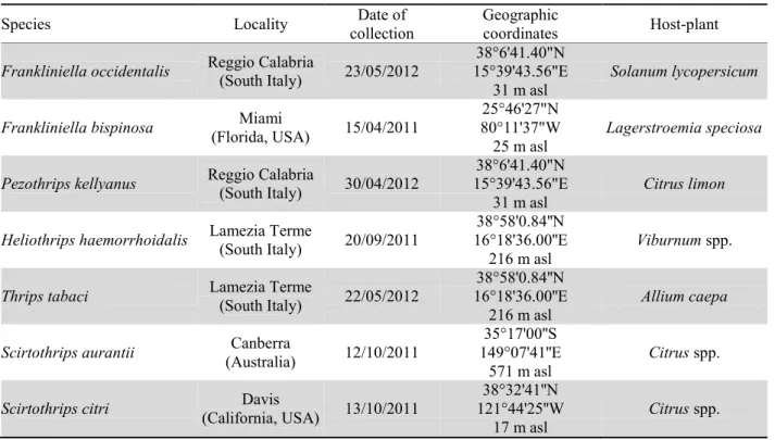
Seventy individual thrips identified as seven different species were collected for this study (collecting details are summarized in table 1). The specimens of F.
bispinosa, S. citri and S. aurantii from field populations,
were provided by Prof. J Funderburk (University of Florida, USA) and Prof. L. Mound (CSIRO, Australia), respectively.
Molecular technique
Ten specimens of each species studied have been tested using a protocol which provides a few phases: - DNA extraction: DNA has been extracted from sin-gle specimens using the Qiagen “DNeasy Tissue” kit (Germany) which allows the carcass of each specimen to be retrieved and mounted onto microscope slides. With the aid of a sterilized needle, we incised the specimen’s abdomen and placed it in a 1.5 ml tube con-taining 20 µl proteinase K. After thoroughly mix by vortexing, the tube was incubated overnight at 55 °C. Then, we added 200 µl Buffer AL (provided with the kit) to the sample and vortexed it. After a short incubation (15 minutes at 70 °C) we added 200 µl ethanol (100%)
2
Table 1. Sampling details for the collected species.
Species Locality collection Date of Geographic coordinates Host-plant
Frankliniella occidentalis Reggio Calabria (South Italy) 23/05/2012 15°39'43.56"E 38°6'41.40"N
31 m asl Solanum lycopersicum
Frankliniella bispinosa (Florida, USA) Miami 15/04/2011 80°11'37"W 25°46'27"N
25 m asl Lagerstroemia speciosa
Pezothrips kellyanus Reggio Calabria (South Italy) 30/04/2012 15°39'43.56"E 38°6'41.40"N
31 m asl
Citrus limon
Heliothrips haemorrhoidalis Lamezia Terme (South Italy) 20/09/2011 16°18'36.00''E 38°58'0.84''N
216 m asl
Viburnum spp.
Thrips tabaci Lamezia Terme (South Italy) 22/05/2012 16°18'36.00''E 38°58'0.84''N
216 m asl Allium caepa
Scirtothrips aurantii (Australia) Canberra 12/10/2011 149°07'41''E 35°17'00''S
571 m asl Citrus spp.
Scirtothrips citri (California, USA) Davis 13/10/2011
38°32'41''N 121°44'25''W
17 m asl Citrus spp.
and mix all again. We inserted the mixture into DNeasy Mini spin column (provided with the kit) placed in a 2 ml collection tube and centrifuged. We discarded flow-through and transferred the spin column to a new 2 ml tube where we added 500 µl Buffer AW1 (provided) and after centrifugation, we again discarded flow-through and transferred the spin column to a new 2 ml tube in which we added 500 µl Buffer AW2 (provided) and cen-trifuged to dry the DNeasy membrane. We placed the DNeasy Mini spin column in a clean 1.5 ml tube and we added 70 µl sterile, ultra pure water directly onto the DNeasy membrane. Finally we incubated at room tem-perature for 1 minute, and centrifuged to elute DNA; - PCR conditions: the ITS regions have been ampli-fied with 2 primer pairs (CS249/CS250:
5’-TCGTAACAAGGTTTCCG-3’; -
GTTAGTTTCTTTTCCTC-3’; 18SMP/28SMP:
5’-TGAACCTGCGGAAGGAT-3’;
5’-TCTCACCTBAACTGAGG-3’) and the polymerase chain reaction mixture contained: 10 µl of template; 5.0 µl of 10x PCR-buffer; 0.4 µl of 25 mM dNTPs; 4.0 µl of 25 mM MgCl2; 1.5 µl of each primer; 0.2 µl of Taq-polymerase and sterile distilled water to a final vol-ume of 50 µl. The amplification is carried out in an Ep-pendorf Mastercycler gradient. The DNA has been de-natured at 95 °C for 3 minutes, followed by 30 cycles of denaturation at 95 °C for 45 seconds. Annealing (the temperature at which the primer anneals or binds to the target site) at 47 °C (CS249-CS250) or 54 °C (18SMP-28SMP) for 45 seconds. and elongation at 72 °C for 2 minutes. The last cycle is followed by 4 minutes of incubation at 72 °C to complete any partially synthe-sized strands;
- Restriction: 7 µl of the PCR-products have been di-gested with 3 units of five enzymes (RsaI, HaeIII, MspI,
HinfI and AluI) (Promega) and observed by electrophore-sis in a 2% agarose gel with ethidium bromide to visual-ize the DNA restriction fragments (Moritz et al., 2000). Results
The technique of RFLP of the amplified internal tran-scribed spacer region of ribosomal DNA (ITS1 and ITS2) enables the production of specific DNA fragment patterns for the identification of thrips species. The identification of a species has to be done through com-parison of different patterns obtained with the same couple of primers (visual key) (figures 1-2).
Figure 1 shows the differences between F. bispinosa,
P. kellyanus, S. citri and T. tabaci whose DNA was
am-plified using the primers 18SMP-28SMP. In all the spe-cies, patterns are represented by a sequence of bands for each restriction enzyme used (table 2). In particular, lane 3 corresponds to restriction fragments obtained by digestion of rDNA of F. bispinosa using RsaI enzyme; lane 4 coincides in restriction fragments by digestion of rDNA with HaeIII enzyme; lane 5 shows the bands ob-tained by MspI enzyme; lane 6 and 7 represent restric-tion fragments obtained with HinfI and AluI, respec-tively. Similarly, lanes 11-15, 19-23 and 27-31, repre-sent the bands obtained through the digestion of rDNA of P. kellyanus , T. tabaci, S. citri, respectively.
Figure 2 reproduces the patterns of F. bispinosa and
F. occidentalis by using the primers CS249-CS250.
Each of the seven species constitutes a distinct pattern. Figure 3 shows the patterns of the second instar larvae of F. occidentalis and P. kellyanus obtained by diges-tion of rDNA with 5 restricdiges-tion enzymes (RsaI, HaeIII, MspI, HinfI and AluI).
Figure 1. Agarose gel showing the ITS-RFLP patterns of F. bispinosa (lanes 3-7), P. kellyanus (lanes 11-15), T.
tabaci (lanes 19-23) and S. citri (lanes 27-31) obtained by digestion of amplified ITS regions of rDNA
(18SMP-28SMP) with RsaI, HaeIII, MspI, HinfI and AluI. Lanes 1, 8, 9, 16, 17, 24, 25 and 32, 100 bp DNA ladder. Lanes 2, 10, 18 and 26, PCR products of F. bispinosa, P. kellyanus, T. tabaci and S. citri respectively.
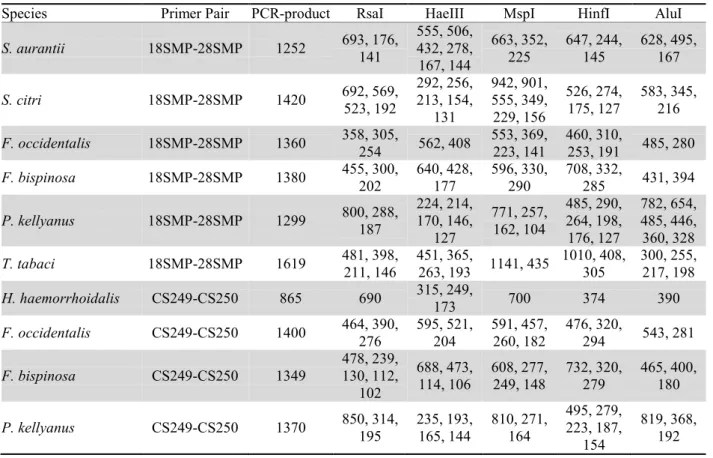
Table 2. Size (in base pairs) of the ITS-RFLP restriction fragments of each species tested.
Species Primer Pair PCR-product RsaI HaeIII MspI HinfI AluI
S. aurantii 18SMP-28SMP 1252 693, 176, 141 555, 506, 432, 278, 167, 144 663, 352, 225 647, 244, 145 628, 495, 167 S. citri 18SMP-28SMP 1420 692, 569, 523, 192 292, 256, 213, 154, 131 942, 901, 555, 349, 229, 156 526, 274, 175, 127 583, 345, 216 F. occidentalis 18SMP-28SMP 1360 358, 305, 254 562, 408 553, 369, 223, 141 460, 310, 253, 191 485, 280 F. bispinosa 18SMP-28SMP 1380 455, 300, 202 640, 428, 177 596, 330, 290 708, 332, 285 431, 394 P. kellyanus 18SMP-28SMP 1299 800, 288, 187 224, 214, 170, 146, 127 771, 257, 162, 104 485, 290, 264, 198, 176, 127 782, 654, 485, 446, 360, 328 T. tabaci 18SMP-28SMP 1619 481, 398, 211, 146 451, 365, 263, 193 1141, 435 1010, 408, 305 300, 255, 217, 198 H. haemorrhoidalis CS249-CS250 865 690 315, 249, 173 700 374 390 F. occidentalis CS249-CS250 1400 464, 390, 276 595, 521, 204 591, 457, 260, 182 476, 320, 294 543, 281 F. bispinosa CS249-CS250 1349 478, 239, 130, 112, 102 688, 473, 114, 106 608, 277, 249, 148 732, 320, 279 465, 400, 180 P. kellyanus CS249-CS250 1370 850, 314, 195 235, 193, 165, 144 810, 271, 164 495, 279, 223, 187, 154 819, 368, 192
Sequences of species determined by ITS-RFLP analysis were used to develop two molecular identifica-tion keys to confirm the identificaidentifica-tion of a species through a series of parameters as: primer pair used, re-striction enzyme, length of PCR-product, fragments ob-tained. In a first key (table 3) six species studied were included, considering differences in size of the PCR products and restriction fragment lengths using the primer pair 18SMP-28SMP. These primers did not work on H. haemorrhoidalis. Therefore, a second key
for the identification of four species (table 4) was obtained using the primer pair CS249-CS250. Keys require the user to make a choice between only two data at a time. The choices are numbered and refer to the size of the ITS band and the number of fragments obtained by a precise restriction enzyme. In addition, for a further confirmation of the correct identification of a species it is possible to connect to the web-data base: http://moritz.zoologie.uni-halle.de/ (Moritz, 2010).
4
Figure 2. Agarose gel showing the ITS-RFLP patterns of F. bispinosa (lanes 3-7) and F. occidentalis (lanes 11-15), obtained by digestion of amplified ITS regions of rDNA (CS249-CS250) with RsaI, HaeIII, MspI, HinfI and AluI. Lanes 1, 8, 9 and 16, 100 bp DNA ladder. Lanes 2 and 10, PCR products of analysed species.
Figure 3. Agarose gel showing the ITS-RFLP patterns of larval stages of F. occidentalis (lanes 3-7) and P. kellyanus (lanes 11-15) obtained by digestion of amplified ITS regions of rDNA (CS249-CS250) with RsaI, HaeIII, MspI, HinfI and AluI. Lanes 2 and 10 represent PCR products. Lanes 1 and 16, 100 bp DNA ladder.
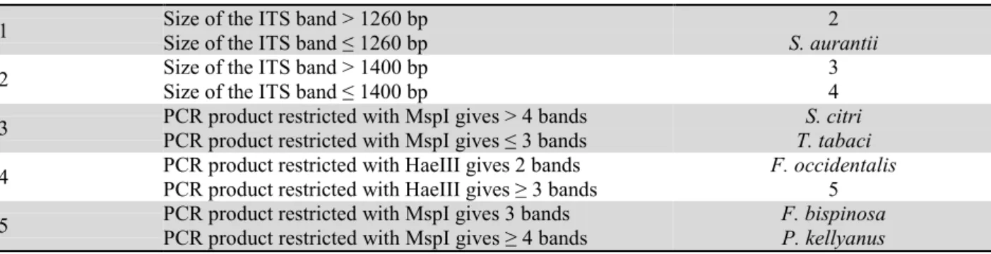
Table 3. Molecular key to thrips species based on ITS-RFLP products obtained with 18SMP-28SMP.
1 Size of the ITS band > 1260 bp Size of the ITS band ≤ 1260 bp S. aurantii 2
2 Size of the ITS band > 1400 bp Size of the ITS band ≤ 1400 bp 3 4
3 PCR product restricted with MspI gives > 4 bands PCR product restricted with MspI gives ≤ 3 bands T. tabaci S. citri 4 PCR product restricted with HaeIII gives 2 bands PCR product restricted with HaeIII gives ≥ 3 bands F. occidentalis 5 5 PCR product restricted with MspI gives 3 bands PCR product restricted with MspI gives ≥ 4 bands F. bispinosa P. kellyanus Table 4. Molecular key to thrips species based on ITS-RFLP products obtained with CS249-CS250. 1 Size of the ITS band > 1000 bp Size of the ITS band < 1000 bp H. haemorrhoidalis 2
2 Size of the ITS band > 1350 bp Size of the ITS band < 1350 bp 3 4
3 PCR product restricted with RsaI gives 3 bands PCR product restricted with RsaI gives > 3 bands F. bispinosa 4 4 PCR product restricted with MspI gives 3 bands PCR product restricted with HaeIII gives > 3 bands F. occidentalis P. kellyanus
Discussion
DNA patterns generated by ITS-RFLP have potential as useful tool in the identification of pest thrips species. Furthermore, we have successfully tested this method on instar stages, for which morphological keys are scanty or difficult to utilize, and a similar system has been applied to identify single eggs (Moritz et al., 2004). The technique might improve the efficacy of pest quarantine diagnoses. The identification to species level of larval stages is usually avoided by quarantine work-ers. Larval thrips are often mistaken for Collembola, or adults are confused with Staphylinid beetles (Vierber-gen, 1995). Such misidentifications might be important in the case of thrips species that have a predatory behav-iour, such as Karnyothrips or Scolothrips species (Ma-rullo and De Grazia, 2013).Therefore, a need exists for a system of identifying adult and larval stages of pest thrips species. Recently, the molecular technique known as loop-mediated isothermal amplification (LAMP) was employed to discriminate the presence of DNA of
T. tabaci from other species, using material from
differ-ent stages of developmdiffer-ent (Fekrat et al., 2015) and to detect Thrips palmi Karny, a pest species under quaran-tine regulation in the European Union. These studies highlighted the sensitivity and the usefulness of LAMP to identify all the stages of the life cycle, including the preimaginal stage, which otherwise could be only dif-ferentiated by expert taxonomists (Przybylska et al., 2015).
In this study, two molecular keys have been presented, one with six of the seven species investigated and the other was developed to include H. haemorrhoidalis that was not represented in the first key. Production of a mo-lecular key that involves members of different genera can be difficult, and certain constraints have to be con-sidered. In the present work analysis, a few technical problems were encountered, such as the absence of re-sults for H. haemorrhoidalis using the primer pair 18SMP-28SMP in order to amplify the ITS regions. Also, the primer pair CS249-CS250 was ineffective for
T. tabaci, S. citri and S. aurantii . A further weakness of
the ITS-RFLP method is that it still requires morpho-logical identification of specimens, as similarly asserted by Rugman-Jones et al. (2006) for the most important pest species of the genus Scirtothrips. This aspect might be an hindrance for non-specialist taxonomists not fa-miliar with thrips' dichotomous and electronic keys (Hoddle et al., 2012; ThripsWiki, 2014).
Another critical consideration is that studies based on PCR are able to detect only a few species at a time. It is essential to develop a more efficient method for simul-taneous screening of mass samples. Possibly, DNA bar-coding methods in combination with the mitochondrial COI gene could be a future way to molecular studies (Mehle and Trdan, 2012). An integrated approach of morphology and DNA barcoding for the invasive pest thrips, Thrips parvispinus (Karni) is proposed from Tyagi et al., 2015, in order to support how DNA bar-coding data may be immense use for accurate species identification and phylogenetic analysis (Mound et al, 2010).
Moroever, recent studies (Chung et al., 2011; Lee et
al., 2013; Yeh et al., 2015) report the microarray assay
as a valid method for insect pest identification. Particu-larly, Yeh et al. (2015), using DNA microarray based on species-specific primers referring to ITS1 sequences of 15 agriculturally important thrips, provided an efficient tool for the simultaneous identification and monitoring of a number of thrips species. In conclusion, the avail-ability of a molecular key could be extremely useful in the identification of species. The advantage of such an identification system over morphology-based taxonomic methods and other molecular techniques is that it is quick, specific, requires only basic laboratory skills, and can be performed with DNA extracted from single indi-viduals, which can be preserved, mounted on slides and used for future reference. On balance, molecular meth-ods for identification of thrips species represent an im-portant possibility when classical morphological meth-ods are difficult, time consuming or visually impossible. Acknowledgements
The Authors are grateful to Laurence Mound, Australian National Insect Collection, CSIRO, Canberra (Austra-lia) for the text revision. The scientific work, in particu-lar the stage of A. De Grazia at Molecuparticu-lar Laboratory of the Biology Institute, Halle-Wittenberg University (Germany), has been carried on through the financial support of GEISCA Project 2010/2011 (MIUR, Ministe-ro dell’Università e della Ricerca Scientifica e Tecnolo-gica, Roma).
References
BRUNNER P. C., FREY J. E., 2010.- Habitat-specific population structure in native western flower thrips Frankliniella
occi-dentalis (Insecta, Thysanoptera).- Journal of Evolutionary Biology, 23: 797-804.
BRUNNER P. C., FLEMING C., FREY J. E., 2002.- A molecular identification key for economically important thrips species (Thysanoptera: Thripidae) using direct sequencing and a PCR-RLFP-based approach.- Agricultural and Forest
Ento-mology, 4: 127-136.
BRUNNER P. C., CHATZIVASSILIOU E. K., KATIS N. I., FREY J. E., 2004.- Host-associated genetic differentiation in Thrips
tabaci (Insecta: Thysanoptera), as determined from mtDNA
sequence data.- Heredity, 93: 364-370.
CHUNG I. H., KANG S., KIM Y. R., KIM J. H., JUNG J. W., LEE S., LEE S. H., 2011.- Development of a low density DNA microarray for diagnosis of target-site mutations of pyretroid and organophosphate resistance mutations in the whitefly
Bemisia tabaci.- Pest Management Science, 67: 1541-1548.
FARRIS R. E., RUIZ-ARCE R., CIOMPERLIK M., VASQUEZ J. D., DE LEON R., 2010.- Development of a ribosomal DNA ITS2 marker for the identification of the thrips, Scirtothrips
dor-salis. - Journal of Insect Science, 10: 1-15.
FEKRAT L., ZAKI AGHI M., TAHAN V., 2015.- Application of the LAMP Assay as a diagnostic technique for rapid identi-fication of Thrips tabaci (Thysanoptera:Thripidae).- Journal
of Economic Entomology, 108 (3): 1337-1343.
HILLIS D. M., HUELSENBECK J. P., CUNNINGHAM C. W., 1994.- Application and accuracy of molecular phylogenies.-
6
HODDLE M. S., HERATY J. M., RUGMAN-JONES P. F., MOUND L. A., STOUTHAMER R., 2008.- Relationships among species of Scirtothrips (Thysanoptera: Thripidae, Thripinae) using molecular and morphological data.- Annals of the
Entomo-logical Society of America, 101: 491-500.
HODDLE M. S., MOUND L. A., PARIS D., 2012.- Thrips of
Cali-fornia.- [online] URL: http://www.lucidcentral.com
JACOBSON A. L., BOOTH W., VARGO E. I., KENNEDY G. G., 2013.- Thrips tabaci population genetic structure and polyp-loidy in relation to competency as a vector of Tomato spot-ted Wilt Virus.- PLoS One, 8 (1): e54484.
KOX L. F. F., VAN DEN BELD H. E., ZIJLSTRA C., VIERBEGEN G., 2005.- Realtime PCR assay for the identification of
Thrips palmi.- EPPO Bulletin, 35: 141-148.
KUCHARCZYK H., 2010.- Comparative morphology of the
second larval instar of the Thrips genus species (Thysanop-tera: Thripidae) occurring in Poland.- Wydawnictwo
Man-tis, Olsztyn, Poland.
LEE W.S.,CHOI H., KANG J.,KIM J.H., LEE S.H., LEE S., HWANG S. Y., 2013.- Development of a DNA microarray for species identification of quarantine aphids.- Pest
Manage-ment Science, 69: 1399-1406.
MARULLO R.,DE GRAZIA A., 2012.- Thripidae, pp. 109-118. In: Integrated control of citrus pests in the Mediterranean
region (VACANTE V.,GERSON U., Eds).- Bentham Science
Publishers, United Arab Emirates.
MARULLO R.,DE GRAZIA A., 2013.- Territorial distribution, classification and relationships amongst Italian Thysanopte-ra.- Bulletin of Insectology, 66 (1): 127-134.
MEHLE N.,TRDAN S., 2012.- Traditional and modern methods for the identification of thrips (Thysanoptera) species.-
Journal of Pest Science, 85: 179-190.
MORITZ G., 2010.- Database ThripsNet.- [online] URL: http://moritz.zoologie.uni-halle.de/
MORITZ G.,DELKER C.,PAULSEN M.,MOUND L.A.,B URGER-MEISTER W., 2000.- Modern methods for identification of Thysanoptera.- EPPO Bulletin, 30: 591-593.
MORITZ G.,MOUND L.A.,MORRIS D.C.,GOLDARAZENA A., 2004.- Pest thrips of the world. CD-ROM.- The University of Queensland, Brisbane, Australia.
MORITZ G.,O'DONNEL C.,PARRELLA M., 2009.- Pest thrips of
North America.- CBIT, The University of Queensland,
Bris-bane, Australia.
MORITZ G.,BRANDT S.,SSERWAGI P.,MYAMBA A.,WAIGANJO M.,SUBRAMANIAN S., 2010. - Entwicklung eines LucID 3.5-Identifikations- und Informationssystems für Schad-Thrips in Ostafrika. - Julius-Kühn-Archiv, 428: 250-251.
MORITZ G., BRANDT S., TRIAPTSYN S., SUBRAMANIAN S., 2013.- Identification and information tools for pest thrips in East Africa.- QBIT, The University of Queensland, Bris-bane, Australia.
MORSE J. G., HODDLE M. S., 2006.- Invasion biology of thrips.- Annual Review of Entomology, 51: 67-89.
MOUND L.A.,WHEELER G.S., WILLIAMS D. A., 2010.- Re-solving cryptic species with morphology and DNA; thrips as a potential biocontrol agent of Brazilian pepper tree, with a new species and overview of Pseudophilothrips (Thysanop-tera).- Zootaxa, 2432: 59-68.
PRZYBYLSKA A.,FIEDLER Z.,KUCHARCZYK H.,OBREPALSKA -STEPLOWSKA A., 2015.- Detection of the quarantine species
Thrips palmi by loop-mediated isothermal amplification.- PLoS ONE, 10 (3): e0122033.
RUGMAN-JONES P.F.,HODDLE M.S.,MOUND L.A.,S TOUTHA-MER R., 2006.- Molecular identification key for pest species of Scirtothrips (Thysanoptera:Thripidae).- Journal of
Eco-nomic Entomology, 99 (5): 1813-1819.
THRIPSWIKI, 2014.- ThripsWiki - providing information on the
World’s thrips.- [online] URL: http://thrips.info/wiki/
TIMM A.E.,STILLER M.,FREY J. E., 2008.- A molecular iden-tification key for economically important thrips species (Thysanoptera:Thripidae) in southern Africa.- African
En-tomology, 16 (1): 68-75.
TODA S.,KOMAZAKI S., 2002.- Identification of thrips species (Thysanoptera:Thripidae) on Japanese fruit trees by polyme-rase chain reaction and restriction fragment length polymor-phism of the ribosomal ITS2 region.- Bulletin of
Entomolog-ical Research, 92: 359-363.
TYAGI K.,KUMAR V.,SINGHA D., CHAKRABORTY R., 2015.- Morphological and DNA barcoding evidence for invasive pest thrips, Thrips parvispinus (Thripidae: Thysanoptera), newly recoded from India.- Journal of Insect Science, 15 (1): 105.
VIERBENGEN G., 1995.- International movement, detection and quarantine of Thysanoptera pests, pp. 119-132. In: Thrips
biology and management (PARKER B.L.,SKINNER M.,LEWIS
T., Eds).- Plenum Press, New York, USA.
VIERBENGEN G.B.,KUCHARCZYK H.,KIRK W. D. J., 2010.- A key to the second instar larvae of the Thripidae of the West-ern Palaearctic region (Thysanoptera).- Tijdschrift voor
En-tomologie, 153: 99-160.
YEH W.B.,TSENG M.J.,CHANG N.T.,WU S.Y.,TSAI Y. S., 2015.- Agronomically important thrips: development of spe-cies-specific primers in multiplex PCR and microarray assay using internal transcribed spacer 1 (ITS1) sequences for identification.- Bulletin of Entomological Research, 105: 52-59.
Authors’ addresses: Alessandra DE GRAZIA (correspond-ing author, [email protected]), Rita MARULLO, Di-partimento di Agraria, Università degli Studi Mediterranea di Reggio Calabria, Località Feo di Vito I-89060 Reggio Cala-bria, Italy; Gerald MORITZ, Institute of Biology, Martin-Luther University, Halle-Wittenberg, Faculty of Natural Sciences 1, Heinrich-Damerow-Str.4, 06120 Halle, Germany.
Received August 31, 2015. Accepted November 18, 2015.
View publication stats View publication stats