UNIVERSITÀ POLITECNICA DELLE MARCHE
FACOLTÁ DI MEDICINA E CHIRURGIA
DOTTORATO DI RICERCA-XXIX° CICLO
Curriculum Scienze Biomediche
Coordinatore: Chiar.mo Prof. A. Giacometti
DIPARTIMENTO DI SCIENZE CLINICHE SPECIALISTICHE E
ODONTOSTOMATOLOGICHE
SEZIONE DI SCIENZE PEDIATRICHE
Direttore: Prof. C. Catassi
MARCATORI DI DANNO INTESTINALE NELLE MALATTIE
INFIAMMATORIE CRONICHE INTESTINALI PEDIATRICHE
Tutor Dottoranda
Prof. CARLO CATASSI Dott.ssa SIMONA GATTI
Anno accademico 2015/2016
Summary
Part I: Overview on pediatric IBD
1. Introduction……….4
2. Epidemiology………..4
3. Pathogenesis………5
4. Clinical presentation………..6
5. Diagnosis……….…6
6. Classification of phenotype……….10
7. Assessment of disease severity……….12
a. The pediatric Crohn’s disease activity index (PCDAI)………..12
b. The pediatric Ulcerative Colitis activity index (PUCAI)………14
8. Treatment……….15
a. Guidelines on the treatment of pediatric Crohn’s disease………15
b. Enteral nutrition in pediatric Crohn’s disease………17
c. Guidelines on the treatment of pediatric Ulcerative Colitis……….18
Part II: Overview on the mechanisms of intestinal damage in IBD
1. Mechanisms of intestinal damage in IBD: the role of microbiota and the intestinal barrier
function………..21
2. Epithelial barrier dysfunction in IBD……….22
3. The role of the microbiota………..23
4.
Effects of the exclusive enteral nutrition on inflammation, microbiota composition and
intestinal permeability……….24
Part III: Investigation
1. Study project and aims……….26
2. General overview of setting, patients and methods………26
3. Statistics………..………..26
4. Specific methodologies for each part of the study………...27
a. Role of the microbiota as a marker of intestinal damage (Microbiota study)………27
b. Role of the diet in IBD and possible influences on the intestinal microbiota (EEN study).28 c. Intestinal barrier and role of the Intestinal permeability test (IPT) as a marker of intestinal damage (IP study)……….28
5. Results……….………29
a. Microbiota study………..29 b. EEN study………33 c. IP study………396. Discussion……….……….…..41
7. Conclusion………...43
PART I: OVERVIEW ON PEDIATRIC IBD
I.1
Introduction
The inflammatory bowel diseases (IBD) are chronic relapsing inflammatory disorders of the gastrointestinal tract, comprising Crohn’s disease (CD), ulcerative colitis (UC), and IBD-unclassified (IBD-U). CD is characterized by a transmural and often granulomatous inflammation that can involve any part of the gastrointestinal tract in a discontinuous manner1, while UC is defined as a chronic inflammatory condition causing continuous mucosal inflammation of the colon, without granulomas on biopsy, affecting the rectum and a variable extent of the colon in continuity.2 The term IBD-U is used for patients presenting with IBD restricted to the colon without the specific features of either CD or UC.2 IBD is often diagnosed in late childhood and early adulthood, but can also occur in very young children3.Several studies reported different disease phenotypes in children with a diagnosis of IBD before 10 years of age compared with children aged over 10 years4-6 adolescents or adults, leading to the concept that pediatric IBD is an different entity from adult IBD. Furthermore it also appears that very-early-onset IBD (VEO-IBD) (age <6 years at diagnosis) might be another distinct form from the classical pediatric IBD7.
I.2 Epidemiology
Approximately 10 – 20% of IBD patients are diagnosed during childhood8-9, and the rest occurs throughout adulthood, peaking in the second and third decades of life.8 The incidence and prevalence of IBD varies greatly worldwide, with the highest disease incidence rates occurring in the Western countries10. In Europe epidemiological studies have shown incidence rates of 0.6 to 6.8 per 100,000 children per year for CD, and 0.8 to 3.6 for UC.6, 12-20 Incidence rates of pediatric CD are usually higher than those for UC9,12-14,16-17,19-20, although data from Finland and Poland have shown the opposite.15, 18 The analysis of the Italian registry of the Italian Society of Pediatric Gastroenterology and Nutrition (SIGENP) showed that the incidence of pediatric IBD in Italy has increased from 0.89/105 subjects aged under 18 years in 1996 to 1.39/105 in 200321. Since IBD are chronic diseases, incident cases increase the number of prevalent cases in the population, eventually resulting in a significant burden of disease in the general population. Part of the variation in incidence rates may be due to heterogeneity of data collection, differences in disease classification, and differences in the age limit used for pediatric patients, but also geographical differences may play a role.22 In the last five decades, the incidence rates of pediatric IBD seem to be increasing, especially of pediatric CD.22 Reasons for this increase are unknown, but may be associated with yet undetermined environmental factors.
I.3 Pathogenesis
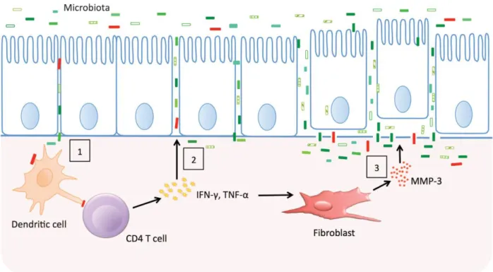
IBD is a complex disorder that is thought to be the result of an aberrant immune response to commensal bacteria in a genetically susceptible host.23 The importance of genetic factors in the development of IBD has first been demonstrated by epidemiological studies. Approximately 25– 30% of pediatric IBD patients has a positive family history for IBD, 24, and first-degree relatives of IBD patients carry a 10-fold increased risk of developing the disease.25 Today, genetic studies and genome-wide association (GWAS) studies have identified almost 100 susceptibility genes that are associated with an increased risk of developing IBD.26-28 Most of these genes code for molecules of the innate or adaptive immune system. Furthermore a small number of monogenic mutations have been identified in children with IBD diagnosis at a very young age. However, concordance rates in monozygotic twins are only 35 – 63% for CD and 16 – 18% for UC28-29, illustrating that genetic factors alone are not sufficient to cause IBD. The importance of other factors, particularly the intestinal microbiota, is supported by the observations that surgical deviation of inflamed intestine ameliorates inflammation30, and that antibiotic treatment can be effective in at least a subset of IBD patients.31-32. Other environmental factors that are believed to be associated with the etiology of IBD are smoking, perinatal events, childhood infections, diet, and domestic hygiene.33 In figure 1 the pathogenetic hypothesis behind IBD is represented.
Figure 1: Epithelial barrier dysfunction and inflammation in inflammatory bowel disease (IBD). Genetically
encoded variation in the epithelial barrier function may allow microbes to cross the barrier and trigger a T-cell response (1). The cytokines produced by activated T T-cells and macrophages loosen tight junctions allowing more antigens to cross (2). Finally, degradation of the basement membrane causes the epithelial
I.4 Clinical presentation
A clinical suspicion of IBD is raised in children with persistent (≥ 4 weeks) or recurrent symptoms, such as abdominal pain, diarrhea, rectal bleeding, and weight loss.34 Other gastrointestinal symptoms may include decreased appetite, nausea, and vomiting. Only 25% of pediatric CD patients presents with the ‘classic triad’ of abdominal pain, diarrhea, and weight loss, as was demonstrated by Sawczenko et al.35 Approximately 25% of children with CD presents with non-specific symptoms such as lethargy and anorexia, which may be associated with only mild abdominal discomfort. In contrast to the diverse symptomatology in pediatric CD, the clinical presentation of pediatric UC is almost uniformly bloody diarrhea (84 – 94% of children).36 Clinical presentation alone is however not sufficient to make a reliable distinction between UC and CD.37 Extra-intestinal manifestations may be a presenting sign in 6 – 17% of pediatric IBD patients, with joint inflammation, skin manifestations, and aphthous stomatitis being most frequently reported.38-39
Perianal lesions can be present in CD, but are not a common feature of UC. These lesions can range from single simple skin tags to complex networks of fistulas and abscesses. Fistulas and abscesses have been reported in 7 – 10% of newly diagnosed pediatric CD patients, while skin tags and fissures can be present in 5 – 20% of patients.40-42 Unique to pediatric-onset IBD is the occurrence of linear growth impairment and pubertal delay. Impaired growth velocity may be the first sign of IBD, and may start several years before the onset of gastrointestinal symptoms.43-44 Growth failure at diagnosis is reported in 10 to 20% of pediatric CD patients35, 45-48, and to a much lesser extent in pediatric UC patients.35, 44, 49-50
I.5 Diagnosis
The diagnosis of IBD is based on a combination of clinical presentation, physical examination, endoscopic appearance, histologic findings, and small bowel imaging studies. In 2005, the IBD Working Group of ESPGHAN (European Society for Pediatric Gastroenterology, Hepatology and Nutrition) published consensus-based criteria for the diagnostic workup of pediatric IBD, the Porto criteria.34 In 2014 the original Porto criteria were reviewed by using an evidence-based approach and new specific practice recommendations for the diagnosis of pediatric IBD (PIBD) were published. 51 First of all, infectious causes of diarrhea need to be excluded by stool cultures, and laboratory screening tests should be performed. Anemia, increased inflammatory markers (erythrocyte sedimentation rate, C-reactive protein), trombocytosis, and hypoalbuminemia are suggestive of IBD52, but normal laboratory screening tests may be present in 21% of children with mild CD, and in 54% with mild UC.53 Serologic markers have been introduced as a possible tool to help diagnose IBD. These markers are antibodies against microbial products that have been found in the blood of IBD patients. Antibodies to anti-Saccharomyces cerevisiae (ASCA) are associated with CD, and are found in approximately 60% of CD patients, 10% of UC patients, and <5% of non-IBD patients. In addition, perinuclear antineutrophil cytoplasmic antibodies (pANCA) are associated with UC, and are present in
approximately 60% of UC patients, 20% of CD patients, and <5% of non-IBD patients.54 Other markers that have been studied, are: anti-E. coli outer-membrane porin C antibodies (anti-OmpC), antibodies to bacterial flagellin (anti-CBir1), antibodies to a bacterial sequence from Pseudomonas fluorescens (anti-I2), and antiglycan antibodies. Non-invasive stool tests, particularly the fecal calprotectin, have become increasingly important as a screening tool in order to avoid more invasive investigations.55 Secondly, endoscopy and histology are of key importance to establish a diagnosis of the type of disease, as well as disease severity, localization, and extent of disease. All children suspected of IBD should undergo a complete endoscopic evaluation (ileocolonoscopy and upper gastrointestinal endoscopy) with multiple biopsies taken from each segment of the gastrointestinal tract.51 Table 1 summarizes endoscopic and histologic features of CD.
Typical macroscopic findings of CD Typical microscopic findings
Mucosal apthous ulcers Non caseating granuloma(s)-must be remote from
ruptured crypt
Linear or serpentine ulceration Focal chronic inflammation, transmural inflammatory
infiltrate, submucosal fibrosis Cobblestoning
Stenosis/structuring of bowel with prestenotic dilatation Imaging or surgical bowel wall thickening with luminal narrowing
Perianal lesions-fistula(s), abscesses, anal stenosis, anal canal ulcers, large and inflamed skin tags
Skip lesions
Jejunal or ileal ulcers
Nonspecific macroscopic findings of CD Nonspecific microscopic findings of CD
Oedema Granuloma adjacent to ruptured crypt
Erythema Mild nonspecific inflammatory infiltrate in lamina propria
Friability Mucosal ulceration/erosion
Granularity Signs of chronicity (eg. Crypt architectural changes,
colonic Paneth cell metaplasia and goblet cell depletion) Exudate
Loss of vascular pattern Isolated apthous ulcers
Perianal lesions-midline anal fissures, small skin tags
The revised Porto criteria underlines as pediatric-onset UC may present with atypical phenotypes such as macroscopic rectal sparing, isolated non-serpiginous gastric ulcers, normal crypt architecture, absence of chronicity in biopsies, or a cecal patch. Patients with acute severe colitis may have transmural inflammation. These individual phenotypes in isolation should not lead to reclassification to CD.51 In table 2 typical and atypical features of UC are reported.
Presentation Macroscopic features Microscopic features
Typical Contiguous disease from the rectum Architectural distortion, basal lymphoplasmacytosis, disease most severe distally, no granulomas Atypical
1. Rectal sparing No macroscopic disease in rectum or rectosigmoid
Same as typical, especially in the involved segment above sparing 2. Short duration Contiguous disease from the rectum, may
also have rectal sparing
May have biopsy with focality, plus signs of chronicity or architectural distortion may be absebt, other features are identical. Usually occurs in young children with a short duration of symptoms.
3. Cecal patch Left-sided disease from rectum with area
of cecal inflammation and normal appearing segment between the 2
Typical: biopsies from the patch may show nonspecific inflammation
4. Upper gastrointestinal involvement (UGI)
Erosions or small ulcers in stomach, but are neither serpiginous nor linear
Diffuse or focal gastritis, no granuloma (except pericryptal) 5. Acute severe colitis
(ASC)
Contiguous disease from the rectum May have transmural inflammation or deep ulcers, other features typical. Lymphoid aggregates are absent, ulcers are V-shaped fissuring ulcers
Table 2. Typical and atypical features of UC.
In order to differentiate colonic CD disease from UC and IBD-U the revised Porto panel proposed further indications summarized in table 3.
Likelihood of occurring in UC Features Diagnostic approach
Cl
ass
1:
No
n
exi
st
en
t
Well-formed granulomas anywhere in the GI tract, remote from ruptured crypt
Di
ag
n
os
e
as
CD
Deep serpentine ulcerations, cobblestoning or stenosis anywhere in the SB or UGi tract Fistulizing disease (internal or perianal)
Any ileal inflammation in the presence of normal cecum (ie incompatible with backwash ileitis) Thickened jejunal or ileal bowel loops or other evidence of significant SB inflammation (more than a few scattered erosions) not compatible with backwash ileitis
Macroscopically and microscopically normal appearing skip lesions in untreated IBD (except with macroscopic rectal sparing and cecal patch)
Large inflamed perianal skin tags
Cl
ass
2:
R
ar
e
w
it
h
UC
(<5%
)
Combined (macroscopic and microscopic) rectal sparing, all other features are consistent with UC
Di
ag
n
os
e
a
s
IBD
-U,
if
a
t
le
ast
1
cl
ass
2
fe
atu
re
e
xi
sts
**
Significant growth delay (height velocity <2 SDS), not explained by other causes Transmural inflammation in the absence of severe colitis, all other features are consistent with UC
Duodenal or esophageal ulcers, not explained by other causes (eg, Helicobacter pylori, NSAIDs and celiac disease)
Multiple aphthous ulcerations in the stomach, not explained by other causes (eg, H pylori and NSAIDs)
Positive ASCA in the presence of negative pANCA
Reverse gradient of mucosal inflammation (proximal >distal (except rectal sparing))
Cl
ass
3:
Unco
m
m
on
(5%
–
10
%)
Severe scalloping of the stomach or duodenum, not explained by other causes (eg, celiac disease and H pylori)
Di
ag
n
os
e
a
s
IBD
-U
if
a
t
le
ast
2
–
3
fe
atu
re
s
e
xi
st
Focal chronic duodenitis on multiple biopsies or marked scalloping of the duodenum, not explained by other causes (eg, celiac disease and H pylori)
Focal active colitis on histology inmore than 1 biopsy frommacroscopically inflamed site Non–bloody diarrhea
Aphthous ulcerations in the colon or UGI tract
Table 3. Diagnostic features in a child with untreated colitis phenotype at diagnosis and diagnostic
indications.
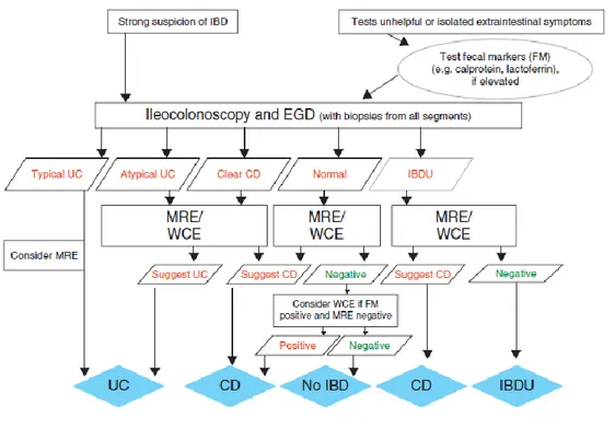
Small bowel investigation is indicated in all patients at diagnosis (except in definitive cases of UC) to guide therapeutic management and to detect strictures that may need surgical resection.51 The revised Porto criteria recommend Magnetic resonance enterography (MRE) as the imaging modality of choice in pediatric IBD at diagnosis. It may detect small intestinal involvement, inflammatory changes in the intestinal wall and identify disease complications (fistula, abscess, stenosis). MRE is preferred over CT and fluoroscopy because of the high diagnostic accuracy and the lack of radiation involved51. Wireless capsule endoscopy (WCE) is a
conventional endoscopy and imaging tools have been non diagnostic or in whom MRE cannot be performed due to young age or in settings where MRI is not available or not feasible. A normal WCE study has a high negative predictive value for active small bowel CD51. The proposed algorithm for a child with suspected IBD is represented in Figure 2. 51
Figure 2. Evaluation of child/adolescent with intestinal or extra-intestinal symptoms suggestive of IBD, as
proposed by the Porto’s group.
I.6 Classification of phenotype
Accurate phenotype classification is important for choosing the most appropriate therapy, assessing disease prognosis, and for a better understanding of the pathophysiology of the different manifestations of IBD. Adult gastroenterologists have therefore developed the Vienna classification for CD, which was revised and extended to UC in the subsequent Montreal classification. As the Montreal classification did not sufficiently capture the dynamic features of pediatric IBD and had only moderate interrater reliability when utilized in pediatric IBD, an international group of pediatric IBD experts has recently developed a pediatric modification of the Montreal classification: the Paris classification.56 The differences between the Paris and Montreal classification for CD and UC are displayed in Figures 3 and 4.
I.7 Assessment of disease severity
Evaluating disease activity in children with IBD requires the use of outcomes that reflect pediatric-specific qualities of the disease. Monitoring pediatric UC and CD is not limited to observing intestinal symptoms, but also involves assessing weight and height gains, sexual maturation, extraintestinal manifestations, and psychosocial well-being. Disease activity is best measured if using multi-item indices, which incorporate clinical symptoms, laboratory parameters, and endoscopic findings. Indices are prediction rules used to measure the activity of disease, and use a combination of history, examination, and laboratory data to develop an objective score that is reproducible between different observers. Currently, validated indices are utilized both in clinical trials of pediatric IBD and in clinical practice.
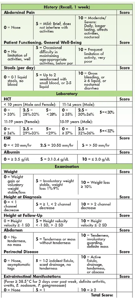
I.7.a The Pediatric Crohn’s Disease Activity Index (PCDAI)
In 1990, the PCDAI was developed and validated solely by a group of experts without the involvement of statistical modeling. The PCDAI, specifically designed for use in children, has several benefits over the previous adult CDAI for this population57-58:
- Calculation does not require a 1-week diary and the historical items can be assessed during the clinic visit,
- The PCDAI includes a child-specific item: the height velocity variable,
- The PCDAI includes the addition of 2 additional laboratory parameters: erythrocyte sedimentation rate (ESR) and albumin level,
- The scoring of hematocrit HCT is adjusted based on age and gender
The PCDAI score can range from 0-100, with higher scores signifying more active disease (see Figure 5). A score of < 10 is consistent with inactive disease, 11-30 indicates mild disease, and > 30 is moderate-to-severe disease. A decrease of 12.5 points is taken as evidence of improvement.
While the PCDAI remains the most widely used index to measure disease activity in pediatric CD patients, it has been criticized by researchers for including laboratory tests and items that may not change fast enough during therapy to make them useful to detect change over time (eg, growth velocity).16 More recently shorter version have been proposed (abbreviated PCDAI, weighted PCDAI).
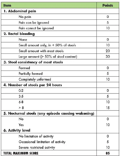
I.7.b The Pediatric Ulcerative Colitis Activity Index (PUCAI)
The PUCAI was developed using a combined judgmental and statistical approach, utilizing 2 prospectively accrued cohorts of 205 children with UC. Item generation, reduction, and grading were performed using a Delphi technique among 36 experts in pediatric UC. To minimize inter-observer variability, the group established logical gradations and clear definitions of items. Proposed gradation schemes for each item of the PUCAI were distributed to the Delphi group, with the final instrument reflecting consensus opinion59. The final version of PUCAI is composed of 6 clinical items (see Figure 6). The PUCAI score ranges from 0 to 85; a score of < 10 denotes remission, 10-34 mild disease, 35-64 moderate disease, and 65-85 severe disease. Weights of the included items were assigned according to a multivariate regression analysis of 157 children with UC, in which rectal bleeding assumed the highest weight. In the validation study, the PUCAI showed excellent correlation with physician global assessment of disease activity, colonoscopic appearance, and the adult-invasive endoscopic Mayo Score. The historical parameters should reflect a daily average of the patient’s last 48 hours. However, if the patient’s condition is changing rapidly, the last 24 hours may be used.
I.8 Treatment
Induction and maintenance of remission are the main goals of treatment in IBD. As pediatric IBD occurs during a critical period of growth and development, special considerations in treatment are needed to reverse linear growth failure, malnutrition, delayed puberty, and deficits of bone mineralization. High quality evidence from clinical trials in children with IBD is still scarce, and treatment decisions were often based on extrapolations from the adult literature in the past.61 More recently European consensus-based guidelines for the treatment of pediatric IBD have been published60-61. The current approach to the treatment of pediatric IBD is based on a step-up strategy, indicating that more aggressive immunosuppressive treatment is only prescribed after milder/less toxic treatment has failed.
I.8.a Guidelines on the treatment of pediatric CD
Induction of remission can be achieved by exclusive enteral nutrition (EEN) or corticosteroids. These treatment modalities have been demonstrated to be equally effective in children with CD, but EEN has significant advantages over steroids due to its beneficial effect on growth, and lack of serious side effects. Thiopurines, such as azathioprine and 6-mercaptopurine, are used for maintenance of remission, and are often introduced at the time of remission induction with either EEN or corticosteroids. Thiopurines have a very slow onset of action and may take 3 – 6 months to reach maximal effect. Methotrexate, another immunomodulator, is an alternative to thiopurines when these drugs are ineffective or not tolerated. When patients are refractory to or intolerant of these conventional treatment regimens, anti-TNF treatment is indicated (Infliximab or Adalimumab). Infliximab is a chimeric monoclonal antibody against TNFα, a proinflammatory cytokine with an increased expression in the inflamed tissues of IBD patients. In the presence of significant fistulizing disease (and perianal), or severe growth retardation in Tanner or 2-3, presence of predictors of severe disease course (deep colonic ulcerations on endoscopy, persistent severe disease despite adequate induction therapy, extensive (pan-enteric) disease, marked growth retardation, severe osteoporosis o stricturing and penetrating disease at onset) treatment with biologics should be condidered earlier60. Elective surgery should be considered in children with refractory CD or strictures, especially in pre-pubertal or early pubertal children with growth failure and localized CD.
Figure 7. Proposed algorithm for the treatment of pediatric CD60.
1. The weighted pediatric Crohn's disease activity index (wPCDAI) can be used to assess disease severity supplemented by serum and fecal inflammatory markers, growth, endoscopic and radiographic evaluation and other lab results. 2. Consider: symptoms due to stenosis, irritable bowel syndrome, lactose intolerance, infection (e.g. C. difficile and CMV), wrong diagnosis, side effects of medication, bacterial overgrowth, and bile-salt diarrhea. 3. EEN should be especially preferred in children with poor growth, low weight and those with catabolic state (e.g. hypoalbuminemia). If EEN is not tolerated orally, a nasogastric tube may be used, however, the emotional and financial implication
4. The use of 5-ASA in Crohn's disease is controversial and generally not recommended. In some selected mild cases, 5-ASA may be considered to supplement induction therapy (50–80 mg/kg/day up to 4 g daily in 2 divided doses) especially in colonic disease. Sulfasalazine may be more effective than the newer regimens but is also associated with higher adverse effect rate. Gradual dose increase of sulfasalazine over 7–14 days may decrease adverse event rate. 5. Prednisone/prednisolone (1 mg/kg once daily up to 40 mg) must be tapered over ~10 weeks. Repeated steroid courses or steroid dependency should not be tolerated. 6. The risk for malignancy and perhaps also infections is higher when anti-TNF is combined with thiopurines (i.e. combo therapy). Since good evidence is lacking to support combo therapy in thiopurine-failure children, thiopurines should be discontinued within 6 months of combination therapy. However, combo therapy is superior to mono therapy in thiopurinenaïve patients and may be considered in high risk patients, especially in girls for whom the risk of lymphoma is smaller. Stepping down to either drug may be considered after a period of sustained deep remission. 7. Immunization status should be checked prior to starting immunomodulator or anti-TNF therapy, similarly when history of chickenpox is unclear, screen immunity and consider immunization of seronegative patients against varicella zoster prior to starting immunomodulator or anti-TNF therapy. In cases of primary anti-TNF failure, the switch to another anti TNF regimen is associated with a low success rate. 8. Surgery is particularly attractive in children with refractory short segment ileal disease without colonic involvement and those with stenotic disease unresponsive to anti-inflammatory therapy. 9. High risk patients include the presence of perianal disease, severe growth retardation, the presence of deep ulcers in endoscopy or extensive disease (including upper GI and proximal small bowel), the need for corticosteroids at diagnosis. 10. Although PEN has been shown to be inferior to EEN in inducing remission, some weak evidence suggest that it may be partially effective in maintaining remission in pediatric Crohn's disease. 11. Oral azathioprine 2–2.5 mg/kg once daily or 6-mercaptopurine 1–1.5 mg/kg once daily. Typical onset of action is 8–14 weeks. CBC and liver enzymes should be closely monitored. Measurement of TPMT (genotyping or enzymatic activity) at baseline and the drug metabolites (i.e. 6-TG and 6-MMP) levels after 2–4 months may aid in optimizing thiopurine dosing.
12. Failure of immunomodulators should be considered in frequent relapses, inability to wean off corticosteroids and may be
considered in an asymptomatic child with signs of significant mucosal inflammation as evident by marked abnormal blood tests, fecal markers, endoscopic or radiographic evaluation. 13. Methotrexate dose is 15 mg/m2 (max 25 mg) once weekly. Subcutaneous administration is likely as effective as intramuscular. There is insufficient data to support oral treatment at any time. Daily folic acid should be prescribed to minimize adverse events. Liver enzymes and complete blood count should be frequently monitored. After sustained remission has been achieved (typical onset of action 2–3 months), MTX dose may be reduced by 40%. 14. Antibiotics may have some role in induction of remission in Crohn's disease such as metronidazole, ciprofloxacin, azithromycin and rifaximin.
I.8.b Enteral nutrition in pediatric CD
Exclusive enteral nutrition (EEN) is the best-known dietary intervention for induction of remission in mild to moderate CD both in children and adults. Case series and randomized clinical trials have demonstrated the ability of EEN to induce clinical remission in approximately 80% of patients. Rates varied depending upon type of study (retrospective or prospective), or type of analysis (per protocol or intention to treat), and seem to be independent of type of formula62–64.Results are summarized in three meta-analyses, with an overall combined remission rate for EEN in pediatric CD of 73% (relative risk (RR) 0.95, 95% confidence interval (CI) 0.67–1.3428 and RR 0.97, 95% CI 0.7–1.429)65-67. Interestingly and in contrast to steroids, EEN has also the potential to induce mucosal healing. Disease severity and luminal disease seem to be the only significant predictors of response to EEN. According to the ECCO/ESPGHAN consensus, EEN should be the first line therapy to induce remission in children with active mild to moderate luminal CD60
initially thought that using EEN should be limited only to patients with small bowel involvement; however results from further meta-analysis have shown no difference in the efficacy of EEN when considering the disease location. Beneficial effects of the EEN in patients with CD, including effects on bone, growth and nutritional deficiencies have extensively been described. Hypothesis on the possible ways of action of EEN include bowel rest, anti-inflammatory effects, restoration of the epithelial barrier and positive changes in the intestinal microbiota. As both polymeric and elemental formulas show similar efficiency, gut rest is unlikely to be the primary involved mechanism. It is reasonably considered that EEN reaches the clinical effects through a combination of the above-cited mechanisms.
I.8.c Consensus-based Guidelines on the Treatment of Pediatric UC
61Treatment of pediatric UC depends on disease extent and severity. Mild or left-sided colitis is usually managed with oral and/or topical aminosalicylates. Oral (and topical) aminosalicylates are also recommended as first line therapy for mild to moderate (pan)colitis, while corticosteroids are used in case of an insufficient treatment response or severe disease. Maintenance treatment consists of aminosalicylates, or thiopurines for patients with relapsing disease. After its recent registration for treatment of pediatric UC, infliximab is increasingly used in children with refractory UC. Colectomy may be indicated in patients with persistently active disease with corticosteroid dependency despite concomitant immunosuppression, or growth retardation despite apparently adequate maintenance therapy. Children with acute severe colitis (PUCAI > 65) should be admitted to the hospital for intravenous corticosteroid therapy. Approximately 30 – 40% of children will not respond to steroid therapy and will require second-line therapy or colectomy. The pediatric UC disease activity index (PUCAI) can assist in determining the need and timing of alternative treatments early during the admission. Second-line treatment options include cyclosporine, tacrolimus, or infliximab, which all seem to have similar short-term response rates. Colectomy is indicated in case a patient is refractory to one salvage therapy or in case of toxic megacolon.
Figure 8. Proposed therapeutic algorithm by the Joint European Society for Paediatric Gastroenterology,
Hepatology and Nutrition (ESPGHAN)-European Crohn’s and Colitis Organization (ECCO) for pediatric ulcerative colitis (UC) 61.
1Medical therapies in UC should be divided into those that induce remission (5-aminosalicylic acid [5-ASA], corticosteroids, anti-tumor necrosis factor [TNF] therapy, calcineurin inhibitors, and likely probiotics), and those that maintain remission (5-ASA, thiopurines, anti-TNF therapy, and selected probiotics). 1In any state of active disease, the following must be ruled out: infectious colitis (including cytomegalovirus [CMV] and C difficile), 5-ASA-related colitis, lactose intolerance, irritable bowel syndrome, wrong diagnosis, celiac disease, and the like. 2 Unlike in adults, endoscopic evaluation of the rectal mucosa is conceived to be more invasive for routine monitoring of disease activity and response to therapy in children. Therefore, these should be based on noninvasive indirect markers of disease activity. Cutoff values of the Pediatric UC Activity Index (PUCAI) for remission, mild, moderate, and severe disease activity have been previously validated in 3 independent cohorts. 35-ASA is dosed 60 to 80mg_ kg_1 _
mg/kg up to 1 g) are more effective than steroid enemas. Enemas should be administered in the left decubitus position. Liquid enemas are more difficult to tolerate than foams and suppositories but work for more extensive colitis. 5If there is lack of improvement (ie, PUCAI decrease of <20 points) after 7 to 10 days or an increase in PUCAI _20 points at any time, consider admission for intravenous steroids or outpatient treatment with anti-TNF therapy, or less often tacrolimus. Steroid dependency should be declared in children achieving remission with corticosteroids but who experience return of symptoms when dosage is lowered or within 3 months following complete taper, or if steroids cannot be stopped within 14 to 16 weeks. Maintenance therapy should be then escalated. 6Turner et al 7Response is defined as a drop in PUCAI of at least 20 points; however, the goal of induction therapy is eventually complete clinical remission (PUCAI<10). 8For example, previous intolerance or resistance to steroids, or when infliximab is indicated anyway for maintenance treatment after failing thiopurines. 9Measuring thiopurine methyltransferase (genotyping or enzymatic activity) at baseline, and 6-TG and 6-MMP levels after 2 to 3 months, may aid in optimizing thiopurine dosing. 10If infliximab has been used in thiopurine-naı¨ve disease, thiopurines may be added and infliximab discontinued after 4 to 8 months if complete remission has been achieved. Stepping down to 5-ASA may be considered in selected cases, if 5-ASA did not fail previously, and after a period of sustained complete remission. 11There is no evidence to support adding thiopurines to infliximab in thiopurine-failure children; however, some discontinue thiopurines after 4 to 8 months of combined therapy.
Part II: Overview on the mechanisms of intestinal damage in IBD
II.1 Mechanisms of intestinal damage in IBD: the role of microbiota and the intestinal barrier
function
The causes of IBD are still not understood, but there is no doubt that the intestinal tissue injury is caused by an excessive immune/inflammatory process in the gut wall. In terms of the relationship between gut barrier function and IBD, the critical question is whether the impaired barrier function is secondary to gut inflammation and damage, or if it is important as an independent event, which may either protect or confer risk of IBD. In healthy subjects homeostasis exist between the intestinal microbiome, mucosal barrier and immune system68. In IBD this homeostasis is disrupted leading to durable alterations in the intestinal microbiome (dysbiosis), altered barrier function (leaky gut) and immune system activation (inflammation) (Figure 9).
Figure 9.The pathophysiological circuit of IBD68
In 1995, Gordon and co-workers developed a chimeric mouse model in which some of the small bowel epithelium expressed N-cadherin instead of E-cadherin, thereby disrupting the E-cadherin homotypic interactions that help maintain barrier integrity69. At the regions of the intestine expressing N-cadherin, the epithelium was leaky and the mice developed focal inflammation in these areas. Several subsequent studies using gut epithelial gene-specific knockout mice confirmed that a dysfunctional epithelial barrier results in spontaneous intestinal inflammation70-71. Markedly, genes associated with uncontrolled cell death seem to be involved. Also, defects in mucus assembly and production can lead to spontaneous development of colitis in mice models73-76. The clear lesson from this work is that if the gut epithelium is disrupted, ingress of bacterial components into the lamina propria is sufficient to trigger IBD. However, many animal models have shown that even in the presence of an apparently normal gut epithelial barrier, changes in immune regulation can result in exaggerated mucosal immune responses and IBD phenotypes. For example, in dirty animal houses, all interleukin-10 (IL-10)-null mice develop small and large bowel inflammation early in life, whereas in clean animal houses, only a colitis develops and the onset of IBD is delayed77. The importance of IL-10 is
enteritis early in life78. Regulation of inflammatory responses is also crucial to intestinal homeostasis, as shown by the identification of IL-23R variants as risk factors for both Crohn’s disease and ulcerative colitis79. Thus, in the absence of immune regulation, the low levels of bacterial antigens that cross into the lamina propria are sufficient to trigger inflammation. In healthy individuals, translocating bacteria and bacterial antigens are mopped up by macrophages in the lamina propria. However, e.g., in children with defects in their ability to deal with low-grade bacterial infections, such as chronic granulomatous disease or glycogen storage disease type 1b, ~ 40% of patients develop a lesion similar to Crohn’s disease80-81.
II.2 Epithelial barrier dysfunction in IBD
There is a widely held perception that a determinant of susceptibility to IBD, especially to Crohn’s disease, is an inherent/genetic defect in the intestinal barrier, which allows greater ingress of luminal antigens into the tissues. Patients with active IBD have clear epithelial barrier defects, exemplified most typically by overt ulceration. When patients enter remission, barrier function improves, however, it rarely returns to normal. This is most probably due to the fact that inflammation continues at a relatively low level.82 One way to determine if there is indeed a tendency for those who develop IBD to have a leaky gut is to study unaffected relatives who are known to have a 30-fold increase in the risk of developing IBD. Permeability tests in these individuals are normal; however, in response to an insult, such as a non-steroidal anti-inflammatory drug (NSAID), a subset of relatives (35%) did show markedly increased intestinal permeability (+2 s.d. of the controls). Overall, these first-degree relatives to Crohn’s disease patients had a 110% increase in intestinal permeability (IP) compared with a 57% increase in healthy controls after the NSAID challenge. Therefore, while the epithelial barrier per se may not be intrinsically leaky in IBD, the response to injury may be impaired, perhaps because of impaired healing or delayed epithelial restitution. A high proportion of IBD patients (up to 70%) is found to have an increased IP using different marker probes (such as PEG, polyethylene glycol, CrEDTA, TcDTPA, CrEDTA/C-mannitol, lactulose, mannitol, rhamnose and cellobiose). The lactulose and mannitol (L/M) excretion test is a simple, non invasive and reliable method used for estimation of IP in clinical practice. Furthermore the IP test results has been correlated to disease activity in IBD and preliminary report suggest also a possible role as marker of mucosal healing after specific treatments84.
II.3 The role of the microbiota
Although the pathogenesis of IBD is still partially unknown, it is evident that there is an interplay between genetic factors (primarily related to defects in some immunological mechanisms), environmental factors and alterations in the intestinal microbiota. The term “microbiota” indicates the microflora resident in the intestine, especially in the ileal portion of the small intestine and in the colon. In particular, the indirect evidence of the implication of the microbiota in the pathogenesis of IBD is derived from some observations
such as: (a) the intestinal inflammation occurs mainly in areas characterized by high bacterial concentration and the antibiotic treatment is associated with clinical improvement85; (b) murine germ-free and free of intestinal flora, do not develop chronic colitis.86 The human intestinal microbiota is composed of 1014 bacterial cells (up to 1000 different bacterial species)87 and varies considerably in different individuals with a high degree of overlap in monozygotic twins. The development of the microbiota is thus linked to genetic factors but also to environmental factors. It is estimated that the human intestinal microbiota contains a number of genes 100-fold higher than the human genome. Ninety percent of the bacteria fall into the two phyla: Bacteroidetes and Firmicutes87. Other phyla, including Actinobacteria, Proteobacteria, Fusobacteria, and Verrucomicrobia, are also present in lower quantities. Diversity estimates place the total number of species between 1000 and 500088, and only a fraction of these, the ‘core’ microbiota, are commonly present in most individuals. The main functions of the intestinal microbiota are: (1) metabolic: digestion of dietary and xenobiotic compounds, fermentation of carbohydrates and production of short chain fatty acids, ethanol, gases, vitamins, absorption of ions, conversion of polyphenols in the diet to active forms. Taken together, the metabolic products of the microbiota constitute the so-called “metabolome”; (2) protection of the intestinal barrier: inhibition of invasion by pathogens and reinforcement of the intestinal barrier by increased expression of some proteins of tight-junctions (zonulin and occludin); (3) immunological: stimulation of the immune system and maintenance of intestinal epithelium homeostasis89.
Many studies have shown that in active IBD there is a dysbiosis of the microbiota, which could be a cause for a disturbed epithelial barrier function. Reduced complexity of the phylum Firmicutes is a common signature of fecal microbiota of Crohn’s disease patients and in particular decreased abundance of Faecalibacterium prausnitzi.90 Several species within the phylum Firmicutes ferment complex carbohydrates in the colon and produce butyrate, which has been reported to increase production of secreted mucus and has other potential barrier-protecting functions 91 Additionally, E. coli pathobionts exhibiting pathogen-like behaviors that disrupt the epithelial barrier are more frequently cultured from IBD patients.92
The relationship between the intestinal microbiome and IBD in children has been extensively reviewed. There is a reduction of some bacterial species in the intestine of children with IBD, particularly of Firmicutes (among these especially the Clostridium group IX and IV) and Bacteroides, and a parallel increase in Enterobacteriacee93. Clostridium and Bacteroides are the main producers of short-chain fatty acids (SCFA) in the human colon. The Clostridium group IV and IX are the major producers of butyrate, which is a major energy source for colon cells acting as an inhibitor of the expression of pro-inflammatory cytokines in the intestinal mucosa94, and is also able to reinforce the intestinal barrier (inducing the production of mucins and antimicrobial peptides and increasing the expression of proteins of the tight-junctions) 95. Reduced levels of butyrate may therefore be involved in the intestinal inflammation that characterizes IBD and this molecule
investigated the gut microbiota of children with IBD in relation to the natural history of disease (at diagnosis or during follow-up) and / or the type of treatment. One of the pivotal studies found a reduced presence of Bacteroides vulgatus in the intestinal mucosa of patients with CD or IBDU96. Another study evaluated the composition of the intestinal microbiota in 69 pediatric patients with IBD, showing a reduction of Faecalibacterium prausnitzii and bifidobacteria in patients with CD and an increase of Escherichia coli (CD in patients with active phase) 97. The reduction in Faecalibacterium prausnitzii has not subsequently confirmed by other studies. More recently some studies related the gut microbiota to therapeutic responses and association were described between fecal microbiota and the response to steroid therapy in pediatric patients hospitalized for severe UC98 and between fecal microbiota and response to antiTNF alpha99. This suggests that characterization of gut microbiota may provide an additional tool in assessment of therapeutic responses, warranting further studies.
II.4 Effect of the exclusive enteral nutrition on inflammation, microbiota composition and
intestinal permeability
The anti-inflammatory effects of EEN have been shown both at a mucosal and systemic level. A few studies have demonstrated a decrease in pro-inflammatory mediators in response to EEN 100 and an increase in anti-inflammatory molecules, such as TGF-β 101. The same data were replicated in an ex-vivo study in which incubation of CD-biopsies with elemental formula led to an increased ratio of IL-1Ra to IL-1 β compared to control samples (non inflammatory controls or UC patients)102. Other authors confirmed the direct effect of a polymeric formula on colonic epithelial cell chemokine responses to the pro-inflammatory cytokine TNF-α, through a direct interaction with components of the NF-kß pathway 103. The anti-inflammatory effect of EEN has also been shown at the mesenteric fat level, where EEN treatment improved adipocyte size, decreased pro-inflammatory adipokines (TNF- α and leptin) levels and increased adiponectin levels 104.
The effects of EEN on intestinal barrier come mostly from in vitro or in animal studies. In human colonic epithelial cells a polymeric formula has been found to maintain trans epithelial electrical resistance, short circuit current, para-cellular permeability and morphological distribution of tight junction proteins, through mechanisms involving inhibition of the long myosin light chain kinase (MLCK)105. In a IL-10 knock out mouse model of colitis (induced by Helicobacter) EEN treatment maintained normal gut barrier function and integrity, reversed inflammatory changes and reduced bacterial load 106.
Initial investigation based on “obsolete” techniques indicated profound changes in composition of mucosal microbiota induced by EEN107-8. Since then, several studies have confirmed major changes in the intestinal microbiota related to EEN. The most frequently reported effect is a reduced diversity of the microbiota, already occurring after few days or weeks of EEN109-112. The persistence of such changes has been variably described, with a study reporting persistent modification at 4 months after EEN109 and another study
indicating a prompt reversibility of all the changes to pretreatment levels on a free diet113. The recent study by Lewis et al. observed a unique effect of EEN in intestinal microbiota, different from the effect induced by anti-TNF and most importantly from partial EN, suggesting that the exclusion of table foods was the primary determinant in changing the gut microbiota and perhaps in mediating the effectiveness. Furthermore the microbiota composition differed at 1 week between ultimate responders and non-responders, evoking measures of microbiota as possible predictor of response114. Recurrence of CD following EEN corresponded with a new increase in operational taxonomic units 115. In contrast, the effect of reducing microbiota special diversity induced by therapeutic diet was disconfirmed by 2 studies: the first demonstrating an increase in species diversity after elemental diet116, the second showing a higher overall microbiome diversity in mice fed by a monotonous diet for 20 days compared to alternating chow fed mice117. Also specific effects on strains or single bacteria were reported by different authors, in particular a significant decrease in Bacteroides induced by EEN was described109-11, 116. A decrease in concentration of Faecalibacterium prausnitzii was observed both in one small adult study117 and in a pediatric cohort110, challenging the previous paradigm of a protective role of Faecalibacterium prausnitzii in CD. In a very recent small report on 8 patients providing complete sets of stool samples, authors observed significantly altered fecal bacterial communities already after 2 weeks of EEN118. In this study EEN decreased the relative sequence abundance of Gram-negative bacteria belonging to the phylum Bacteroidetes, including members of the family Bacteroidaceae, Porphyromonadaceae, and Rikenellaceae, in agreement with results of previous studies. At the phylotype level, in terms of operational taxonomic units (OTUs) results are contrasting with some authors reporting no effect of the dietary intervention118 and others reporting a reduction in the number of OTUs112. Results regarding the intestinal metabolic profile are heterogeneous. Tjellstrom119 described a decreased level of pro-inflammatory acetic acid and increased concentrations of anti-inflammatory butyric acids and valeric acids, while Gerasimidis110 reported a decrease in butyric acid and an increase in fecal sulfide after EEN. In the latter study these effects were more pronounced, once patients who failed treatment were excluded. From a meta-genomic point of view a single study described a reduced abundance in genes involved in biotine and thiamine biosynthesis following EEN and an increase in genes involved in spermidine/putrescine biosynthesis, which have a major role in cell growth and renewal and possibly in tissue healing113.
PART III: INVESTIGATION
III.1 Study project and aims
With this project, we overall aimed to investigate three different mechanisms of intestinal damage in pediatric IBD. The study considered the following aspects:
1. Role of microbiota as a marker of intestinal damage: we aimed to characterize the fecal microbiota in relation to different IBD type, disease activity and levels of inflammation.
2. Role of the diet in IBD and possible influences on the intestinal damage: we particularly focused on microbiological, therapeutic and clinical effects induced by a course of exclusive enteral nutrition (EEN) in children with CD.
3. Intestinal barrier and role of IPT as a marker of intestinal damage: we investigated the intestinal barrier through the double sugars intestinal permeability test (IPT) in relation to IBD type, severity and effect of specific treatments.
III.2 General overview of setting, patients and methods of the study
In the period 2013- 2016 fifty IBD patients were followed-up at our pediatric gastroenterology unit. Twenty-seven were children with a new diagnosis, 23 had an already established diagnosis prior of the study period. Twenty-five had a diagnosis of UC, 23 of CD and 2 of IBD-U. Diagnosis was established following the Porto criteria. The mean age at diagnosis was 9,3 years (± 3,8) and female to male ratio was 1,7:1.The patients were generally admitted to the hospital at the moment of diagnosis and then seen as outpatients during the follow-up. Treatment of patients with IBD generally followed the European guidelines. Specifically in children with CD we offered the EEN as induction treatment at diagnosis.
Medical information of all the patients enrolled in the study were collected including age, gender, family history of IBD in first degree relatives, disease type (Porto’s criteria), disease location and phenotype according to the Paris classification. At the moment of enrollment disease activity (by the PCDAI or the PUCAI) was assessed and clinical parameters (including height and weight), laboratory parameters (including fecal calprotectin, ESR and CRP) were collected. Anthropometric data were interpreted as age and sex specific standard deviations based on the Italian reference values (Cacciari, 2006).
III.3 Statistics
Continuous variables are presented as means (± SD) or medians (range). Non-continuous parameters are depicted as frequency and percentage. Quantitative paired data were compared using the Wilcoxon matched-pairs signed rank test and unpaired data using the Mann-Whitney test or the t-test, as appropriate.
test for quantitative data. The correlation significance was determined by means of Spearman and Pearson correlation analyses. Statistical analysis were performed using GraphPad Prism version 6 (Graph-Pad Software Inc., San Diego, CA, USA). P-values below 0.05 were considered statistically significant.
III.4 Specific methodologies for each part of the study
III.4.a Role of microbiota as a marker of intestinal damage (Microbiota study)
Specifically for this part of the study, subjects were enrolled in the period 2013-2014. We aimed to enroll both patients at diagnosis (possibly treatment naïve) and patients during follow-up. Clinical information and use of drugs (particularly antibiotics and probiotics in the month before enrollment) were carefully recorded. Patients were asked to collect a stool sample within few hours before the clinic visit. All stool samples were homogenized with 1 ml of RNA later and then immediately frozen (at - 80 ° C). Samples were subsequently sent in dry ice at the Institute for Genome Sciences in Baltimore for analysis. The analysis of the intestinal microbiota were conducted according to the following procedure. Total DNA was extracted from 0.25 grams of stool samples that had been preserved and transported in RNA later with PowerSoil DNA isolation kit (Mobio). Amplification of the V3-V4 regions of the 16S rRNA gene was performed according to Fadrosh et al120 using a dual-barcode system with fusion primers 338F and 806R. Amplicons were subsequently sequenced on Illumina MiSeq using the 300 bp paired-end protocol. Raw reads were preprocessed to remove the first 3 and last 3 bases if their phred score was lower than 3, read end was trimmed if the average phred quality score of 4 consecutive bases are below 15. Paired reads were retained if their length is at least 75% of their original length after trimming, which were then assembled using FLASH121 with overlap by ~90bp on average. Assembled reads were de-multiplexed by binning sequences with the same barcode and quality trimmed in QIIME (version 1.8.0) 122 (for details, please refer to Fadrosh et al. 123. Similar sequences with less than 3% dissimilarity were clustered together using USEARCH (v5.2.32) 124 and de novo chimera detection was conducted in UCHIME v5.1125. The taxonomic ranks were assigned to each sequence using Ribosomal Database Project (RDP) Naïve Bayes Classifier v.2.2126, using 0.8 confidence values as the cutoff to a pre-built greengenes database of 16S rRNA sequences (Aug, 2013 vers.) 119. The analysis of variance (ANOVA) was carried out on transformed data followed by separation of means with Tukey’s HSD, using the statistical software Statistica for Windows (Statistica 6.0 per Windows 1998, StatSoft, Vigonza, Italy). Letters indicate significant different groups (P<0.05) by Tukey’s test. Data (Unifrac distance metric and taxonomic abundance) were analyzed by Principal Component Analysis (PCA) and permutation analysis to assess the bacterial composition of samples using the statistical software Statistica for Windows (Statistica 6.0 per Windows 1998, StatSoft) and PermutMatrix. Interpretation of the results and part of the statistical analysis were conducted in collaboration with the Section of Alimentary Microbiology oft he
III.4.b Role of the diet in IBD and possible influences on the intestinal damage (EEN study)
This part of the study included 2 different projects. In a retrospective manner we firstly collected data of all the newly diagnosed pediatric CD patients during the period 2011-2016, and subsequently followed-up at our institution, and initiating EEN as induction therapy. Clinical, laboratory, endoscopic and radiologic data were recorded before and after a course of EEN. Information on the type of formula, route of administration, duration of the liquid diet and adherence to EEN treatment was also recorded (when available), as well as the initiation of immunomodulatory maintenance therapy or biologics or surgery. The PCDAI, anthropometric, clinical and laboratory data were compared before and after the EEN course. Furthermore in a subgroup of patients, newly diagnosed with CD and included in the microbiota study, fecal samples were prospectively collected at the beginning and at the end of a 8 weeks course of EEN and analyzed for comparisons. Furthermore the composition of microbiota was compared with another group of CD patients (included in the microbiota study) that had completed a course of EEN from at least one year and was on a normal diet at the moment of enrollment.
III.4.c Intestinal barrier and role of the Intestinal Permeability Test (IPT) as a marker of intestinal
damage (IP study)
For this specific pilot study in 2016 a subgroup of IBD patients was enrolled (5 CD and 4 UC patients). In 4 patients longitudinal samples were collected (before and after an induction treatment). The IPT was performed as previously described128. After an overnight fast and bladder emptying, an oral solution containing 5 g of lactulose and 2 g of mannitol was administered. Urine was collected during the following 5 hours and quantity registered. An aliquot was preserved at −20 °C. Urinary excretion of each sugar was assessed using a high performance anion-exchange Chromatography (Dionex DX-500). The ratio of recovered to ingested sugar was reported as ratio of lactulose% to mannitol% (L/M). According to our own reference values, a urinary L/M ratio > 0.08 was considered abnormal. All IPTs were performed in the Laboratory of the Department of Pediatrics, Università Politecnica delle Marche, Ancona. Results were compared between groups (CD versus UC, before and after a specific treatment).
III.5 Results
III.5.a Microbiota study
In the study period, 17 IBD patients and 2 subjects with other GI conditions (IBS and reflux oesophagitis) were enrolled for the microbiota study. In total 28 fecal samples were analyzed for the microbiota composition (26 samples from IBD patients, 2 samples from donors with other GI conditions). Eleven samples were collected from UC patients and 15 samples from CD patients. In the latter group, 4 patients received the EEN treatment at diagnosis and longitudinal samples were collected during the treatment (results are presented in the EEN study chapter). Clinical features at enrollment of the microbiota group are described in Table 4.
UC (n=9 ) CD (n=8) Other GI conditions (n=2) p Gender (male/female) 5/4 3/5 2/0 0,657 Age at enrollment (median, range) 13,2(7,1-16,7) 12,2(8,1-16,3) 10,6 0,6826 Age at diagnosis (median, range) 11,3 (o,7-15,6) 10,9 (8-16,3) 0,5411 Fecal calprotectin level at enrollment (median, range) 163 (6,25-1000) 143 (43-426) 0,9626 Disease activity Remission Mild Active Severe 5/9 (55,5%) 1/9 (11,2%) 3/9 (33,3%) 0/9 (0%) 2/8 (25%) 1/8 (12,5%) 4/8 (50%) 1/8 (12,5%) 0,7895 Disease behavior S0 6/9 (66,6%) S1 3/9 (33,3%) B2 8/8 (100%) n.a. Disease location E1 1/9 (11,2%) E2 1/9 (11,2%) E3 2/9 (22,4%) E4 5/9 (55,5%) L2 2/8 (25%) L3 4/8 (50%) L3L4a1/8(12,5%) L3L4b 1 (12,5%) n.a.
Table 4. Clinical characteristics of patients enrolled for the microbiota study.
The total number of sequences written was 348681 and the total number of input sequences was 94760274. After filtration the median sequence length was 318 bp (range: 214-29867 bp). In Figure 9 the heatmap of the OTUs is represented.
The individual sample relative abundance at phylum level is represented as stacked bars in Figure 10.
Figure 10. Relative abundance of the most represented phyla (Actinobacteria, Bacteroidetes, Firmicutes,
Fusobacteria, Proteobacteria, TM7 and Verrucomicrobia) in all the samples collected.
Interestingly, comparison between UC and CD samples revealed significance in phylum level diversity for the Firmicutes phylum (p=0,046), more represented in CD than UC. In table 5 and figure 11 the comparison between UC and CD is summarised.
UC (mean, SD) n=9 CD (mean, SD) n=8 p Actinobacteria 26,64±19,90 16,50±16,19 0,114237 Bacteroidetes 15,16±14,11 15,73±17,26 0,456353 Firmicutes 51,23±12,98 62,79±11,99 0,046113 Fusobacteria 0,54±1,52 0,04±0,11 0,177364 Proteobacteria 11,31±11,09 9,73±13,92 0,409701 TM7 0,02±0,028 0,024±0,053 0,360389 Verrucomicrobia 0,61±1,22 0± 0 0,099373
Table 5. Comparison of the mean proportions of different bacteria phylum in feces of children with UC and
*
C D
A c t in o b a c t e r ia B a c t e r o id e te s F ir m ic u te s F u s o b a c t e r ia P r o te o b a c t e r iaU C
A c t in o b a c t e r ia B a c t e ro id e te s F ir m ic u te s F u s o b a c t e ria P r o te o b a c t e riaFigure 11. Mean proportions of the main bacteria phylum represented in stool samples from CD and UC
patients (*= p< 0,05).
We further analyzed IBD samples in correlation to disease activity (active disease: PUCAI or PCDAI > 10, disease in remission PUCAI or PCDAI < 10) and level of inflammation (fecal calprotectin < o > 100 ug/g faeces) and no statistical difference was found comparing active UC versus inactive UC and active CD versus inactive CD at phylum level. In Figure 12 a and B the comparison of different microbiota composition in correlation to disease activity is shown.
Ac tin ob ac teri a Ba cte roid ete s Fir mic ute s Fu so ba cte ria Pro teo ba cte ria TM 7 Ve rru co mic rob ia 0 5 0 1 0 0 1 5 0 r e la ti v e a b u n d a n c e ( % ) U C r e m U C a c t
Figure 12a. Composition of the fecal microbiota in children with UC in correlation with disease activity (UC
rem= PUCAI < 10, UC act= PUCAI>10). Data are presented as median and range.
Ac tin ob ac teri a Ba cte roid ete s Fir mic ute s Fu so ba cte ria Pro teo ba cte ria TM 7 Ve rru co mic rob ia 0 2 0 4 0 6 0 8 0 1 0 0 r e la ti v e a b u n d a n c e ( % ) C D r e m C D a c t
Figure 12B. Composition of the fecal microbiota in children with CD in correlation with disease activity (CD
rem= PCDAI < 10, CD act= PCDAI>10). Data are presented as median and range.
Even though no difference was observed at the phylum level, an interesting result was detected at species level, where Faecalibacterium prausnitzii was found to be increased in CD act compared to CD rem (p=0,02).
III.5.b EEN study
In the period of observation (2011-2016) EEN was prescribed in 28 CD patients for induction of remission at diagnosis. Clinical and laboratory data were available for 25 patients that represent the EEN study group, with a mean age of 10,3 ± 1,3 years. Baseline patient characteristics are presented in table 6.
Baseline patients characteristics prior to EEN
Gender (male/female) 13/13
Age at diagnosis 10,36 ± 1,3
Positive family history 1/26 (3.8%)
Growth abnormalities 3/26 (11.5%) Disease location L1 L2 L3 L4a L5b Pan-enteric 1/26 (3,8%) 6/26 (23%) 13/26 (50%) 3/26 (11,5%) 2/26 (7,6%) 1/26 (3,8%) Disease behavior B1 B2 B3 B2B3 p 17/26 (65,3%) 8/26 (30,7%) 0/26 (0%) 1/26 (3,8%) 7/26 (26,9%)
Table 6. Baseline clinical and disease characteristics of the EEN group.
At baseline 9/25 patients (36%) had moderate to severe disease activity. Three patients discontinued EEN (2 for lack of compliance and one for worsening of symptoms and need of surgery). Twenty-two patients completed an EEN course (rate of adherence: 88%). Type of formula prescribed, duration, route of administration, energy (Kcals) prescribed and really taken by patients are summarized in table 7.