UNIVERSITÀ DEGLI STUDI DI CATANIA
FACOLTÀ DI MEDICINA E CHIRURGIA
DOTTORATO IN BIOLOGIA, GENETICA UMANA E
BIOINFORMATICA: BASI CELLULARI E MOLECOLARI DEL
FENOTIPO-XXIV CICLO
DIPARTIMENTO DI ANATOMIA, PATOLOGIA DIAGNOSTICA,
MEDICINA LEGALE, IGIENE E SANITÀ PUBBLICA
G.F. INGRASSIA
UNITÀ DI BIOMEDICINA MOLECOLARE GENOMICA E DEI SISTEMI COMPLESSI, GENETICA, BIOLOGIA COMPUTAZIONALE
Luisa Statello
__________
Specific Alterations of miRNA Transcriptome and Global
Network Structure in Colorectal Cancer After Inhibition of
MAPK/ERK Signaling Pathway
_______________
Tesi di Dottorato
______________________________
Coordinatore e Tutor: Chiar.mo Prof. MICHELE PURRELLO
Table of contents
TABLE OF CONTENTS
1 Abstract 1
2 Introduction 4
Genetic alterations in CRC 7
Prevention and screening tests 9
Epidermal growth factor receptor signaling and EGFR-targeted
therapy 11
Molecular predictors of EGFR-targeted therapy efficacy 14
New therapeutic perspectives in RAS/RAF/MEK/ERK pathway
targeting 16
MicroRNAs 19
Genomic location of miRNA genes 20
miRNA biogenesis 22
Connection between miRNA generating complex and
MAPK pathway 25
miRNA mediated post-transcriptional repression 26
miRNAs and cancer 29
Circulating miRNAs as a powerful tool for cancer diagnosis 32
miRNAs in CRC 35
miRNA targeting as a new possible therapeutic approach in CRC 39
3 Materials and Methods 42
Cell lines 42
Treatments with drugs and inhibitors impairing MAPK pathway 42
Western blot analysis of ERK1/2 and pERK1/2 45
RNA isolation, reverse transcription and miRNA profiling
Table of contents
Expression data analysis 47
Cetuximab treated samples 47
MAPK Inhibitors treated samples 48
Tissue samples 49
Detection of KRAS mutations and miRNA expression assays
in FFPE CRC samples 50
miRNA target prediction and gene ontology term analysis 50
Cetuximab treated samples 51
MAPK Inhibitors treated samples 51
Computational genomic analysis 54
Network analysis 54
Cetuximab treated samples 54
MAPK Inhibitors treated samples 55
In vitro modulation of miRNA expression 55
Cell viability and apoptosis assays 56
Cell migration assay 58
4 Results 60
Alterations of miRNA transcriptome and global network
structure in colorectal carcinoma after cetuximab treatment 60
Sensitivity of colon cancer cells to cetuximab treatment 60
Analysis of miRNA transcriptome after cetuximab treatment 61
Correlation between KRAS mutational status and miRNA
expression in FFPE CRC samples 64
Identification of putative conserved TFBS for co-regulated
miRNAs 65
miRNA targets and biological networks 70
Alterations of miRNA transcriptome and global network structure in colorectal carcinoma after treatment with
Table of contents
Effects of FR180204, U0126, WAY265506 on CRC cells 74
Alterations of miRNA profile in CRC after treatment
with MAPK/ERK inhibitors 76
Correlation between KRAS mutational status and miRNA
expression in FFPE CRC samples 80
Analysis of miRNA targets 81
Alteration of targets expression after miRNA modulation 83
Alteration of targets expression after treatment with MAPK
inhibitors 85
Identification of transcription factors regulating differentially expressed
miRNAs and correlation of their expression with miRNA targets 87
Network based on TFs regulating miRNAs and their targets 90
Effects of drug treatments and DE miRNAs modulation on cell
proliferation, migration, apoptosis 92
Analysis of cell proliferation 92
Analysis of apoptosis 94
Analysis of cell migration 97
5 Discussion 99
6 Conclusions and future perspectives 110
Abstract
1
1. ABSTRACT
Colorectal cancer (CRC) is one of the most frequent malignancies affecting the western societies. Currently, the gold standard of CRC treatment is cetuximab, belonging to the class of monoclonal antibodies (mAb), alone or in combination with chemotherapy. Not all patients positively respond to cetuximab therapy: the analysis of KRAS mutational status at tumor site, a highly invasive analysis, is the only universally accepted genetic predictor for patient’s response. For reasons not clarified yet, some KRAS wild type patients considered as potential good responders don’t benefit from this therapy. Many efforts are being made to overcome these obstacles, primarily focusing on the identification of new biomarkers easily detectable in circulating blood or other body fluids that can be used for diagnosis as well as for predicting the response to certain therapies. miRNAs, small RNA molecules involved in all aspects of cellular metabolism through their important activity as regulators of gene expression, have been identified as new important biomarkers for many diseases and cancers, including CRC. On the other hand, the scientific research is investigating on new molecules providing high specificity for the key players of the main cellular pathway affected in cancer. The main pathway involved in CRC is MAPK/ERK signaling pathway, which members are considered good targets for the design of new specific inhibitors that could importantly help to overcome the problems related to non-responsive patients to EGFR-targeted therapy. This thesis is primarily focused on the relationship between the response to certain drugs and miRNA transcriptome changes in CRC human cellular models, based on their KRAS mutational status, an issue that was unexplored so far. We profiled the expression of 667 miRNAs in 2 human CRC cell lines (Caco-2, KRAS wild type, and HCT-116, KRAS
Abstract
2
mutated), and 745 miRNAs in 3 CRC cell lines (Caco-2, HCT-116 and SW-620, another KRAS mutated cell line) after cetuximab treatment and after subministration of three specific inhibitors of MAPK pathway, respectively. Our aim was the identification of typical miRNA transcription profiles associated to cetuximab response, as well as the investigation on the global involvement of miRNAs within MAPK/ERK pathway. The first analysis led us to the identification of substantially unique subsets of differentially expressed miRNAs in the sensitive cell line compared to the resistant one. Global network functional analysis on their targets suggested a role of these miRNAs in cancer related processes and reveals the presence of hubs involved in EGFR internalization. With the second analysis we identified six differentially expressed miRNAs (miR-372, miR-663b, miR-1226*, miR-92a-1*, miR-135b*, and miR-720), that we have demonstrated to be involved in cell proliferation, migration, apoptosis, and to globally affect the regulation circuits centered on MAPK/ERK signaling. We evaluated the expression of the main candidate miRNAs identified in both studies in biopsies from CRC patients that we had previously categorized for the presence/absence of KRAS mutation: two miRNAs from the first study (miR-146b-3p and miR-486-5p) and four from the second (miR-92a-1*, miR-135b*, miR-372, miR-720) resulted significantly highly expressed in biopsies from CRC patients than in normal controls. Moreover, the last four miRNAs are overexpressed in CRC patients with mutated KRAS respect to wild-type genotypes. The identification of miRNAs, which expression is linked to the efficacy of therapy, should help to predict the patients’ response to treatment and possibly lead to a better understanding of the molecular mechanisms of drug response. On the other hand, our results contribute to deepen current knowledge on some features MAPK/ERK pathway, pinpointing new oncomiRs in CRC and allowing their translation into clinical practice and CRC therapy. Data shown in this thesis were published in 2010 in Molecular Cancer Therapeutics (Ragusa M,
Abstract
3
Majorana A, Statello L, et al. Specific alterations of microRNA transcriptome and global network structure in colorectal carcinoma after cetuximab treatment. Mol Cancer Ther. 2010 Dec; 9:3396-409), and in 2012 in the Journal of Molecular Medicine (Ragusa M, Statello L, Maugeri M, et al. Specific alterations of the microRNA transcriptome and global network structure in colorectal cancer after treatment with MAPK/ERK inhibitors. J Mol Med (Berl). 2012 Jun 4).
Introduction
4
2. INTRODUCTION
Colorectal cancer (CRC) is the fourth most prevalent cancer and the second leading cause of death from cancer in both sexes in western society, at present comprising 11% of all cancers [1]. During 1998-2002, colorectal cancer was the 4th most frequently diagnosed cancer among males (11.3% of all cancers) and the 3rd among females (11.5%) in Italy, while it was the second most relevant among cancer deaths in both sexes (http://www.registri-tumori.it). It is also one of the most preventable and curable cancers if diagnosed in early stages since about 85% of tumors arise from premalignant polyps; it becomes highly lethal, and only partially controlled by therapy when it overgrows the basal membrane, giving rise to metastasis. Invasive CRCs are caused by the abnormal growth of colic mucosa; epidemiologically, colon and rectal cancers can be easily distinguished, since they show different incidence and mortality rates: this suggests that they could have related but not fully overlapping causes, and different biological features. CRC is a heterogeneous syndrome, arising as the cumulative effect of multiple genetic mutations and epigenetic alterations within the cell after prolonged exposures to several different environmental factors and lifestyles, which promote and induce DNA alterations in intestinal mucosa epithelial cells: around 80% of all CRC cases are defined, indeed, sporadic. The main environmental leading causes for sporadic CRC are i) wrong diet (high assumption of saturated fats and low amount of fibers) and related obesity, ii) smoking (long-time smokers are more likely to develop CRC compared to non-smokers), iii) inactive lifestyle, all social features strongly associated to the ‘western way of life’. The remaining 15-20% of CRC cases has a dominant inherited pattern, with several conditions associated to an increased risk of developing CRC; hereditary non-polyposis colorectal cancer
Introduction
5
(HNPCC) and Familial adenomatous polyposis (FAP) account for 5% of all CRC with a genetic bases [2]. The stage of a cancer is one of the most important factors in determining prognosis and treatment options: The American Joint Committee on Cancer (AJCC) staging system, also known as TNM system for CRC is the most commonly accepted classification based on the histopathological features of the tumor, and it represents a more detailed CRC grouping respect to the previous staging systems like Duke’s and Aster-Coller’s (Table 1). Three different parameters (T, N and M), each associated with ascending numeric values, are used in this standardized system: T indicates how far the primary tumor has grown into the wall of the intestine and whether it has grown into nearby areas, N describes the extent of spread to nearby lymph nodes, and M tells whether the cancer has spread to other organs of the body (in case of CRC the most common metastasis sites are liver and lungs).
Table 1. Anatomic stages and prognostic groups for CRC.
Stage T N M Dukes MAC
0 Tis N0 M0 -- -- I T1 N0 M0 A A T2 N0 M0 A B1 IIA T3 N0 M0 B B2 IIB T4a N0 M0 B B2 IIC T4b N0 M0 B B3 IIIA T1-T2 N1/N1c M0 C C1 T1 N2a M0 C C1 IIIB T3-T4a N1/N1c M0 C C2 T2-T3 N2a M0 C C1/C2 T1-T2 N2b M0 C C1
IIIC T4a N2a M0 C C2
T3-T4a N2b M0 C C2
T4b N1-N2 M0 C C3
IVA Any T Any N M1a -- --
Introduction
6
However, CRC is a heterogeneous multifactorial disease, and it is not unlikely that histologically identical tumors may have drastically different prognosis and/or response to treatment. In order to enhance tumors categorization, but also prediction of prognosis and response to treatment, it is important to improve CRC classification by taking in account also the main molecular hallmarks of CRC genomic alterations: chromosomal instability (CIN), microsatellite instability (MSI), and CpG island methylator phenotype (CIMP). CIN, characterized by karyotypic variability resulting from gains and/or losses of whole/portions of chromosomes is commonly found in 50 to 85% of CRCs [3]. MSI, due to inactivation of DNA mismatch repair genes was found in 15% of sporadic CRCs, and is associated to TGFβRII, EGFR, and BAX genes mutations; many other genes, involved in cell proliferation, apoptosis and DNA repair are often affected by MSI [3]. In addition to genomic instability, epigenetic instability appears to be frequent in CRC. The alteration of methylation patterns is crucially involved in the transcriptional silencing of regulators of tumor suppression, cell cycle, DNA repair, and apoptosis: 35-40% of CRC are positive to CIMP [3]. The molecular pathways are determined at an early evolutionary stage and are fully established within precancerous lesions. Based on these molecular features, CRCs can be further classified in 5 molecular subtypes:
1) Type 1 (CIMP-high ⁄MSI-H ⁄ BRAF mutation);
2) Type 2 (CIMP-high ⁄ MSI-L or MSS ⁄ BRAF mutation) 3) Type 3 (CIMP-low ⁄ MSS or MSI-L ⁄ KRAS mutation) 4) Type 4 (CIMP-neg ⁄ MSS)
Introduction
7
Genetic alterations in CRC
Notwithstanding the molecular differences among sporadic and hereditary cancer forms, all CRCs are considered to evolve through a similar histological progression from adenoma to carcinoma, accompanied by a linear sequence of genetic alterations, as it was proposed by Vogelstein: this process is known as the adenoma to carcinoma sequence [4] (Figure 1). CRC originates from small benign outgrowths due to hyper-proliferation of the intestinal mucosa, called precancerous polyps. Some of them, based on their histological features (villous component, high grade dysplasia), are considered high-risk polyps, 25% of which can turn to malign tumor after accumulation of several genetic mutations.
__________________________________________________________________ Figure 1. Schematic representation of the adenoma to carcinoma sequence, showing the mutational
events associated with histopathological changes.
With the exception of HNPCC, where DNA instability initiates tumorigenesis, the majority of CRCs appear to progress through the selection of a series of mutations
Introduction
8
involving several key players in CRC carcinogenesis. The first molecular event associated to adenomatous polyps formation is the inactivating mutation of the APC (Adenomatous Polyposis Coli) gene, implicated in the development of FAP and observed in 80% of sporadic CRCs. Its gene product is involved in the regulation of differentiation, adhesion, polarity, migration, development, apoptosis, and chromosomal segregation [5]. APC has a main role in inhibition of Wnt signaling pathway, which is known to play a major role in cancer development through regulation of cell cycle genes: it helps the suppression of Wnt-signaling pathway through β-catenin binding, promoting its targeting for ubiquitin-mediated degradation. In the absence of this regulation, β-catenin escapes degradation and translocates to the nucleus where it initiates the transcription of a wide variety of genes involved in the development and progression of colorectal carcinoma, including cyclin D142 and the oncogene MYC [6]. Larger adenomas and early carcinomas acquire mutations in the small GTPase KRAS, which mutations in exon 2 (codons 12 and 13) and exon 3 (codon 61) occur in 40-50% of CRCs and contribute to the development of colorectal adenomas and hyperplastic polyps, and are considered as predictive markers for CRC [7]. Mutations in each of the three codons compromise the inactivation of KRAS, thus leading to upregulation of RAS function and constitutive activation of its downstream signaling cascades: this in turn increases the invasive properties of mutated cells [8]. KRAS mutation, with promotion of adenomatous growth is followed by biallelic loss of chromosome 18q in up to 70% CRCs, associated to the loss of tumor suppressor genes as DCC (a cell-surface receptor for neuronal protein netrin-1 important in cell adhesion and apoptosis), SMAD2 and SMAD4 (functioning in the TGF-β-signaling pathway), and TP53 mutations in the switch from late adenoma to early carcinoma [4]. Development of CRC in MSI cancers involves different, but analogous, genetic changes to those involved in chromosomal instability. The first steps of tumorigenesis involve Wnt pathway
Introduction
9
impairment instead of APC mutations, while BRAF mutations generally occur in place of KRAS ones: the two mutations are mutually exclusive as they are intimately connected in the RAS-RAF-MAPK pathway [9]. MSI cancers are positively selected for mutations affecting microsatellites in several genes, including BAX, which provides a TP53-independent mechanism of progression to carcinoma [10]. The tumor suppressor gene PTEN encodes a dual-specificity phosphatase that negatively regulates the PI3K/AKT-dependent cellular survival pathway, which is responsible of AKT activation, a proto-oncogene involved in several functions such as cell proliferation, cell growth and inhibition of apoptosis [8]. AKT pathway is frequently hyperactivated in CRC, through different genetic mechanisms. Inactivating mutations of PTEN, which is a late event in CRC carcinogenesis correlated to advanced metastatic tumors, occurs in one third of CRCs [8]. The PIK3CA gene encodes for a lipid kinase that regulates, alongside with KRAS, signaling pathways downstream of the EGFR: gain of function mutations of the PIK3CA gene upstream the pathway, occurring in 20% of CRCs, cause AKT signaling even in absence of growth factors; moreover, since this kinase can be activated by RAS signaling, the co-presence of KRAS mutation in CRC can hyperactivate AKT pathway [9].
Prevention and screening tests
For CRC patients showing clear clinical features and a family history consistent with a given familial syndrome, diagnostic tests can be easily performed to identify the germline mutation in patients and other members of the family to check if they have inherited the mutation. In case of sporadic CRC, however, screening and prevention are the main instruments for precocious diagnosis and organization of the best therapeutic strategy. For the identification and subsequent
Introduction
10
asportation of adenomatous polyps colonoscopy remains the gold standard for CRC screening, though less than 60% of individuals at risk usually undergo this test, because of its high invasivity. For this reason, and because many early lesions cannot be easily identified by colonoscopy, there is increased interest in the development of other alternative methods for CRC screening. So far, the two main biological specimens in which targets for earlier molecular detection of CRC have been developed are feces and blood. Fecal occult blood testing is a noninvasive method conducted as a first-line screening for CRC, which has reduced CRC mortality by 15–33% [11]. A wide variety of genetic and epigenetic alterations have been suggested as markers to indicate neoplastic growth including KRAS, TP53, APC, and COX-2. Tumor cells circulating in the peripheral blood may be reflective of tumor genetic features and, therefore, useful in predicting metastatic and recurrence potential; at present, around 40 serum proteins have been identified as potential CRC biomarkers [8]. Notwithstanding, mortality rates for CRC are still high, claiming the need of developing new diagnostic testing comprising a wide spectrum of biomarkers, given the high genetic complexity of this tumor, allowing the earlier detection of CRC and the possibility to develop more efficient and personalized therapeutic regimens. Unbiased high-throughput screening, including genome wide association studies (GWAS) and analysis of gene expression signatures, opened new roads to the identification of new markers for CRC, aiming at the identification of discrete and quantitative markers, respectively. Several GWAS identified highly robust SNPs that are significantly associated with CRC risk [4]. It is interesting to note that five of them affect genes connected to the TGF-β-signaling pathway, involved in tumor suppression functions, that play a role in CRC biology: SMAD7, GREM1, BMP2, BMP4 and RHPN2 [12]. This overrepresentation suggests a master role of TGF-β pathway in common inherited predisposition to CRC. Gene expression analysis holds great promise for the understanding of the functional differences between tumor tissue
Introduction
11
and normal tissue, having the potential to identify specific tumor tissue signatures compared to the normal counterpart. This kind of analysis can be performed at mRNA level, but it has also been applied to microRNA (miRNA) research, which is at present among the most expanding research areas regarding tumors, and will be deeply discussed later in this thesis. This kind of studies are affected by several limits, one among the others the need of strong statistical validation required for a huge amount of expression data in generally not so big cohorts of patients, leading to a high number of false-positive and false-negative discovery rate. The increased knowledge of gene expression-based technologies and the efforts made to improve the design of such studies giving more statistical strength to the results has allowed reaching good results for breast cancer so far, and encourages the researchers to pursue this road. Anyway, at present, the only validated and reliable marker with sufficient evidence to justify routine clinical assessment is KRAS mutational analysis for selection of patient’s EGFR-targeted therapy.
Epidermal growth factor receptor signaling and EGFR-targeted
therapy
The epidermal growth factor receptor (EGFR) displays a master role as regulator of cell growth in normal conditions, and alterations of the pathways triggered by this receptor contribute to malignant growth of several tumor types, including CRC [13]. EGFR belongs to ErbB receptors family, which is comprised of transmembrane proteins complexes forming tyrosine kinases receptors activated by different ligands, such as EGF, TGF alpha, Neuregulin family and some others [14]. Ligand binding to the extracellular portion of the receptor induces autophosphorylation of its intracellular tyrosin-kinase domain, which in turn leads to EGFR homodimerization or heterodimerization with other ErbB members and
Introduction
12
activation of the molecular signaling cascades within the cell, including the RAS-RAF-MAPK, JAK-STAT and the PIK3-AKT pathways [13]. Through the EGFR-regulated pathways, different extracellular stimuli can drive specific cellular responses such as proliferation, migration, differentiation and apoptosis through selective translocation of various transcription factors to the nucleus, where they can regulate gene expression in a specific direction (Figure 2).
________________________________________________________________________
Figure 2. Epidermal growth factor receptor activated cellular pathways, and its main inhibition
strategies in oncology (from PM Harari et al. Endocr Relat Cancer December 1, 2004; 11: 689-708).
Introduction
13
EGFR-mediated cell signaling is frequently and importantly hyperactivated in CRC, mainly due to EGFR overexpression as well as receptor ligands, transforming growth factor-a (TGF-a), heparin-binding EGF-like growth factor (HB-EGF) and amphiregulin [15]. EGFR, overexpressed in 25-77% of CRCs, is associated with a poor prognosis and has been suggested as a potential target for antitumor agents both for its position on the top of several cancer-related pathways and because it’s easily reachable by drugs. Two classes of EGFR antagonists, small molecule tyrosine kinase inhibitors (TKIs) and monoclonal antibodies (mAb), have been approved by the Food and Drug administration (FDA) and the European Medicines Evaluation Agency (EMEA) for the treatment of metastatic non-small cell lung cancer (mNSCLC), CRC, squamous-cell carcinoma of the head and neck and pancreatic cancer [16] (Figure 2). Gefitinib and Erlotinib are reversible inhibitors of EGFR tyrosine kinase activity, able to inhibit the receptor autophosphorylation and block downstream signal transduction. The most recent therapeutic approach to colorectal cancer is based on the use of monoclonal antibodies against EGFR, able to compete with its natural ligands within the extracellular space. Since 2004, the introduction of cetuximab (a recombinant, chimeric, IgG1) and panitumumab (a fully humanized IgG2 antibody) in clinical practice, either in combination with chemotherapy or as single agent, has shown to improve the outcome of metastatic CRC patients [16]. Cetuximab is a chimeric human murine derivative IgG1 mAb that binds extra-cellular domain of the EGFR with higher affinity that that of its natural ligands. Binding of cetuximab to EGFR triggers the internalization of the receptor without activation of its tyrosine kinase activity; this results in downregulation of EGFR, which prevents further receptor binding and activation by the natural ligands [17]. The mechanisms through which cetuximab expresses its antitumor activity after downregulating EGFR are numerous and not completely cleared yet. Since cell progression into S-phase is strongly dependent on the interaction of growth factors with their receptors at the
Introduction
14
cell membrane, the first evident effect displayed by cetuximab is inhibition of cell cycle progression, leading in some cases to apoptosis [17]. Activity of the EGFR signal transduction pathway is required for stimulation of angiogenesis by malignant cells: in this context, it was demonstrated that cetuximab decreases tumor cell production of angiogenic growth factors such as vascular endothelial growth factor (VEGF), bFGF and interleukin-8 (IL-8). This correlates with a significant decrease in microvessel density and an increase in apoptotic endothelial cells [17]. Through inhibition of both expression and activity of MMPs, a family of zinc-dependent endopeptidases involved in tumor progression and metastatization and known to be strongly expressed in malignant cells, cetuximab inhibits the invasive and metastatic ability of different tumor types. Finally, cetuximab antitumor effect can be driven by triggering antibody-dependent cellular cytotoxicity (ADCC), through activation of cytotoxic host effector cells [18].
Molecular predictors of EGFR-targeted therapy efficacy
In early clinical trials, EGFR expression status on tumor specimens was thought to be a predictive factor for cetuximab efficacy; notwithstanding, further clinical studies failed to identify a strong association between clinical outcome and EGFR status, since high clinical responses have been registered also in patients with undetectable EGFR protein [14]. Currently, the only confirmed molecular marker for cetuximab response in CRC treatment is KRAS mutational status. KRAS encodes a membrane-associated GTPase playing a major role in many signal transduction pathways, which acts as on/off switch molecule for the recruitment and activation of proteins necessary for the propagation of many signals, such as RAF1 and PI3-kinase [19]. KRAS activation is thrilled by ligand binding to
Introduction
15
EGFR; activated KRAS is involved in the hydrolysis of GTP to GDP, after which it is turned off. Activating mutations of the KRAS gene occurring mainly at codons 12 and 13 have been detected in about 50% of CRC patients. KRAS mutation lead to a constitutive downstream activation of the RAS/MAPK pathway, which is independent from EGFR receptor: this leads to an increase of cell proliferation signals that cannot be significantly inhibited by cetuximab, since this mAb acts upstream the KRAS protein (Figure 3).
________________________________________________________________________
Figure 3. EGFR targeted therapy and effect of KRAS and BRAF mutations in the constitutive
activation of MAPK pathway.
The idea that failure of EGFR-targeted therapy could be due to mutations in molecules downstream EGFR has directed to the search of BRAF and PIK3CA activating mutations and on PTEN deregulation. Similar to KRAS mutations, BRAF V600E mutation induces structural changes in RAF protein, which is the first effector identified downstream of RAS, increasing its kinase activity [20]. BRAF mutations are responsible for an additional 12-15% of patients who fail to respond to anti-EGFR treatment. This finding suggests that testing for the BRAF V600E mutation complements KRAS mutation analysis and may be as important
Introduction
16
as KRAS testing for treatment decisions, though the relationship between BRAF mutation and cetuximab resistance is not as clear as in KRAS case [16]. Mutations in the PIK3CA gene occur in about 15% of CRCs: while KRAS and BRAF mutations are mutually exclusive, PIK3CA mutations can be found in the same tumor together with either KRAS or BRAF ones: for this reason it is difficult to evaluate the predictive role of these mutations in CRC patients. Patients with mutation in KRAS or BRAF and PIK3CA do not respond to cetuximab while KRAS and BRAF wild type patients mutant for PIK3CA may have different sensitivity, depending on the kind of mutation they harbor [21].
New therapeutic perspectives in RAS/RAF/MEK/ERK pathway
targeting
As discussed above, the main alterations leading to the malignant transformation of adenomatous polyps to adenocarcinoma span the regulatory system of cell cycle, which is affected both by external stimuli and alterations of internal cell signaling, such as EGFR overexpression and RAS mutations. Mitogen-activated protein kinases (MAPK) belong to a large evolutionarily conserved family of serine-threonine kinases kinase linking extracellular signals to the machinery that controls fundamental cellular processes such as growth, proliferation, differentiation, migration and apoptosis in response to several extracellular and intracellular stimuli, such as the presence of mitogen agents (e.g. growth factors), LPS, oxidative stress and inflammation [19]. These stimuli are able to trigger MAPK pathway through a signaling cascade starting from specific receptor molecules that lead to the consecutive activation of the kinases RAS/RAF/MEK/ERK, involved in the control of growth signals, cell survival, and invasion in cancer. MAPK/ERK pathway could also influence the activity of
Introduction
17
miRNAs through the modulation of transcription factors, which control their expression [22]. Moreover, it was recently demonstrated that through ERK-mediated TRBP phosphorylation, MAPK pathway can positively influence the activity of the miRNA generating complex [23]. This pathway includes several proto-oncogenes and is deregulated in about 30% of all cancers, including CRC [24] (Figure 4).
__________________________________________________________________ Figure 4. The RAS/RAF/MEK/ERK signaling pathway (from CA Pratilas et al. Clin Cancer Res
Introduction
18
Epidermal growth factor receptor (EGFR) is the main switch of MAPK pathway through activation of RAS protein. Beyond EGFR other receptors, such as TRK A/B, fibroblast growth factor receptor (FGFR) and PDGFR can activate MAPK pathway via the adaptor protein GRB2 [25]. Activated RAS binds to RAF (A-RAF, B-(A-RAF, or RAF-1) which is recruited to the cell membrane. RAF proteins directly activate kinases MEK1 and MEK2 via phosphorylation of multiple serine residues. MEK1/2 in turn activate ERK1/2 through threonine and tyrosine residues phosphorylation. ERK has multiple targets through which it displays diverse cellular functions, including regulation of cell proliferation, survival, mitosis, and migration [26]. Aberrant activation of this pathway is very frequent in CRC as well as other human cancers, and can be driven either by ligand-induced stimulation of membrane associated receptor tyrosine kinases or by ligand-independent mechanisms such as mutational activation of proteins that constitute the pathway, one among the others KRAS and BRAF mutations. Given the well-established role of this pathway in CRC, and the unsolvable question related to mutated KRAS patients treatment, its key components downstream RAS protein represent good putative targets for treatment of colorectal cancer, even though one major problem in this field is that since these pathway displays fundamental functions in many physiological processes, it is not unlikely to find that inhibition of these proteins could lead to unacceptable toxicity [15, 27, 28]. Since the high frequency of RAF gene mutations has been reported, we assisted a significant increase in the design of therapeutic strategies targeting RAF, with varying selectivity profiles. Sorafenib (BAY 43-9006), affecting tumor signaling and the tumor vasculature, is the first oral RAF inhibitor which was approved for the treatment of advanced renal cell carcinoma in 2005 by Food and Drug Administration (FDA), while in 2011 a selective inhibitor against mutant (V600E) BRAF tumors, Vemurafenib, was approved by FDA for the treatment of metastatic melanoma [29, 30]. The option of targeting MEK1 and MEK2 is a promising
Introduction
19
therapeutic strategy, since for these kinases no substrates other than ERK1 and ERK2 have been identified, and this suggests the possibility of a selective inhibition of the pathway with minor collateral effects on other cellular processes [31]. Highly selectivity was reported both for PD98059 and U0126, the latter showing increased potency compared to PD98059 [32]. Notwithstanding, their chemical features make them not exploitable as in vivo therapeutic agents, even if their application in research gave a high contribution in understanding the molecular mechanisms of MAPK inhibition in human tumors; recent clinical trials have identified Selumetinib (AZD6244) as one the most promising MEK inhibitors capable of eliciting oral anticancer activity for biliary cancers [31]. Doubtless, since ERK is the final effector of MAPK pathway directly responsible of transcription regulation downstream the RAS pathway, its selective inhibition would be the ideal therapeutic approach for treatment of cancer and other diseases. One of the first highly selective ERK inhibitors, FR180204, was identified in 2005, while AEZS-131 represents one of the most promising highly selective orally active ERK 1/2 inhibitors so far [33]: notwithstanding, no valid therapeutic options are currently available for a direct targeting of ERK1/2.
MicroRNAs
MicroRNAs (miRNAs) are short 20–22 nucleotides (nts) RNA molecules that function as negative regulators of gene expression in eukaryotic organisms. These highly conserved RNAs regulating gene expression constitute about 1-5% of predicted genes in animals genomes, and 10-30% of protein-coding genes are probably regulated by miRNAs. Recently, many small non-coding RNAs have been also identified in prokaryotic organisms and viruses. Since they were recognized in 2001, the biological significance of these newly identified small
Introduction
20
RNAs was elucidated, because more and more evidences suggest that miRNAs play an essential role in multiple biological processes through negative regulation of gene expression at a post-transcriptional level; they perform their functions by partial pairing with one or more 3’UTR (3’ untranslated region) of mRNA targets to promote their degradation or transcriptional repression [34]. The majority of known miRNAs are evolutionarily conserved among species, demonstrating that miRNA mediated gene silencing pathways have essential roles in development, cell differentiation, cell proliferation, cell death, chromosome structure and virus resistance, signaling transduction, disease and cancer [35, 36]. From a functional point of view, it has been demonstrated that most of miRNAs are able to recognize several mRNA targets, on the other hand, one specific mRNA can be regulated by more than one miRNA [37]. Moreover, studies from the last years have demonstrated that there is altered expression of miRNA genes in many human malignancies.
Genomic location of miRNA genes
It was estimated that the human genome contains 1600 miRNA loci, encoding for 2042 mature miRNAs, distributed in all chromosomes except for Y chromosome, according to the latest release of miRBase database, the primary online repository for all microRNA sequences and annotation, where all known and newly identified miRNAs are deposited (http://www.mirbase.org). The identification of miRNA genes is the result of a combination of directional cloning together with computational approaches, based on the examination of genomic sequences to identify phylogenetic conservation of known miRNA genes, taking in account the high level of 5’ region conservation in all miRNA sequences. An integrative approach leading to the identification of a significantly higher number of miRNAs than previously expected in humans combined bioinformatics predictions with microarray analysis and sequence-directed cloning, allowing the identification of a
Introduction
21
huge amount of non-conserved miRNAs [38]. Around 50% of the known miRNAs are organized in genomic clusters and are transcribed as polycistronic primary transcripts. Generally, miRNAs belonging to the same cluster are structurally and functionally related to each other, demonstrating their origin as a result of duplication events during evolution; on the other hand, some miRNAs lying in close genomic regions can be unrelated to each other. One possible explanation for the existence of clustered miRNAs with related functions or structure is the possibility that they may play a synergic role in regulating the same genes, or different genes involved in the same pathway [37]. Despite it was originally inferred that most of miRNA are located in intergenic regions, more recent analysis have shown that a vast majority of mammalian miRNA genes is located in well-defined transcription units, many of them within introns of coding genes in the sense orientation. Based on these findings, miRNAs can be categorized as follows:
a) intronic miRNAs in coding regions; b) intronic miRNAs in non-coding regions; c) exonic miRNAs in coding regions;
d) exonic miRNAs in non-coding regions (Figure 5).
Based on the splicing pattern, some miRNAs have mixed features; moreover, for miRNAs comprised within coding regions, as expected for genes sharing the same promoters, miRNAs usually have similar expression profiles to their host transcript [37].
Introduction
22
__________________________________________________________________ Figure 5. Genomic organization of miRNA genes (from F Wahida et al. BBA 2010; 1803(11):
1231–1243).
miRNA biogenesis
miRNA biogenesis is a multistep process where a mature miRNA is generated from a miRNA gene, with several enzymes playing critical roles in the process (Figure 6). As it was already discussed, intronic miRNAs share their regulatory elements and primary transcript with their pre-mRNA host genes, and are undoubtedly transcribed by RNA-polymerase II (pol II). This gives a possible mechanism for coordinated miRNA and protein-coding gene expression [39].
Introduction
23
__________________________________________________________________ Figure 6. miRNA biogenesis pathways and their regulation. (from J Winter et al. Nature Cell
Biology 2009; 11:228-234).
All the characterized promoters for the other miRNA genes contain general RNA polymerase II transcriptional regulatory elements previously found in protein-coding genes: this, together with other experimental evidences, strongly suggests
Introduction
24
that all miRNAs are pol II products [40]. miRNA gene transcription by pol II produces a pri-miRNA of several hundreds of nts in length, with a 5’ cap and a polyadenylated 3’, which forms a hairpin stem-loop secondary structure within the nucleus and enters a large complex called microprocessor complex (500–650 kDa). The main components of this complex are Drosha (an RNase III endonuclease) and the essential cofactor DGCR8/Pasha (containing two double-stranded RNA binding domains). Drosha asymmetrically cleaves both strands of the hairpin stem at sites near the base of the primary stem loop thus releasing a 60- to 70-nucleotides pre-miRNA with a 5′ phosphate and a 2-nucleotide 3′ overhang (Figure 6). This process is highly specific and pre determines the sequence of mature miRNA [40]. The pre-miRNAs are then transported to the cytoplasm by Exportin-5 (Exp5) (a member of the Ran transport receptor family): this transport process requires energy, a specific hairpin secondary structure, and is Ran-GTP dependent. When the pre-miRNA reaches the cytoplasm, one of its ends has already been predetermined by Drosha cleavage site selection. Once in the cytoplasm, the pre-miRNAs enters the RISC loading complex (RLC), made by Dicer (a second RNase endonuclease), TRBP (able to bind double stranded RNAs), PACT (protein activator of PKR) and Ago-2 [41-43]. Dicer recognizes the 3’ overhangs of the pre-miRNA through the PAZ domain, and its cleavage 22-nt from the end of the substrate releases a mature double-stranded miRNA (miRNA:miRNA* duplex) with 5’ phosphates and a 2-nt 3’ overhang [44]. Finally, miRNA:miRNA* duplex is unwound by helicase into two single strands: in the majority of cases, miRNA*, which is the less thermodynamically stable strand at 5’ end, is degraded, while the leading strand is incorporated into a ribonucleoprotein effector complex known as RNA-induced silencing complex (RISC) which induces gene silence at a post-transcriptional level [36].
Introduction
25
Connection between miRNA generating complex and MAPK
pathway
It has been widely demonstrated that miRNAs have a master role in many cell related processes; therefore, understanding the relationship between miRNA machinery and the other cellular systems cannot prescind from a deep knowledge of the mechanisms by which miRNA pathway itself is governed. A first demonstration of a direct connection between a cell signaling pathway and the core miRNA machinery was provided by Paroo et al, who demonstrated that the ERK pathway enhances the stability of the miRNA-processing complex by phosphorylating TRBP, and that the consequent reprogramming of miRNA expression might mediate ERK signaling [23]. Active TRBP is phosphorylated on Ser142, Ser152, Ser283 and Ser286: it was shown that TRBP and phosphorylated ERK1 and ERK2 interact in vivo, and that recombinant MKK1 and ERK2 are required for the phosphorylation of TRBP in vitro. Furthermore, treatment of cells stably expressing TRBP with a mitogen that induces ERK activation causes the phosphorylation and accumulation of TRBP, and this is blocked by the MKK1 inhibitor U0126. All these evidences strongly suggest that TRBP activation is intimately connected to MAPK pathway [23]. The use of TRBP phospho-mimic is able to induce overexpression of some growth promoting miRNAs, such as miR-17, 92a and 20a, and downregulation of the tumor suppressor let-7a, while an opposite effect is observed in cancer cell lines treated with the inhibitor U0126: this implies that ERK-induced phosphorylation of TRBP and the subsequent alteration in the expression of miRNAs, might actively contribute to the growth-promoting actions of the ERK signaling pathway, and is a potential target of MAPK-targeted therapy [23].
Introduction
26
miRNA mediated post-transcriptional repression
After being processed, miRNAs are assembled into ribonucleoprotein complexes called RISC (RNA-induced silencing complexes), which identify miRNA targets by the perfect or nearly perfect complementarity between the miRNA and the mRNA. The better characterized components of RISC are Argonaute (AGO) proteins, with PAZ and PIWI domains: in mammals, four AGO proteins (AGO1 to AGO4) function in the miRNA repression but only AGO2 functions in RNA interference (RNAi), which is the process guiding the endonucleolytic cleavage of the target mRNA. Beyond Ago proteins, the complex contains other proteins playing critical roles as regulators of the main effectors of RISC inhibitory functions [45]. The complementary sites for miRNAs associated with RISC reside in the 3‘untranslated regions (UTRs) of target mRNAs and are generally present in multiple copies as a necessary condition to reach an efficient translational inhibition [46]. The specific site of mRNA recognition in miRNA molecules is called seed region, and is comprised between nucleotides 2-8; generally, in metazoan miRNAs bind the target mRNA with mismatches and bulges, in contrast, most plant miRNAs bind with near-perfect complementarity to sites within the coding sequence of their targets (Figure 7) [47].
__________________________________________________________________ Figure 7. Principles of microRNA–mRNA interactions in metazoan. The base pairing is perfect at
the seed region and at miRNA 3’ half, while bulges and mismatches can be present in the central region (from W Filipowicz et al. Nat Rev Gen 2008; 9:102-114).
Introduction
27
Once associated to the target mRNA, RISC inhibitory functions can take place through several possible mechanisms:
- Translational repression, in presence of mismatches in the core part of the seed region;
- mRNA deadenylation and decay in case of perfect complementarity with the target mRNA (Figure 8).
__________________________________________________________________ Figure 8. Mechanisms of miRNA-mediated post-transcriptional repression: a) repression of
transcription initiation; b) inhibition of protein chain elongation; c) lysis of nascent peptide; d) mRNA deadenilation (from RS. Pillai et al. Trends Cell Biol. 2007; 17(3):118-26).
Translational repression can occur at the initiation or at post-initiation steps. Initiation of translation of most cellular mRNAs starts with the recognition of the mRNA 5′-terminal-7-methylguanosine (m7G) cap by the eukaryotic translation initiation factors (eIF) 4E, 4F and 4G, which together with PAPB1 are responsible
Introduction
28
for the association between the mRNA ends, triggering the translation process [48]. Ago 2 and related proteins can compete with eIF4E for m7G binding, preventing translation. An alternative mechanism of miRNA action was recently proposed at a post-initiation step, since studies with reporter mRNAs targeted by either synthetic or endogenous miRNAs have shown that repressed mRNAs were associated with active polysomes; notwithstanding, how miRNAs could modulate the elongation or termination process remains unclear [49]. In eukaryotes, mRNA degradation can follow two pathways, both of them initiated by a gradual shortening of the mRNA poly(A) tail. The mRNA body can then be degraded by progressive 3′→5′ decay, which is catalysed by the exosome, or by the removal of the cap followed by 5′→3′ degradation, catalysed by the exonuclease XRN1. The final steps of mRNA degradation occur in P-bodies, cytoplasmic structures enriched in proteins participating in miRNA repression functions such as AGO proteins and GW182, and miRNAs themselves [48]. Recent findings demonstrated that lowered mRNA levels account for most (≥84%) of the decreased target’s protein production: by using ribosome profiling to measure the overall effects on protein production and compare these to simultaneously measured effects on mRNA levels, Guo et al. showed that changes in mRNA levels closely reflect the impact of miRNAs on gene expression and indicate that destabilization of target mRNAs is the predominant reason for reduced protein output [50]. Under certain conditions, or in specific cells, miRNA-mediated repression can be effectively reversed or prevented, and miRNPs or their components can even act as translational activators: this is the case of CAT1, targeted by miR-122 in hepatoma cells, which is released from P-bodies and recruited to polysomes in a process dependent on ELAVL1, or LIMK1 in neurons, repressed by miR-134, which is translated at synapse level [48, 51].
Introduction
29
miRNAs and cancer
Like protein coding genes, some miRNAs function as tumor suppressors when their loss of function contributes to the malignant transformation of normal cells; in the same way other miRNAs can function as oncogenes, when their upregulation promotes cancer development or progression. A list of experimentally validated miRNAs acting as tumor suppressor or oncogenes is shown in Table 2.
Table 2. list of validated tumor suppressor or oncogenetic miRNAs in cancer (from R Garzon et al.
Introduction
30
An example of miRNAs acting as tumor suppressor is given by the cluster of miR-15a and miR-16-1, located at 13q14.2 region, frequently deleted and mutated in CLL patients. Evidence of a tumor suppressor role for this cluster derives from the discovery that the antiapoptotic gene BCL-2, which is widely overexpressed in CLL by unknown mechanisms, is a target of miR-15a/miR-16-1 cluster [52]. Similarly, the let-7 family is downregulated in several tumors: many let-7 members are located in fragile genomic areas associated with lung, breast and cervical cancers, and are involved in the repression of several known oncogenes, such as members of the RAS family, HMGA and MYC, and induce apoptosis and cell cycle arrest when overexpressed in lung and colon cancer [53-55]. Among the oncogenic miRNAs, there is strong evidence of miR-21 playing a role in promoting tumorigenesis: it is upregulated in a wide variety of hematological malignancies as well as solid tumors, including AML, CLL, glioblastoma, and cancers of pancreas, prostate, stomach, colon, lung, breast, and liver, where it plays its main functions through transcriptional repression of three main tumor suppressor genes: PTEN, PDCD4 and TPM1 [55]. Other important oncomiRs are members of miR-17-92 cluster, amplified and overexpressed in many solid tumors, and miR-372 and miR-373, inducing proliferation and tumorigenesis in cooperation with RAS through direct inhibition of the expression of the tumor suppressor gene LATS2 [56, 57]. Since miRNAs play their functions through the spectrum of targets mRNA they interact with, the effect on the processes above mentioned are strongly dependent on the cell type and the cellular context in a given developmental stage of the cell itself; this means that the classification of miRNAs as oncomiRs or tumor suppressors cannot be stated a priori, and that a number of miRNAs could play an oncogenic role in one setting but suppress tumor formation in a different scenario [58]. MiRNA expression is tissue specific and strongly dependent on the cellular differentiation stage, that’s why the presence of significant alterations of miRNA expression in tumor tissues could be considered a
Introduction
31
secondary consequence of processes more deeply implied in tumorigenesis, rather than a direct casual event in tumor progression itself. Since 2002, when it was highlighted for the first time a link between miRNAs and cancer in chronic lymphocytic leukaemia (CLL), several groups have demonstrated that miRNAs are extensively involved in the pathogenesis of many solid tumors, both at steady state and after specific treatments, showing for the most of tumors specific signatures related to tumor classification, diagnosis, and progression [59]. In addition to the identified abnormal levels of specific miRNAs in certain types of human cancers, the biological evidence that miRNAs play important roles in cancer development and progression derives from experimental validations in animal models [58]. A number of cancers with a certain importance, because of their incidence in the population or because of their lethality, are well defined by the presence of highly specific miRNA expression signatures: this is the case of breast, lung, colon, stomach, prostate and pancreatic cancer (Figure 9) [59].
__________________________________________________________________ Figure 9. Clustering of miRNA expression profiles derived from 228 miRNAs in 6 solid tumors
Introduction
32
Distinguishable abnormalities in miRNA genes and expression patterns were identified in almost all types of cancer, thus providing a strong rationale for the application of miRNAs as diagnostic/prognostic biomarkers. Expression profiles of miRNAs have been found significantly altered in numerous types of human cancers when compared with their corresponding normal tissues, among different subtypes within the same type of cancers, or among individual patients suffering from a same type of cancer but having different prognoses [60, 61].
Circulating miRNAs as a powerful tool for cancer diagnosis
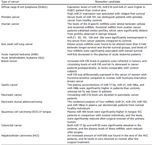
The analyses of miRNA signatures are in general limited to tissue biopsies; however, in the last few years several studies have shown the diagnostic and prognostic usefulness of circulating miRNAs, released by some cell types both under normal and pathological conditions (Table 3) [62]. One of the first studies measuring miRNA levels in serum demonstrated that levels of miR-21 were associated with relapse-free survival in patients with diffuse large B-cell lymphoma [63]. In parallel, many other studies have been published in this field: by measuring the serum levels of miR-141, Mitchell and colleagues could distinguish patients with prostate cancer from healthy subjects; a wider study on miRNA expression profiling in lung cancer, colorectal cancer, and diabetes patients in comparison to related healthy controls found that cancer patients had more elevated serum levels of miR-25 and miR-223 [62, 64]. Currently, one of the most complete studies highlighting the role of circulating miRNAs was carried out on a cohort of 303 non-small-cell lung cancer (NSCLC) patients through Solexa sequencing, and led to the identification of eleven serum miRNAs significantly altered between longer-survival and shorter-survival groups, while some of them were associated to overall survive [65].
Introduction
33
Table 3. Serum miRNAs as biomarkers for cancer (from N Kosaka et al. Cancer Sci
2010; 101(10):2087-92).
To be able to use circulating miRNAs as a diagnostic marker, we need to gain a better understanding of the mechanisms by which miRNAs are released in the bloodstream. There is evidence that serum miRNAs are particularly resistant to RNases digestion, since they can be detected in this body fluid containing high levels of these enzymes; this implies that they should be released in the blood stream in a form that protects them from degradation. Some hypotheses have proposed that they are secreted in protected protein–miRNA complexes (AGO2,
Introduction
34
NPM1, and other RNA binding proteins); another hypothesis is that they are produced as by-products of dead cells [66, 67]. Recent studies have demonstrated a novel mechanism of cell communication using miRNA released in microvesicles (up to 1 m), or in small membrane vesicles called exosomes (10-100 nm) [68] (Figure 10). Exosomes have been detected in many biological fluids, where they are secreted both under physiological and pathological conditions (e.g. blood plasma, urine and cancerous pleural effusions); besides they are actively produced by many cultured cell types [69]. Among the possible biological functions exerted by exosomes, they have been demonstrated to play a significant role in signalling, immune response, and tumor development [70-72]. The increased levels of tumor-derived exosomes in plasma and malignant effusions of patients with cancer suggest that exosomes can be a rich source for the discovery of blood-based diagnostic biomarkers of disease.
__________________________________________________________________ Figure 10. Origin of circulating miRNAs (from J Xu, et al. J Mol Med (Berl), 2012; 90(8):865-75).
Introduction
35
One of the first reports showing the existence of miRNAs in exosomes was released by Valadi et al., who reported that exosomes released from human and murine mast cell lines contain mRNAs and miRNAs [73]. Since then, many studies started to focus on the expression profiling of miRNAs from exosomes isolated from plasma of diseased patients, in order to identify new miRNA markers that are easily measurable in blood samples. Two pivotal studies led to the identification of eight putative miRNA markers for the diagnosis of ovarian cancer (miR-21, miR-141, miR-200a, miR-200c, miR-200b, miR-203, miR-205, and miR-214) and twelve specific miRNAs (miR-17-3p, miR-21, miR-106a, miR-146, 155, 191, 192, 203, 205, 210, 212, and miR-214) for lung cancer, respectively: in both cases it was demonstrated that miRNA profiling could be performed in the absence of tissue and accurately reflects the tumor’s profile [74, 75].
miRNAs in CRC
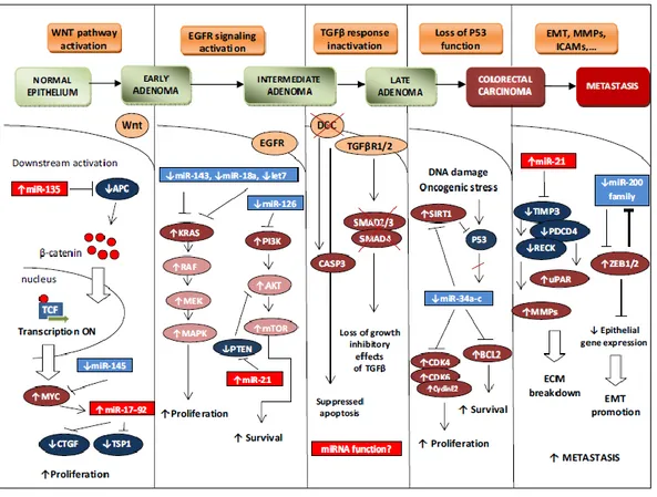
As the result of the numerous alterations involved in CRC pathogenesis, up regulation or reduced expression of several miRNAs, due to transcriptional alterations as well as amplifications, deletions, epigenetic silencing or defects in their biogenesis, have been described in the carcinogenesis of CRC. Two main approaches have been applied to the study of miRNAs involvement in CRC: functional analysis and profiling studies spanning the entire miRNA transcriptome. The first approach is intimately connected to the well-defined alterations leading to impairment of several important cellular pathways in CRC, such as Wnt/ catenin, MAPK, and P53 pathways (Figure 11).
Introduction
36
_______________________________________________________________________________
Figure 11. miRNAs involvement in colorectal cancer histopathogenesis (from O Slaby et al. Mol
Cancer 2009; 14;8:102).
For example, it was recently shown that mir-135 family alterations can be among the early events in CRC pathogenesis: miRNAs belonging to that family are upregulated in CRC, and were demonstrated to target APC tumor suppressor in vivo [76]. EGFR signalling cascades are profoundly associated to miRNA deregulation: KRAS gene, which role in CRC has been widely unveiled, is a direct target of let-7, miR-143 and miR-18a*, which show an inverse expression pattern in CRC samples respect to their target and are significantly down regulated in CRC; loss of miR-126 in CRC is associated with aberrant PI3K signalling, since we assist to accumulation of its target p85β, directly involved in the activation of the pathway [77]. Another important negative regulator of the PI3K pathway is the
Introduction
37
tumor suppressor gene PTEN, strongly repressed by miR-21, which is the most frequently upregulated miRNA in CRC [78, 79]. The tumor suppressor gene p53, mutated in 50-70% of CRCs, is a known expression activator, but numerous reports have indicated that p53 also represses the expression of specific genes either directly or indirectly, through molecular mechanism that still need to be characterized: one of these could be the transcriptional activation of miR-34a-c family, which is a well know family of tumor suppressor miRNAs [80]. The study of miRNA connections with cellular pathways led to a better understanding of the role of those signalling pathways in cancer and other diseases, identifying some miRNAs as new potential therapeutic targets, beyond their already established role in diagnosis. Up to some years ago, the most common method for quantification of miRNAs, as well as other types of transcripts, was Northern blot analysis. Over the last years, with the advent of high-throughput (HT) technologies, several new approaches have been developed to screen entire transcriptomes at once: techniques such as cDNA arrays, Real-Time PCR and bead-based miRNA expression rapidly led to the identification of a wide number of putative miRNA markers for CRC. The main advantage of real-time PCR respect to the other techniques is that, since it requires miRNAs amplification, it is more quantitative and more sensitive, allowing the identification of less abundant molecules that couldn’t be identified otherwise. The first study of this kind compared the expression of a miRNA set in match paired tumoral and non tumoral tissues from CRC patients and CRC cell lines, leading to the identification of a reliable set of 13 miRNAs significantly deregulated in CRC [78]. The most interesting deregulated miRNAs were miR-31, miR-96, miR-135b, miR-183 (up regulated) and miR-133b, miR-145 (downregulated). These data were also consistent with those obtained by using an experimental approach called miRNA serial analysis of gene expression (miRAGE), which had previously shown a systematic alteration of CRC miRNA expression profiles in CRC. Moreover, it was one of the first
Introduction
38
studies on CRC showing that differential miRNA expression between CRC samples could be a link between CRC carcinogenesis and alterations of the RAS/RAF/MEK/ERK pathway. For example, among several proteins with potential oncogenic functions, putative miR-145 targets are MAPK transduction proteins such as MAP3K3 and MAPK4K4, while among targets of miR-133b, the most notable oncogenic target is KRAS [81]. In addition, the expression level of miR-31 was also correlated with the stage of CRC tumor, suggesting that this miRNA could contribute to tumorigenesis and the acquisition of a more aggressive phenotype in CRC, maybe through the inhibition of genes such as FOXC2 and FOXP3, members of the forkhead family of transcription factors which are involved in the silencing of antiapoptotic genes and are known to be modulated, among other pathways, also by ERK/MAPK pathway [82]. In 2005 Volinia et al. identified a cancer-specific signature which included significant up regulation of miR-21, miR-17-5p, miR-191, miR-29b, miR-223, miR-128b, miR-24, miR-155, miR-20a, miR-107, miR-32, miR-30c, miR-221 and miR-106a in CRC. Most of these miRNAs have been later confirmed to be up-regulated in CRC by other profiling studies [59]. MiRNA profiling has also been used to classify CRC on the basis of their microsatellite status: Lanza and colleagues showed that microRNA profiling clearly discerned between MSI and microsatellite stable (MSS) tumors [83]. This was subsequently confirmed by another group that also showed the prognostic value of miR-320 and miR-498 in predicting recurrence-free survival [84]. From then, several other studies focused on the use of microRNA profiling to provide prognostic informations and to direct patients to specific treatments. For example, Schetter et al. in 2008 confirmed the promising clinical potential of microRNA profiling in clinical management of CRC patients by performing a microRNA microarray expression profiling in more than 200 paired tumor and non-tumor tissues. They found 37 differentially expressed miRNAs in CRC patients, with miR-20a, miR-21, miR-106a, miR-181b, and miR-203 associated
Introduction
39
also with poor survival; further validation showed that miR-21 was significantly associated with poor prognosis, and this association was independent of age, sex, and tumor location [79]. This study demonstrated miR-21 role in CRC pathogenesis, showing its involvement in tumor progression and metastasis: it was shown indeed that its expression was progressively increasing in the adenoma-carcinoma sequence. This action can be performed through the modulation of the tumor suppressor genes PDCD4 (that inhibits TPA-induced tumor transformation and progression), which expression resulted inversely correlated with miR-21 in a cohort of 22 CRC patients [85]. The importance on circulating miRNAs in CRC patient screening was first pinpointed on a study by Ng et al., in 2009: miR-92 upregulation was identified as one of the best predictive markers for CRC with a sensitivity of 89% and a specificity of 70%, which was demonstrated by strong decrease in its expression after tumor resection [86]. The importance of miR-17-92 cluster, involved in cancer progression through stimulation of proliferation, had been already identified in CRC, through miRNA expression profiling in cancer tissues that identified a significant up regulation of these miRNAs in tumor samples [87]. Similar results were obtained by Huang et al., who measured the expression of 12 candidate miRNAs in plasma samples from patients with advanced colorectal neoplasia and healthy controls using real-time RT-PCR. They found that plasma miR-29a and miR-92a have significant diagnostic value for advanced neoplasia [88].
miRNA targeting as a new possible therapeutic approach in CRC
Another important question for management of CRC patients is the possibility of predicting therapy response based on miRNA expression patterns; moreover, it has been shown that specific treatments are able to induce or repress the expression of