Current Alzheimer Research, 2018, 15, 1-8 1
RESEARCH ARTICLE
1567-2050/18 $58.00+.00 © 2018 Bentham Science Publishers
Assessment of the Autonomic Response in Alzheimer's Patients During the
Execution of Memory Tasks: A Functional Thermal Imaging Study
David Perpetuini
1,*, Daniela Cardone
1, Roberta Bucco
2, Michele Zito
2and Arcangelo Merla
11Infrared Imaging Lab, Centro ITAB – Institute for Advanced Biomedical Technologies, University G. d’Annunzio, Chieti, Italy and Department of Neurosciences, Imaging and Clinical Sciences, University G. d’Annunzio, Chieti-Pescara, Italy; 2Department of Medicine and Science of Ageing, University G. d’Annunzio, Chieti-Pescara, Italy
A R T I C L E H I S T O R Y Received: October 31, 2017 Revised: January 28, 2018 Accepted: April 23, 2018 DOI: 10.2174/1871529X18666180503125428
Abstract: Background: Alzheimer’s disease (AD) is a form of dementia characterized by failure of memory that becomes more severe with the progression of the disease. The Free and Cued Selective Reminding Test (FCSRT) is a clinical test used to evaluate such a deficit. However, since the cognitive performances could depend also on the psychophysiological state of the individual, it is important to monitor that state through the peripheral autonomic activity during the execution of the test. Thermal infrared imaging has been used for this kind of assessment in order to preserve the free and unbiased interaction between doctor and patient, thanks to the contactless features of the technique.
Objective: To investigate whether the variation of facial temperature parameters during the FCSRT is
indicative of different autonomic states in the early AD with respect to healthy controls (HC).
Results: At a group level, a greater sympathetic activity for the HC with respect to AD during the
execu-tion of the test was found, indicative of a suppression of anxiety associated with the performances of the FCSRT in AD patients.
Conclusion: These results indicate that AD and HC may present different autonomic activity associated
with the execution of a cognitive task, thus suggesting a different modulation of high-cognition and emotion network.
Keywords: Alzheimer's disease, FCSRT, thermal imaging, memory deficit, visuospatial deficit, autonomic response. 1. INTRODUCTION
Alzheimer’s disease (AD) is a form of dementia charac-terized by failure of memory that becomes more acute with the progression of the disease [1]. In fact, according to the International Working Group on Alzheimer’s [2], the core diagnostic criterion is the progressive memory deficit. In order to assess this kind of impairment, many tests and bat-teries have been proposed.
These tests could be a cause of anxiety and stress for the participants, both for AD and healthy controls (HC) as anxi-ety and stress may be associated with the execution of learn-ing and cognitive tasks [3]. Furthermore, it is known that the emotional state could affect efficient functioning of the at-tentional system and the performance of tasks, especially under test conditions [3-6].
The aim of this study is to investigate whether the execu-tion of cognitive/mnemonic tasks elicits a different arousal involvement in AD patients with respect to HC, as revealed *Address correspondence to this author at the Infrared Imaging Lab, Centro ITAB – Institute for Advanced Biomedical Technologies, University G. d’Annunzio, Chieti, Italy and Department of Neurosciences, Imaging and Clinical Sciences, University G. d’Annunzio, Chieti-Pescara, Italy; E-mail: [email protected]
by autonomic modulation, and if this may impact the per-formances in the execution of the tests. In fact, currently, the clinical diagnosis of AD is carried out by interviews and administration of tests. It is therefore relevant to investigate the effect of the autonomic system on the performance.
One of the most effective tests for discriminating AD pa-tients from HC is the Free and Cued Selective Reminding Test (FCSRT) [7, 8]. This test shows a high sensibility and specificity in distinguishing AD patients from HC [9, 10], from other forms of dementia [11] and from Mild Cognitive Impairment (MCI) patients non-converters [12].
It starts with a phase of encoding, during which the pa-tient is required to remember 12 pictures of everyday life shown four at a time. After that, the Immediate Free Recall (IFR) follows, during which the patient is requested to recall all these figures. If he/she cannot remember independently all the figures, the Immediate Cued Recall (ICR) follows. During this phase, the doctor reminds the semantic fields of the not retrieved pictures. This procedure is repeated for three times. After a period of 30 minutes, the test provides the Delayed Free Recall (DFR) during which the patient has to remind all the pictures shown previously. If he/she forgets some of them, the Delayed Cued Recall (DCR) follows, dur-ing which all the semantic fields of the missdur-ing pictures are reminded by the doctor.
In order to fill the time between the Immediate Recall and the Delayed one, some filler tests are used. Usually, these tests are chosen according to their ability to discrimi-nate some particular impairment typical of the investigated dementia. In particular, the constructive and visuo-spatial impairments are typical of the AD, and, in order to discriminate these deficits, in the present study the Clock Drawing Test (CDT) [13] was used. During this test, the patient is requested to draw a circle, then to insert the num-bers like it was a clock and, finally, to set the clock hands at ten past ten.
Then, Digit Span Test (DST) was administrated to evalu-ate short-term memory and verbal working memory impair-ments [14]. For this test, the participant is asked to repeat sequences of digits verbally presented with a pace of 1 sec-ond. The test started with a series of two digits and every time the series is repeated correctly, a new series with one digit more is presented. If the participant cannot remember a sequence, another one of the same length is proposed. If the participant is not able to repeat two sequences of the same length, the test ends.
To evaluate the visuospatial memory deficit the Corsi Tapping Test (CTT) [15] was proposed. During this test, the experimenter has to tap some cubes starting with a sequence of two cubes and, if the participant can repeat correctly the sequence, the number of cubes is progressively increased with a step of one. If the participant cannot repeat two sequences of the same length the test ends. The cubes are touched with the index finger at a rate of 1 cube per second.
For the evaluation of visuo-spatial impairments, the Benton Visual Retention Test (BVRT) [16] was added. It is composed of three Forms (C, D, and E) and can be adminis-tered in four different ways (Administrations A, B, C, and D) [17]. In this study, the administration C (Form D) was used, during which the examinee has to reproduce a design in the presence of stimuli, without limits of exposure time.
Finally, the Trial Making Test, versions A e B (TMT A-B) [18] was proposed. TMT-A requires the participant to draw lines connecting sequentially 25 encircled numbers on a sheet of paper. For TMT-B the subject has to alternate be-tween numbers and letters (e.g., 1, A, 2, B, 3, C, etc.) [18].
In order to replicate the ecological conditions under which the tests are administered in the clinical routine (with-out any associated recording of vital signs and autonomic activity, i.e. by not using any contact sensing probe), func-tional thermal infrared imaging (fIRI) was used to assess indices of peripheral autonomic activity while performing the test.
fIRI (or thermography) is one of the most suitable tech-niques to be considered for this purpose: it is a non-invasive technique used to measure the peripheral autonomic activity through the modulation of the cutaneous temperature, which is a known expression of the psychophysiological state of the subject [19]. In fact, stress, anxiety or fatigue can produce changes in skin temperature [20]. fIRI has been widely used to study the effect of workload [21] and learning process [3] on the facial skin temperature. In addition, fIRI is an impor-tant tool for the detection of the emotional state through the
noninvasive and touchless assessment of the autonomic ac-tivity [22, 23].
According to the literature, this is the first time that auto-nomic activity is monitored through fIRI during the admini-stration of cognitive/mnemonic tasks in AD. Since all the above-mentioned tests request abilities compromised in AD, a different autonomic response for patients with respect to HC is expected.
2. MATERIALS AND METHODS 2.1. Participants
Fourteen healthy people (mean age ± SD: 68.4±6.3 years; M/F: 11/3) and sixteen patients (mean age ± SD: 75.5±5.4 years; M/F: 8/8) with a diagnosis of Mild probable Alz-heimer’s disease, according to the Diagnostic and Statistical Manual of Mental Disorders, 5th edition (DSM-5) partici-pated in the study. The exclusion criteria were moderate-severe cognitive impairment (MMSE <25/30) [24], vascular dementia, behavioral or psychiatric disorders, hydrocepha-lus, brain lesions, history of stroke or traumatic brain injury and circulatory diseases that could impact the thermal meas-urement. The Research Ethics Board of the University of Chieti-Pescara approved this study, conducted according to the Declaration of Helsinki. Before the experiment, every participant signed the informed consent and they could with-draw from it at any time. All sessions were scheduled at the same time of the day to mitigate possible effects due to cir-cadian rhythm variations [25].
2.2. Experimental Design
The FCSRT was proposed by Buschke [26] and it was validated for the AD diagnosis by Grober [11]. It consists of an encoding and a recall phase. The immediate and delayed recall were separated by some filler test (CDT, DST, CTT, TMT A-B, BVRT). If the participant took less than 30 min-utes for the execution of the filler tests, TMT A-B was pro-vided. Hence, since it was not administered to all subjects, it was excluded from the analyses.
During the administration of this battery, the patient and the examiner were seated in front of each other and they had to interact. In order to separate the different tests and to have a baseline to compare the signal measured during the tasks, 1 minute of rest was provided to separate one test and the next one. The experimental design is shown in Fig. (1).
For the thermal measurements, the guidelines suggested in Merla and Romani (2006) [27] Ring and Ammer (2012) [28], and Diakides, Bronzino and Peterson (2012) [29] were followed.
2.3. Functional Thermal Imaging Measurement
For each subject, the facial temperature was recorded during the administration of the tests, by means of a digital thermal infrared camera FLIR SC660 (640 x 480 bolometer FPA, sensitivity/Noise Equivalent Temperature Difference: < 30 mK @ 30°C, FOV: 24° x 18°). The camera was placed at 60 cm from the participant and pointed toward the face of the subject (Fig. 2). The sample frequency was 1 Hz. To re-move the effects related to the potential drift/shift of the
sen-sor’s response and optical artifacts, the camera was black-body-calibrated.
Fig. (1). Experimental paradigm: FCSRT (blue blocks), filler tests (yellow blocks) and rest periods (green blocks) added to separate the different phases.
Fig. (2). Thermogram of a representative participant.
2.4. Thermal Data Processing and Statistical Analysis
Since the participant could freely move during the ex-periment, the quality of all the recorded thermal videos was preventively checked by visual inspection. No video was rejected.
Seven regions of interest (ROI), reported as indicative for the evaluation of autonomic activity [19], were selected: corrugator, up nose, nose tip, right and left perioral, right and left chin (Fig. 3).
Fig. (3). ROIs position of a representative participant: corrugator (ROI 1), upper nose (ROI 2), nose tip (ROI 3), right and left pe-rioral (ROIs 4 and 5 respectively), right and left chin (ROIs 6 and 7 respectively)
The time courses of the average temperature were ex-tracted from each ROI. Since the participants could move the head without restriction, a soft-tissue tracking algorithm was used to track each ROIs across all the images of the video, in order to properly consider the temperature from each ther-mogram. The tracking software has been developed under Matlab environment and validated in Manini et al (2013) [30]. It is based on the 2D cross-correlation between a tem-plate region, chosen by the user on the first frame, and a similar ROI in a wider searching region, expected to contain the desired template in each of the following frames.
The thermal signal was further corrected from residual motion artifacts. Motion errors were visually detected and corrected by substituting them with the mean value of the 6 samples before and after the artifact.
For each time series of each ROI, the mean value of the temperature during every experimental condition was evalu-ated. Then, the difference between the mean value of tem-perature during each test ( ) and the mean value of the temperature during the previous baseline ( ) was com-puted (Equation 1). Moreover, the slope of the thermal signal was computed as the ratio between the maximum variation of the signal during the experimental phase and the time taken to get this variation (Equation 2).
(1) (2) Furthermore, in order to investigate the different contri-bution of the sympathetic and parasympathetic system to the thermal fluctuations, the power of the thermal signal in the low frequencies (LF) band (0.03-0.15 Hz) and in the high frequencies (HF) band (0.15-0.35 Hz) has been evaluated. In fact, while the LF band has been assessed to be indicative for the sympathetic system, the HF band is suggestive for both the sympathetic and parasympathetic system, thus the ratio LF/HF is considered an indicator of the balance of the acti-vation of the two systems [31].
Since the minimum duration of each test was greater than the inverse of the lower frequency of the LF band, the LF/HF ratio was evaluated for each task.
For each experimental phase, a Shapiro Wilk Test was performed to assess dataset normality for both time and fre-quency domain analysis. Since data resulted not normally distributed, Wilcoxon-Mann-Withney test was used to evaluate group differences between AD and HC.
Finally, in order to better investigate the capability to discriminate the two groups by means of the autonomic re-sponse during these tests, a Linear Discriminant Analysis was carried out [32].
3. RESULTS
3.1. Behavioural Results
The scores of all the FCSRT and the filler tests discrimi-nate the two groups, except for the DST and CTT. Group differences tested through an independent t-test between HC and AD. are summarized in Table 1.
Table 1. Behavioural results: t-stat and p-value for the scores of each test between HC and AD.
t test HC vs AD t stat p value
IFR 6,9527 1,47*10-7 ICR 7,4339 0,43*10-7 CDT 3,2879 0,0027 DST 0,8151 0,4219 CTT 1,2384 0,2258 BVRT 4,9438 3,87*10-5 DFR 5,4102 9,05*10-6 DCR 5,8000 3,14*10-6
3.2. Time Domain Results
Time series of the thermal signals from the considered ROIs are shown in (Fig. 4) for a representative subject.
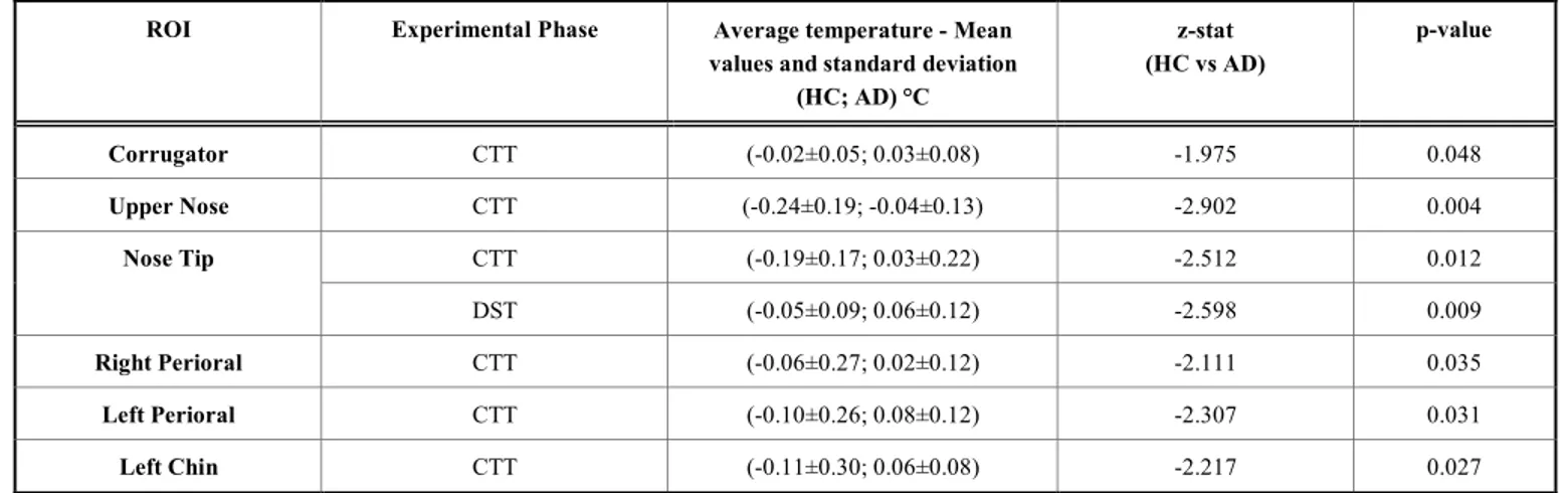
The statistically significant differences, during the differ-ent experimdiffer-ental phases, for the two groups between the mean values during the test with respect to the previous rest period, are reported in Table 2.
Table 3 resumes the group differences between the slopes of the temperature during the different experimental phases.
3.3Frequency Domain
The statistically significant differences between the LF/HF ratio for the two groups during the different experi-mental phases are summarized in Table 4.
3.4. Classification
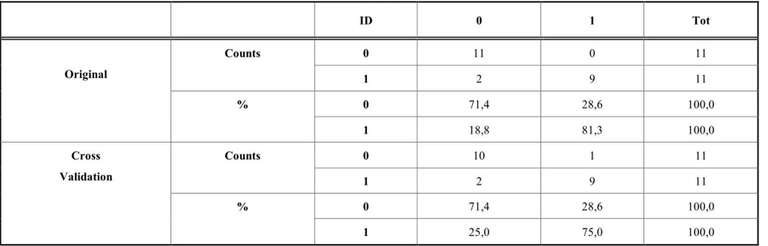
Since the ROI on the Nose Tip resulted to be significant for the CTT for all the three parameters investigated, a dis-criminant analysis using these variables has been carried out. The results are presented in Table 5. The original cases are classified properly for the 76,7 %. With the cross validation, the percentage of the cases correctly classified is 73,3%.
4. DISCUSSION
The aim of this study was to assess whether differences in the autonomic system activity during the performance of cognitive/mnemonic tests exist between AD and HC. Since, these tests are created to evaluate some specific impairments, their administration could elicit a state of stress or anxiety that could have, in turn, an effect on the performance. In order to avoid the effect of contact-sensor instrumentation on the performance, it would be advisable to control the psy-chophysiological state of the participants in a non-invasive way; to this purpose, thermal infrared imaging appears to be one of the most suitable tools. In particular, in this study, the variation of the facial temperature and the LF/HF ratio of the thermal time series have been evaluated, to quantitatively assess the metrics of the autonomic activity of the partici-pants. The main findings are that the temperature variations were lower in the HC group than the AD patients for Nose Tip, Upper Nose, Corrugator, Left and Right Perioral and
Table 2. Mean values: group differences between HC and AD patients for each ROI during the different experimental phases.
ROI Experimental Phase Average temperature - Mean
values and standard deviation (HC; AD) °C z-stat (HC vs AD) p-value Corrugator CTT (-0.02±0.05; 0.03±0.08) -1.975 0.048 Upper Nose CTT (-0.24±0.19; -0.04±0.13) -2.902 0.004 CTT (-0.19±0.17; 0.03±0.22) -2.512 0.012 Nose Tip DST (-0.05±0.09; 0.06±0.12) -2.598 0.009 Right Perioral CTT (-0.06±0.27; 0.02±0.12) -2.111 0.035 Left Perioral CTT (-0.10±0.26; 0.08±0.12) -2.307 0.031 Left Chin CTT (-0.11±0.30; 0.06±0.08) -2.217 0.027
Table 3 Slopes. Group differences between HC and AD patients for each ROI during the different experimental phases.
ROI Experimental Phase Slope - Mean values and standard de-viation (HC; AD) °C z-stat (HC vs AD) p-value Nose Tip CTT (2.41·10-5± 1.02·10-4; -1.68·10-4± 3.38·10-4) 2.681 0.007 Upper Nose DST (-0.01±0.01; 0.01±0.07) -2.640 0.008 Right Perioral DST (-0.05±0.09; 0.01±0.04) -2.103 0.036 Left Perioral DST (-0.15±0.32; -1.74·10-4±0.07) -2.260 0.024
Table 4 LF/HF ratio. Group differences between HC and AD patients for each ROI during the different experimental phases as-sessed using Wilcoxon-Mann-Whitney test.
ROI Experimental Phase LF/HF - Mean values and
standard deviation (HC; AD)
z-stat (HC vs AD) p-value Corrugator CTT 19.33±7.80 14.22±6.15 2.029 0.043 Encoding 14.93±5.96 12.38±1.37 2.684 0.007 Upper Nose CTT 20.76±9.46 13.82±6.14 2.073 0.038 Nose Tip CTT 20.78±9.48 14.23±6.15 2.117 0.027
Right Perioral Encoding 15.90±4.61
12.52±1.14
3.128 0.002
Left Perioral Encoding 16.66±4.65
12.71±1.17 3.293 9.92*10-4 CTT 21.29 ±9.97 14.19±6.15 2.203 0.028 Right Chin Encoding 14.72±5.90 12.48±1.12 2.802 0.005
Left Chin Encoding 16.77±4.59
12.68±1.06
Table 5. Linear Discriminant Analysis for fIRI parameters on the Nose Tip during CTT. ID 0 1 Tot 0 11 0 11 Counts 1 2 9 11 0 71,4 28,6 100,0 Original % 1 18,8 81,3 100,0 0 10 1 11 Counts 1 2 9 11 0 71,4 28,6 100,0 Cross Validation % 1 25,0 75,0 100,0
Left Chin (Table 2), while the LF/HF ratio was higher for the HC than the AD for all the considered ROIs, as shown in Table 4. More precisely, the HC group showed a lower variation of temperature for the CTT and DST. A lower skin temperature variation could be caused by a superficial vaso-constriction that is due to a major activation of the sympa-thetic system [33], which therefore resulted larger in HC than AD patients. This was confirmed by the LF/HF results; in fact, the HC group showed a higher ratio with respect to AD. This ratio is indicative of the balance between the sym-pathetic and parasymsym-pathetic system, hence the HC group showed a higher sympathetic activation.
Many authors have already investigated autonomic dis-orders in AD patients [34]. Differences in the heart rate be-tween AD patients and controls were assessed by evaluating the LF/HF ratio and resulting in a hypersympathetic activity for the AD group during 5 minutes of rest in different posi-tions (upright and supine posture) [35]. Conversely, Melling-sæter et al (2015) found a lower LF/HF ratio for the heart rate variability in AD patients with respect to healthy con-trols during a head-up tilt test [36]. In addition, vasomotor sympathetic functions were investigated, monitoring the re-duction of finger pulse amplitude during Valsava manoeu-vre, revealing an autonomic dysfunction concerning para-sympathetic, as well as vasomotor para-sympathetic, functions in AD [37]. Since the vasomotor regulation influences the skin temperature, it is valid to suppose an influence of the disease on the thermal signal. In fact, it is known from the literature that AD patients exhibit impaired variations in the circadian temperature [38] and in the thermoregulation [39]. However, the cited studies were carried out during a resting condition and they did not involve any cognitive or mnemonic task, thus the results are clearly related to a functional dysregula-tion due to the disease. In the present study, whereas, the subjects were requested to perform a battery of cognitive tests, thus the results were dependent mainly on the perform-ance and the possible expectation associated with them. In addition, as no differences were found between the two groups during the resting period, it is possible to suppose that the observed different activation of the autonomic sys-tem was mostly related to the test execution rather than to a dysregulation caused by the disease. However, it is worth noticing that the absence of differences between HC and AD
during the rest period could have been due to the short length of this phase (1 minute). This is an aspect that needs further studies for being clarified.
The greater sympathetic activity assessed for the HC dur-ing both DST and CTT could have been due to a stress con-dition. In fact, it is known that verbal learning depends on experimental stress [40]. Although differences in the thermal signal were found, the scores of these tests did not show sig-nificant differences between the two groups (Table 1). The CTT and DST are indeed selective for the short term mem-ory deficit, but not very discriminant for AD dementia [41]. The reported results suggested that the performances of the DST and CTT might depend on the psychophysiological state of the participant. However, further studies are neces-sary to better investigate the relationship between stress and performance and to clarify if this difference in autonomic activation between AD and HC could be indicative of early AD diagnosis.
LF/HF ratio showed significant differences between the two groups also during the encoding phase. During this ex-perimental phase, the participant was asked to memorize the pictures shown by the experimenter. HC participants exhib-ited higher LF/HF ratio with respect to AD patients, hence a greater activation of the sympathetic system. Deficits in en-coding and semantic memory in AD have been investigated so far and it seems that they are related to a lack of attention and decrease of cognitive effort [42]. In addition, it is known that the sympathetic activity during the encoding phase can influence the retrieval [43], thus monitoring the autonomic system during this process could be of great interest, because it influences the recall, from which the AD diagnosis is car-ried out in the FCSRT.
Concerning the slope of the signal, statistical differences were found for the Nose Tip, Upper Nose, Right and Left Perioral during the CTT and DST. In particular, the HC group exhibited a larger slope for the CTT and a lower slope for the DST with respect to the AD patients (Table 3). The slope is an indicator of the speed of the changes in the ther-mal signal. Thus, during the DST the variation of the therther-mal signal was larger but slower in the HC group than the AD. This test becomes progressively more difficult, starting with a sequence of two digits and adding one more digit every
time the participant could properly repeat the sequence. Thus, this result seemed to suggest that healthy people had a sympathetic response delayed towards the end of the test, while the AD exhibited an early response. It was probably caused by a short memory deficit of the AD patients [1]. This effect was not present during the CTT. A possible ex-planation is that it came soon after the DST and their struc-ture is quite similar. Hence, it is valid to suppose that there was a habituation effect that shifted the response towards the start of the test.
During the other experimental phases, no significant dif-ferences were found. This finding suggested the absence of clear differences in autonomic activity between the two groups during these phases.
Furthermore, the possibility to discriminate the two groups by means of the autonomic response during the CTT, considering the Nose Tip, was investigated by means of lin-ear discriminant analysis. An accuracy of 73.3% was ob-tained. This result, although preliminary, seemed to suggest that autonomic response in AD patients during the execution of cognitive/mnemonic tests could be highly indicative of the disease. However, the limited number of participants did not allow to carry out an accurate and generalizable classifica-tion. In fact, the purpose of future studies is to increase the number of participants to improve the discrimination of AD patients from HC. In addition, it could be of great interest to combine thermal infrared imaging with physiological meas-urements, such as heart rate and galvanic skin response to ground-truth the results here presented, through the integra-tion of different physiological informaintegra-tion. In particular, it could be worth focusing on the encoding phase to search a possible precursor of the AD that could be useful to improve the current diagnosis of the pathology.
Moreover, it might be interesting to investigate the DST and CTT randomizing the order of administration to clarify if the effect showed in this study is related to the test itself or to the sequence of administration.
Finally, it could be worth increasing the length of the rest period, in order to assess if the thermal signal could give important information about the autonomic disorders in AD patients, thus providing a useful tool for the AD diagnosis.
CONCLUSION
Referring to the actual state of the art, this study repre-sents the first time that the autonomic activity is recorded by means of thermal response in AD patients during the admini-stration of cognitive/mnemonic tests, in completely ecologi-cal conditions.
The results seemed to show a greater activation of the sympathetic system in HC with respect to the AD patients during the CTT, DST and Encoding phase. The findings suggested that the administration of clinical tests could elicit different autonomic responses in AD patients with respect to HC, but further studies are necessary to better investigate the influence of the psychophysiological state to the perform-ance, hence to the AD diagnosis.
ETHICS APPROVAL AND CONSENT TO PARTICI-PATE
The Research Ethics Board of the University of Chieti-Pescara.
HUMAN AND ANIMAL RIGHTS
No animals were used in this research. All humans re-search procedures followed were in accordance with the standards set forth in the Declaration of Helsinki principles of 1975, as revised in 2008 (http://www.wma.net/ en/20activities/10ethics/10helsinki/).
CONSENT FOR PUBLICATION
All participants gave their informed consent to take part in the study.
CONFLICT OF INTEREST
The authors declare no conflict of interest, financial or otherwise.
ACKNOWLEDGEMENTS.
This study is supported by ASTONISH european project (ref. H2020 ECSEL2015 - 692470).
REFERENCES
[1] Dubois B, Feldman HH, Jacova C, Dekosky ST, Barberger-Gateau P, Cummings J, et al. Research criteria for the diagnosis of Alz-heimer's disease: revising the NINCDS-ADRDA criteria. Lancet Neurol 6(8): 734-46 (2007).
[2] Cummings JL, Dubois B, Molinuevo JL, Scheltens P. International Work Group criteria for the diagnosis of Alzheimer disease. Med Clin 97(3): 363-68 (2013).
[3] Eysenck MW, Derakshan N, Santos R, Calvo MG. Anxiety and cognitive performance: attentional control theory. Emotion 7(2): 336 (2007).
[4] Eysenck MW, Calvo MG. Anxiety and performance: the process-ing efficiency theory. Cogn. Emotion6(6): 409-34 (1992). [5] Sanders AF. Towards a model of stress and human
perform-ance. Acta Psychologica 53(1): 61-97 (1983).
[6] Ashkanasy NM. Emotion and performance. Hum Performan 17(2): 1317-44 (2004).
[7] Lemos R, Simões MR, Santiago B, Santana I. The free and cued selective reminding test: Validation for mild cognitive impairment and Alzheimer's disease. J Neuropsychol 9(2): 242-57 (2015). [8] Buschke H. Cued recall in amnesia. J Clin Exp Neuropsychol 6(4):
433-40 (1984).
[9] Grober E, Sanders AE, Hall C, Lipton RB. Free and cued selective reminding identifies very mild dementia in primary care. Alz-heimer Dis Assoc Disord 24(3): 284 (2010).
[10] Perpetuini D, Bucco R, Zito M, Merla A. Study of memory deficit in Alzheimer’s disease by means of complexity analysis of fNIRS signal. Neurophotonics 5(1): 011010 (2017).
[11] Grober E, Hall C, Sanders AE, Lipton RB. Free and cued selective reminding distinguishes Alzheimer’s disease from vascular demen-tia. J Am Geriatr Soc 56(5): 944-46 (2008).
[12] Grober E, Buschke H. Genuine memory deficits in dementia. De-velo Neuropsychol 3(1): 13-36 (1987).
[13] Ricci M, Pigliautile M, D'Ambrosio V, Ercolani S, Bianchini C, Ruggiero C, et al. The clock drawing test as a screening tool in mild cognitive impairment and very mild dementia: a new brief method of scoring and normative data in the elderly. Neurol Sci 37(6): 867-73 (2016).
[14] Gerton BK, Brown TT, Meyer-Lindenberg A, Kohn P, Holt JL, Olsen RK, et al. Shared and distinct neurophysiological
compo-nents of the digits forward and backward tasks as revealed by func-tional neuroimaging. Neuropsychologia 42(13): 1781-87 (2004). [15] Binetti, Giuliano, et al. Executive dysfunction in early Alzheimer's
disease. J Neurol Neurosur Psychiat 60(1): 91-3 (1996).
[16] Sivan, Abigail Benton. Benton visual retention test. San Antonio, TX: Psychological Corporation, 1992.
[17] Benton, Arthur Lester. Contributions to neuropsychological as-sessment: A clinical manual. Oxford University Press, USA, 1994. [18] Tombaugh TN. Trail Making Test A and B: normative data strati-fied by age and education. Arch Clin Neuropsychol 19(2): 203-14 (2004).
[19] Cardone D, Merla A. New frontiers for applications of thermal infrared imaging devices: computational psychopshysiology in the neurosciences. Sensors 17(5): 1042 (2017).
[20] Genno H , Ishikawa K , Kanbara O, Kikumoto M, Fujiwara Y, Suzuki R, et al. Using facial skin temperature to objectively evalu-ate sensations. Intern J Indus Erg 19(2): 161-71 (1997).
[21] Veltman JA, Vos WK. Facial temperature as a measure of operator state. (2005).
[22] Salazar-López E, Domínguez E, Juárez Ramos V, de la Fuente J, Meins A, Iborra O, et al. The mental and subjective skin: Emotion, empathy, feelings and thermography. Consciousness Cogn 34: 149-62 (2015).
[23] Clay-Warner J, Robinson DT. Infrared thermography as a measure of emotion response. Emo Rev 7(2): 157-162 (2015).
[24] Folstein MF, Folstein SE, McHugh PR. Mini-mental state: a prac-tical method for grading the cognitive state of patients for the clini-cian. J Psychiatric Res 12: 189-98 (1975).
[25] Marins JCB, Formenti D, Costa CMA, Fernandes AA , Sillero-Quintana M. Circadian and gender differences in skin temperature in militaries by thermography. Infrared Phy Technol 71: 322-28 (2015).
[26] Buschke H. Cued recall in amnesia. J Clin Exp Neuropsychol 6(4): 433-40 (1984).
[27] Merla A, Romani GL. Biomedical applications of functional infra-red imaging. Engineering in Medicine and Biology Society, 2005. IEEE-EMBS 2005. 27th Annual International Conference of the. IEEE, 2006.
[28] Diakides M, Bronzino JD, Peterson DR, eds. Medical infrared imaging: principles and practices. CRC press, 2012.
[29] Ring EFJ, Ammer K. Infrared thermal imaging in medicine. Physiol Meas 33: 3 R33 (2012).
[30] Manini B, Cardone D, Ebisch SJH, Bafunno D, Aureli T, Merla A. Mom feels what her child feels: thermal signatures of vicarious
autonomic response while watching children in a stressful situation. Front Hum Neurosci 7: 299 (2013).
[31] Malliani A, Lombardi F, Pagani M. Power spectrum analysis of heart rate variability: a tool to explore neural regulatory mecha-nisms. Brit Heart J 71(1): 1 (1994).
[32] Hair, Joseph F., et al. Multivariate data analysis. Vol. 5. No. 3. Upper Saddle River, NJ: Prentice hall, 1998.
[33] Jänig, Wilfrid, and Elspeth M. McLachlan. "Neurobiology of the autonomic nervous system." Autonomic Failure: A Textbook of Clinical Disorders of the Autonomic Nervous System Ox-ford (2013): 21-34.
[34] Femminella GD, Rengo G, Komici K, Iacotucci P, Petraglia L, Pagano G, et al. Autonomic dysfunction in Alzheimer's disease: tools for assessment and review of the literature. J Alzheimer's Dis 42(2): 369-77 (2014).
[35] Aharon-Peretz J, Harel T, Revach M, Ben-Haim SA. Increased sympathetic and decreased parasympathetic cardiac innervation in patients with Alzheimer's disease. Arch Neurol 49(9): 919-22 (1992).
[36] Mellingsæter MR, Wyller TB, Ranhoff AH, Bogdanovic N, Wyller VB. Reduced sympathetic response to head-up tilt in subjects with mild cognitive impairment or mild Alzheimer's dementia. Demen Geriatr Cogn Disord Extra 5(1): 107-15 (2015).
[37] Algotsson A, Viitanen M, Winblad B, Solders G. Autonomic dys-function in Alzheimer's disease. Acta Neurologica Scandi-navica 91(1): 14-18 (1995).
[38] Prinz PN, Christie C, Smallwood R, Vitaliano P, Bokan J, Vitiello MV, et al. Circadian temperature variation in healthy aged and in Alzheimer's disease. J Gerontol 39(1): 30-35 (1984).
[39] Diamond PT, Diamond MT. Thermoregulatory behavior in Alz-heimer's disease. J Am Geriatr Soc 39(5): 532-32 (1991).
[40] Spielberger Charles D., and Lou H. Smith. "Anxiety (drive), stress, and serial-position effects in serial-verbal learning." Journal of
Ex-perimental Psychology72.4 (1966): 589.
[41] Pasquier, Florence. "Early diagnosis of dementia: neuropsychol-ogy." Journal of neurology 246.1 (1999): 6-15.
[42] Dick, Malcolm B., Mary-Louise Kean, and Dan Sands. "Memory for internally generated words in Alzheimer-type dementia: Break-down in encoding and semantic memory." Brain and Cognition 9.1 (1989): 88-108.
[43] Smeets, Tom, et al. "True or false? Memory is differentially af-fected by stress-induced cortisol elevations and sympathetic activ-ity at consolidation and retrieval." Psychoneuroendocrinology 33.10 (2008): 1378-1386.
DISCLAIMER: The above article has been published in Epub (ahead of print) on the basis of the materials provided by the author. The Editorial Department reserves the right to make minor modifications for further improvement of the manuscript.