UNIVERSITÁ DEGLI STUDI DI CATANIA
Dottorato di Ricerca in Scienze Geologiche, Biologiche e Ambientali
XXIX Ciclo
Settore scientifico disciplinare (SSD) BIO/06
Ph.D. Thesis
Toxicity evaluation of new engineered nanomaterials in model
organisms
Dott.ssa Roberta Pecoraro
Coordinatore: Prof.ssa Agata di Stefano
Tutor: Prof.ssa Maria Violetta Brundo
Co-tutor: Dott.ssa Maria Antonietta Buccheri
The present work has been supported by the VII FP project Winning
Applications of nanoTEchnologies for Resolutive hydropurification
(WATER) funded by the European Commission (Grant 316082).
Summary
ABSTRACT ... 1
1. INTRODUCTION ... 5
1.1. Classification of Engineered Nanomaterials (ENMs) ... 6
1.2. Applications of nanoparticles ... 7
1.3. Routes of entry, uptake and bioavailability into living systems ... 8
1.4. Risks to human health via water ... 9
1.5. Harmful effects of NPs and possible consequences on biological systems ... 9
1.6. Mechanism of toxicity ... 10
1.7. Silver nanoparticles (AgNPs) ... 11
1.7.1. Properties and applications ... 12
1.7.2. Environmental concern ... 12
1.7.3. Natural and anthropogenic source of AgNPs ... 13
1.7.4. Fate, transport and distribution of AgNPs in environment ... 13
1.7.5. Mechanism of toxicity ... 14
1.8. Gold nanoparticles (AuNPs) ... 16
1.8.1. Synthesis of AuNPs ... 16
1.8.2. Role of gold NPs in biomedical applications ... 17
1.8.3. Gold nanoparticles toxicity ... 18
1.9. Titanium dioxide nanoparticles (TiO2) ... 19
1.9.1. Properties and applications ... 19
1.9.2. Ecotoxicological studies ... 20
1.9.3. Toxicity of nanosized TiO2 to freshwater and marine invertebrates ... 20
1.9.4. Toxicity of nanosized TiO2 to freshwater fish ... 21
1.9.5. Environmental concerns... 21
1.10. Graphene and its derivatives ... 22
1.10.1. Properties and application of graphene-based nanomaterials... 23
1.10.2. Origin of graphene oxide (GO) ... 24
1.10.3. In vivo studies about GO ... 24
1.10.4. Environmental toxicity and toxicity mechanisms... 25
1.11. Artemia salina: a model organism in environmental field ... 26
1.11.1. Historical notes on genus Artemia (Crustacea: Branchiopoda) ... 26
1.11.2. Morphological features of Class Branchiopoda ... 27
1.11.3. Morphological features of Order Anostraca ... 27
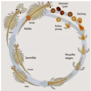
1.11.4. Artemia salina Leach, 1819 ... 28
1.11.6. Cysts morphology ... 30
1.11.7. Physiology of the hatching process ... 31
1.11.8. Artemia salina in aquaculture: an excellent food for newly-hatched fish larvae ... 32
1.12. Danio rerio: an emerging model organism in the environmental field... 33
1.12.1. Morphological features of Danio rerio ... 33
1.12.2. Embryonic development of zebrafish ... 34
1.12.3. Zebrafish as a model system for biomedical studies and human disease modeling ... 37
2. ECOTOXICOLOGY ... 40
2.1. The ecotoxicity tests ... 40
2.2. Aquatic toxicity tests ... 40
2.3. The importance of biomarkers as predictive tools in ecotoxicology ... 41
2.3.1. Classification of Biomarkers ... 42
2.4. Metallothioneins (MTs) ... 42
2.5. Nitric Oxide Synthase (NOS) ... 44
2.6. Heme Oxygenase (H-O) ... 45
2.7. The use of Artemia salina in ecotoxicological testing ... 46
2.8. The use of Danio rerio as a model vertebrate in ecotoxicological testing ... 48
3. MATERIALS AND METHODS ... 51
3.1. Synthesis of oxide nanoparticles by Pulsed Laser Ablation in Liquids (PLAL) ... 51
3.2.Characterization of nano-TiO2 powder ... 51
3.3. Synthesis of graphene oxide (GO) and reduced graphene oxide (rGO) ... 52
3.4 Atomic Layer Deposition (ALD): synthesis of engineered nanomaterials (ENMs) ... 54
3.5 Graphene oxide and titania loaded Nafion based nano-composites ... 56
3.6. Artemia salina acute toxicity test (24 h) ... 56
3.6.1. SEM protocol on Artemia salina nauplii ... 58
3.7. Fish embryo toxicity (FET) test on zebrafish with GO and nanocomposites... 58
3.7.1 Fish Embryo Toxicity (FET) Test with Nanomaterials “TiO2/Au” and “Au/TiO2” compared to Au andTiO2 free NPs ... 60
3.7.1.2 SEM Analysis (Day 0/Day 12) of the Nanomaterials “TiO2/Au” and “Au/TiO2” ... 61
3.8. Ecotoxicological long-term testing on adult zebrafish with AgNPs ... 61
3.8.1 AgNPs Characterization ... 62
3.8.2 Silver quantification through ICP-MS analysis ... 63
3.8.3. Istological protocol on adult zebrafish ... 64
3.8.4. Immunohistochemical protocol on zebrafish adult ... 64
3.9. Immunofluorescence protocol on zebrafish larvae ... 65
3.10. Protein expression protocol performed by Western blot ... 66
3.11. Gene expression protocol performed by Real-Time PCR method ... 67
3.11.1 Reverse Transcription ... 68
3.11.2 Real Time PCR preparation ... 69
4. RESULTS and DISCUSSIONS ... 70
4.1. Discussion on short-term toxicity test with Artemia salina ... 70
4.2. Discussion on FET test with Nanomaterials (“TiO2/Au” - “Au/TiO2”) versus free Au and TiO2 nanoparticles ... 74
4.3. FET test with graphene oxide (GO) and Nafion polymer combined with various fillers ... 78
4.4. Discussion on long term exposure with AgNPs and adults of zebrafish ... 82
5. CONCLUSIONS ... 89 6. REFERENCES ... 90 RESEARCH ACTIVITY ... 100 Papers. ... 100 Abstracts/Posters ... 100 RINGRAZIAMENTI ... 101
1
ABSTRACT
According to the definition adopted by European Commission in 2011 a nanomaterial (NM) is “a natural, incidental or manufactured material containing particles, in an
unbound state or as an aggregate or as an agglomerate and where, for 50% or more of the particles in the number size distribution, one or more external dimensions is in the size range 1 nm-100 nm” (European Commission, 2011/696/EU).
NM exhibit peculiar characteristics (e.g. small size, large surface area to mass ratio, shape, surface charge, reactive surface groups, state of agglomeration) that confer them properties substantially different from those of the bulk particles of the same composition.
Due to their widespread use in the consumer and industrial products, NMs can be released into the environment and it has been raised concern of the scientists about this question (Royal Society and the Royal Academy of Engineering, 2004).
The effects that NMs have on aquatic organisms depend on their characteristics influenced by environmental parameters. NMs enter the aquatic organisms mainly through the epithelial surfaces (such as gills, skin) or direct ingestion (Moore, 2006). After crossing the cell membrane, NMs may be stored in vesicles, mitochondria and additional organelles within epithelial cells. They may generate reactive oxygen species, oxidative stress, cytotoxicity, apoptosis and necrosis (Oberdörster et al., 2005).
Ecotoxicological tests of NMs should first consider the behaviour of NMs in the aquatic environment and the conditions that may influence aggregation state. For example, some NMs are almost impossible to disperse in water by physical methods such as sonication or stirring and may require the use of a dispersing agent. The choice of dispersant is problematic since some of the best dispersants from a chemistry point of view are also toxic to organisms.
The potential for NMs to cause oxidative injury in fish and invertebrates remains controversial. Bar-Ilan et al., 2009 showed that silver nanoparticles (AgNPs) induced almost 100% mortality in larvae of Danio rerio after acute exposure and a variety of embryonic morphological malformations were observed.
A study showed that gold nanoparticles (AuNPs) are non toxic at the employed concentrations and do not cause obvious abnormalities in developing zebrafish embryos (Asharani et al., 2010).
Zhu et al. (2009) observed effects on mortality and immobility on D. magna in the case of titanium nanoparticles (TiO2NPs) nanoparticles smaller than 20 nm.
Currently there is a lack of knowledge about long-term risks and potential mechanisms of toxicity of NMs; the industrial-scale application of engineered nanomaterials in many areas of daily life raises the question of the security of these systems because the nanodimensions are able to overcome natural barriers, resulting in potential biological damage.
The main aim of this Ph.D. Thesis was the evaluation of the potential toxic effects of several NMs on aquatic organisms used as models considering their increasing use in
2
the product market. Short and long-term ecotoxicological assays developed, searching for specific biomarkers of exposure by immunohistochemistry, western blotting and gene expression analysis.
3
In accordo alla definizione adottata dalla Commissione Europea nel 2011 per nanomateriale s’intende “un materiale naturale, casuale o prodotto contenente particelle, in uno stato slegato o come aggregato o come agglomerato e dove, per il 50% o più delle particelle nella distribuzione delle grandezze numeriche, una o più dimensioni esterne sono nell' intervallo di grandezza 1-100 nm”.
I nanomateriali esibiscono peculiari caratteristiche (ad es. dimensioni piccole, area di superficie molto grande, forma, carica di superficie, gruppi reattivi presenti in superficie, stato di agglomerazione) che conferiscono loro delle proprietà molto diverse da quelle possedute dal materiale della stessa composizione da cui essi derivano. A causa dell’uso diffuso nei prodotti di consumo e nei prodotti industriali, i nanomateriali possono essere rilasciati nell’ambiente e ciò ha sollevato molte preoccupazioni da parte degli scienziati (Royal Society and the Royal Academy of Engineering, 2004).
Gli effetti che i NM esercitano sugli organismi acquatici dipendono dalle loro caratteristiche che possono essere influenzate da parametri ambientali. I NM entrano negli organismi acquatici principalmente attraverso le superfici epiteliali (come branchie, pelle, cornea) o mediante ingestione diretta (Moore, 2006). Dopo aver attraversato la membrana delle cellule, possono essere immagazzinati in vescicole, mitocondri e altri organelli all' interno delle cellule epiteliali. Possono generare specie reattive dell’ossigeno, stress ossidativo, denaturazione delle proteine, citotossicità, apoptosi e necrosi (Oberdörster et al., 2005).
I test ecotossicologici sui NM dovrebbero innanzitutto considerare il comportamento dei NM in ambiente acquatico e le condizioni che possono influenzare lo stato di aggregazione.
Ad esempio, alcuni NM sono quasi impossibili da disperdere in acqua con i metodi fisici quali sonicazione o agitazione e può essere richiesto l’uso di un agente disperdente. La scelta dell’agente disperdente è problematica poiché alcuni dei disperdenti migliori da un punto di vista chimico sono anche tossici per gli organismi.
La possibilità che i NM possano causare stress ossidativo nei pesci e negli invertebrati non è del tutto chiara. Bar-Ilan et al. (2009) hanno dimostrato che le nanoparticelle d’argento inducono quasi il 100% di mortalità in larve di Danio rerio dopo esposizione acuta e diverse malformazioni a carico dell’embrione sono state osservate. Uno studio ha dimostrato che nanoparticelle d’oro (AuNPs) non sono tossiche alle concentrazioni testate e non causano anomalie nello sviluppo di embrioni di zebrafish (Asharani et al., 2010).
Zhu et al. (2009) hanno osservato eventi di mortalità ed effetti di immobilizzazione su esemplari di Daphnia magna esposta a nanoparticelle di biossido di titanio (TiO2NPs) aventi dimensioni inferiori ai 20 nm.
Attualmente vi è una penuria di informazioni circa i rischi a lungo termine e i potenziali meccanismi di tossicità delle nanoparticelle; l’applicazione su scala industriale dei NM in molti settori della vita quotidiana solleva la questione circa la sicurezza di tali sistemi in quanto le dimensioni nanometriche sono in grado di attraversare le barriere naturali arrecando danni ai sistemi biologici.
Scopo del presente progetto di tesi è la valutazione dei potenziali effetti tossici di nanomateriali ingegnerizzati su organismi acquatici impiegati come modello, mediante
4
saggi ecotossicologici a breve e lungo termine, la ricerca di specifici biomarkers di esposizione mediante analisi immunoistochimica, western-blotting e analisi dell’espressione genica.
5
1.
INTRODUCTION
Worldwide research on nanotechnology has grown considerably over the last decades. “Nanoscience” is the ability to observe, measure and manipulate the physical matter from an atomic and molecular point of view, while “nanotechnology” is the set of processes and techniques necessary to produce nanometric materials.
Nanotechnology was born in 1959 when American Physicist Richard Feynman, during the American Physical Society's annual meeting at the Californian Institute of Technology, held a celebrated speech entitled “There's plenty of room at the bottom” proposing the possibility of manipulating atoms to build infinitely small objects. Later in 1986, the American Engineer Kim Eric Drexler, defined nanotechnology as “a
molecular technology that will allow us to put every atom where we want it to be. We call this ability as nanotechnology because it works on the nanometer scale of 1 billionth of a meter”. Drexler, considered the true father of nanotechnology,
underlined the technological importance of nano-scale phenomena.
According to a Recommendation on the definition of nanomaterials of 18 October 2011 adopted by European Commission (2011/696/EU), Nanomaterials means a “natural, incidental or manufactured material containing particles, in an unbound state
or as an aggregate or as an agglomerate and where, for 50% or more of the particles in the number size distribution, one or more external dimensions is in the size range 1 nm-100 nm”. Nanomaterials (NMs) have peculiar chemical and physical properties
depending on their size and differ from those found in materials of conventional size (called bulk materials). NMs can have a natural origin, such as those produced by natural combustion processes (volcanoes, spontaneous fires) or have an anthropogenic origin. In this case, we distinguish those involuntarily produced (originating from vehicular traffic, in particular from diesel engines, incinerators, etc.) and those built voluntarily or engineered nanomaterials (ENM), specifically produced by nanotechnology to find application in various scientific and industrial fields (Tab. 1).
NATURALS MANUFACTERED
Non Engineered Engineered
Fires Internal combustion engines Carbon-based NM Volcanoes Power stations Metallic NM
Electric engines Dendrimers Incinerators Composites Jet airplanes
Fumes steel industries
Tab. 1. Major sources of nanomaterials.
There are two fundamental ways to build ENM: bottom up (bottom to top) and top down (top to bottom).
6
The bottom up method consists of assembling the nanostructured material from the nanoparticles that will allow the precursor to grow in dimensions and characteristics. The top-down method, which is currently the most consolidated one, consists of making of nanostructures from massive blocking of bulk material by physical methods that reduce the size of the initial structures, bringing them to a nanometric level.
1.1. Classification of Engineered Nanomaterials (ENMs)
There are several types of engineered nanomaterials; according to a commonly used classification, it is possible to subdivide them into four groups:
Carbon-based nanomaterials Metal oxide nanomaterials Dendrimers
Composites.
Carbon-based nanomaterials are mainly made of carbon, usually spherical (fullerenes) or cylindrical (nanotubes). They often used for selective and controlled transport of drugs (Figs. 1);
Figs. 1. Carbon-based nanomaterials: A) Fullerene; B) Nanotube.
Metallic nanomaterials made up of metals include nanogold, nanosilver and various metal oxides such as titanium dioxide (TiO2), zinc oxide (ZnO), silicon dioxide (SiO2) and aluminum oxide (Al2O3);
Dendrimers are polymers made up of branched units with numerous terminal chains that can be engineered and/or functionalized to be used, for example, in catalysis processes (Fig. 2);
7 Fig. 2. Structure of a dendrimer.
Composites, obtained by combining solids of different nature bound by a matrix (metallic, polymeric or ceramic) reinforced with nanometric particles; they are used to build electronic components of cars and packaging materials.
Engineered nanomaterials are also used in sporting goods, stain-resistant clothing, sunscreens, cosmetics and electronics. The unusual physicochemical properties are attributable to their small size, chemical composition (purity, crystallinity, electronic properties, etc.), surface structure (large surface area, surface reactivity, surface groups, inorganic or organic coatings), solubility, shape and aggregation. Although impressive from a physicochemical viewpoint, the properties of NM raise concerns about adverse effects on biological systems, some studies suggest that NM are not inherently benign and that they affect biological behaviors at the cellular, subcellular, and protein levels (Oberdörster et al., 2005; Royal Society and Royal Academy of Engineering, 2004).
Moreover, some nanoparticles readily travel throughout the body, deposit in target organs, penetrate cell membranes and may trigger injurious responses. Manufactured nanoparticles (NPs), due to the size of the particles itself and its chemical reactivity, can lead direct toxicity, and biodegradability may be a further significant factor in governing harmful biological effects (Hoet et al., 2004).
1.2. Applications of nanoparticles
Because of the nanoscale nature of nanoscience and nanotechnology, they already bridge many fields including medicine, pharmaceuticals, manufacturing technologies, electronics and telecommunications (Royal Society and the Royal Academy of Engineering, 2004). In medical therapeutics, many drug candidates fail to reach their targets at appropriate concentrations, which can severely limit their effectiveness (Brigger et al., 2002). However, when drugs are encapsulated into nanoscale particles and treated to prevent clumping, the result is often a stable and water-soluble material, due to the very large surface to volume ratio (Panyam and Labhasetwar, 2003). Other nanoparticle-based medicines are also being investigated for use in cancer therapies and diagnosis where the nanoscale properties facilitate entry and intracellular targeting to specific sites (Brigger et al., 2002).
8
1.3. Routes of entry, uptake and bioavailability into living systems
Uptake of nanoparticles by inhalation or ingestion are likely to be the major routes in terrestrial organisms (Dowling, 2004; Warheit, 2004; Moore, 2002). However, in aquatic animals there may be other routes of entry such as direct passage across gills and external surface epithelia. At the cellular level, most internalisation of nanoparticles will occur via endocytosis (Fig. 3). Endocytotic pathways into cells can either lead to the endosomal and lysosomal compartments (Panyam and Labhasetwar, 2003). At the cellular level, prokaryotes like bacteria may well be largely protected against the uptake of many types of nanomaterials, since they do not have mechanisms for the bulk transport of supramolecular and colloidal particles across the cell wall. However, eukaryotes have highly developed processes for the cellular internalisation of nanoscale (100 nm or less) and microscale (100-100.000 nm) particles, namely endocytosis and phagocytosis respectively (Panyam and Labhasetwar, 2003; Synnes et al., 1999). These processes are integral to key physiological functions such as intracellular digestion and cellular immunity. In invertebrate animals, the cellular immune system, gut epithelium and hepatopancreas (digestive or midgut gland), where present, is likely to be targeted. The hepatopancreas is involved in uptake and digestion of food and storage of nutrient reserves; and is frequently specialised for intracellular lysosomal digestion of food via internalisation by endocytosis (Moore et al., 2004; Moore, 2002). The liver is a probable target following endocytotic transport across the intestinal epithelium into the hepatic portal blood system followed by endocytosis into hepatocytes.
Fig. 3. Diagrammatic representation of the various routes of uptake (both diffusion and endocytotic) of xenobiotic chemicals into the cell that can result in their accumulation in the lysosomal compartment, based on their physical chemical characteristics (from Moore et al., 2004).
9
Endocytotic routes of entry to the cell may allow pollutant nanoparticles to embed themselves within the functional machinery of the cell in ways that are toxicologically quite different from conventional toxic chemicals. Nanoparticles situated in the endoplasmic reticulum, Golgi and endo-lysosomal system could conceivably act as foci of oxidative damage that could not readily be expelled from the cell; while generation of radicals could lead to organelle dysfunction.
1.4. Risks to human health via water
Accidental spillages or permitted release of industrial effluents in waterways and aquatic systems may result in direct exposure to nanoparticles of humans via skin contact, inhalation of water aerosols and direct ingestion of contaminated drinking water or particles adsorbed on vegetables or other foodstuffs (Daughton, 2004). More indirect exposure could arise from ingestion of organisms such as fish and shellfish (i.e. molluscs and crustaceans) as part of the human diet. Filter-feeding molluscs are prime candidates for uptake of manufactured nanoparticles from environmental releases, if it transpires that some of these nanomaterials will associate with natural particulates (Fig. 4).
Fig. 4. In the aquatic environment, nanoparticles can react with various organisms.
1.5. Harmful effects of NPs and possible consequences on biological systems
The very large surface area of ultrafine particles can result in the direct generation of reactive oxygen species (ROS) leading to cell oxidative stress: it can cause cell injury by attacking DNA, lipids, proteins, membranes and metabolic activities. The toxic effects of ENPs essentially depend on several key factors such as their intrinsic nature and
10
capacity to form larger aggregations, the route of exposure, dose response, exposure time, the response of the receptor organisms to the lack of biocompatibility of ENPs, and the interactions in the mechanisms involved in the physiological process of uptake. ENPs enter the environment via different exposure routes, including solid and liquid waste from domestic sources and industrial activity, accidental spillages, and atmospheric emissions. Additional types of source that need to be assessed are those resulting from the use of products that contain ENPs (i.e. sunscreens and paints). Industrial products and wastes tend to end up in waterways (e.g. drainage ditches, rivers, lakes, estuaries and coastal waters) despite safeguards (Daughton, 2004; Moore
et al., 2004; Moore, 2002). Consequently, it is inevitable that nanoscale products will
enter the aquatic environment (Moore et al., 2004; Royal Society and Royal Academy of Engineering, 2004).
In natural water ecosystems, water travels over the surface of the land, through ground or underground until it reaches estuarine water (EW) or saltwater (SW), passing through conditions of increasing water salinity, ionic concentration and differences in pH. During this physical process, ENPs can be degraded, transformed, carried and accumulated. One main effect is that the ENPs could form colloidal suspensions by association with substances originating from animals or from human activity, and could undergo processes of agglomeration or self-aggregation (Lapresta et
al., 2012). During the transport of ENPs to their final destination in the ocean, colloid
mobilization is also promoted by chemical perturbations (e.g. ionic strength and pH) (Petosa et al., 2010) and by the physical conditions of the water system (e.g. temperature). Due to the high specific surface area and binding constants of ENPs, they tend to absorb small colloids that may aggregate and even separate out from the water column by differences in settling velocity, resulting in a transfer of chemicals (e.g., metals and polymers) to the sediments where the ENPs can also be ingested by organisms. In soils, ENPs may be degraded by biotic or abiotic processes and transported to water bodies through run-off, leaching and drain flow (Boxall et al., 2007). Colloidal surface properties are therefore key aspects for tracking the fate and the behavior of ENPs in the environment. In the final agglomerated/aggregated entity, the particle reactivity decreases because the surface area of the individual particles and the interfacial free energy are reduced during the aggregation process (e.g. humic acid in water often mitigates the toxic effect of heavy metals). The aggregation of ENPs is therefore an important factor that may influence their toxicity, either mitigating or increasing the effect.
1.6. Mechanism of toxicity
One of the most important factors related to the toxicity of ENPs is oxidative stress. ROS and oxidative stress have been explained in detail by Lushchak (2011). In aquatic organisms, it is possible to measure the effect of ROS on lipids by monitoring the intermediate (primary an secondary products) species of lipid peroxidation (tightly
11
associated with exposure to NPs) and end products. ROS-induced DNA damage may bring physiological consequences that impair reproduction, inhibit growth, and damage both lysosomes and mitochondria. DNA damage leads to the liberation of toxic cations, inhibits algal photosynthesis and affects electron and/or ion transport. The disruption of ion transport changes membrane permeability and increases the probability of NPs gaining entry inside the cell, which may lead ultimately to cell death. The effects of ENPs on aquatic ecosystems are produced essentially by oxidative damage (internalization by cells) and interaction between ENPs and the cell membrane (without internalization). The properties of the NPs affect the uptake of NPs, which are bioaccumulated predominantly in the gills, intestine, liver, kidneys and blood (Scown
et al., 2010). The latter three organs seem to be mainly due to internalization through
the gills and the intestine. The main target of the toxicity mechanism is to disrupt the osmoregulatory function. Thus, the main adverse effects are related to an increase in the diffusive permeability of the membrane leading to a disturbed ion transport, as well as changes in cellular morphology, mitochondrial function, mitochondrial membrane potential and DNA damage-related gene expression that induce potential inflammation, necrosis and apoptosis of cells.
According to these results, ecotoxicology research is urgently required to explain and clarify the behavior of ENMs and ENPs in their different forms and their environmental impact on the ecosystems. ENPs tested in this study are: silver (AgNPs), gold (AuNPs) and titanium dioxide (TiO2NPs), and they will be investigated hereinafter. Moreover ENMs and nanocomposites will be considered later.
1.7. Silver nanoparticles (AgNPs)
Metallic silver (Ag) is a durable transition element and because of its rarity (67th in abundance among the elements) and its attractive white metallic luster, silver has long been used as jewellery, currency coins and silverware. Silver nanoparticles (AgNPs) showed excellent performance in antibacterial application; it was reported that AgNPs show biocidal action by the slow release of Ag+, and by multiple mechanisms (such as interaction with thiol groups in proteins and enzymes, inhibition of DNA replication, induction of oxidative stress) making it more difficult for bacteria to produce resistant strains (Luoma, 2008).
As early as 1889, Lea had reported the first synthesis of a silver colloid stabled by citrate (Lea, 1889). Though not formally registered or under the name of “nano”, literature shows that silver colloids have been used in the medical area for more than 100 years since 1897 by the name of “Collargol”. The first biocidal silver product “Algaedyn” was registered in 1954 in the U.S., which is still used in disinfectants today (Nowack et al., 2011). During the past two decades, the advancement of nanotechnology has opened new avenues for AgNPs. Being in the nano-scale dimension, AgNPs exhibit novel properties relative to the bulk metal, which has aroused great interest in the development of new applications.
12 1.7.1. Properties and applications
Pure silver has high thermal and electrical conductivity and relatively low contact resistance, which makes it a popular material in electronics. Silver nanoparticles have been used to fabricate thin-film transistor electrodes, as pastes and inks for printed circuit boards, optoelectronics, data storage devices and battery-based intercalation materials (Yu et al., 2013). Thanks to their extremely small size, AgNPs possess large surface area, which provide them high surface energy and more possible reactive sites. These characteristics qualify AgNPs as one of the most promising materials in catalysis. AgNPs are capable of catalyzing several reactions, such as CO and benzene oxidation, reduction of 4-nitrophenol in the presence of NaBH4 (Yu et al., 2013).
For years, knowledge about nanosilver's ability to kill harmful bacteria has drawn extensive attention, making it popular for incorporation into various products. Nanosilver exhibits a broad spectrum of antimicrobial activity, and can inhibit the growth of both Gram-positive and Gram-negative bacteria (including Escherichia coli,
Pseudomonas aeruginosa and Staphylococcus aureus) (Birla et al., 2009). Nanosilver is
also an effective fungicide: AgNPs can kill a number of fungal strains, including
Aspergillus fumigatus, Mucor, Saccharomyces cerevisiae and Candida tropicalis (Wright et al., 1999). Nanosilver also has antiviral properties; it was reported that AgNPs
synthesized in Hepes buffer could inhabit HIV-1 replication, and the anti-HIV activity (98%) was much higher than that of gold nanoparticles (6-20%) (Sun et al., 2005).
1.7.2. Environmental concern
The widespread application of AgNPs in our daily life will inevitably increase human and ecosystem exposure. Many literature reports have shown data on the leaching and fate of AgNPs. Benn and Westerhoff revealed that AgNPs and Ag+ could be easily leached into water by simply immersing commercial AgNPs containing socks into water with shaking. Some brands of socks could even lose nearly 100% of the total silver contents after four consecutive washings (Benn and Westerhoff, 2008). Consumer silver nanotextiles were subjected to surfactants, oxidizing agents, different pH, washing machines and simulated perspiration fluids to test the normal laundering process and human skin sweat on the release of AgNPs (Geranio et al., 2009). It was shown that low pH, mechanical stress and the presence of bleaching largely enhanced the dissolution of Ag, and the nature of incorporation determined the amount and form of Ag release. The considerable release of Ag in simulated perspiration fluids suggested the potential human risk in the use of textiles containing AgNPs. As AgNPs are widely used in consumer spray products for their antibacterial ability, there is a risk that nanoparticles may be directly inhaled and deposited in the respiratory tract during product use. Quadros and Marr explored the emission behavior of three consumer spray products containing AgNPs. It was shown that emitted aerosols ranged from nanoscale up to 10 µm, and 0.24-56 ng of silver could be released per
13
spray action. It is also estimated that up to 70 ng of silver may deposit in the respiratory tract according to the usual use of the spray products (Quadros and Marr, 2011).
1.7.3. Natural and anthropogenic source of AgNPs
The increasing use of nanosilver has generated enthusiasm in developing methods to fabricate different AgNPs, which may eventually increase the amount of nanosilver in the environment (Yu et al., 2013). However, it seems that not all AgNPs are produced by humans. It is reported that silver as nanoparticles was found in an old silver mining area of Mexico (Gomez-Caballero et al., 2010) and before the manufacture of engineered AgNPs, colloidal and particulate silver was also discovered in river and estuarine waters of Texas (Wen et al., 1997).
AgNPs could be formed naturally but also by anthropogenic activities. AgNPs are used as electronic and medical devices, incorporated into textiles, dressing or directly added into disinfectants. However, during the production and manufacturing of nanosilver products, they could be directly released into the environment. The synthesis process often involves mixing centrifugation and filtration steps to remove impurities and the wastewater may be directly discharged into the environment. The uncontrollable release of silver during the use, recycling and disposal process gives rise to public concerns. Ideally the released AgNPs and Ag+ would undergo “Wastewater Treatment Plants” (WWTP) processing. Through previous study (Benn and Westerhoff, 2008) has shown that WWTP biomass is able to partition high levels of heavy metals and with treatment largely reduce the silver concentration in the effluent stream, unfortunately, in some instances untreated sewage sludge is often used as an agricultural additive or fertilizer, which results in the recycling of AgNPs. This would be an hazardous source of AgNPs to the environment.
1.7.4. Fate, transport and distribution of AgNPs in environment
Once released into the environment, AgNPs would undergo different pathways during transport. They may remain as individual particles in suspension and be delivered long distances, or tend to aggregate at high ionic strength. The behavior of the AgNPs depends on the surface properties of the nanoparticle themselves and the surrounding environment, involving electrolyte composition, solution ionic strength, pH and natural organic matter (NOM). After contact with oxygen and other oxidants, partial oxidation and Ag+ dissolution is also expected. Most probably, AgNPs would react with sulfide, chloride or other natural substances, altering the original properties of the nanoparticles (Luoma, 2008). Due to the large surface area and high surface energy of nanoparticles, they are prone to coalesce to form larger clusters. NOM, ubiquitously existing in aquatic systems, is also a key factor influencing the fate of AgNPs. The presence of various functional groups of NOM facilitates surface binding of AgNPs, resulting in more stable AgNPs suspensions. AgNPs coated by NOM are able to stay
14
dispersed for months (Akaighe et al., 2011). As mentioned above, AgNPs can escape from the manufacturing factory during the production process, including drying the solution, mechanical grinding, mixing and packaging, resulting in the release of AgNPs into the atmosphere. Also, the widespread use of AgNPs in disinfection sprays promotes the emission of AgNPs to the air. Surface disinfectants, which can be used on walls, tables, floors, often cause AgNPs to deposit on these surfaces, and AgNPs would probably transfer to a duster cloth after cleaning and then go down the drain during laundering. Added in anti-odor sprays and used in rooms, AgNPs could also find their way in the open air by transport. According to Fick's first law that the diffusion coefficient is inversely related to the particle diameters, AgNPs would diffuse rapidly because of their small size. If they are stable enough, long distance mobility in the air would be expected. Additionally, their large surface area provides abundant reactive sites for dusts, microbes and pollution, making the AgNPs much more toxic than the original particles. The airborne particles may also coalesce to large agglomerates during transport, and deposit on surfaces by gravitation, or be washed down to terrestrial or aquatic systems by rain. AgNPs may enter the aquatic system in several ways:
Silver leached out from nanosilver consumer products, and eventually end up in streams and rivers (Benn and Westerhoff, 2008);
Suspended AgNPs in air finally depositing on water;
Runoff scouring AgNPs polluted soils or landfill sewage sludge could result in AgNPs migrating to surface water.
On the other hand, divalent cations (e.g. Ca2+ and Mg2+), commonly present in natural waters, could easily induce the aggregation of AgNPs. Colloidal clusters would probably deposit in the sediment, reducing the bioavailability for aquatic organisms and plants. As AgNPs are not highly stable and can easily be oxidized, a slow dissolution of Ag+ would be expected in aquatic environments (Liu and Hurt, 2010). In marine waters, as sodium chloride is the dominant salt, Ag+ associates with Cl- to a great extent, and the main species observed are AgCl2-, AgCl3-, and AgCl4-, making silver more mobile in seawater (Barriada et al., 2007). Due to the large surface area and relatively high surface energy, once released into the environment, AgNPs could be highly dynamic and different transformations such as sulfurization, chlorination will readily occur, which would greatly affect the behavior of the AgNPs. Silver sulfide nanoparticles were also discovered in the final stage sewage sludge materials of a full-scale municipal wastewater treatment plant (Kim et al., 2010).
1.7.5. Mechanism of toxicity
The antibacterial activity of AgNPs is a complex process, and several possible modes of action are proposed, involving:
15
Production of reactive oxygen species (ROS);
Direct attachment to cell membrane and disruption of membrane integrity; Changes in membrane permeability;
Interaction with proteins and disruption of their regular function; Interference with DNA replication and causing DNA damage.
However, in another report that assessed the effect of AgNPs on rainbow trout hepatocytes, no excess ROS was detected after exposure. The cytotoxicity was mainly associated with the reduced mitochondrial activity and membrane integrity (Farkas et
al., 2010). Membrane disruption related toxicity of AgNPs was also reported in other
studies. The formation of pits and pores in the cell membrane of fungus C. albicans was observed, and the authors suggested that AgNPs may attack cell membrane lipid bilayers and destroy the membrane permeability barrier, resulting in the leakage of ions, formation of pores and cell death (Kim et al., 2008).
E. coli membrane damage caused by AgNPs was also confirmed by Sondi and
Salopek-Sondi (2004). TEM/SEM images all showed that AgNPs accumulated on cell membranes and some were successfully incorporated into the lipid bilayer structure, forming irregular shaped pits in the outer membrane. It was speculated that AgNPs may lead to the progressive release of lipopolysaccharide molecules and proteins, causing changes in membrane integrity and permeability, and finally inducing cell disfunction and death.
Existing results have showed that AgNPs can cause potential toxicity to test animal models. Lankveld and co-workers also demonstrate that when three different sized AgNPs (20, 80, 110 nm) were intravenously injected into rats at a concentration of 23.8 µg/ml for 5 days, the concentration of Ag was reduced rapidly in the blood, and AgNPs redistributed to liver, lung, brain and all other organs, showing a systemic distribution (Lankveld et al., 2010). A number of non-mammals animals have also been used to test the adverse effect of AgNPs and most of these studies are based on aquatic organisms. Zebrafish or Danio rerio, as a correlative and predictive model, has been used in many studies to evaluate the effects of AgNPs. Earlier in vivo study (Lee et al., 2007) demonstrated that single AgNPs (11.6 ± 3.5 nm) could transport into zebrafish embryos through the chorion pore canal, and AgNPs were detected inside embryos at each developmental stage. In another study, four different sized AgNPs were synthesized to test their toxicity to zebrafish embryos, and only a few differences were observed between them. It is reported that AgNPs induced almost 100% mortality after exposure for 120 h at 250 µM, and a variety of embryonic morphological malformations were observed at a dose of 100 µM (Bar-Ilan et al., 2009).
However, different from previous studies that smaller particles were more toxic, some authors found that larger AgNPs (41.6 ± 9.1 nm) produced higher toxic impacts and more severely deformed zebrafish than the smaller ones (11.6 ± 3.5 nm) at the same concentration (Lee et al., 2012). Toxicity of AgNPs in rainbow trout gill cells showed
16
that nanoparticles were taken up into cells and lead to cytotoxicity related with membrane integrity disruption and oxidative stress. Furthermore, different kinds of morphological malformations such as edema, spinal abnormalities, finnfold abnormalities, heart malformations and eye defects emerged after long time exposure, indicating the developmental toxicity of AgNPs (Wu et al., 2010).
1.8. Gold nanoparticles (AuNPs)
Gold was one of the first metals discovered by humans, and the history of its study and application is estimated to be a minimum of several thousand years old. Looking in the past, gold has been already reported as a health adjuvant. In the Middle Ages in Europe, colloidal gold was studied and employed in alchemists’ laboratories. In 1583, alchemist David de Planis-Campy, surgeon to the King of France Louis XIII, recommended his “elixir of longevity”, an aqueous colloidal gold solution, as a means of life prolongation (Dykman and Khlebtsov, 2012). Chinese and Indian culture reported the use of gold compounds in the form of “Swarna Bhasma” for treatment of various health needs like increasing vital power and curing male impotence (Mahdihassan, 1985). Myocrism™ (sodium aurothiomalate) and Solganol™ (aurothioglucose) are water soluble gold complexes present which show beneficial effect in rheumatoid arthritis. Earlier in 1978 cisplatin was approved by the Food and Drug Administration (FDA) as an antitumor agent. Gold nanoparticles (AuNPs), are one of the promising inorganic nanoparticles that have attracted scientific and technological interests due to their ease of synthesis, chemical stability and excellent optical properties that make them appealing tools for cancer diagnosis and treatment (Dykman and Khlebtsov, 2012; Jain et al., 2012). Literature investigations have demonstrated that these inorganic nanoparticles are non toxic (Fard et al., 2015). Evaluation of AuNPs toxicity is essential because of broad spectrum application of these in biomedical sciences.
1.8.1. Synthesis of AuNPs
The production of nanoparticles consist of reduction of size (top-down technique). Gold nanoparticles can be produced by reduction of gold salts in the presence of stabilizing agents in order to prevent agglomeration. The gold NPs can be prepared in three different methods:
Physical methods like ultra violet (UV) radiation, sonochemical method, laser ablation, thermolytic process (Giorgetti et al., 2007);
Chemical methods in an aqueous medium using citrate or sodium borohydride as a reducing agent;
Biological methods take into consideration microrganisms as a source used to produce NPs (Alivisatos, 2004). These ones have been used as an alternative in
17
order to avoid the use of organic solvents, thus making biological process an eco-friendly nanotechnological approach.
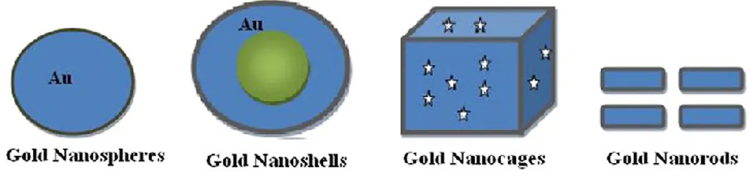
There are different types of gold NPs (Fig. 5):
Gold nanospheres: their size lie in the range of 2-100 nm and can be synthesized by controlled reduction of an aqueous solution of HAuCl4 using a reducing agent like citrate (Khan et al., 2013);
Gold nanoshells: spherical with diameters ranging in size from 10 to 200 nm. They possess a remarkable set of optical, chemical and physical properties, which make them ideal candidates for enhancing cancer detection, cancer treatment, cellular imaging and medical biosensing (Khan et al., 2013);
Gold nanocages: hollow porous gold NPs, size basically ranges in between 10 and 150 nm and consists of hollow interiors and porouswalls. They are generally produced by silver nanoparticles with chloroauric acid (HAuCl4) in boiling water;
Gold nanorods: used as a drug delivery vehicle due to its low cytotoxicity, excellent stability, biocompatibility and suitable physiochemical parameters.
Fig. 5. Different types of gold NPs with their shapes (from Khan et al., 2013).
1.8.2. Role of gold NPs in biomedical applications
Several pharmacological sectors have got approval from the Food and Drug Administration (FDA) for the use and development of nanotechnology-based drugs in the last few years. AuNPs are an important tool for drug delivery, as a tracer for diagnostic imaging which will give structural information on very molecular level, and treatment of major life threatening diseases. Because of their small particle size, diversity in surface chemistry, unique physical properties, adaptable absorption, emission properties, flexibility in synthesis, AuNPs have a unique approach in biomedical technology. They are used in different fields:
18
They can easily penetrate to the intracellular cell wall and, if monitored, they can give a plenty of useful information which can lead researchers for cancer diagnosis and gene therapy (Bhattacharyya et al., 2011);
AuNPs have the tendency to absorb and scatter visible and near infrared (NIR) light resonantly upon excitation of surface plasmon oscillation. Detection of biological events at the single molecular level has been used for biosensing by exploiting the distance dependent scattering properties of the AuNPs. Upon getting excited plasmon resonance band can be spectrally modified over a wide range. The outcome is that the sense of light scattering signal is more profound (highly intense) and comparatively much brighter than the chemical fluorophores. This property can be useful in imaging techniques (Sönnichsen et
al., 2002);
Can be coated with polyethylene glycol (PEG) to impart antibiofouling properties, which extends their lifetime in the blood stream. Studies conducted by Kim et al. (2007) showed the advantages of AuNPs as a contrast agent compared to iodine-based compounds which show several limitations like short imaging times due to rapid renal clearance, renal toxicity and vascular permeation. The results of the X-ray absorption coefficient in vitro show that there is an increase in the X-ray absorption behaviour of AuNPs as compared to a iodine-based compound called “Ultravist”;
Can be bound with biological protein and peptides for the fabrication of biosensors which can be used in new signal transduction technologies (de la Escosura-Muniz et al., 2009).
1.8.3. Gold nanoparticles toxicity
Gold, in its bulk form, has long been considered an inert and noble metal with some therapeutic value. However, some researches revealed the toxicity of gold NPs. As with the toxicity observed for AgNPs at the cellular level, the possible adverse effects of AuNPs can be attributed to:
Their interaction with the cell membrane; Oxidative stress leading to cytotoxicity effects;
The inhibition of metabolic activity (e.g., leading to mitochondrial damage); Possible damage to the nuclear DNA.
To date, very few data are available on the ecotoxicity of AuNPs. A study (Lovern et al., 2008) made on the presence and distribution in Daphnia magna exposed to sub-lethal concentrations of AuNPs for 1, 6, 12 or 24 h, and the highest concentration of AuNPs was found after 12 h. Earlier reports on metal and metal oxide nanoparticles and have emphasized the deleterious effects of nanoparticles in vivo and in vitro (Zhu et al., 2008). It has been observed that the toxic output of nanoparticles in vitro differ significantly from in vivo models. Similarly AuNPs is considered as non-toxic in vitro,
19
whereas injection of AuNPs in mice has shown tissue damage and apoptosis in liver (Cho et al., 2009). A study showed that gold nanoparticles are non toxic at the employed concentrations and do not cause obvious abnormalities in developing zebrafish embryos (Asharani et al., 2010). Indirect human exposure is possible through the consumption of fresh water fish exposed to nanowastes. Hence it is necessary that extreme care must be taken for nanowaste disposal and in vivo applications of nanoparticles. The data gathered from experiments will help future researchers to unravel the mechanisms of nanoparticle toxicity, which still remains unclear.
1.9. Titanium dioxide nanoparticles (TiO2)
Titanium dioxide (TiO2) nanoparticles are the most extensively studied metal oxide nanoparticles (Menard et al., 2011; Cattaneo et al., 2009). One of the reasons for the large amount of ecotoxicity data on nanosized TiO2 is the adoption of this nanomaterial by a variety of industries. It is a fine, white, crystalline, odorless, low-solubility powder which exhibit relatively low toxicity (Sager et al., 2008). It is a natural, thermally stable and nonflammable, nonsilicate mineral oxide found primarily in the form of the minerals rutile, anatase, brookite, and as the iron-containing mineral ilmenite (FeTiO3) (Duan et al., 2010). Anatase phase exhibits the highest photocatalytic activity, so is used in catalysis and photocatalysis applications. Rutile is known as white pigment providing opacity to paints, papers, inks, and consumer products such as toothpaste. Because of their properties TiO2NPs have been used in several applications, but this widespread use led to a significant environmental exposure to TiO2 nanoparticles (Hall et al., 2009). With regard to its potential adverse health effects, several studies have defined TiO2 as biologically inactive and physiologically inert in both humans and animals. Recently, pulmonary inflammation, fibrosis, epithelial hyperplasia, and tumorigenesis were reported in animals under conditions of substantial TiO2 particle lung burden due to sufficiently high dose and/or duration of exposure. Thus, in 2006, the International Agency for Research on Cancer (IARC), classified and in 2010 reassessed TiO2 as “possibly carcinogenic to humans” on the basis of the sufficient evidence of carcinogenicity in experimental animals and inadequate evidence in humans (Group 2B) (IARC, 2010, 2006).
1.9.1. Properties and applications
It has excellent physicochemical properties, such as good fatigue strength, resistance to corrosion, machineability, biocompatibility, whitening and photocatalysis, as well as excellent optical performance and electrical properties (Iavicoli et al., 2012; Chen et
al., 2009). Nanosized TiO2 is used extensively in sunscreen cosmetics as an inorganic UV absorbant that can allow a film transparent to visible light to be applied to human skin (Jaroenworaluck et al., 2005). A surface coating, for example silica and other compounds, can also be added to nanosized TiO2 to decrease its photoreactivity so that nanoTiO2 can be used to protect human skin, plastic, and other objects from UV
20
radiation. In cosmetic products, rutile phase is used as a pigment and thickener and it is used in plastics and for its ultraviolet (UV) light absorbing properties (Mueller and Nowack, 2008). TiO2NPs are widely used in paints, printing ink, rubber, paper, cleaning air products, decomposing organic matters in wastewater.
1.9.2. Ecotoxicological studies
Currently few data exist regarding observed environmental concentrations of TiO2 nanoparticles (Griffitt et al., 2008). Evidence that TiO2NPs can leach from exterior facade paints and discharge into surface waters has been provided. The concentrations of metallic Ti found in the surface runoff were as high as 600 mg/l (Kaegi et al., 2008). Two studies modelled the quantities of TiO2NPs released into the environment (Gottschalk et al., 2009; Mueller and Nowack, 2008). Aggregated particles are generally less mobile and can interact with filter feeders and sediment-dwelling organisms (Farré et al., 2009). The extent of aggregation is governed by pH, ionic strength, and the nature of the electrolytes (Navarro et al., 2008). Much knowledge already exists on the effects of TiO2 nanoparticles on biological systems. NanoTiO2 is photoinducible, redox active and thus a generator of ROS at its surfaces. The precise mechanisms of toxicity of nanosized TiO2 and other metal nanoparticles are largely unknown (Griffitt et al., 2008), but recent studies have shown that the toxicity of nanoparticles is generally governed by properties such as particle size, shape, and surface properties (Navarro et al., 2008). Upon irradiation by a photon possessing an energy larger than its band gap, an electron associated with the TiO2 nanoparticle can be excited to the conduction band, thereby creating a positively charged hole. This hole can then remove an electron from a molecule (such as water) that is in contact with the surface of the TiO2, thereby resulting in the formation of reactive oxygen species (Fujishima and Honda, 1972). Ultrafine TiO2 nanoparticles (<20 nm) can enter the human body through different routes, including inhalation, ingestion, dermal penetration and injection (Oberdörster et al., 2005).
1.9.3. Toxicity of nanosized TiO2 to freshwater and marine invertebrates
There is an emerging literature on the ecotoxicity of nanosized TiO2 with invertebrates. According to Baun et al. (2008) invertebrate tests are well suited to generate reproducible and reliable nanotoxicity data. Nanoecotoxicology may take advantage of the fact that use of invertebrates as experimental animals is not subject to legal restrictions. The ecotoxicological effects of nano-TiO2 on freshwater invertebrates have been summarised in several papers. There are a lot of data available for freshwater invertebrates especially for crustaceans like Daphnia magna, Daphnia
pulex, Ceriodaphnia dubia, Chydorus sphaericus, Thamnocephalus platyurus (Cattaneo et al., 2009; Farré et al., 2009; Baun et al., 2008). Zhu et al. (2009) observed effects on
mortality and immobility on D. magna in the case of particles of ≤ 20 nm. Lovern et al. (2007) observed significant differences in toxicity to daphnids when filtration of the
21
TiO2 suspension permitted testing of smaller 30 nm particles. When the suspension of particles was no filtered, aggregates of size 100-500 nm were present and no toxicity to daphnids was observed. Sublethal effects of nanosized TiO2 on D. pulex after exposure of 24 h have been observed. It was found that nanoTiO2 at 500 mg/l elevated the activity of the antioxidant enzymes catalase and glutathione-S-transferase (Klaper
et al., 2009). One study reported a 24-h test with mussels bivalve Mytilus galloprovincialis showing that nanoTiO2 induced lysosomal membrane destabilisation in the hemocytes and digestive gland. It was also found that nanoTiO2 induced increases in lysosomal accumulation of neutral lipids and enhancement of catalase and glutathione transferase activity in the digestive glands. No effect on catalase and glutathione transferase activities in the gills and no mortality was observed (Canesi et
al., 2010).
1.9.4. Toxicity of nanosized TiO2 to freshwater fish
The ecotoxicological effects of nanoTiO2 on fish have been summarised in some papers in recent years (Farré et al., 2009) and toxicity data are available for Danio rerio (Griffitt et al., 2008; Zhu et al., 2008), Pimephales promelas (Hall et al., 2009),
Oncorhynchus mykiss (Federici et al., 2007; Warheit et al., 2007) and Cyprinus carpio
(Hao et al., 2009). Sublethal effects of nanosized TiO2 have been reported on juvenile rainbow trout (Oncorhynchus mykiss) after 14 days waterborne exposure (Federici et
al., 2007). The Na+/K+-ATPase activity in the gills, brain and intestine decreased after 14 days; after 8 weeks this effect was observed only in the brain. Total glutathione in the gills and liver was unaffected by nanoTiO2. Lipid peroxidation increased in the gill, intestine and brain after 14 days, but decreased after 8 weeks. Other observed effects included gill pathologies, such as edema and thickening of the lamellae, and a few foci of lipidosis and condensed nuclear bodies in liver cells. Federici et al. (2007) concluded that TiO2NPs caused respiratory distress and sub-lethal toxicity involved oxidative stress, organ pathologies and the induction of antioxidant defense system. Zhu et al. (2008) compared the toxicity of several metal oxide NPs with their bulk counterparts suspensions to zebrafish at early developmental stage by a 96h embryo-larval bioassay and showed that neither TiO2NPs nor bulk TiO2 caused any toxicity to zebrafish embryos and larvae.
1.9.5. Environmental concerns
To date, TiO2NPs tested in ecotoxicity studies published have different physico-chemical properties, such as diameter, crystalline form, coating, surface area, zeta potential and purity. The variability of the tested material was further enhanced as a result of preparation and suspension of the nanoparticles in test media, for example by sonication, filtration, and use of different solvents and surfactants. Physicochemical characteristics of nanoTiO2 can explain the high variability in the toxicity data for nanoTiO2 obtained by different authors conducting similar experiments with the same
22
species. However, the toxicological profile of TiO2NPs is not completely understood and several concerns have emerged on the potential undesirable effects, in regard to the harmful interactions with biological systems and the environment (Nel et al., 2006). The recently recognized occupational carcinogenic potential of the inhaled TiO2NPs have ulteriourly enhanced these scientific concerns. Therefore, an appropriate assessment of the risks for the exposed population requiring TiO2NPs hazard identification and dose-response data seems necessary (Cho et al., 2010). In this regard, a deeper knowledge about TiO2NPs mechanisms, obtained from in vitro and in
vivo experiments, respectively, could be useful to identify indicators of early biological
events and to establish appropriate methods for their detection.
1.10. Graphene and its derivatives
Materials of the graphene family include few-layer graphene, graphene nanosheets, graphene oxide (GO) and reduced graphene oxide (rGO) (Sanchez et al., 2012). Graphene-based nanomaterials vary in the layers number, lateral dimensions, surface chemistry, defect density or quality of the individual graphene sheets and purity. The structural features of these materials affect the physico-chemical properties of the resulting materials and, probably, they determine also the main biological effects (Pretti et al., 2014).
The recent biomedical applications of graphene and derivatives have determined a rapid increase of the studies related to the biological interactions of these materials (Sanchez et al., 2012); for example graphene dispersed in air might represent a danger to people daily handling these materials, either by contact or inhalation, and studies on this topic are therefore necessary. Nevertheless, accidental spills and effluent discharges can determine an increased risk of release of these exogenous nanoparticles (NPs) into aquatic environment and even though emissions of graphene oxide (GO) to aquatic environment should be low (if any), their expected low degradability requires adequate investigation.
Graphene is composed of sp2-hybridized carbon atoms hexagonally arranged in a two-dimensional structure, resulting in a large surface area on both sides of the planar axis (Novoselov et al., 2004). It has gained a considerable scientific interest starting from its discovery in 2004 as a result of its electrical, mechanical and optical properties proposed for use it in a wide range of applications ranging from flexible electronics to DNA sequencing (Buccheri et al., 2016) (Figs. 6).
23 Figs. 6. Structural models of graphene: A) Graphene oxide (GO); B) Reduced graphene oxide (rGO).
Because of the formation of hydrogen bonds between polar functional groups on the GO surface and water molecules, a stable GO colloidal suspension can be reached suggesting advantages for potential biomedical applications of GO compared with other carbon-based materials (Bitounis et al., 2013).
The level of toxicity that graphene might reach in a biological system and the degree of safety for its use are important to explore.
However, prior to the use of graphene-based materials, it is imperative to establish a heedful approach for these materials by evaluating their potential toxicity, which is unknown compared with that of other carbon nanostructures, such as carbon nanotubes (Seabra et al., 2014).
1.10.1. Properties and application of graphene-based nanomaterials
Thanks to the surprising electrical, mechanical, thermal and reactivity, graphene-based nanomaterials are exploited in many applications:
They are applied for bio-sensing to make preventive diagnosis of diseases and cancer;
The use of carbon-based nanomaterials is suitable for the filtration of micro-organisms as well. In fact, graphene-based nanomaterials have been found to show antibacterial properties;
They display the ability to adsorb heavy metals if their surfaces are adequately functionalized. In particular, GO and GO-based nanocomposites were recently proposed for the removal of pollutants (Yeh et al., 2013) and for the adsorption of organic dyes from water and they are increasingly used for wastewater treatment in hybrid nanocomposite membranes (Filice et al., 2015);
Thanks to the ability to behave as a conductors or semiconductors, they are used in electronic field.
24 1.10.2. Origin of graphene oxide (GO)
Graphitic oxide has been known for almost a century. It was first prepared by Brodie in 1859 by repeated treatment of Ceylon graphite with an oxidation mixture consisting of potassium chlorate and fuming nitric acid. Since then, many other procedures have been devised for forming graphitic oxide nearly all dependent upon strong oxidizing mixture containing one or more concentrated acids and oxidizing materials. Fourty years after, Staudenmaier (1898) improved this procedure, but the most accomplished method is the one created by Hummers and Offeman (1958). In their method, the oxidation of graphite to graphitic oxide is governed by treating graphite with essentially a water free mixture of concentrated sulfuric acid, sodium nitrate and potassium permanganate.
GO is composed of graphene flakes mainly with hydroxyl and epoxy groups at the surfaces and carboxyl, carbonyl and hydroxyl functional groups at the edges. In particular, the presence of such oxygen functional groups makes the GO hydrophilic and highly dispersible in water (Buccheri et al., 2016).
Moreover, GO shows semiconducting properties and the energy gap can be tuned by a reduction of the oxygen functional groups making the valence and conduction band of GO suitable, respectively, for O2 and H2 evolution from water decomposition. The reduction of the oxygen content can be achieved in several ways such as chemical or thermal treatment or UV light irradiation (Buccheri et al., 2016). Moreover, literature data report that visible laser irradiation of GO flakes in water solution can cause a modification or a reduction in the oxygen content (D’Angelo et al., 2015). The conductive properties of reduced graphene oxide (rGO) become similar to those of pure graphene but with lower electron mobility, because rGO flakes show defects in the deoxygenated graphene domains (Mattevi et al., 2009).
1.10.3. In vivo studies about GO
The great majority of in vivo studies of graphene-related nanomaterials is based on evaluation of tissue distribution (bioaccumulation) and excretion. Zebrafish has been established as an important preclinical model for in vivo toxicity studies of nanomaterials due to its close homology with the human genome. Furthermore, zebrafish embryos are more sensitive to chemical agents compared with adult organisms (Seabra et al., 2014). Gollavelli and Ling (2012) studied the in vivo toxicity of graphene to Danio rerio (zebrafish) embryos microinjected with multifunctionalized graphene (coated with polylactic acid and fluorescein o-methacrylate). The authors did not observe significant abnormalities or changes in the survival rate of fish embryos, although graphenes were found to be largely biodistributed in zebrafish. The results showed that the graphene complexes were not toxic toward the development from the embryo to the larvae stages. This in vivo biocompatibility can be explained by the presence of capping functionalities on the graphene surface. Because graphene is not a degradable material, its solubility and biocompatibility can be improved by
25
chemically coating it with hydrophilic polymers, such as polyethylene glycol (PEG), which will decrease its hazard (Liu et al., 2013).
Recent studies indicate that graphene-based materials could be used in antimicrobial products because of their versatility. Hu et al. (2010) found a useful bactericidal effect of GO on E. coli because it caused bacterial membrane damage. Direct interaction of bacteria with graphene sheets induced the loss of bacterial membrane integrity and glutathione oxidation, suggesting that the GO antimicrobial action contributes to both membrane disruption and oxidative stress.
1.10.4. Environmental toxicity and toxicity mechanisms
Recently, the environmental and biological toxicities of graphene and graphene composites applied in environmental remediation have been discussed (Kemp et al., 2013). Many biological activities were significantly impacted by the presence of GO in an activated sludge. In this sense, graphene is a promising material for environmental applications, and its phytotoxicity should be further investigated. It was suggested that the generation of ROS could be one of the responsible mechanisms for the toxic effect of GO (Ahmed et al., 2013). Many reports have proposed that oxidative stress is one of the mechanisms involved in the toxic effects of carbon nanomaterials.
Due to its hydrophobic surface, graphene can significantly interact with cell membrane lipids, causing toxicity. Cell membrane damage through physical interaction with graphene possessing sharp edges is another possible mechanism of toxicity (Hu et al., 2010).
Toxicity also depends on the physicochemical properties of graphene-based materials, such as the density of the functional groups, size, conductivity, and chemical nature of the reducing agent used for deoxygenation of GO, as well as on the cell types exposed to the materials.
It is clear from the recent literature that due to the increasing importance of graphene-related materials, there is a need for more detailed and accurate in vitro and in vivo studies of toxicity of the graphene family.