https://doi.org/10.1007/s11739-020-02614-7 IM - ORIGINAL
Serial KL‑6 measurements in COVID‑19 patients
Miriana d’Alessandro1,2,3 · Laura Bergantini1,2 · Paolo Cameli · Giuseppe Curatola1,2 · Lorenzo Remediani1,2 ·
David Bennett1,2 · Francesco Bianchi1,2 · Felice Perillo1,2 · Luca Volterrani1,2 · Maria Antonietta Mazzei1,2 ·
Elena Bargagli1,2 on behalf of Siena COVID Received: 17 November 2020 / Accepted: 18 December 2020 © The Author(s) 2021
Abstract
SARS-CoV2-induced direct cytopathic effects against type II pneumocytes are suspected to play a role in mediating and perpetuating lung damage. The aim of this study was to evaluate serum KL-6 behavior in COVID-19 patients to investigate its potential role in predicting clinical course. Sixty patients (median age IQR, 65 (52–69), 43 males), hospitalized for COVID-19 at Siena COVID Unit University Hospital, were prospectively enrolled. Twenty-six patients were selected (median age IQR, 63 (55–71), 16 males); all of them underwent follow-up evaluations, including clinical, radiological, functional, and serum KL-6 assessments, after 6 (t1) and 9 (t2) months from hospital discharge. At t0, KL-6 concentrations were signifi-cantly higher than those at t1 (760 (311–1218) vs. 309 (210–408) p = 0.0208) and t2 (760 (311–1218) vs 324 (279–458),
p = 0.0365). At t0, KL-6 concentrations were increased in patients with fibrotic lung alterations than in non-fibrotic group
(755 (370–1023) vs. 305 (225–608), p = 0.0225). Area under the receiver operating curve (AUROC) analysis showed that basal KL-6 levels showed good accuracy in discriminating patients with fibrotic sequelae radiologically documented (AUC 85%, p = 0.0404). KL-6 concentrations in patients with fibrotic involvement were significantly reduced at t1 (755 (370–1023) vs. 290 (197–521), p = 0.0366) and t2 (755 (370–1023) vs. 318 (173–435), p = 0.0490). Serum concentrations of KL-6 in hospitalized COVID-19 patients may contribute to identify severe patients requiring mechanical ventilation and to predict those who will develop pulmonary fibrotic sequelae in the follow-up.
Keywords KL-6 · Biomarker · COVID-19 · Prognosis Abbreviations
KL-6 Krebs von den Lungen-6 ILDs Interstitial lung diseases
Introduction
Krebs von den Lungen-6 (KL-6) is a serum high molecular weight glycoprotein, increased in many interstitial lung dis-eases (ILDs), including idiopathic pulmonary fibrosis and hypersensitivity pneumonitis [1–3]. It is mainly produced by damaged or regenerating alveolar type II pneumocytes and its serum concentrations are regarded as biomarker of lung epithelial damage (reff). The prognostic value of peripheral KL-6 in ILD has been validated as well as its promising value in predicting the response to antifibrotic therapies [4,
5]. Moreover, KL-6 has been also proposed as bioindicator of acute respiratory distress syndrome (ARDS) and infective pneumonia (ref). Since the outbreak of the severe acute res-piratory syndrome coronavirus 2 (SARS-CoV-2) pandemic, KL-6 has also been proposed as a prognostic marker for this disease [6–9]. The pathogenesis of COVID-19 (as the lung disease caused by SARS-CoV-2 is defined) is not entirely clear [10], although it is postulated that elevated serum concentrations of proinflammatory cytokines and oxidative
Supplementary Information The online version contains supplementary material available at https ://doi.org/10.1007/s1173 9-020-02614 -7.
* Miriana d’Alessandro
1 Respiratory Diseases and Lung Transplantation, Department
of Medical and Surgical Sciences & Neurosciences, Siena University Hospital, Siena, Italy
2 Diagnostic Imaging Section, Department of Medical
and Surgical Sciences & Neurosciences, Siena University Hospital, Siena, Italy
3 Dipartimento Di Medicina Clinica E Scienze
Immunologiche, UOC Malattie Respiratorie, Policlinico Le Scotte, Viale Bracci, 53100 Siena, Italy
stress mediators contribute to lung injury, facilitating the onset of an acute respiratory syndrome (similar to ARDS) [11]. Host susceptibility and virus-induced direct cytopathic effects against type I and II pneumocytes are suspected to play a crucial role in mediating and perpetuating lung dam-age [12–14].
Our research group first reported elevated serum con-centrations of KL-6 in critical COVID-19 patients and our results were soon confirmed by two other papers [8, 9]. However, no data are currently available on KL-6 role in the follow-up of COVID-19 patients or its potential predictive value for fibrotic lung alteration development.
The aim of this study was to evaluate serum KL-6 behav-ior in a population of COVID-19 hospitalized patients along the 9-month follow-up of our Centre, to investigate its poten-tial role in predicting clinical course of disease.
Materials and methods
Study populationSixty patients (median IQR, 65 (52–69), 43 males), all hospitalized for COVID-19 at Siena COVID Unit Univer-sity Hospital, were prospectively enrolled. Fifteen of these patients need intensive care unit (ICU) and mechanical ventilation. Our sixty patients underwent serum sampling specifically for KL-6 assessment on admission (t0). Patients who did not give informed consent to the study or who had a previous diagnosis of interstitial lung disease or chronic obstructive pulmonary disease were excluded.
After 6 (t1) and 9 (t2) months from hospital discharge, twenty-six patients (median age (IQR) 63 (55–71) years, 16 males) underwent follow-up evaluations including physi-cal examination, lung function tests, diffusing capacity of the lung for carbon monoxide (DLCO), blood gas analysis and high-resolution computed tomography (HRCT) of the chest. CT features (fibrotic interstitial lung abnormalities, ground-glass opacities, and air-trapping) were evaluated by on-site radiologists, experienced in interstitial lung diseases. All patients gave their written informed consent to the study for clinical data collection. The study was approved by our local ethics committee (C.E.A.V.S.E Markerlung 17,431).
KL‑6 assay
Serum samples were obtained from all patients on admis-sion, before any biological treatment or intravenous infusion of high-dose steroids or invasive ventilation, and from 26 patients at follow-up, 6 and 9 months after discharge from hospital. Serum concentrations of Krebs von den Lungen-6 were measured by KL-6 reagent assay (Fujirebio Europe, UK), as previously reported [2, 3, 9, 15, 16]. The principle of
the assay is agglutination of sialylated carbohydrate antigen with KL-6 mAb by antigen–antibody reaction. The change in absorbance was measured to determine serum concen-trations of KL-6, which were expressed in U/ml. A cut-off value of 465 U/ml was applied, as previously reported [17].
Lung function tests
Lung function tests were performed according to ATS/ERS standards [18] using a Jaeger body plethysmograph with cor-rections for temperature and barometric pressure. Forced expiratory volume in the first second (FEV1), forced vital capacity (FVC) and diffusing capacity of the lung for carbon monoxide (DLCO) were recorded.
Statistical analysis
The data did not show a normal distribution. Results were reported as median (IQR), unless otherwise stated. One-way ANOVA nonparametric test (Kruskal–Wallis test) and Dunn’s post test were used for multiple comparisons. The Mann–Whitney test was used to compare pairs of variables. Wilcoxon matched pairs signed rank test was used to com-pare variables of the same patient at t0 and t1. The Chi-squared test was used for categorical variables as appropri-ate. Statistical analysis and graphic representation of the data were performed with GraphPad Prism 8.0 software.
Results
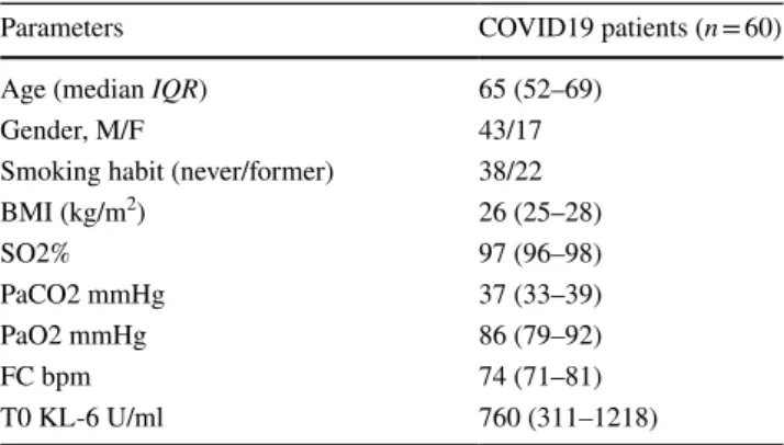
Table 1 shows the main characteristics of our COVID-19 population at t0 (hospital admission). Serum concentrations of KL-6, PFT and blood gas analysis parameters were col-lected (Table1).
At each sampling time, the study population did not differ significantly in terms of sex, age or comorbidities,
Table 1 Main characteristics of COVID19 population including age,
gender, smoking habit, BMI, blood gas analysis values, and KL-6 serum concentrations
Parameters COVID19 patients (n = 60)
Age (median IQR) 65 (52–69)
Gender, M/F 43/17
Smoking habit (never/former) 38/22
BMI (kg/m2) 26 (25–28) SO2% 97 (96–98) PaCO2 mmHg 37 (33–39) PaO2 mmHg 86 (79–92) FC bpm 74 (71–81) T0 KL-6 U/ml 760 (311–1218)
whereas serum KL-6 concentrations were higher at hospitalization (t0) than at t1 (760 (311–1218) vs 309 (210–408) p = 0.0208) and t2 (760 (311–1218) vs 324 (279–458), p = 0.0365). About PFT parameters, we did not collect these data at t0 in hospitalized patients affected by COVID19.
According to CT radiological features, patients were classified according to the evidence of fibrotic lung altera-tions (including ground-glass opacities, linear thickening, or organizing pneumonia areas).
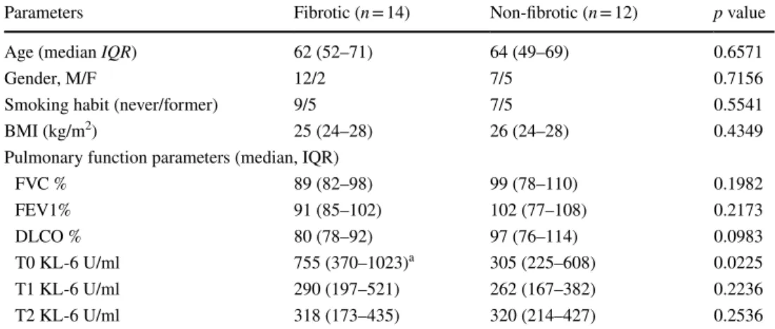
At t0, KL-6 serum concentrations were significantly increased in fibrotic (n = 14) than non-fibrotic (n = 12) group (755 (370–1023) vs 305 (225–608), p = 0.0225) (Fig. 1). Area under the receiver operating curve (AUROC) analysis showed that basal KL-6 had good accuracy in discriminating patients with HRCT evidence of fibrotic interstitial lung abnormalities (AUC = 85%, Std. Error 0.1080, 95%CI 64–100, p = 0.0404) (Fig. 2). The best cut-off value for KL-6 concentrations was 455U/ml (75% sen-sitivity and 80% specificity). At t1 and t2, no differences of demographic features, blood gas analysis values, and PFT parameters were observed between the two groups (data available). KL-6 concentrations in fibrotic group signifi-cantly reduced at t1 (755 (370–1023) vs 290 (197–521),
p = 0.0366) and t2 (755 (370–1023) vs 318 (173–435), p = 0.0490) (Table 2). Interestingly, three patients main-tained very high KL-6 concentrations after 6 and 9 months of follow-up (median IQR, t0: 1006 (1000–1011); t1: 595 (570–1799); t2: 527 (498–1373)), showing simultaneous HRCT evidence of interstitial lung involvement (Fig. 1S). Patients with no fibrotic lung sequelae at HRCT of the chest showed normal KL-6 concentrations at follow-up.
Concerning respiratory function, no significant dif-ferences in FVC and/or DLCO percentages were found between patients with and without fibrotic lung alterations at CT scan.
Fig. 1 KL-6 concentrations at t0 in serum from COVID19 patients
divided according to radiological fibrotic interstitial lung abnormali-ties after 6 months of hospital discharge
Fig. 2 Receiver operating curve analysis showed that, basal KL-6 had good accuracy in discriminating patients with HRCT evidence of fibrotic interstitial lung abnormalities
Table 2 The main
characteristics of our COVID-19 population at follow-up divided according to fibrotic abnormalities, including serum concentrations of KL-6
All data were expressed as median IQR. aKL-6 concentrations significantly reduced at t1 (p = 0.0366) and
t2 (p = 0.0490)
Parameters Fibrotic (n = 14) Non-fibrotic (n = 12) p value
Age (median IQR) 62 (52–71) 64 (49–69) 0.6571
Gender, M/F 12/2 7/5 0.7156
Smoking habit (never/former) 9/5 7/5 0.5541
BMI (kg/m2) 25 (24–28) 26 (24–28) 0.4349
Pulmonary function parameters (median, IQR)
FVC % 89 (82–98) 99 (78–110) 0.1982 FEV1% 91 (85–102) 102 (77–108) 0.2173 DLCO % 80 (78–92) 97 (76–114) 0.0983 T0 KL-6 U/ml 755 (370–1023)a 305 (225–608) 0.0225 T1 KL-6 U/ml 290 (197–521) 262 (167–382) 0.2236 T2 KL-6 U/ml 318 (173–435) 320 (214–427) 0.2536
Discussion
This report first described KL-6 peripheral concentrations in a population of COVID-19 patients after 6 and 9 months from hospitalization discharge together with radiological and functional parameters. Serum concentrations of KL-6 at hospital admission were reported to be significantly increased in severe patients admitted to intensive care unit and requiring intubation with mechanical ventilation, but not in mild-moderate patients with less severe respiratory impairment [9]. This mucin protein has been widely stud-ied in idiopathic pulmonary fibrosis and ARDS patients, but limited data are available on its prognostic potential in infectious viral pneumonia [11–13]. Our interest aroused from the observation that KL-6 was associated with prog-nosis in ILD and ARDS, reflecting type I and type II alveolar pneumocyte damage. SARS-CoV-2 is known to have specific tropism for alveolar epithelial cells, causing interstitial lung damage, epithelial lung alterations, and regenerative processes, mainly in the acute phase [9]. A significant increase in serum concentrations of KL-6 was demonstrated in patients critically ill with COVID-19 [8,
9] and was further confirmed in our study.
Interestingly, our results demonstrated for the first time a normalization of serum concentrations of KL-6 (< 465U/ ml) 6 and 9 months after discharge from hospital. Lung damage due to COVID-19 seemed not to be necessar-ily progressive, unlike in idiopathic pulmonary fibrosis, where KL-6 tends to increase with IPF progression [1,
4, 5]. Moreover, in the majority of patients with fibrotic sequelae, the decrease and substantial normalization of serum KL-6 concentrations may reflect the amelioration of the clinical status, suggesting that fibrotic interstitial lung abnormalities related to COVID-19 are not associ-ated with persistent active epithelial damage and conse-quent aberrant fibrogenesis. On the contrary, those patients who developed severe persistent fibrotic lung sequelae at HRCT showed persistent high levels of KL-6 in the follow-up, implicating that this biomarker may be use-ful in the mid-long-term management of these patients. However, these data will be further evaluated in a longer follow-up to be confirmed. Moreover, in our population, lung volumes were almost preserved at follow-up as well as DLCO percentages, differently from a recent follow-up study reporting functional alterations in more than 25% of patients, albeit after a shorter follow-up of 3 months [19], and this aspect may surely influence the heterogeneity of our population.
Thus, this study contributes to the definition of the nat-ural course of COVID-19 as the normalization of periph-eral KL-6 concentrations, recorded 6 and 9 months after the acute phase of SARS-CoV-2 infection, suggesting a
non-progressive fibrotic lung involvement in the majority of patients.
Serum concentrations of KL-6 seemed to reflect lung involvement in COVID-19 patients as reflected by HRCT features at 6-month follow-up.
In conclusion, increased serum concentrations of KL-6 in hospitalized COVID-19 patients may help to early dis-criminate severe patients requiring mechanical ventilation and predict those developing fibrotic lung sequelae in the follow-up.
Author contributions MD, LB, PC, and EB conceived the study and supervised all aspects. MD, PC, GC, LR, FB, and MAM collected data and built the database. MD, PC, LB, EB, and MAM oversaw data analysis and interpretation of results. All authors drafted and revised the paper.
Funding Open Access funding provided by Università degli Studi di
Siena. The study was performed at Siena University without funding or sponsors.
Compliance with ethical standards
Conflict of interest All the authors have no conflicts of interest to de-clare.
Statement of human and animal rights The research was approved by the local ethics committee (MARKERLUNG n° 17431, C.E.A.V.S.E n° 18712).
Informed consent All subjects gave their written informed consent to the study.
Open Access This article is licensed under a Creative Commons Attri-bution 4.0 International License, which permits use, sharing, adapta-tion, distribution and reproduction in any medium or format, as long as you give appropriate credit to the original author(s) and the source, provide a link to the Creative Commons licence, and indicate if changes were made. The images or other third party material in this article are included in the article’s Creative Commons licence, unless indicated otherwise in a credit line to the material. If material is not included in the article’s Creative Commons licence and your intended use is not permitted by statutory regulation or exceeds the permitted use, you will need to obtain permission directly from the copyright holder. To view a copy of this licence, visit http://creat iveco mmons .org/licen ses/by/4.0/.
References
1. d’Alessandro M, Bergantini L, Cameli P, Vietri L, Lanzarone N, Alonzi V et al (2020) Krebs von den Lungen-6 as biomarker for disease severity assessment in interstitial lung disease: a compre-hensive review. Biomark Med 68(6):414–421
2. Lanzarone N, Gentili F, Alonzi V, Bergantini L, d’Alessandro M, Rottoli P, et al. Bronchoalveolar lavage and serum KL-6 con-centrations in chronic hypersensitivity pneumonitis: correlations with radiological and immunological features. Intern Emerg Med. 2020.
3. d’Alessandro M, Bergantini L, Cameli P, Lanzarone N, Antonietta Mazzei M, Alonzi V et al (2020) Serum KL-6 levels in pulmonary Langerhans’ cell histiocytosis. Eur J Clin Invest 20:e13242 4. Wakamatsu K, Nagata N, Kumazoe H, Oda K, Ishimoto H,
Yoshimi M et al (2017) Prognostic value of serial serum KL-6 measurements in patients with idiopathic pulmonary fibrosis. Respir Investig 55(1):16–23
5. Bergantini L, Bargagli E, Cameli P, Cekorja B, Lanzarone N, Pianigiani L et al (2019) Serial KL-6 analysis in patients with idiopathic pulmonary fibrosis treated with nintedanib. Respir Investig 57(3):290–291
6. Awano N, Inomata M, Kuse N, Tone M, Takada K, Muto Y et al (2020) Serum KL-6 level is a useful biomarker for evalu-ating the severity of coronavirus disease 2019. Respir Investig 58(6):440–447
7. Xue M, Zheng P, Bian X, Huang Z, Huang H, Zeng Y et al (2020) Exploration and correlation analysis of changes in Krebs von den Lungen-6 levels in COVID-19 patients with different types in China. Biosci Trends 14(4):290–296
8. d’Alessandro M, Cameli P, Bergantini L, Franchi F, Scolletta S, Bargagli E. Serum concentrations of Krebs von den Lungen-6 in different COVID-19 phenotypes. J Med Virol. 2020.
9. d’Alessandro M, Cameli P, Refini RM, Bergantini L, Alonzi V, Lanzarone N et al (2020) Serum KL-6 concentrations as a novel biomarker of severe COVID-19. J Med Virol 92(10):2216–2220 10. D’alessandro M, Bennett D, Montagnani F, Cameli P, Perrone A,
Bergantini L et al (2020) Peripheral lymphocyte subset monitoring in COVID19 patients: a prospective Italian real-life case series. Minerva Med. https ://doi.org/10.23736 /S0026 -4806.20.06638 -0
11. Sato H, Callister MEJ, Mumby S, Quinlan GJ, Welsh KI, duBois RM et al (2004) KL-6 levels are elevated in plasma from patients with acute respiratory distress syndrome. Eur Respir J 23(1):142–145
12. Nakamura H, Tateyama M, Tasato D, Haranaga S, Yara S, Higa F et al (2009) Clinical utility of serum beta-d-glucan and KL-6
levels in Pneumocystis jirovecii pneumonia. Intern Med Tokyo Jpn 48(4):195–202
13. Arai Y, Obinata K, Sato Y, Hisata K, Tadokoro R, Tawa T et al (2001) Clinical significance of the serum surfactant protein D and KL-6 levels in patients with measles complicated by interstitial pneumonia. Eur J Pediatr 160(7):425–429
14. Kawasaki Y, Aoyagi Y, Abe Y, Go H, Imamura T, Kaneko M et al (2009) Serum KL-6 levels as a biomarker of lung injury in respira-tory syncytial virus bronchiolitis. J Med Virol 81(12):2104–2108 15. d’Alessandro M, Carleo A, Cameli P, Bergantini L, Perrone A,
Vietri L et al (2020) BAL biomarkers’ panel for differential diag-nosis of interstitial lung diseases. Clin Exp Med 20(2):207–216 16. d’Alessandro M, Perillo F, Metella Refini R, Bergantini L, Bellisai
F, Selvi E et al (2020) Efficacy of baricitinib in treating rheu-matoid arthritis: modulatory effects on fibrotic and inflammatory biomarkers in a real-life setting. Int Immunopharmacol 86:106748 17. Ohnishi H, Yokoyama A, Kondo K, Hamada H, Abe M, Nishimura K et al (2002) Comparative study of KL-6, surfactant protein-A, surfactant protein-D, and monocyte chemoattractant protein-1 as serum markers for interstitial lung diseases. Am J Respir Crit Care Med 165(3):378–381
18. Culver BH, Graham BL, Coates AL, Wanger J, Berry CE, Clarke PK et al (2017) Recommendations for a standardized pulmonary function report an official American thoracic society technical statement. Am J Respir Crit Care Med. 196(11):1463–1472 19. Zhu C, Zhao YB, Kong LF, Li ZH, Kang J (2016) The expression
and clinical role of KL-6 in serum and BALF of patients with dif-ferent diffuse interstitial lung diseases. Zhonghua Jie He He Hu Xi Za Zhi Zhonghua Jiehe He Huxi Zazhi Chin J Tuberc Respir Dis 39(2):93–97
Publisher’s Note Springer Nature remains neutral with regard to