NF-KB
and Rel: Participants in a Multiform
Transcriptional
Regulatory System
Mariagrazia Grilli,
Jason J.-S.
Chiu, and Michael
J.Lenardo
Laboratory of Immunology, National Institute for Allergy and Infectious Diseases, National Institutes of Health, Bethesda, Maryland 20892
1. Introduction
Molecular regulatory pathways were originally identified as series of cova- lent enzym;rtic modifications of substrate molecules during intermediary metabolism. Recent gene regulation research has revealed subtle and com- plex molecular regulation by information pathways involving noncovalent interactions between macromolecules. High affinity interactions between proteins with DNA sequences play a key role in governing gene transcrip- tion. Regulatory interactions also occur between transcription factors and RNA polynierases. In an increasing number of examples, protein-protein association\ between various types of transcriptional regulatory factors determine the specificity of gene activation. These noncovalent informa- tion pathways are also intimately interlaced with enzymatic control points such as phosphorylation or other forms of post-translational modification. Together these pathways form a network that shuttles information throughout the cell allowing physiological needs to be met by selective gene expression.
Modulation of gene expression by the NF-KB/Rel family of protein complexes illustrates many of the newly emerging themes of intracellular regulation. Associations of the binding and regulatory subunits of NF-KB operate at the center of a variety of different signal pathways. NF-KB can process and integrate instructions that come from the extracellular milieu as well as detect intracellular events originating in either the cytoplasm or the nucleus. NF-KB then transmits this information with astonishing rapidity to the transcriptional machinery by directly binding to a range of different DNA sequences in gene control regions. NF-KB comprises members of a family of dimer-forming proteins with homology to the re1 Inrernurionul Kcuiciv oJ Cvrnlopy. Vol. 143 1 Copyright D 1993 hy Academic Press, Inc. All rights of reproduction in any form reserved.
2 MARIAGRAZIA GRILLI Er AL
oncogene. Its binding, location in the cell, and transcriptional properties are regulated by a second Family of proteins that contain repeats of a domain characteristically found in the erythrocyte protein ankyrin. The DNA sequences which bind NF-KB are also recognized by a third family of structurally distinct proteins bearing the “zinc-finger’’ DNA interaction domain. This highly evolved system controls a wide diversity of genes and thus provides an interesting paradigm for several areas of molecular and cellular research.
II. NF-KB Function in B Cells
In 1986, Sen and Baltimore first identified NF-KB as an apparently tissue- restricted factor that activated the Ig K light chain intron enhancer during B-lymphocyte development. They used an electrophoretic mobility shift assay to detect a protein complex with a DNA sequence which was then
called the B site (Sen and Baltimore, 1986a) (see Fig. I ) . The B site was a 10-nucleotide DNA sequence in a region of the large intron of the K light chain gene that had been defined by various functional assays as a B-cell- specific enhancer element (Queen and Baltimore, 1983; Bergman et a/., 1984). A hint to the importance of this sequence was that it was also present in the enhancer of the simian virus-40 (SV-40) DNA tumor virus (Emorine et ul., 1983; Sen and Baltimore, 1986a).
NF-KB had the striking appearance of being constitutively present only in B cells of the “appropriate” stage for Ig K light chain gene expression (mature B cells and plasma cells, but not pre-B cells)(Sen and Baltimore, 1986a). Even more remarkable was that NF-KB was inducible in pre-B cells. For example, in the pre-B tumor line 702/3, NF-KB could be induced by agents such as lipopolysaccharide (LPS), active phorbol esters, cyclo- heximide, IL-1, tumor necrosis factor-a (TNF-a), and virus infection (Sen and Baltimore, 1986b; Atchison and Perry, 1987; Shirakawa et ul., 1989; Lenardo ef ul., 1989). Because these same agents stimulated transcription of the K locus, inducible NF-KB in pre-B cells strengthened the correlation with K light chain gene expression (Nelson eta/., 1985). Chromatin changes and occupancy of the NF-KB site were directly demonstrated in intact nuclei from LPS-treated 70Z/3 cells (Gimble and Max, 1987; Hromas
et a / . , 1988). More recent studies have also found poor activation of
NF-KB in genetic variants of the 70Z/3 cell line that display defective K
gene expression in response to LPS (Briskin rt ul., 1990; Rooney et ul., 1990a, b) .
The observation that cycloheximide induced NF-KB was especially pre- scient (Sen and Baltimore, 1986b). NF-KB induction apparently did not
NF-KBIRel PROTEIY COMPLEXES 3
Namal wa
,,,,
I I-
sv
1
2
Namalwac
Namalwa,,
-
- + -
+
3 4 5 6
I I0
1mM
7 8
FIG 1 Mobiliiy shift electrophoresis analysis of extracts from Namalwa human mature B
lymphocytes using the KB binding site from the Ig K gene. Lanes 1 and 7 are nuclear extracts
from unstimulated cells and lane 8 is the same with 1 mM GTP added to the binding reaction; lane 2 is nuclear extract from Sendai virus-induced cells. Lanes 3 to 6 are cytosolic extracts in which sodium deoxycholate treatment (+, lanes 4 and 6) has been used to free NF-KB from I-KB. These results and the methods for these experiments have been previously described in Lenardo rf a/. (1989).
require new protein synthesis; in fact, cycloheximide and other stimulants caused a superinduction of NF-KB (Sen and Baltimore, 1986b). This corre- lated with the lack of a requirement for new protein synthesis for K gene
expression (Wall et al., 1986) and led to the proposal that a preexisting form of NF-KB, which was controlled by a labile inhibitory protein, might reside in the cell prior to induction (Baltimore, 1987).
The functional role of NF-KB was demonstrated by mutational analysis of the K intronic enhancer (Lenardo et al., 1987) (see Fig. 2). Alterations
B
SITE
MUTANT
NO
ENHANCER
WILD TYPE
MOCK NONE + P M A NONE +LPS +PMA NONE +LPS +PMA
FIG. 2 Analysis of transcriptional activity conferred by the KB binding site in the Ig K gene enhancer in mouse PD-31 pre-B
lymphocytes. The left panel shows the bacterial chloramphenicol acetyltransferase activity for the truncated c-fos reporter plasmid with no enhancer. The middle panel shows the bacterial chloramphenicol acetyltransferase activity conferred by the 470-bp version of the Ig K enhancer in unstimulated cells (none). or in cells stimulated with LPS or phorbol ester (PMA). The right panel shows
the Ig K enhancer with multiple point mutations in the KB binding site that eliminate binding of NF-KB in unstimulated cells (none),
or in cells stimulated with LPS or PMA. These results and the methods for these experiments have been previously described in
NF-KBIRel PROTEIN COMPLEXES
5
of the B sxte that interfered with NF-KB binding destroyed both basal enhancer function in mature B cells and inducibility in pre-B cells. The mutated enhancer was completely inactive. Further experiments revealed that the NF-KB binding site on its own could act independently as a strong tissue-specific and inducible enhancer element (Pierce et a/. ,' 1988; Wirth and Baltimore, 1988). In fact, two copies of an oligonucleotide containing only the NF-KB site stimulated transcription as well as the entire K en- hancer in transient transfection assays.
The initial conceptualization, based on the correlation to K light chain gene expression, was that NF-KB was the key regulatory molecule for light chain gene activation. It was even imaginable that NF-KB orches- trated the entire transition from pre-B to mature B phenotype by turning on a set of differentiation genes. Four pieces of evidence have since suggested 1 hat the true picture is more complicated and more interesting. First, y-interferon can activate K gene expression and not alter the level of NF-KB binding activity in pre-B cells (Briskin et al., 1988; Bornsztyk et al., 1990). Second, the endogenous K light chain gene has been found to be expressed at high levels in myeloma cell lines that contain little NF- KB and show no K intronic enhancer activity (see below) (Atchison and Perry, 1987). Third, NF-KB does not seem to regulate the recently deline- ated enhancer elements in the Ig A light chain loci (Hagrnan et a/., 1990). Fourth, as described below, elements other than the intron enhancer contribute to gene expression and do not involve NF-KB. Taken together, these findings would best portray NF-KB as one part of a transcriptional differentiat ion program that has multiple, perhaps sequential, but function- ally similar elements. Further work will be needed to describe the interplay between the elements of this program and how they coordinate the matura- tion of pre-B to B cells.
One interesting paradox in Ig gene regulation that illustrates the com- plexity of eukaryotic gene analysis has been recently resolved. Early studies of the Ig heavy and light chain genes suggested that the intronic enhancer elements were dispensable for high level gene expression (Schaffner, 1988). As mentioned above, a myeloma line with little NF-KB was found to have robust Ig K gene expression (Atchison and Perry, 1987). This led to the hypothesis that once the Ig genes were triggered, the enhancers and their associated binding proteins might not be needed for the maintenence of transcription (Klein et a / . , 1985; Atchison and Perry,
1987). Preservation of transcription might be attained by the methylation status of the gene or particular chromatin configurations (Kelley et a/., 1988; Atchison and Perry, 1988). Evidence against this idea was that NF-
KB was only rarely absent in mature cells of the B lineage (Sen and Baltimore, 1986a). Moreover, a continuous requirement for the enhancer element was found for the Ig heavy chain gene (Grosschedl and Marx,
6 MARIAGRAZIA GRILLI E r AL.
1988). A coherent picture of Ig gene control subsequently emerged with the discovery of additional enhancer elements for both the Ig heavy chain and K genes at significant distances downstream of the constant region (Meyer and Neuberger, 1989; Pettersson et al., 1990). In the case of the K gene, the downstream enhancer was stronger than the intronic enhancer and essential to achieve high enough expression of K genes in transgenic mice to cause allelic exclusion (Meyer and Neuberger, 1989; Meyer et
ul., 1990). Significantly, the downstream K enhancer is not activated by NF- KB, indicating that multiple pathways lead to high level K gene expression (Meyer et al., 1990; M. Atchison, personal communication).
Although constitutively present in the nucleus, NF-KB can be further modulated in B cells. For instance, higher levels of nuclear NF-KB can be induced in human Namalwa cells by virus infection (Lenardo et al., 1989). At least part of the NF-KB that is induced in certain B cells seems to come from the “masked” cytoplasmic form (see below). It also becomes evident
in whole cell extracts from Daudi cells with a mild denaturant (Visvanathan and Goodbourn, 1989), or in mouse S194 myeloma cells by HTLV-I tax
gene product (Mauxion er uf., 1991). However, preexisting but nonbinding NF-KB does not appear to be present in all mature B cells (Baeuerle et
al., 1988). Recently, Marcuzzi et ul. (1989) and Liu et al. (1991) reported that in murine splenic B cells, NF-KB, which is already present to some degree constitutively, can be induced further after Ig crosslinking.
NF-KB has been implicated in the transcriptional activation of a variety of genes other than the K gene in B cells. These include the MHC class I and p2 microglobulin genes (Yano et ul., 1987; Baldwin and Sharp, 1988).
as well as the genes for MHC class 11 and its associated invariant chain Ii (Blanar et al., 1989; Pessara and Koch, 1990; Zhu and Jones, 1990), and other early activation genes (Anderson et al., 1992). A motif similar to the consensus binding sequence of NF-KB has been found in the promoter of the gene for the murine B-cell tyrosine kinase hck (Lock et al., 1990).
As in other cell lineages, NF-KB has to be viewed in B cells as an eclectic mediator of gene transcription that facilitates rapid genetic interpretation of extracellular signals. Two important regulatory issues are highlighted by the study of NF-KB in the B lineage. First, how does NF-KB turn on specific genes in the B lineage despite the fact that it induces transcription of many genes in many lineages? Second, how is NF-KB binding activity maintained constitutively in mature cells of the B lineage? One explanation for the B-cell-restricted response of certain genes to NF-KB has come from
a study of the Ig K intronic enhancer. Pierce et al. have shown that NF- KB’S effect on the K enhancer is under the influence of a negative element (1991). They identified a 232-base pair (bp) fragment located 5‘ of the NF-
KB binding site that makes the enhancer nonresponsive to NF-KB in non-B cells. Strikingly, this silencer element works in a distance- and orientation- independent fashion.
NF-KBIRel PROTEIN COMPLEXES 7
The issue of how B cells maintain a certain fraction of NF-KB in a nuclear binding form has not yet been adequately explained. There is some evidence that NF-KB is constitutively present in some T-cell lines and thymocytes (Baeuerle et al., 1988; Bohnlein et al., 1988; Leungand Nabel, 1988; Cross etal., 1989; Lin et al., 1990; Korner etal., 1991)and possibly in mature macrophages (Griffin et al., 1989). The mechanism for constitutive activity in any cell lineage has not been determined. Evidence from studies with the cloned genes indicates that the mRNAs for the NF-KB binding subunits p50 and p65 are not elevated as cells mature in the B lineage (Ghosh et al., 1990; Nolan et al., 1991). The simplest model would entail modulation of the level of the cytoplasmic inhibitor, I-KB, but the distribu- tion and functional status of this molecule and its relatives have not yet been evaluated in different lineages.
111. NF-KB as an Inducible Transcriptional Activator
The property that gives NF-KB a widespread significance in cellular regula- tion is its role as a mediator of inducible gene transcription. The key features of NF-KB transcriptional control are that it is fast and versatile and is used in many different gene systems. It also has the important ability to carry signals from the cytoplasm into the nucleus and trans-activate specific genes by binding directly to their promoters.
The versatility of the NF-KB system is evident in the large number of
agents able to up-regulate the levels of NF-KB binding in the nucleus. Unlike transcription factors which respond to a limited number of inducers such as the heat shock factor or specific steroid receptors, signals from a number of directions converge on NF-KB to be conveyed icio the nucleus. Importantly, signals that pass through NF-KB appear to be launched from different parts of the cell (Lenardo and Baltimore, 1989; Baeuerle, 1991). They may emanate from cell surface molecules such as the receptors for cytokines (IL-I, TNF-a, and TNF-P), lectins, or LPS. They may come from elements of second messenger pathways that are associated with events at the cell membrane such as phorbol esters or calcium ionophores. They may arise in the cytoplasm through kinase induction by dsRNA or by inhibition of protein synthesis. They may also involve triggering by intranuclear proteins such as the Tax trans-activator of the HTLV-I virus that induces NF-KB. Finally, and perhaps most surprisingly, induction may even be signaled at the DNA level such as by UV light and other agents thali cause DNA damage. A list of the known inducing agents for NF-KB is given in Table I.
The kinetics of the NF-KB response to inducing agents indicate that it can be extremely rapid and quite sensitive. For example, early studies
8 MARIAGRAZIA GRILLI Er AL. TABLE I
Inducers of NF-KB
Inducer Cell type Species Reference
Lipopol y sacc haride
Protein synthesis inhibitors Phorbol ester Anti-IgM HTLV-I tt/x dsRNA TNF-a L ymphotoxin Interleukin- I Interleukin-1 Interleukin-2 Pre-B (70213; 3-1) Splenic B Fibrosarcoma (WEHI 164) Monocytelmacrophage (mono-mac-6) Pre-B (70213) Splenic B Pre-B (70213) T (Jurkat) HeLa Splenic B Monoc ytelmacrophage Splenic B Plasmacytoma T (Jurkat) Osteosarcoma (MG63) HeLa Fibrosarcoma (WEHI 164) Fibroblast L929 Pro-monocytic (U937) Rat-2 fibroblast M yelomonoc ytic (WEHI 38) T (Jurkat) Fibrosarcoma (WEHI 164) Hepatoma (HepG2) Erythroleukemia (KS62) Macrophages Myelomonocytic T (YT) T (Jurkat) Foreskin fibroblast Hepatoma (HepG2) T (SPB2I) T (EL-4-El) m m m h m m m h h rn h/m m m m h h h m m h r m h m h h m h h h m h h h
Sen and Baltimore, 1986b Atchison and Perry, 1987 Liu et nl., 1991
Gromkowski et a/., 1990 Haas et a/., 1990 Sen and Baltimore, 1986b Liu et d . , 1991
Sen and Baltimore, 198611 Sen and Baltimore, 1986b Sen and Baltimore, 1986b Liu e/ el., 1991
Griffin et d . , 1989
Marcuzzi P I d., 1989
Liu et d., 1991 Mauxion et a / . . 1991
Leung and Nabel, 1988; and others (see text) Visvanathan and Goodbourn, 1989 Visvanathan and Goodbourn, 1989 Gromkowski et d . , 1990 Lenardo e t a / . , 1989 Griffin er u / . , 1989 Pessara and Koch, 1990 Pessara and Koch, 1990 Osborn e t a / . , 1989 Gromkowski et a/., 1990 Banerjee et d . , 1989 Meichle rt a/.. 1990 Collart et d . , 1990 Hohmann et d . , 1990a, Freimuth rt a/., 1989 Osborn er a/., 1989 Novak ct a/., 1990 Zhang et a/., 1990 Nonaka and Huang, 1990 Hazan r / a/., 1990
1991b
NF-KBIRel PROTEIN COMPLEXES TABLE I (continued)
9
Inducer Cell type Species Reference
Ph ytohemagglutinin T (Jurkat)
Pro-monocytic (U937) Concanavalin A T (Jurkat)
Calcium ionophore T (EL-4-El)
Anti-CD2 Anti-CD3 Anti CD28 Anti-ap TCH: Antigen C-AMP UV light 4-Nitroquinoline oxide H!Oz
Theilera parva (parasite) Serum Okadaic acid Monocytes T (SPBZI) T (Jurkat) T (SPB2I) T (SPBZI) Pre-B (702/3) HeLa HeLa T (Jurkat) T cells BALBIc 3T3 T (Jurkat) T (AR-5) T (A-E7) Viruses CMV T (Jurkat) or HF Adenovirus Fibroblast (3T3) (foreskin) Cervical carcinoma (HeLa)
Hepatitis B Hepatocyte (HepG2)
Sendai Fibroblast (L929)
Herpes simplex, type I T (Jurkat) Human herpes virus, T (Molt3) type VI h h h m h m h h h h m m h h h b m h h m h h m h h
Sen and Baltimore, 1986b Libermann and Baltimore, I990 Rattner et a/., 1991 Novak et d., 1990 Bressler ct al., 1991 Jamieson et a/., 1991 Hazan et a / . , 1990 Verweij e t a / . , 1991 Hazan et a/., 1990 Hazan et a/., 1990 Kang et a/., 1992a Shirakawa et a/., 1989 Stein e t a / . , 1989a; b Stein e t a / . , 1989a Schreck e t a / . , 1991 lvanov et a / . , 1989 Baldwin e t a / . , 1991 Thevenin e t a / . . 1990 Sambucetti et u/., 1989 Shurman et a/., 1989 Nielsch e t a / . , 1991 Twu e t a / . , 1989a; Faktor and Shawl, 1990 Lenardo et a/., 1989 Gimble e t a / . , 1988 Ensoli er a/., 1989
indicated that phorbol esters gave maximal induction in 30 min and later studies showed significant induction occurred at much earlier times (Sen and Baltimore, 1986b; vide infra). Also, the kinetics were not identical for different inducers: phorbol stimulation of 702/3 cells remained maximal from 0.5 to 6 hr and was gone at 8 hr, whereas LPS induction became maximal at 2 hr and was still apparent at 8 hr (Sen and Baltimore, 1986b).
10 MARIAGRAZIA GRILLI E r AL.
Careful studies of induction by TNF-a showed that NF-KB was detectable within 2 min and maximal after 10-15 min (Hohmann e f d . , 1991b). NF- KB can also be detected in nuclear extracts from 70Z/3 cells within 15 min after either LPS or IL-1 treatment (Shirakawa et al., 1989). The sensitivity of NF-KB induction was also demonstrated by the finding that occupancy of only 5-2096 of the TNF-cu surface receptors was required in HL-60 and HepG2 cells (Hohmann et al., 1991 b). The rapidity of the response seems due to the fact that the immediate induction by many stimuli is independent of new protein synthesis (Sen and Baltimore, 1986b; Osborn et nl., 1989;
Banerjee et al., 1989; Zhang et d . , 1990; Liu et al., 1991) and appears to be directly connected to kinase pathways in the cell (see below).
There is also some evidence that NF-KB may respond differently to a given inducing agent depending on the cell type. Phorbol esters alone will strongly induce NF-KB in 70Z/3 pre-B lymphoma cells, but do not induce NF-KB at all in nontransformed CD4+ T-lymphocyte clones (Kang et al.,
1992a). Another example is that IL-I or active phorbol esters will induce NF-KB in 70213 cells or LBRM T cells but neither will induce NF-KB in TH-2 T-lymphoma cells (Osborn et nl., 1989). Although many of the pathways that induce NF-KB have been only incompletely mapped, it is clear that NF-KB lies at the intersection of a large number of control routes traversing the cell. NF-KB may therefore serve as an integration node. Consistent with this idea, there are instances where inducers synergize to activate NF-KB. Mimicking T-cell receptor stimulation by phytohemagglu- tinin (PHA) or phorbol ester (PMA) alone can modestly induce NF-KB in Jurkat T-lymphoma cells, but more significant increases are seen if both are used together (Hoyos et nl., 1989). Similarly, PMA
+
TNF-a induce NF-KB more strongly than either alone, though it is not clear whether the effect is additive or synergistic (Osborn et a/., 1989). Though these examples are artificially contrived costimulatory situations, they indicate that multiple signals can be integrated and then transduced by increased levels of NF-KB in the nucleus. The role of protein kinase C and other serinelthreonine kinases during the induction of NF-KB will be discussed below.One area of research for the future will be to more fully explore effects on NF-KB induction pathways by signal transduction pathways such as those mediated by tyrosine kinases, phosphotidyl inositol breakdown products, or prostaglandin and related molecules. One interesting recent development is evidence that protein modifications such as methylation and isoprenylation may be important events in the induction of NF-KB by LPS but not PMA (Law et a[., 1991). Proteins with these adducts are typically associated with the membrane during LPS induction and may be among the earliest events in the signaling pathway. It will be interesting to see how these modified proteins connect either to kinases or possibly to the latent form of NF-KB in the cytoplasm.
NF-KBIRel PROTEIN COMPLEXES 11 Once the NF-KB signal has activated a gene, how is it stopped? Some attenuation may be due to the natural turnover of the proteins but the complex of NF-KB and its cognate DNA motif appears to be quite stable (Zabel and Baeuerle, 1990). Another possibility is that IKB or other proteins actively quench NF-KB binding. In uitro binding studies indicate that the half-life of an NF-KB complex with DNA can be dramatically reduced by the presence of I-KB, the specific inhibitor of NF-KB binding (see below) (Zabel and Baeuerle, 1990). For this mechanism to be effective, IKB would be required to attack NF-KB complexes in the chromatin and possibly sequester them in the nucleus, ferry them back to the cytoplasm, or facilitate their degradation (for a discussion see Baeuerle, 1991). An- other posbibility is that there are nuclear inhibitors that can maintain complexes in the nucleus in a nonbinding form. These possibilities have not yet been addressed experimentally.
It is important to note that a second tier of regulation seems to occur at the level of expression of the mRNAs encoding the subunits of NF-KB (Irving et
d.,
1989; Bours et a / . , 1990, 1992; Meyer rt a/., 1991) as will be discussed further below. Perhaps all members of the gene family that includes NF-KB can be induced to some degree at the RNA level by many of the same agents that seem to activate NF-KB. This effect could play a role in sustaining a gene activation signal at times long after the initial release of NF-KB from cytoplasmic stores by agiven stimulus. It is interest- ing to note that genes encoding proteins that inhibit NF-KB binding are also induced soon after certain cell stimuli (Haskill etd.,
1991). Complex gene regulatory interactions could therefore result from the long-lived production of certain NF-KB subunits and inhibitors and the exclusion of others.IV. A Wide Variety of Genes Are Regulated by NF-KB
The target genes of NF-KB are numerous and most share the common feature of being quickly induced in response to an extracellular stimulus. NF-KB is able to signal the occurrence of many extracellular events and its effects are remarkably pleiotropic. Hence, a rise in nuclear NF-KB is not very informative for understanding a program of gene expression in response to a particular inducing agent. Perhaps the major role of NF-KB is simply to send a signal into the nucleus that an acute process that requires a genetic response is under way. A significant body of work has
been carried out on genes induced by NF-KB and related binding proteins during T-lymphocyte activation or other aspects of the immune response. Several other genes have no obvious relationship to the immune response.
12 MARIAGRAZIA GRILLI E r AL.
NF-KB induction of specific genes will be discussed in association with the physiologic role that they play.
A. T-Lymphocyte Activation
An important subset within the wide variety of genes controlled by NF- KB is the collection of genes that are induced during T-cell activation. Over 100 genes appear to be transcribed in T cells after stimulation by antigen in the context of an appropriate major histocornpatability complex (MHC) molecule or agents that mimic this interaction (Zipfel er al., 1989;
Irving et al., 1989; Crabtree, 1989). Therefore understanding gene control during T-cell activation should provide significant insights into the general molecular regulation of this process (Ullman et al., 1990). NF-KB in coop- eration with other transcriptional regulators (activators and repressors) appears to be involved in the expression of many genes especially during the early phases of the response to T-cell receptor stimulation (Jamieson
et al., 1991; Lenardo and Baltimore, 1989). Cytokine gene expression provides a good model for understanding NF-KB’S role early in T-cell activation. mRNAs for cytokines, or more specifically interleukins, are usually undetectable in T cells unless the appropriate stimuli have been administered (DeMaeyer and DeMaeyer-Guignard, 1988). Though there has been some controversy regarding whether transcriptional or post- transcriptional mechanisms are the prevailing determinant in elevating mRNA levels, careful analysis of cytokine regulation in T cells indicates
a major role for transcriptional control (Brorson et al., 1991). The process initiates rapidly since increases in cytokine mRNAs are detected within 2 hr and in some cases peak at
4
hr. NF-KB is well suited for cytokine control because of the speed of its activation and its independence from new protein synthesis.The best studied examples of T-cell cytokine gene regulation, IL-2 and the IL-2 receptor a chain genes, illustrate how NF-KB plays only one part in an ensemble of regulatory factors. A region of approximately 300 bp located immediately upstream of the transcription start site of the IL-2 gene, variously called the enhancer or the promoter, has been shown to strongly respond to T-cell activation signals (Brunvand et ul., 1988; Fujita et al., 1986; Durand e? al., 1987; Novak et a1.,1990). This DNA segment contains binding sites for NF-KB and other nuclear trans-activat- ing proteins including the nuclear factor of activated T cells (NF-AT), octamer factors, AP-1, purine-rich binding factors, and, at least in human cells, a CD28-responsive complex (Lenardo et al., 1988; Durand et al.,
1988; Hoyos el al., 1989; Muegge et al., 1989; Randak et al., 1990; Fraser et al., 1991; Granelli-Piperno and Nolan, 1991; Hentsch et al., 1992).
NF-KB/Rel PROTEIN COMPLEXES 13
Deletion studies suggest that multiple protein binding sites must be occu- pied in order to allow full IL-2 enhancer activity (Durand et al., 1988, Serfling et ul., 1989; Hoyos PI al., 1989). Certain of these are factors are tissue-specific, such as NF-AT; however, others such as AP-1 and NF-KB do not have any obvious tissue restriction but are important in regulatory states such as clonal anergy (Kang et al., 1992b). The molecular switch that activates the IL-2 gene appears to be the constellation of trans- activators rather than any one taking a leading role.
Studies in which point mutations were introduced into the promoter clearly indicated a potentially important role for KB binding motif (IL-2- KB), presumably due to trans-activation by NF-KB or other Re1 family members (Ballard et al., 1988; Shibuya et al., 1989; Shibuyaand Taniguchi, 1989). Studies have also provided evidence that in certain cell lines the IL-Z-KB site may function in a tissue-specific manner (Radler-Pohl el
al., 1990; Briegel et al., 1991). Recently, evidence from the study of
nontransformed murine T-cell clones indicates that more than one distinct
re/ family member acts at the IL-~-KB sequence (Kang et al., 1992a). A complex, termed NF-KC, that comprises the p50 subunit of NF-KB and is distinct from the NF-KB p50/p65 heterodimer is present in unstimulated T cells but disappears after activation. Activation signals that lead to loss
of NF-KC induce IL-2 mRNA. By contrast, conditions that elevate nuclear NF-KB or NF-AT levels but do not lead to loss of NF-KC fail to induce IL-2 mRNA. NF-KC removal appears to be due to its sequestration in the nucleus by a novel inhibitory protein, which we informally refer to as I- K C , whose synthesis is tightly linked to antigen stimulation and sensitive to cyclosporin A. One molecule that may serve an J-KC-like function is the Bcl-3 protein that is located in the nucleus and inhibits the p50 homodimer complex (Wulczyn et al., 1992; G. P. Nolan and D. Baltimore, unpublished results). This pathway illustrates a mechanism by which NF-KB-related complexes can be regulated exclusively within the nucleus during activa- tion of T cells. It also maintains tight regulation of the IL-2 gene, since NF-KB can be activated in response to a variety of signals but the loss of the NF-KC protein is directly linked to antigen stimulation (Kang et al.,
1992a). Recent results in human cells suggest that the mechanism de- scribed by Kang er al. may govern the transcription of HIV-1 and that Bcl-3 serves as an inhibitor of the NF-KC or p50 homodimer complex (Franzoso et al., 1992). In contrast to antigen, stimulation of nontrans- formed T cells with anti-CD3e does not stimulate transcription through the IL-Z-KB site (Jain et al., 1992). Interestingly, the induction of NF-KB seems to be only weakly blocked by cyclosporin A at physiological doses, but there may be other mitogen-induced complexes binding to KB sites that are sensitive to this immunosuppressant (Randak et al., 1990; Schmidt
14 MARIAGRAZIA GRILLI E r AL al., 1990; Brini et d . , 1990). In tumor cells, induction of NF-KB and the transcription activity of KB-binding elements stimulated by PMA appear to be more resistant to effects of cyclosporin A than inducing agents that cause increases in intracellular Ca2+ such as ionomycin and lectins (U.
Siebenlist, personal communication).
The IL-2 receptor a chain (IL-2R) gene also displays an interesting regulatory pattern centered on an NF-KB binding motif in its promoter. Elevated cell surface expression of the IL-2 receptor a chain is a critical step in generating high affinity receptors for IL-2 during the response of
T lymphocytes to antigen (Waldmann, 1986). Several groups have shown that the p65/p50 heterodimer of NF-KB or a related complex transduces T-cell activation signals into increased activity of the IL-2R promoter (Leung and Nabel, 1988; Bohnlein et al., 1988, 1989b; Lowenthal et al.,
1989a,b; Cross et al., 1989; Lin et ul., 1990). Significantly, IL-2R promoter activation by the Tax trans-activator gene of HTLV-1 or TNF-a also works through NF-KB (Fig. 3 ) (Cross et ul., 1987, 1989; Siekevitz et al.,
1987a; Ruben et ul., 1988; Lowenthal et al., 1989a). By contrast, phorbol ester inducibility of the IL-2R promoter is mediated by a distinct sequence and did not involve the NF-KB binding site (see below) (Ballard ef al.,
1989; Lin et al., 1990). The KB motif in the IL-2R gene only weakly binds NF-KB and manifests a poorer response to trans-activation than other KB motifs (Shibuya et al., 1989). Recently it has been found that the activity of the KB motif in T cells is due to cooperation between NF-KB and the serum responsive factor (SRF) and other promoter factors such as Spl (Pomerantz rt ul., 1989; Ballard et al., 1989; Toledano et ul., 1990; Roman et ul., 1990). Serum responsive factor appears to be present at very high levels in both resting and activated T cells and binds adjacent to NF-KB in the promoter region (Kuang et ul., 1993). Constitutively higher levels of NF-KB are associated with deregulation of IL-2 and its receptor in certain T-lymphoma lines that depend on IL-2 for autocrine growth (Hemar et al., 1991a). It is notable that a putative transcription regulatory
molecule unrelated to NF-KB, but that binds to an IL-2R promoter se- quence encompassing the NF-KB site, has been recently cloned (Adams et ul., 1991).
A variety of other cytokines that are produced by activated T cells also appear to involve NF-KB in their transcriptional control. These include IL-6, which stimulates B cells to proliferate, differentiate and produce Ig (Shimizu et al., 1990; Libermann and Baltimore, 1990); GM-CSF, which sustains proliferation of bone marrow precursors for macrophages and granulocytes (Sugimoto et ul., 1990; Shreck et al., 1990); TNF-a and
-p,
which are inflammatory mediators (Messer etd.,
1990; Paul et ul., 1990; Collart et a / . , 1990; Goldfeld and Maniatis, 1989; Shakhov ot ul., 1990; Hass et ul., 1990; Drouet et al., 1991; Kuprash and Nedospasov, 1992;NF-KBIRel PROTEIN COMPLEXES 15
I
MT-2
7
JURKAT
1 % I € \z
0c s s z
5.-
KB
Competitor
z'
nq:
0
2
8
32 0
2 8
32
1 2 3 4
5 6 7 8
9
10
11
12
IL-
2R
NF-KB
FIG. 3 Mobility shift electrophoresis analysis of nuclear extracts from the HTLV-1 infected human T-lymphoid tumor line MT-2 or the human T-lymphoma cell Jurkat. Lanes 1-4 show
the complex detected with the K B binding site from the Ig K gene, competed with increasing
amounts of the same sequence to indicate specificity of the nucleoprotein complex. Lanes
5-8 show that a comigrating specific complex is formed with the KB binding site from the
IL-2 receptor (IL-2R) a chain promoter. Lanes 9-12 are the complexes detected using the
KB binding site from the Ig K gene in nuclear extracts from Jurkat cells with unstimulated,
stimulated with phorbol ester and phytohemagglutinin (PMA/PHA), o r stably transfected
with expression plasmids containing the HTLV-I Tax coding sequence in the sense (tax) or
antisense orientation (astax). These results have been previously described in Cross et ul. (19891, and Leonard0 et ul. (1988).
16 MARIAGRAZIA GRILL1 ET At
Zuckerman and Evans, 1992); and proenkephalin, a precursor for neuro- peptides whose mRNA is expressed, for unknown reasons, at high levels
in activated T-helper lymphocytes (Rattner et id., 1991). Interestingly, in the case of TNF-a and $3, these same cytokines are potent NF-KB inducers indicating that an autostimulatory loop could be set into motion in the response to antigen (Griffin eta/., 1989; Hohmann et
d.,
1991a). Therefore NF-KB appears to form part of a coordinated program that underlies the immune response.Precisely how NF-KB connects to signals initiated by the antigen recep- tor is still obscure. Different answers are obtained in various cellular models that have been studied. In tumor cells, often a relatively simple signal will suffice. For example, in EL-4 murine thymoma cells, PMA or lectin alone will induce NF-KB (Lacoste er af., 1990; Shibuya and Tani- guchi, 1989). PMA alone also induces NF-KB in non-T cells (Nelson et a/.,
1988). In Jurkat both PMA and phytohemagglutinin together give an even greater induction than either agent alone (Hoyos et al., 1989). The fact that induction of NF-KB by PMA and phytohemagglutinin is not blocked by physiologic concentrations of cyclosporin A in this system has been taken to indicate that NF-KB itself is not involved in the immunosuppres- sion caused by this drug (Emmel et al., 1989; Mattila et af., 1990). In evaluating other lines, especially nontransformed T lymphocytes, PMA does not induce NF-KB (Kang et ul., 1991). Stimulation of the T-cell antigen receptor by physiologic ligands appears to activate NF-KB (Jamieson et al., 1991; Kang et al., 1992a). Signaling pathways through other T-cell surface markers such as CD28 also activates NF-KB (Gruters et af., 1991; Verweij et al., 1991; Granelli-Piperno and Nolan,
1991).
In many types of cells, including T cells, a single cytokine such as IL-
1 or TNF-a will induce NF-KB (Krasnow et al., 1991 ; Lacoste et
d.,
1990; Hohmann et af., 1990b). It is unclear, however, how many and what types of different signal transduction pathways are activated following engagement of the receptors for these cytokines. Some evidence suggests that TNF induction does not go through protein kinase C, at least in Jurkat T cells (Meichle et al., 1990). Furthermore, the pathways activated by an individual cytokine may differ between cell types: TNF induction of NF- KB is increased by cycloheximide in Jurkat cells, but blocked by cyclohex- mide in HepG2 cells (Banerjee et af., 1989). As in the case of NF-KB induction in B cells, other signal transduction pathways in T cells, espe- cially tyrosine phosphorylation, have not been evaluated.One interesting observation is that a decrease in the level of intracellular thiols can be one of the steps in NF-KB induction (Staal et af., 1990). For example, the induction of NF-KB by TNF-a or phorbol ester can be blocked by N-acetylcysteine, an agent that will maintain high levels of
NF-KBIRel PROTEIN COMPLEXES 17 glutathione and other thiol compounds. It has been recently reported that this may be a useful intervention to prevent the activation of HIV transcription (Roederer et al., 1990; Kalebic et al., 1991). Reducing agents have the capacity to release NF-KB from I-KB in cytosolic extracts (Toled- ano and Leonard, 1991). It has also been found that H202 can induce NF-
KB and the replication of a latent HIV provirus in a human T lymphoma. This presumably occurs by generating reactive oxygen intermediates, and can be blocked with N-acetylcysteine, dithiocarbamates, and metal chela- tors (Schreck et al., 1991, 1992).
6. Other Genes Involved in Immune or Inflammatory Function
Certain cytokines that are primarily derived from leukocytes that are not lymphocytes are also controlled by NF-KB. These include G-CSF, a growth cytokine for granulocytes (Nishizawa et al., 1990; Nishizawa and Nagata, 1990); TNF-a and IL-6, made primarily by monocytes but also by T cells (Collart et al., 1990; Shakhov et al., 1990; Libermann and Baltimore, 1990; Zhang et al., 1990; Shimizu et al., 1990); and IL-8, a chemotactic factor that recruits neutrophils into inflammatory sites (Mukaida et al., 1990). Recent evidence has shown that NF-KB physically interacts with the NF-IL-6 transcription factor which is an important regulator of the IL-6 and IL-8 genes (K. LeClair, personal communica- tion). The involvement of NF-KB in p-interferon (@-IFN) gene induction was first reported by Visvanathan and Goodbourne (1989). They found that double-stranded RNA induced a binding complex with characteristics of NF-KB and pointed out the possibility that multiple kinases, including DI kinase, may release NF-KB from I-KB. Similar work by others showed that virus infection can also induce NF-KB (Lenardo et af., 1989; Fujita et
al., 1989b; Hiscott et al., 1989). NF-KB binds to and positively activates a site called PRD-I1 in the @-interferon promoter. As such it is part of a
group of virus-regulated events including activation by two positive ef- fectors and the release of a repressor. Because the @-interferon gene can be induced in almost every cell type, NF-KB is likely involved in gene regulation in perhaps all nucleated cells. Curiously, another factor that binds to the PRD-I motif of the @-interferon promoter, IRF-1, is itself regulated by NF-KB, but its role in @-interferon activation is controversial (Fujita et al., 1989a; Baeuerle, 1991; Pine et al., 1990; Reis et al., 1992). Other investigators have provided evidence that the distinct expression of cr- and @-interferons is due to different virally induced transcription factors since the uI-interferon promoter is not regulated by NF-KB (MacDonald et al., 1990).
18 MAAIAGRAZIA GRIM E r AL
Key tasks of immune recognition may be carried out by NF-KB. For example, the ability of specialized cells such as B cells, macrophages, and dendritic cells to participate in antigen activation of
T
cells seems to involve gene control by NF-KB. This regulation embraces the genes for molecules, mostly receptors, that play key roles in the events of antigen processing, presentation, or recognition: MHC class 1 and 11; µ- globulin molecules; MHC class 11 molecules: the invariant chain, Ii; and the T-cell antigen receptor0
chain (Baldwin and Sharp, 1988; Israel et al., 1987; Jamieson rt ul., 1989; Blanar et al., 1989; Mauxion et a/., 1990; Schwemmle et al., 1991; Pessara and Koch, 1990; Blanchet et ul., 1992). Another inflammatory response that involves NF-KB and gene activa- tion is the acute phase response of hepatocytes (Edbrooke ctd.,
1989,1991). During this process, various cytokines (IL-I, IL-6, and TNF-a) acting at the liver cell membrane and steroid hormones acting at the nucleus produce an increase in the expression of particular genes and NF- KB is the mediator for elevated expression of the gene for serum amyloids A2P and A1 (Edbrooke et al., 1989; Li and Liao, 1991). SAA2P is the precursor of amyloid A protein subunits of amyloid fibrils in secondary amyloidosis (Edbrooke et al., 1989). Other acute phase genes-angiotensi- nogen, a precursor for the potent pressor hormone angiotensin 11, and a,-
acid glycoprotein, a nonspecific immunosuppressant-are also controlled by NF-KB or a trans-activator of related specificity (Ron et a/., 1990a,b,c; Brasier et a/., 1990; Won and Baumann, 1990).
Other hepatocyte genes loosely included under the rubric of immune/ inflammatory genes which are apparently regulated by NF-KB include the complement factor 4 and factor B genes ( Y u et a/., 1989; Nonaka and Huang, 1990). The factor B protease is a member of the complement cascade that plays a critical role in the activation of the alternative pathway and its induction by TNF-a seems due to NF-KB (Nonaka and Huang,
1990).
Data supporting a role for NF-KB in the direct regulation of cell growth are emerging. Renan (1989) and Duyao et a / . (1990) have both suggested that NF-KB takes part in a novel type of growth gene regulation. By binding to a site in the first exon of the c-myc mRNA, NF-KB could act to relieve transcriptional blockade which governs the level of full-length mRNA produced. NF-KB would interrupt a stable stem-loop structure in this region of the c-myc message that appears to halt transcriptional elongation by RNA polymerase (Duyao et a/., 1990). Another set of growth-related genes, the gro family, which are transiently induced by serum stimulation and may participate in the growth of melanomas, involve NF-KB when actuated by IL-1 and TNF-a (Anisowicz et
at.,
1991). Lastly, Baldwin and colleagues have found that serum stimulation of BALB/c 3T3 cells leads to the presence of a binding complex resembling NF-KB ( I 991). Collectively,NF-KB/Rel PROTEIN COMPLEXES 19 these findings support the notion that NF-KB may be a central regulator of genes involved in cell growth in both lymphocytes and nonlymphoid cells.
Other genes whose expression is influenced by NF-KB or its congeners cannot be characterized as strictly immunological or inflammatory. These diverse targets for NF-KB complete the picture of a very versatile tran- scription factor. NF-KB or a member of the family of zinc-finger proteins that bind to &-related motifs may regulate a site in the gene for the al- crystallin protein that is a constituent of the lens of the eye (Nakamura et
al., 1990). ’The promoter of the urokinase-type plasminogen activator has at least one or possibly multiple &-like sites that bind complexes of the c-re1 protein and p65 (Novak et al., 1991; Hansen et af., 1992). Also, NF- KB binding sites have been postulated by sequence homologies within a novel transcription unit in the rat insulin-like growth factor gene I1 (Matsuguchi et al., 1989), the mouse perforin gene (Youn et al., 1991), and the mouse MHC class 11 EP gene (Shenkar et al., 1991). Finally many viral promoters and enhancers contain functional NF-KB sites which will be discussed further below.
V. Structure of NF-KB Sites: Clues for Specificity?
One of the features of NF-KB binding motifs is that they can differ quite significantly in their primary nucleotide sequence compared to the Ig-KB sequence (originally defined as the 10-nt 5’-GGGACTTTCC-3’). These variant sites can differ in their affinity for NF-KB and may be functionally distinct depending on the structure of the nucleoprotein complex formed (Lenardo and Baltimore, 1989; Baeuerle, 1991; Zabel et al., 1991; Kunsch
et al., 1992). These sites have been summarized in Table 11. At a glance,
it can be noticed that most NF-KB motifs are asymmetric. The first three nucleotides are invariably guanine and the last two are virtually always cytosine. As one moves from 5’ to 3‘ through the nucleotide sequence, the complexion of the motif shifts from primarily purines to pyrimidines. The nucleotides in the very middle are quite variable (but they tend not to be G in the alignments depicted) and may contribute very little to binding. For example, in comparing the NF-KB motifs from the Ig K genes in different species, they appear as versions of the mouse site in which either one (rabbit) or both (human) of the central nucleotides have been lost (see Table 11) (Emorine et al., 1983). Nonetheless, the central nucleotides may play a role in the ultimate structure of the nucleoprotein complex (Schreck et al., 1990).
20
TABLE II NF-KB Target Genes
MARIAGRAZIA GRILLI E r AL.
~ ~~
Gene Sequence" Retcrcnce Well detined a. Surface molecule'; involved in immune function b. Soliihlc rnolccules involved in immune function
c. Acute pharc response genes
d. Oncogene\
e. Viruses
I g K light chain (mouse)
Ig K light chain (human)
Ig K light chain (rahhit)
IL-2Ru chain TCR p chain MHC class I (H-ZKh) MHC class II (En") ( F.K Invariant chain li gZ microglobulin ELAM-I IL-2 IL-h 1 I A (human) C-CSF TNF-n (cachectin) TNF-g (lymphotoxin) a-interferon Proenkcphalin (rat) Serum amyloid A (SAA) Angiotensinogen (rat) GGGACTTTCC GGGCATTTCC GGGGTII ccc GGGAATCTCC GGGAGATTCC GGGGATTCCC GGGGAAGCCC GGGACTTCCC GGGAATTTCC GGGACITTCC GGGGATTTCC GGGATTTCAC G G G A T T T K C GGAATTTCCT GGGAACTACC CGGGAATCTC GG(i(AAlAT(CAC) GGGGClTTCC GtiG A ATTCAC GGGGAATCCT GGGGCTCCCC' GGG AATTTCC GGGGC rrccc GGGAAATTCC GGGACG rccc GGGACTT I CC CGGA ITTCCC Cumplemenl Cactoi B (moiicel GGGAATCCCC Complement C4 protein GGGAAGTCCC c-niw GCGAAAACCC H I V sv-40 GGGACTTTCC GGGACTTrCC tiGGAAGTACC Cytoinegaliivirus GGGACTII'CC (;GGGATTTCC Adenovirus GGGACTTTCC
Sen and Baltimore. 1986a Emorine L'I ul.. IYXS
Ballard C I o l . . IYKX
Leung and Nabel. IYXX Jamieson rl ol , 1989 Baldwin and Sharp. 1988 Israel rr N I . , 1989 Ulanai- el ( I / , , IYX9
Pe\sara and Koch. 1990 Whelan PI ul., 1991
Lcnardo 1'1 ( I / . 19x8
ShimiLu C I ul., lYY(J
Lieherman and Baltimore, IYYI)
Mukaida C I <I/., IYY0 Mahe PI d., 1991
Sugimoto P I (I/ , I990 Schreck 1'1 ol.. IWO
Nishiznwa P I o / . , 1990 Israel Y l a / . , 1989 Shakhov P I o l . , 1990
Collart C I (11 , 1990
Kuprarh and Ncdospdwv, 1992
Paul r1 I!/., 1990
Messer P I ol , 1990 Vicvanathan and Cioodhourn. Fu,iila C I ( I / , IYX9h I.enardo P I d., IYSY Hiscott ri ill., 1989 Rattner P I ol.. 1991 Edbrooke P I u l . , 1989 Kon P I d.. IYYOa Nonaka and Huang. 1990
Visvanathan and Goodbourn. Yu ,,I n l . . 1989
1989
1989 ~ u y a o ri ( I / . , 1990 Nahcl and Baltimore. 1987 Kanno P I ol.. 1989 Macchi C I o l . . 1989 Samhucctti C I u / . , IYKY
Williams P I u / , IYY(1 ( c o n tin ires )
NF-KBIRel PROTEIN COMPLEXES TABLE II (confiliued)
21
Gene Sequence" Reference
f. Others Invariant chain GGGAATTTCC Pessara and Koch. 1990
Vimentin GGGGCTTTCC I.ilienhaum cr nl., 1990
Gro Anisowicz er o l . , 1991
Interferon regulatory factor GGGGAATCCC Baeuerle. 1991 Consensus: GGGRNNYYCC Nucleotide distribution Position: I 2 3 4 5 6 7 8 9 10 4 0 4 0 4 0 1 3 I 3 2 0 0 OG 0 0 1 2 7 2 1 8 I 2 3 OA 0 0 0 0 1 3 I 2 1 8 3 6 3 8 C 0 0 o o 5 2 8 3 5 2 0 I Z T Undefined (genes containing potential NF-KB sites)
Perforin
Insulin-like growth factor I1 1731 LTR (retrotranipuson) TGF 02 Apolipoprotein Clll Urokinase ICAM-I CD7 GGGCCTTTCA TGGGGAGCCCCC AGGCiGCTCCCCAG AGGCAATTTCCA GGGCATTGAC GGGAAATCCC GGAGGATTCC CGGGAAGTCC GGGAGTCCCT GGGACGCCCT Youn er ul., 1991 Matsuguchi P r a / . , 1989 Ziarczyk cf id., 1989 Malipiero er ul., 1990 Reue er ul.. 1988 Novak "1 o l . , 1991: Hansen er ol.. 1992 Vordherger er ul., 1991 Stade cr a / . . IWO Schanberg cr a / . , 1991
' Note that additional variant 10-bp binding sequences can be obtained by shifting the site
one or two nucleotides upstream or downstream or by the sequence encoded by the opposite
strand in many genes.
As will be recounted in more detail below, NF-KB comprises two sub- units, p50 and p65, which can bind individually as homodimers or as a p50/p65 heterodimer. A careful comparison of the binding of either p50 or p65 homodimers to the ~ 5 0 1 ~ 6 5 heterodimer indicates that p50 apparently binds to the 5' (more conserved) half site and p65 binds to the 3' (less conserved) half site (Urban et al., 1991). Does this mean that different versions of the KB motif prefer p50 in combination with different partners from the Re1 family? Direct evidence is just emerging that this will likely be an important theme in the future (Perkins et al., 1992). As might be expected, the binding of homodirners of Re1 proteins is strongest to sites that are palindromic or otherwise highly symmetric (Urban et al., 1991).
In any case, it is clear that variations in the nucleotide sequence of the KB motif have an important influence on function.
How could specificity be encoded in the various versions of the KB motif? Ont obvious way is simple affinity-if a site was of high affinity, it would re*;pond to lower levels of NF-KB, whereas a weaker site would
22 MARIAGRAZIA GRILLI E r AL
be relatively insensitive to NF-KB. This type of functional distinction has been pointed out (Cross et ul., 1989; Kuang et al., 1992). In fact, weaker binding may lead to a greater dependence of the KB motif on other factors within a given control element (Kuang et al., 1992). A second means to
achieve specificity would be the ability of a given site to discriminate between different members of the KB binding protein family (Mauxion and Sen, 1989; Perkins et al., 1992). A third mechanism could be an alteration of the structure of the DNA binding site caused by factor binding. Evidence has been obtained that bending of the KB DNA sequence is induced by interactions with various Re1 proteins (Schreck et ul., 1990; Kuprash and Nedospasov, 1992). Moreover, the angle of the bent site differs depending
on whether NF-KB or other rrl complexes are bound (Schreck et al., 1990; Kuprash and Nedospasov, 1992). Fourth, the sequences surrounding an NF-KB site could have effects on both binding and function of NF-KB.
Finally, a more subtle and perhaps more powerful mechanism would involve the DNA site acting as a ligand that causes an allosteric change in the Re1 protein complex. Depending on the primary sequence of the KB binding motif, it could be envisioned that the bound factor could adopt different structures that have different functional consequences for transcription. Evidence supporting such a mechanism has been re- cently obtained in studies using purified NF-KB subunits in tran- scription and binding assays in uitro (Fujita et al., 1992). They find that p50 homodimers activate transcription through the KB site from the MHC class I gene but not that from the P-interferon gene even though binding
affinity for both is equivalent. The NF-KB p50/p65 heterodimer did not show differential activation of the two sites. This type of “induced fit” may also be determined to some extent by structural variations for re1
proteins in solution: p50 appears to adopt a globular shape in solution though p65 or ~ 5 0 1 ~ 6 5 heterodimers seem to be more elongated (Schreck et ul., 1990).
VI. The Biochemistry of NF-KB
The cardinal feature of NF-KB regulation is that at least two different cellular forms of NF-KB exist: (a) a form that is competent to bind DNA that is constitutively present in mature B lymphocytes and some T lympho- cytes and monocytes (Sen and Baltimore, 1986a; Cross et al., 1989; Griffin et a!., 1989) and (6) a non-DNA-binding form that is present in the cyto- plasm of many, or perhaps all, cell types. The cytoplasmic form can be induced to translocate to the nucleus and bind DNA following a great variety of cellular stimuli (Sen and Baltimore, 1986b; Baeuerle and Balti- more, 1988a). A key question, therefore, was how these forms of NF-KB
NF-&Re1 PROTEIN COMPLEXES 23
differ biochemically. Considerable light was shed on this problem by the description of a highly specific and reversible protein inhibitor of NF-KB (Baeuerle and Baltimore, 1988a, b; Baeuerle et al., 1988). This was first revealed by using dissociating treatments such as formamide or sodium deoxycholate to separate NF-KB from its inhibitor, I-KB, in cytosolic extracts (Baeuerle and Baltimore, 1988a; Harel-Bellan et al., 1989). I-KB appeared to be noncovalently associated with NF-KB. The presence of a
metastable protein inhibitor of NF-KB fits well with the fact that protein synthesis inhibitors cause superinduction of NF-KB (presumably by pre- venting new synthesis of the inhibitor) (Sen and Baltimore, 1986b). Also, the finding that phorbol ester induction reduced the amount of cytosolic NF-KB and increased the nuclear form gave rise to the idea that preexisting NF-KB in the cytoplasm was freed from an inhibitor and shuttled into the nucleus (Baeuerle and Baltimore, 1988b). Because NF-KB and I-KB do not freely exist in the cytosolic extracts, it is likely that each is present in stoichiometrical amounts (Baeuerle and Baltimore, 1988b). Though the true range of regulatory interactions is much more complex than this simple formulation, it represents a central theme in the inducible control of NF-KB.
Original purifications of NF-KB indicated that the binding activity was contained in proteins in the 40,000-60,000 molecular weight (MW) range (Kawakami et al., 1988; Lenardo et al., 1988). These analyses also showed that NF-KB was a positive regulator of transcription in vitro (Kawakami eta/. , 1988) and revealed the interesting property that NF-KB binding could be dramatically stimulated by the addition of nucleoside triphosphates (Lenardo c't af., 1988). Other studies showed that several agents will
increase binding including spermine, spermidine, and other multivalent cations (Schreck et al., 1990). Subsequent biochemical analysis revealed that the NF-KB mobility shift complex comprised two distinct subunits. These components of 50,000 and 65,000 MW have been referred to as p50 and p65, respectively (Baeuerle and Baltimore, 1989). Initially, it was proposed that p50 but not p65 was a DNA binding subunit and that together
they formed a heterotetrameric complex in which two p50 subunits con- tacted each half of the NF-KB binding site (Baeuerle and Baltimore, 1989). However, subsequent studies have determined that p65 does interact with DNA and, in fact, may dictate the binding strength of the p50/p65 hetero- dimer to NF-KB motifs of certain sequence (see below; Nolan et al., 1991; Urban et ul., 1991). Importantly, p65 and p50 each interact with different forms of I-KB introducing an important mode of regulation that has the capacity to distinguish KB site binding complexes of divergent subunit compositions (Kerr et al., 1991; Inoue et a/., 1992a).
The p5Oip65 complex obtained from the nuclei of stimulated cells was biochemically indistinguishable from that obtained from the cytosol of unstimulated cells (Baeuerle and Baltimore, 1988~; Baeuerle et al., 1988).
24
MARIAGRAZIA GRILLI Er AL.Complexes from either source could be inhibited by partially purified
I-
KB. Thus, the induction process appeared to result from a change in I-KB rather than NF-KB. Evidence for biochemical modulation of I-KB came from studies in which treatment of cytosolic extracts with a variety of kinases including protein kinase A or C (PKA or PKC), or heme-regulated eIF-2 kinase (HRI) released NF-KB from its inhibition by I-KB (Shirakawa and Mizel, 1989; Ghosh and Baltimore, 1990). Moreover, whereas purified and unmodified I-KB had potent NF-KB inhibitory activity, I-KB that had been phosphorylated by either PKC or HRI kinases lost all of its NF-KB inhibitory activity (Ghosh and Baltimore, 1990). However, PKA did not phosphorylate or abrogate the inhibitory activity of purified I-KB. This suggested that PKA might release NF-KB from a complex with I-KB through an indirect mechanism (Ghosh and Baltimore, 1990). Interestingly, a 65-kDa protein that binds to KB sites has been shown to be physically associated with a serinelthreonine kinase (Ostrowski et al., 1991). In summary, there appears to be no single kinase or underlying mechanism that governs all NF-KB induction events (see below). For example, though PMA induces NF-KB in many cell types, NF-KB induction by IL-1 or LPS seems not to be mediated by PKC (Bomsztyk et id., 1991). Also,dephosphorylation of I-KB may also accentuate its release from NF-KB (Hohmann ef al., 1992; Link et al., 1992). Nonetheless, the release of NF-KB via 1-KB phosphorylation provides an elegant explanation for the missing link between NF-KB regulation of nuclear genes and signal trans- duction pathways from the cell surface.
Biochemical analysis of I-KB purified from cytosolic extracts of human placenta revealed that it exists in two isoforms that can be separated chromatographically: I-KBCY, a 35- to 37-kDa polypeptide (Ghosh and Balti- more, 1990; Zabel and Baeuerle, 1990), and a minor form (-20-30% of the placental inhibitory activity), I-KBP, a 43-kDa polypeptide (Zabel and Baeuerle, 1990). Interestingly, an early report of an I-KB activity from the cytosol of unstimulated 70Z/3 cells that had a molecular size of -60 kDa by gel filtration was likely a distinct form of I-KB (I - K B ~ ) that was recently found to be specific for lymphoid cells (Baeuerle and Baltimore, 1988b; lnoue et al., 1992a). I - K B ~ and I-KBP appear to interact with p65 but not the p50 subunit. Other studies showed that I-KBP also bound to and inhibited c-re1 DNA binding (Kerr et a/., 1991). Importantly, p50 hetero- dimers with either p65 as well as c-re1 and p65 or c-re1 homodimers, but not p50 homodimers, are targets for inhibition (Urban and Baeuerle, 1990; Kerr et al., 1991; G. Nolan and D. Baltimore, personal communication). Therefore p65 or Re1 appear to be “receptors” for I-KBCY and I-KBP. By contrast, I-KBY interacts with p50 homodimers, p50/p65 heterodimers, or Re1 (Inoue et ul., 1992a). The presence or absence of various I-KBS may determine the expression of complexes that comprise only p50 or p50 paired with other re1 proteins.
NF-KBIRel PROTEIN COMPLEXES 25 Though both the a and the
p
isoforms of I-KB are modulated by phos- phorylation, interesting differences have been found. Apparently the inhib- itory activity of I-KBP can be abrogated by phosphorylation with PKA and PKC, whereas I-KBCI is only affected by PKC (Kerr et al., 1991). Furthermore, phosphatase treatment is able to extinguish the NF-KB inhib- itory activity of I-KBP but does not affect I - K B ~ (Baeuerle, 1991). In uitro experiments have shown that either isoform of I-KB can dissociate a preformed complex between NF-KB and its DNA binding site (Zabel and Baeuerle, 1990). It is possible that I-KB could actively reverse NF-KB- mediated gene regulation, but it is not clear whether I-KB molecules have nuclear localization signals that would allow them to reach NF-KB bound in the chromatin (Zabel and Baeuerle, 1990; Haskill et al., 1991). Further experiments will be required to elucidate the various cellular roles of I-KB isoforms. Possible mechanisms for controlling the nuclear transport of NF-KB and other gene regulatory proteins have been recently discussed (Silver, 1991; Schmitz et al., 1991).VII. Molecular Cloning of NF-KB and I-KB
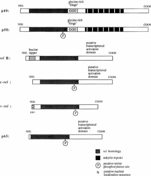
The recent molecular cloning of cDNAs encoding p50, p65, and I-&-like molecules has substantially enhanced our overall understanding of this transcription regulatory system (Ghosh et al., 1990; Kieran et al., 1990; Bours et al., 1990, 1992; Nolan et al., 1991; Meyer et al., 1991; Ruben et al., 1991; Haskill etal., 1991; Davis et al., 1991; Inoue et al., 1992b). Most significantly, the binding subunits of NF-KB are members of a family of proteins that are related to the re1 oncogene (for an overview see Gilmore, 1990) (Figs. 4 and 5). These genes are related to the Drosophila dorsal
gene product as well as a recently identified Xenopus gene, demonstrating that the Re1 DNA binding protein structure arose early in evolution (Steward, 1987; Kao and Hopwood, 1991). Molecular cloning studies have now identified a number of Re1 family members including p49 or p50B, p50, p65, relB, and c-ref (Schmid et al., 1991; Ghosh et al., 1990; Nolan
et al., 1991; Kieran et al., 1990; Meyer et al., 1991; Bours et al., 1990, 1992; Ryseck et al., 1992; Wilhelmsen et al., 1984). Significantly, proteins that have an I-KB-like inhibitory activity for NF-KB share a structural domain known as the “cell cycle” or “ankyrin” repeat. The binding subunits of the Re1 family can form both homodimeric and heterodimeric complexes that differ in both their binding and their transcriptional proper- ties. Heterodimeric regulatory systems allow for a greater combinatorial array of distinct gene regulatory molecules (Murre et al., 1989; Bohmann
et al., 1988). Also, the Re1 protein sequence does not match previously
26 p49: p50: re1 B : c-re1 : v-re1 :
MARIAGRAZIA GRILL1 ET AL. glycme-nch COOH N H Z "hinge"
1
- o w1 L I I I I I - 1
I
--Gr- gl ycine-rich CMlH MI2 "hinge" leucine NHZ zipper putative transcriptional activation domain COOH NH2 putative transcriptional activation domain COOH NH26
COOH(3
env N H Z putative transcriptional activation domain COOH p65: rel homology dyrinrepeats putative serine@
phosphorylation site localization sequence N putative nuclearFIG. 4 Shown schematically are the primary structures of the molecularly defined members of the Re1 gene family. Only two (pS0 and p49/pSOB) have bipartite structures that include Re1 and ankyrin repeat domains.
ture that governs the interactions among Re1 subunits, the ankyrin repeat proteins, and DNA will be of great interest. Finally, it is notable that a chromosomal translocation juxtaposing the gene encoding p49/pSOB, called lyt-10, with the immunoglobulin Ca-1 locus has been described in
apatient with B-cell lymphoma (Neri et a / . , 1991). The fusion gene created by the translocation encodes a re1 DNA binding domain lacking the ankyrin