Peripheral airway stenting: it is worth the effort?
A clinical experience.
INDICE
Abstract... ... 3
Introduction...5
Materials and methods...7
Results...11
Discussion...15
Table and Figure...19
Peripheral airway stenting: it is worth the effort? A clinical experience.
Abstract
Background: At the time of diagnosis or in the follow up, the patients with lung cancer often
present locally advanced disease involving peripheral airway. Although they are pauci-symptomatics, they are at risk of disease progression and complication seriously interfering with the administration of additional treatments and worsening of performance status. While the role of endobronchial interventions is clear in the treatment of central airway obstruction, there are few data regarding the feasibility and the efficacy of the stenting in the peripheral airway disorders.
Objectives: To report and analyze our experience in the management of airway disorders involving
lobar bronchi and secondary carina.
Methods: We retrospectively reviewed medical records of patients underwent placement of airway
stents due to a stenosis below the main carina at the Interventional Pulmonology Unit, La Maddalena Cancer Center, (Palermo, Italy), between November 2008 and October 2013.
Results: Stents were placed in 52 patients with malignant (n=49) and benign airway obstruction
(n=1), broncho-esophageal (n=1) and broncho-mediastinal fistula (n=1). Stents were inserted in the left lower lobar bronchus (n=33), in the left upper lobar bronchus (n=1), in the right lower lobar bronchus (n=1) and on the secondary carina at right site (n=15) and at left site (n=2). Both self-expandable metallic stents (n=26) and silicone stents (n=26) were used. All patients were symptomatic with dyspnoea (n=27, 50%) of moderate degree (MMRC 2.4±1.1) and cough (n=22, 41%). Besides one procedural dislocation, the deployment was successful in all patients without procedure related complications with immediate significant improvement of symptoms (MRC p<0,001) and objective radiographic improvement (60%). The mean follow-up duration was 123 days±157. Complications observed were stent migration (9.6%), tumour overgrowth (9.6%), infections (26%), granulation tissue formation (7.6%) and obstruction due to tenacious secretions (1.9%). The overall 3-month and 6-moth survival were 63% and 40%. Factors significantly
influencing survival using Kaplan-Meyer long-rank analysis were the availability of post-procedural adjuvant treatment (p<0,0001).
Conclusions: The stenting of peripheral airways is technically feasible, effective and acceptable
safe. We believe that the proactive stenting in these patients could alter the natural history of the disease and improve survival .
Introduction
Given the incurable nature of lung cancer, it is considered a terminal illness with a five-year survival rate of approximately 16%1 . Furthermore untreated lung cancer patients live on average
for 7,15 months2. Although most patients succumb to systemic metastatic disease, a significant
number suffer from symptoms of locally advanced malignancy.
Over 20% to 30% of patients with lung cancer will develop symptomatic central airway obstruction. The survival of patients with untreated malignant central airway obstruction is very poor and ranges from 1 to 2 months4,5.The quality of life of these patients is also extremely poor, with a significant
number dying of asphyxia in palliative care facilities or on mechanical ventilatory support. The cost associated with providing end of life care to these patients, especially those in an acute care setting, is substantial6.
The patients with central airway obstruction are an heterogeneous group of subjects3.
It involves both the patients with critical airway obstruction (greater than 90% area reduction of the trachea or main bronchus), in life-threatening conditions with respiratory failure needing ventilatory support and the subgroup of patients with significant airway narrowing (greater than 75% area reduction of the trachea or main bronchus). These last are pauci-symptomatics with symptoms ranging from exertional shortness of breath to severe shortness of breath at rest, stridor, atelectasis of a lobe or entire lung, postobstructve pneumoniae and they have an intermediate performance status.
Central airway obstruction develops secondary to endoluminal disease, external compression by a mediastinal or hilar tumor, bulky lymphadenopathy, or a combination of endoluminal and extrinsic disease. There are a variety of treatments options that can restore airway patency including external beam radiation and endobronchial interventions such as brachytherapy, laser therapy, photodynamic therapy, cryotherapy and stents.
Airway stenting has become a strengthened bronchoscopic procedure for the treatment of tracheobronchial disorders, that it has been used for almost 90 years8.
Several studies have proved stents’ effectiveness as both palliative and curative treatment of lung cancer, in fact they lead to a significant improvement not only in symptoms and in lung function9,10,11 but also in quality of life11,12,13 and survival, if it is timely carried out6,14 in the subgroup
of patients with not critical airway central obstruction.
In fact, if the stenting of the airway is performed timely, it promotes the prevention or delay of the lethal complications of malignant central airway obstruction such as postobstructive pneumonia, sepsis and respiratory failure, consequently the improvement in performance status can develop6.
Furthermore, post-stenting improvement in performance status allows the patients to be considered for adjuvant systemic chemotherapy and (or) external beam radiation and surgical therapy18,17 and as
a result it improves the survival6.
Moreover, the most important prognostic factors for the effectiveness of brochoscopic intervention in advanced lung or esophageal cancer are the available post-procedural additional treatment together with the treatment-naïve status and an intact proximal airway14 (any acute infection is a
contraindication to administer chemotherapy and is always also an exclusion criteria in clinical trials15,16).
In our clinical practice, we often have to deal with patients that, at the time of diagnosis or in the follow up, present locally advanced disease involving lobar bronchi and secondary carina and that are nonsurgical candidates.
We have found few data in the literature regarding the incidence, the prognosis and the management of this kind of patients. The last ACCP lung cancer guidelines has hinted at the possibility that distal obstruction lent itself to radiotherapy approaches31.
Most of the experience reported in the literature about endoscopic interventions has concerned the management of central airway disorders including the trachea, the right and the left main stem bronchus and the bronchus intermedius. In few cases, self expandable metallic stents have been use for stenting of the left lobar bronchus for the treatment of a stenosis due to a compression of a tumor of the left upper lobe19,20 without complications. Recently, Oki has introduced a new
bifurcated silicone stent dedicated for the treatment of the stenosis around the primary carina, showing, in cases series of 10 patients, that its placement is feasible, effective and acceptably safe26.
Although it could be thought that the stenting of the distal airways is technically more complicated than the stenting of the proximal airways and it might cause a higher complication rate, these few reported experience showed encouraging results19,20,26.
As the patients with distal airway obstruction are not critically ill patients, we have hypothesized that they could be equalled to the patients with pauci-symptomatic central airway obstruction and therefore they could benefit from an early stenting.
In this retrospective study, we analyzed our experience in the stenting of the peripheral airways including secondary carina and lobar bronchus and then we compared our results with the data reported in the literature about the stenting of the proximal airways obstruction.
Materials and methods
We retrospectively reviewed all patients referred to the Interventional Pneumology Unit (La Maddalena Cancer Center, Palermo, Italy) between November 2008 and October 2013, submitted to the insertion of airway stent due to malignant or benign stenosis below the main carina involving peripherals lobar bronchi and peripherals carina,
We considered peripherals lobar bronchi: the left upper lobe bronchus (LUL), the left lower lobe bronchus (LLL) and the right lower lobe bronchus (RLL). Peripherals carina were: the one between the bronchus to the right upper lobe (RUL) and the bronchus intermedius (BI) (primary right carina RC1), the other one between the bronchus to the right middle lobe (RML) and the RLL (secondary right carina RC2) and the other still between the bronchus to the LUL and the LLL (secondary right carina LC2).
Before the airway stent placement, flexible bronchoscopy was performed (model 1T-180; Olympus America Inc; Melville, NY) under local anaesthesia to evaluate airway anatomy and to plan the best approach for treatment. Preliminary CT-scan was also investigated to evaluate the presence of
sparing lung parenchyma distal to the stenosis.
Metallic and silicone stents were used. The more metallics stents were the fully covered self-expandable metallic stent Silmet® by Novathec and the covered Ultraflex® by Boston Scientific and in few cases the Palmaz by Corning/Johnson & Johnson stent were placed.
Between silicone stents we used the Dumon stents, both straight and Y-shape and the Oki stent by Novatech.
All stents were placed by rigid bronchoscopy (Dumon-Harrell type; Bryan Corp; Woburn, MA) under general anesthesia and jet ventilation. Previously, the airway lumen was re-established combining mechanical debulkig, balloon dilatation and laser (λ 980 nm Ceralas D50/980/600; Biolitech; Siemensstrasse Bonn; Germany) and then the stent was inserted, in case of best patency of distal bronchi was achieved. The diameter and length of the stenotic airway were measured using the rigid aspirator or flexible bronchoscope.
If silicone Oki stent was judged appropriated for insertion, it was customized on site according to measurements taken. Generally the limb of the stent for the RUL or LUL and the RML was cut shorter than the limb for the BI and LLL. In the stenosis around RC2 the main limb was cut skim to the opening for the RUL. For treatment of stenosis around RC1,RC2 and LC2 firstly both limbs of the stent were inserted respectively into BI and into LLL and then the stent is grasped with the rigid forceps and slowly pulled back until a limb slips into the target bronchus. Finally, the stent is pushed to fit on the bifurcation.
If the lesion could not be maintained with only one stent (eg, extensive stenosis including main carina, right main stem bronchus and secondary carina), Double Y-stenting was performed as described before21,22. Metallic stent were inserted without fluoroscopic guidance under direct vision
by passing the delivery device through the rigid bronchoscope as described before23.
In the delivery catheter of Silmet stent the proximal tip of the stent is market by blue ring. This device was advanced inside the stenotic area with the blue ring skimming the proximal end of the stenosis, than stent is slowly pushed out. Finally is grasped with the rigid forceps and slowly pulled
back until cover the stenosis over 5 mm as recommended3.
After the procedure the patients were moved to a high dependency unit for 1-2 hours and than transferred to ordinary ward area.
After stenting, to prevent retention of secretions, the patients with metallic stents were instructed to perform domiciliary inhalation (beclomethasone 0.8 mg plus saline solution 2 ml plus ipatropium 937.5 mcg and salbutamol 187 mcg) with a simple inhaler three-times a day and the patients with silicone stents were instructed to perform aerosol therapy trough T-PEP for 15 minute and three-times a day.
The temporary positive expiratory pressure device (UNIKO T-PEP) is a new modality to mechanically deliver a low positive expiratory pressure level at the mouth during spontaneous breathing. This technique produces a 1 cmH2O increase in airway pressure along the respiratory
cycle until immediately before the end of expiration. This increase in low pressure was created through a pulsatile flow approximately 42 Hz in frequency. The mouthpiece design incorporates a one-way valve so that there is little or no escape of medication into the atmosphere, ensuring that the patient receives the optimum supply.
TPEP works by detaching and removing secretions from the peripheral airways, and can increase the deposition of aerosolised drugs deeper into the lungs by ~30%, compared with more traditional systems24.
The gathered data included: patient demographics, clinical presentation, diagnosis, the stage of the cancer, number of comorbilities, indications for stent, characteristic of stenosis, type, size and position of stent, duration of procedure, endoscopic intervention used other than stenting (mechanical debulking and/or laser, balloon dilatation), complications, stent related treatment with temporary positive expiratory pressure (T-PEP) and/or simple inhaler.
Symptoms related to the clinical presentation of airway obstruction were grouped in dyspnoea, cough, haemoptysis, occurrence of obstructive infection.
(ECOG) performance status measure (0-5) score, the Barthel Index21 measurement of activity
limitations (0-100) were used to evaluate quality of life.
Additionally any anti-cancer treatment before and after intervention were investigated.
The ASA (American Society of Anesthesiologists) physical status classification system was used for preoperative risk assessment.
According to the protocol of our department, patient’s follow up program consisted of 4 complete visits (V1 at 24 hours after stent placement; V2 at 1 month after procedure; V3 at 3 months after procedure; V4 at 6 months after procedure) during which pulmonary function, Barthel Index and Medical Research Council (MRC) dyspnea scale were measured to assess patients’ clinical status and improvement. During every visit, flexible bronchoscopy was also performed under local anaesthesia, to evaluate stent position and potential complications such as migration, granulation tissue formation at the edges of stent, stent fracture and secretion plugs.
Chest radiography was usually performed the day before and after the procedure to check for complication and to highlight a radiographic improvement in terms of resolution of atelectasis/infilitrates or presence of mediastinal shift.
Complications were grouped into the following categories: procedure-related respiratory failure, implantation-related complication, development of endobronchial granuloma, infection, haemoptysis, chronic cough, migration, tumor ingrowths or/and outgrowths, mucostasis. And infections.
The amount of secretion plugs were classified using a four points endoscopic score (0-no secretions; 1-moderate amount of secretions easily removable by suction; 2-severe amount of secretions removable using a biopsy forceps, mucolytic agent instillations or other devices in addition to the suction; 3-complete stent obstruction or deposit of thick and non-removable secretions).
The overall complication rates was calculated by dividing the total number of complications by the total number of follow-up months of stent use by all patients.
post-stenting chest radiography and beginning candidate to adjuvant chemotherapy or radiotherapy. Qualitative and quantitative variables are presented as count, percentage (%) and mean (± standard deviation SD) respectively. Absolute values in each outcome variable were compared by Student t test. Wilcoxon and Friedman test were applied for non-parametric variable.
Time zero for the survival analysis was defined as the date of the rigid bronchoscopy. Patients were followed until death or the last documented follow-up, if patients were still alive.
The duration of overall survival was analyzed using Kaplan-Meier method. Differences in overall survival were assessed using the long-rank test and the relation with different variables with regression model. All tests were two-sides, and p value less than 0.05 was deemed to indicate statistical significance. Data were analyzed using Statview software®.
Results
We enrolled a total of 52 patients (mean age 67 yr, range 45-85 years, male 41) who underwent stent placement in the peripheral airways for the treatment of the malignant (n°51) and benign (n°1) disorders. The baseline clinical characteristics are summarizes in table 1.
All patients were not candidate to surgery with a TNM stage between IIIB and IV and were symptomatics. The majority of them complained dyspnea (n=27, 50%) of moderate degree (MRC 2.4±1.1) and cough (n=22, 41%).
Except for two cases (Non Hodgkin Lymphoma with obstruction on both left and main bronchus, involvement of main carina and the RC1; undifferentiated carcinoma of the mediastinum with tracheobroncho-oesophageal fistula and the involvement of RC1) there were not critically ill patients.
Oxygen saturation was 95±2, performance status score was 1.8±0.7, Barthel Index 85±10 and number of comorbilities 2.3±1.4.
The preoperative risk assessed with ASA score was high for all 3±0.5.
the patients.
Overall 36 (69%) patients were treatment-naïve status upon bronchoscopic intervention while 16 patients had previously undergone various therapeutic modalities (n°12 chemotherapy, n°1 surgery and n° whole brain radiotherapy; n°3 radio-chemotherapy).
The main cause of airway disorders was primary lung cancer (n° 10 SCLC, n° 19 NSCLC squamous and n°15 NSCL). In five cases the diagnosis was metastatic cancer (n° 2 colon, n°1 kidney and n° 2 laryngeal cancer), one patient had an Hemangiopericytoma and one was affected by Non Hodgkin’s Lymphoma.
The stents were placed to treat intrinsic obstruction (n=6), extrinsic compression (n=13), combination of endoluminal and extrinsic disease (n=29) and to seal a broncho-esophageal (n=1) and a broncho-mediastinal fistula (n=1).
In two patients with malignant post-obstructive abscess of the left lower lobe, stents were placed such as support drainage system with cleaning, sterilization and recovery of the residual lung. In the study there was only one benign stenosis secondary to left post-pneumectomy syndrome (it was the torsion of right upper bronchus and intermedium bronchus due to the shift of the mediastinum secondary to the left pneumectomy for mesotelioma)
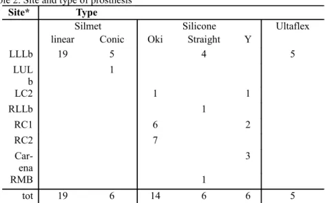
Site and type of prosthesis are summarized in table 2 and size in table 3.
In six patients a double Y-stenting was performed with one prothesis in the central airways and one in the peripheral airways displaced in telescopic fashion:
-in four patients a Dumon Y stent was placed on the main carina plus an Oki stent on the RC1 in two patients and on the RC2 in the other two;
-in one patients an Oki stent was placed on the RC2 plus a straight Dumon into the RMB in order to cover the upper lobar bronchus completely replaced by the tumour;
-in the last one an Oki stent was positioned on the LC2 with a Silmet stent inside the limb for the left lower lobar bronchus to reach the basal pyramid.
sub-segmentary bronchus of right lower lobe above the stenosis during stenting procedure, with no possibility of recovery; the patient was still alive at the 4th month of follow up and he had performed the first few cycle of chemotherapy with no stent-related complications recorded.
No other procedure related complications (such as death, stent rupture, severe bleeding, prolonged hypoxia, airway perforation, pneumothorax) were recorded.
Symptoms improved significantly with a significant difference in MRC dyspnea score between V0 (mean 2.6±0.8) and V1 (mean 1.2±0.5; p<0,001). There were not statistical significant difference in oxygen saturation (p<0,08) and barthel index (p<0,07) between V0 and V1 (fig 1).
In 60% (29/51) of patients radiographic improvement was demonstrated by changing in computed tomography or chest radiography (fig. 2).
The mean duration of the procedure (including mechanical debulking and stenting), was 60 minute±27 (range 20 to 140; mediana 57) and laser assisted mechanical dilatation was used in 36% of cases. Patients were discharged 2 days ±3 after the procedure.
Modalities and outcomes of bronchoscopic intervention are summarized in table 4.
Early complication (<24h) was recorded in five patients (10%). Three patients need mechanical ventilation in the post-operatory for less than 8 hours, two experienced atrial fibrillation treated with pharmacological cardioversion with success. One patient who underwent thoracoscopy and rigid bronchoscopy with stenting in the same section developed a pneumonia ab ingestis.
The mean follow-up duration was 123 days ± 157 (range 15 to 653 dys; mediana 59 dys ).
26 patients (50%), could be evaluated up to the 60th , with a drop out of 50% secondary to the fatal
evolution of their malignant disease.
The overall complication rate of stent placement was 15%.
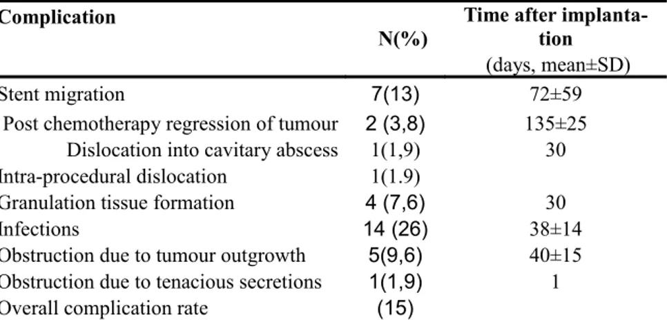
The complication rate and time to detect complications after stent placement are summarized in table 5.
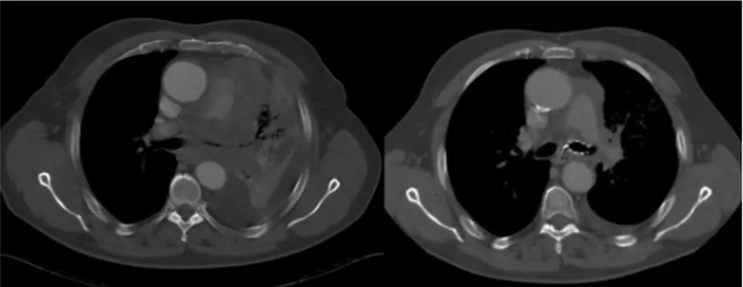
Stent migration was observed in 13% of cases (1% silicone stent; 11% SEMS). In two cases, the migration occurred after chemotherapy due to a significant reduction in tumor volume (fig 4, fig 5).
One stent, placed as drainage system at the top of post-obstructive pneumonia abscess in the LLLb, migrated into the cavity after 1 week. The patient underwent a subsequent rigid bronchoscopy. Removal of stent was possible without complication and with the persistent patency of the left lobar bronchus.
Four patients developed a small granuloma (7.6%) that did not require laser treatment.
Stents’ obstruction due to tenacious secretions was recorded in only one patient and was easily resolved with endoscopic toilette. On average, the endoscopic secretion score was 0.3±0.6 at V0, 0.8±0.9 at V1, 1.1±0.9 at V2, 1.3±1 at V3 and 1±0 at V4. A statistically significant difference was observed between the V0 and V1,V2,V3,V4 (tied p value<0.001 ). The endoscopic score was similar between V1,2,V3 and V4 (tied p value>0.05 ) (fig 5).
Bronchoaspirate was performed in 26 patients and microbiological cultures were positive in 14 out 26 ( 22% at 1 day , 38% at 30 days and 46% at 90 days). Of these 14 patients, 3 performed T-PEP therapy and 11 performed nebulizer therapy alone.
Clinical or radiological signs of infection were presents in about 7 out of the 14 patients (50%) and required targeted antibiotic therapy.
The pathogens isolated on bronchoaspirate were: Pseudomonas (n=3), Staphilococcus (n=6), Candida (n=6), Sfingomonas paucimobilis (n=1), Enterobacteriacoe (n=3), Haemophilus (n=1), Streptococcus (n=1), Alcaligenes xylosoxidans (n=1), Hafnia (n=1)
It was possible to remove stents in six patients (15%) (n=4 regression of tumor; n=1 infection; n=1 thermoterapy) after an average of 307 days ±117 (range 118-653 days) without complications, significant bleeding or stent rupture.
Stents were replaced in case of recurrence (n=1) after bronchoscopic debulking.
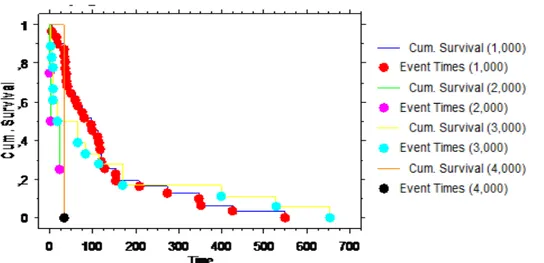
Using the Kaplan Meier method, the overall mean survival was 118±20days. The overall 3-month and 6-moth survival were 63% and 40%. Longer survival was observed in patients who received additional treatment after airway stenting, compared with those who did not (152 days±76 vs 21 days±9; p<0.03). (fig 6)
The survival of patients whit a double stents was worse than patients with stents in LC2, LUL, RLL, while patients with a stent in LLL and RC1 had the better survival (p<0.02)
Discussion
This study represents a single referral institution experience in the peripheral airway stenting.
We have observed that the patients with distal airway obstruction are usually pauci-symptomatic with chronic cough or mild dyspnoea or recurrent infection, they have a well lung function, a fair performance status and independence daily activity. Airway obstruction is often detected at the diagnosis, while post obstructive abscess develops after anti-cancer treatment.
In this study we are able to demonstrate, in a relatively large number of patients, that the stenting of distal airways is technically feasible, effective and acceptably safe.
Previously, Breitenbücher et al9 in a study on the management of complex malignant airway
stenosis placed the Ultraflex stents in the lobar bronchi of 7 patients. They reported that survival after distal stent placement was longer than after proximal stent. Moreover they recommended stent placement in distal airways only in cases of extrinsic compression and not in those with endoluminal growth27, because the short Ultraflex stents were not covered.
Unlike Breitenbücher et al9 we were able to treat with success stenosis secondary not only to
extrinsic compression but also intrinsic obstruction and the complex one.
In fact, in the tight and short stenosis, we implanted the Silmet® stents, that are fully covered in all the available lengths (from 20 to 60 m, in increments of 10 mm), eluding possible tumor ingrowths. While in the presence of complex stenosis involving multiple lobar bronchi and the secondary carina we usually inserted the silicone Oki stent with success, confirming the encouraging results of previous reports26.
We adopted the technique of double Y-stenting described by Oki to seal both broncho-esophageal and mediastinal fistula involving both the proximal and the distal airways21,22.
At least we used the stents to drain a post-obstructive abscess of left lower lobe obtaining the negativity of microbiological culture and the absence of respiratory symptoms at the 2th month of follow up.
Peripheral stenting was well tolerated with almost 70% of the patients experiencing no significant complication after a mean follow up of 4.1 months.
The rate of stent migration at 30 days, not related to the tumor regression, was 9.6% and it was similar to that reported for the other SEMS such the Ultraflex (4.7-7.6%) 6,12,18,27-29 and the Wallsten
stents (12%)28,12 and for the silicone Dumon stents (9.5%)20,30.
Overall granulation incidence (7.6%) was within the limit of range previously reported in the literature both with the SEMS (2.9 to 15.2%) 6,12, 18,27-29 and the Dumon stents (7.8%)20,30, furthermore
laser treatment was not necessary in our patients.
A possible explanation for such a low rate is that the 89% of our patients underwent post-procedural treatment such as chemo and radiotherapy. The immunosuppressive effect of these therapies could have reduced the inflammatory response and as consequence the tissue granulation formation too. Obstruction due to tenacious secretions occurred in 1.9% of cases less than reported in the literature with the SEMS (9 to 38%)27,29,12 and the Dumon stents (3.6%)20.
It is well known that stents impair mucus clearance, thus accumulation of secretions can be considered a slightly annoying problem or a severe complication.
We introduced, in our practice, an endoscopic score of four points to quantifies the amount of accumulated secretions at different visits. We believe this score is less operator dependent because is based on the method used to clean the stent, rather than on the endoscopic appearance of accumulated secretions.
It is interesting that the increased amount of secretion, measured by the endoscopic score was observed from visit 0 to visit 1 but not during the following assessments. A stent, as a foreign body, itself enhances mucus secretion and impairs mucociliary clearance but the daily treatments with aerosol therapy can enhance the drainage of secretions.
The rate of colonization observed in our patients was 26%, whereas the evidence of infections was founded only in 13% of patients and these data are in accord with the literature28-29.
In our series, none of the patients had tumor ingrowth, while it is reported as a quite common complication with the Ultraflex (5 to 21%) and the Wallsten (10%) stents. This could be probably related to the better bio-compatibility of the Silmet stents, due to the mesh silicon cover and polyester external layer down to the edge of the device, compare to the other covered SEMS that have both their tips uncovered.
The rate of tumor outgrowth was 9.6% within the limit of that reported for SEMS ranging from 5% to 20%19,30.
The mean survival after stent placement was 4.4 moth, similar to that reported in the management of malignant central airway obstruction with the SEMS (range from 4.2 to 5.3) 6,14,19 and the silicone
stents (range from 3.4 to 4) 19,26,30 .
As suggested by previous reports14, patients with double stenting involving the main carina have the
worst prognosis.
We observed that, in patients with distal airways obstruction the preoperative ECOG performance status score and dyspnoea MRC scale score did not influence survival. Taking into account that our patients affected by distal airways obstruction had a mean ECOG score of 3, our results are in accord to the previous studies where the intermediate performance group (ECOG<3;MRC<4) of patients with central airway obstruction was associated with a better survival 6,14.
As suggested by previous reports14, the main factor, that significantly influences survival, is the
availability of post-procedural adjuvant treatment. That is strictly linked to the preserved performance status, the absence of acute infection and the absence of the respiratory failure, that are the lethal complications of the evolving malignant airway obstruction.
We can conclude that patients with distal airway obstruction should be assimilated to the patients with pauci-symptomatic central airway obstruction; like they are at risk of disease progression, complication and worsening of performance status, therefore they can benefit from an early
stenting.
We suggest an early diagnosis of distal airways obstruction. It depends on a multidisciplinary approach to the oncologic patients in which the interventional pulmonologist, radiologist and oncologist work together to achieve the best effective approach.
The current study has several limitations. One limitations is that its team has extensive practical experience in performing stenting procedures. All stenting procedures were performed by one of the current authors with 10 years of experience in stenting procedures, who was familiar with SEMS insertion and Y stent placement on the main carina and/or the primary right carina. The endoscopic management of distal airway stenosis and the placement of the Oki stent, needed a certain amount of experience and skill. A multicentre trial is necessary to evaluate the feasibility of peripheral stenting.
Another limitation is that we have compared the results of our study on the stenting of distal airway with the data on the stenting of the proximal airway reported in the literature. A controlled, prospective studies comparing distal to proximal airway stenting could be amenable to better evaluate the effectiveness of stenting and to better describe this group of patients. More studies are necessary to evaluate its utility in benign stenosis.
Table 1: Baseline characteristics of enrolled patients (n=52)
Variables mean±SD or no (%)
Age mean±SD 67±10
Gender,male No(%) 41 (78)
BMI 24±4
Smoking status before/after stenting
Smoker 27/11 Ex-smoker 15/31 Never smoker 10/10 Oxigen saturation 95±2 Etiology No(%) Benign 1(1,9) Malignant 51 (98) Lung cancer 44 (83) NSLC 19 (36) NSCLC(squamos) 15 (28) SCLC 10 (19) LHN 1(2,3) Metastasis 5(11) Hemangiopericitoma 1 (2,3) Stage IIIA/IIIB/IV 9(17)/13(25)/30(57) ECOG mean±SD 1,8±0,7 Comorbilities mean±SD 2,3±1,4 ASA mean±SD 3±0,5
Barthel Index mean±SD 85±10
Symptoms No(%)
Dyspnea/ MRC 27 (50)/2,4±0,7 Cough 22 (41) Hemoptysis 7 (13) Chest pain 5 (9) Treatment-naive status upon bronchoscopic
intervention 36 (69) Pre-procedural treatment 16 (30) Post-procedural treatment 40 (75) Chemotherapy 33(63) Radiotherapy 2(3) Surgery 4(7)
Table 2. Site and type of prosthesis
Site* Type
Silmet Silicone Ultaflex
linear Conic Oki Straight Y
LLLb 19 5 4 5 LUL b 1 LC2 1 1 RLLb 1 RC1 6 2 RC2 7 Car ena 3 RMB 1 tot 19 6 14 6 6 5
*RMB=right main bronchus; LLLb=Left lower lobar bronchus; LULb=left upper lobar bronchus;
LC2=left secondary carin; RLLb= right lower lobar bronchus; RC1= right primary carina; RC2=right secondary carina.
Table 3 Size of stent for site.
Site Lenght mm(mean±DS) Diameter mm(mean±DS)
RMB LMB RMLb R/L-ULb R/L-LLb T RMB LMB RMLb R/L-ULb R/L-LLb T LLLb 25 ± 8 10 ± 1 LULb 25 8 LC2 15 ± 10 9 ± 1,4 18 ± 4 12 ± 2 9,5 ± 0,7 10 RLLb 21 ± 1 11 ± 1 RC1 17 ± 6,4 7 ± 3,5 21 ± 7 13 ± 0,4 9,1 ± 0,4 10 ± 0 RC2 25 ± 8,8 8 ± 1,7 12 ± 5 9 ± 0 10 ± 0 Carena 29 ± 1,4 15 ± 0 35 ± 14 15 12 ± 0 15 ± 0 RMB 23 23 Mean±SD 24 ± 5,6 20 ± 5 8 ± 1,7 8 ± 2,5 19 ± 5 35 ± 14 14 ± 0,4 16 ± 1 9 ± 0 8,9 ± 0,5 10 ± 0 15 ± 0
Length are expressed in millimetre as mean± standard deviation
*RMB=right main bronchus; LLLb=Left lower lobar bronchus; LULb=left upper lobar bronchus; LC2=left secondary carin; RLLb= right lower lobar bronchus; RC1= right primary carina; RC2=right secondary carina; RULb=right upper lobar bronchus; T=trachea
340 360 380 400 420 440 460 480 V0 V1 Box Plot -4,356 44 -1,843 ,0720 Mean Diff. DF t-Value P-Value V0, V1 Paired t-tes t Hypothesize d Difference = 0 20 30 40 50 60 70 80 90 100 110 U ni ts V0 V1 Box Plot 4 3 -5,086 <,0001 -5,214 <,0001 # 0 Differences # Ties Z-Value P-Value Tied Z-Value Tied P-Value
Wilcoxon Signed Rank Test for Colum n 1, Colum n 1.2
2 38,000 19,000 39 823,000 21,103 Count Sum Ranks Mean Rank # Ranks < 0
# Ranks > 0
Wilcoxon Rank Info for Colum n 1, Colum n 1.2
-,5 0 ,5 1 1,5 2 2,5 3 3,5 4 4,5 Column 1 Column 1.2 Box Plot V0 V1 a) b) c) 340 360 380 400 420 440 460 480 V0 V1 Box Plot -4,356 44 -1,843 ,0720 Mean Diff. DF t-Value P-Value V0, V1 Paired t-tes t Hypothesize d Difference = 0 20 30 40 50 60 70 80 90 100 110 U ni ts V0 V1 Box Plot 4 3 -5,086 <,0001 -5,214 <,0001 # 0 Differences # Ties Z-Value P-Value Tied Z-Value Tied P-Value
Wilcoxon Signed Rank Test for Colum n 1, Colum n 1.2
2 38,000 19,000 39 823,000 21,103 Count Sum Ranks Mean Rank # Ranks < 0
# Ranks > 0
Wilcoxon Rank Info for Colum n 1, Colum n 1.2
-,5 0 ,5 1 1,5 2 2,5 3 3,5 4 4,5 Column 1 Column 1.2 Box Plot V0 V1 a) b) c)
Figure 3. Chest radiography performed pre and after stenting showed maximal expansion and recovery of the residual lung
Table 4. Modalities and outcomes of bronchoscopic intervention
Variables No.(%) mean±SD
Type of stenosis Intrinsic obstruction 6 (11) Extrinsic obstruction 13 (25) Complex 29 (56) Distorsion 1(1) Broncho-oesophageal fistula 2(2) Duration procedure 60±27
Procedure other than stenting
Mechanical dilatation 31(59) Mechanical dilatation +Laser 19(36)
Successful palliation 50(99)
Complications after bronchoscopic in tervention
Atrial fibrillation 2
Non invasive ventilation 3
Pneumonia ab ingestis 1
Recovery days after stenting 2±3
Radiographics riespansion after stent
ing 27(51)
Mucostasis prevention
T-PEP 17
Nebhulizer 35
Table 5: Complication rates and time to detect complications after stent placement.
Complication
N(%)
Time after implanta tion
(days, mean±SD)
Stent migration 7(13) 72±59
Post chemotherapy regression of tumour 2 (3,8) 135±25
Dislocation into cavitary abscess 1(1,9) 30
Intra-procedural dislocation 1(1.9)
Granulation tissue formation 4 (7,6) 30
Infections 14 (26) 38±14
Obstruction due to tumour outgrowth 5(9,6) 40±15
Obstruction due to tenacious secretions 1(1,9) 1
Figure 4Significant reduction in tumor volume at CT scan pre and post chemotherapy
Figure 5 Loss of stents’ contact due to tumor of Silmet® stent placed into the left lower lobar bronchus after chemiotherapy. 3 4 8 6,600 ,0858 9,240 ,0263 DF # Groups # Ties Chi Square P-Value
Chi Square corrected for ties Tied P-Value
48 cases w ere omitted due to missing values. Friedm an Tes t for 4 Variables
7 14,000 2,000 7 19,000 2,714 7 24,000 3,429 7 13,000 1,857 Count Sum Ranks Mean Rank V1
V2 V3 V0
48 cases w ere omitted due to missing values. Friedm an Rank Info for 4 Variables
-,5 0 ,5 1 1,5 2 2,5 3 3,5 U ni ts V0 V1 V2 V3 Box Plot -1 -,5 0 ,5 1 1,5 2 2,5 3 3,5 U ni ts V0 V1 V2 5,000 4,000 3,000 2,000 1,000 Box Plot
Split By: 1=SILM ET;2=DUM ON;3=OKI;4=Y;5=M ETALLICA 3 4 8 6,600 ,0858 9,240 ,0263 DF # Groups # Ties Chi Square P-Value
Chi Square corrected for ties Tied P-Value
48 cases w ere omitted due to missing values. Friedm an Tes t for 4 Variables
7 14,000 2,000 7 19,000 2,714 7 24,000 3,429 7 13,000 1,857 Count Sum Ranks Mean Rank V1
V2 V3 V0
48 cases w ere omitted due to missing values. Friedm an Rank Info for 4 Variables
-,5 0 ,5 1 1,5 2 2,5 3 3,5 U ni ts V0 V1 V2 V3 Box Plot -1 -,5 0 ,5 1 1,5 2 2,5 3 3,5 U ni ts V0 V1 V2 5,000 4,000 3,000 2,000 1,000 Box Plot
Split By: 1=SILM ET;2=DUM ON;3=OKI;4=Y;5=M ETALLICA
Figure7 Survival curve using Kaplan-Meier estimates based on the post-broncoschopic additional treatment (p<0,0001) 0=no treatment, 1=chemotherapy, 2=radiotherapy, 3=chemo and radiotherapy.
Figure8 Survival curve using Kaplan-Meier estimates based on type of stent. 1=SEMS;2=straight Dumon; 3=Oky;4=Y-shape Dumon;
Figure9. Survival curve using Kaplan-Meier estimates based on site of stent
RMB=right main bronchus; LLL=Left lower lobar bronchus; LUL=left upper lobar bronchus; LC2=left secondary carin; RLL= right lower lobar bronchus; RC1= right primary carina; RC2=right secondary carina; CARENA= main carena
1. American Cancer Society: Cancer Facts & Figures 2012. Atlanta: American Cancer Society; 2012
2. Survival of patients with non-small cell lung cancer without treatment: a systematic review and meta-analysis. Systematic Reviews 2013 2:10
3. Ernst A, Feller-Kopman D, Becker HD, Mehta AC. Central airway obstruction. Am J Respir
Crit Care Med 2004;169:1278–97; 8. Smith RA, Glynn TJ. Epidemiology of lung cancer. Radiol Clin North Am 2000;38:453–70.
4. Walser EM, Robinson B, Raza SA, Ozkan OS, Ustuner E, Zwischenberger J. Clinical outcomes with airway stents for proximal versus distal malignant tracheobronchial obstructions. J Vasc Interv Radiol 2004;15:471–7.
5. Macha HN, Becker KO, Kemmer HP. Pattern of failure and survival in endobronchial laser
resection. A matched pair study. Chest 1994;105:1668 –72.
6. Connery Airway Obstruction Timely Airway Stenting Improves Survival in Patients With Malignant Central Ann Thorac Surg 2010;90:1088-1093
7. Outcome of Treated Advanced Nonsmall Cell Lung Cancer With and Without Central Airway Obstruction Prashant N. Chhajed, MD, CHEST 2006; 130:1803–1807
8. Freitag L. Tracheobronchial stens in Interventional bronchoscopy, Bolliger, Karger 2000 pp 171-186
9. Vergnon JM, Costes F, Bayon MC, Emonot A. Efficacy of tracheal and bronchial stent placement on respiratory functional tests. Chest 1995; 107: 741–746.
10. Wood DE, Liu YH, Vallières E, Karmy-Jones R, Mulligan MS. Airway stenting for malignant and benign tracheobronchial stenosis. Ann Thorac Surg 2003;76:167–72.
11. P. Leland Oviatt, MD, David R. Stather, MD, Gae¨tane Michaud, MD, Paul MacEachern, MD, and Alain Tremblay, MDCM Exercise Capacity, Lung Function, and Quality of Life After Interventional Bronchoscopy . J Thorac Oncol. 2011;6: 38–42.
12. Amjadi K, Voduc N, Cruysberghs Y, Lemmens R, Fergusson DA, Doucette S, Noppen M. P. Impact of interventional bronchoscopy on quality of life in malignant airway obstruction. Respiration. 2008;76(4):421-8.
13. Anton Vonk-Noordegraaf, MD, PhD; Pieter E. Postmus, MD, PhD, FCCP; and Tom G. Sutedja, MD, PhD, FCCP Tracheobronchial Stenting in the Terminal Care of Cancer Patients With Central Airways Obstruction CHEST 2001; 120:1811–1814
14. Jae-Uk Song et all. Prognostic factor for bronchoscopic intervention in advanced lung cancer or oesophageal cancer patients with malignant airway obstruction. Annals of thoracic medicine 8;2;2013 86-92
15. Van Meerbeeck JP, Gaafar R, Manegold C, et al. Randomized phase III study of cisplatin
with or without raltitrexed in patients with malignant pleural mesothelioma: an intergroup study of the European Organisation for Research and Treatment of Cancer Lung Cancer Group and the National Cancer Institute of Canada. J Clin Oncol 2005; 23:6881–6889
16. Thomas P, Robinet G, Gouva S, et al. Randomized multicentric phase II study of carboplatin/gemcitabine and cisplatin/ vinorelbine in advanced non-small cell lung cancer GFPC 99–01 study (Groupe francais de pneumo-cancerologie)Lung Cancer 2005
17. Jin Hyoung Kim Palliative treatment of inoperable malignat tracheobtonchial obstruction: temporary stenting combined wit radiation therapy and/or chemotherapy. AJR 2009;193:
W38-W42
18. Wallace MJ, Charnsangavej C, Ogawa K, Carrasco CH, Wright KC,McKenna R, McMurtrey M, Gianturco C. Tracheobronchial tree:expandable metallic stents used in experimental and clinical applications.Work in progress. Radiology 1986;158:309–312
19. Breitenbücher A, Chhajed PN, Brutsche MH, Mordasini C, Schilter D, Tamm M. Long-term
follow-up and survival after Ultraflex stent insertion in the management of complex malig nant airway stenoses. Respiration. 2008;75(4):443-9.
20.Mahoney FI, Barthel D. “Functional evaluation: the Barthel Index.” Maryland State Med Journal 1965;14:56-61. Used with permission
21. Oki M , Saka H , Kitagawa C , et al . Double Y-stent placement for tracheobronchial stenosis. Respiration 2010; 79( 3): 245- 249.
22. Oki M , Saka H Double Y-stenting for tracheobronchial stenosis . Eur Respir J . 2012; 40(6):
1483 - 1488
23. Miyazawa T, Yamakido M, Ikeda S, et al. Implantation of Ultraflex nitinol stents in malignant tracheobronchial stenoses Chest 2000; 118:959–965
24. Clini E. Positive expiratory pressure techniques in respiratory patients: old evidence
and new Insights. Breathe ;2009:6:2;153-159
25. Breitenbuucher A, CHHAJED Long term follow up and survival after ultraflex stent insertion in the management of complex malignant stenoses. Respiration 2008;75:443-449
Primary Right Carina CHEST 2013; 144(2):450–455
27.Chung FT, Lin SM, Chen HC, Chou CL, Yu CT, Liu CY, Wang CH, Lin HC, Huang CD, Kuo HP: Factors leading to tracheobronchial selfexpandable metallic stent fracture. J Thorac Cardiovasc Surg 2008, 136:1328-1335.
28. Rieger J, Hautmann H, Linsenmaier U, et al. Treatment of benign and malignant tracheobronchial obstruction with metal wire stents: experience with a balloon-expandable and a self-balloon-expandable stent type. Cardiovasc Intervent Radiol 2004;27:339–43.
29. Amjadi K, Voduc N, Cruysberghs Y, et al. Impact of interventional bronchoscopy on quality of life in malignant airway obstruction. Respiration 2008;76:421– 8.
30. Miyazawa T, Miyazu Y, Iwamoto Y, et al. Stenting at the flow-limiting segment in tracheobronchial stenosis due to lung cancer. Am J Respir Crit Care Med
2004;169:1096 –102
31. Michael J. Simoff at al. Symptom Management in Patients With Lung Cancer Diagnosis and Management of Lung Cancer, 3rd ed: American College of Chest Physicians


