Review Article
(Cumulative Stress): The Effects of Maternal and
Neonatal Oxidative Stress and Oxidative Stress-Inducible Genes
on Programming of Atopy
Sara Manti,
1Lucia Marseglia,
2Gabriella D’Angelo,
2Caterina Cuppari,
1Erika Cusumano,
2Teresa Arrigo,
1Eloisa Gitto,
2and Carmelo Salpietro
11Unit of Pediatric Genetics and Immunology, Department of Pediatrics, University of Messina, 98125 Messina, Italy
2Neonatal and Pediatric Intensive Care Unit, Department of Pediatrics, University of Messina, 98125 Messina, Italy
Correspondence should be addressed to Sara Manti; [email protected] Received 25 January 2016; Revised 27 May 2016; Accepted 22 June 2016 Academic Editor: Daniela Giustarini
Copyright © 2016 Sara Manti et al. This is an open access article distributed under the Creative Commons Attribution License, which permits unrestricted use, distribution, and reproduction in any medium, provided the original work is properly cited. Although extensive epidemiological and laboratory studies have been performed to identify the environmental and immunological causes of atopy, genetic predisposition seems to be the biggest risk factor for allergic diseases. The onset of atopic diseases may be the result of heritable changes of gene expression, without any alteration in DNA sequences occurring in response to early environmental stimuli. Findings suggest that the establishment of a peculiar epigenetic pattern may also be generated by oxidative stress (OS) and perpetuated by the activation of OS-related genes. Analyzing the role of maternal and neonatal oxidative stress and oxidative stress-inducible genes, the purpose of this review was to summarize what is known about the relationship between maternal and neonatal OS-related genes and the development of atopic diseases.
1. Introduction
Allergic diseases including atopic dermatitis, allergic rhinitis, and asthma are some of the most common chronic diseases in the world [1]. Although extensive epidemiological and lab-oratory studies have been performed to identify the environ-mental and immunological causes of atopy, genetic predispo-sition seems to be the biggest risk factor for allergic diseases [2, 3]. It is known that several foetal adaptive responses to environmental factors are mediated by epigenetic changes which, impacting early-life morbidity, may exercise effects on the immune system, lung development, airway remodelling, allergen predisposition, and atopic and nonatopic inflam-mation, through numerous pathways [2, 4]. In particular, the onset of atopic diseases may be the result of heritable changes of gene expression, without any alteration in DNA sequences, occurring in response to early (prenatal) or later (perinatal) environmental stimuli [4]. Findings suggest that the establishment of a peculiar epigenetic pattern may also be generated by oxidative stress (OS) and perpetuated by the
activation of OS-related genes [5]. Reactive oxygen species (ROS), known to be important cell-signalling molecules [6], could, in fact, set up a positive-feedback loop that induces and perpetuates atopic injury. OS, also influencing T-cell signal transduction and gene expression [7], modulates T-cell polarization toward a T helper- (Th-) 2 T-cellular subset [8] which might be, in turn, a further source of ROS.
OS is a specific setting also occurring in normal events such as pregnancy and birth.
Pregnancy is a physiological period associated with enhanced OS related to high metabolic turnover and elevated tissue oxygen requirements [9]. During pregnancy, increased oxygen demand augments the rate of production of ROS, and women, even during normal pregnancies, experience elevated serum OS levels [9]. Increased OS levels and reduced antioxidative capacities may contribute to the pathogenesis of perinatal [10, 11] and postnatal disorders [12, 13], such as atopic diseases [14, 15], as newborns are more prone to OS than individuals later in life [16]. Moreover, it has also been reported that OS-related maternal genetics, independently of Volume 2016, Article ID 8651820, 7 pages
transmission of specific alleles, may influence a child’s atopic risk beginning in the uterus [17, 18].
Also during pregnancy, newborns are also continually exposed to elevated levels of ROS. At birth, newborns transit from a hypoxic intrauterine to a normoxic extrauterine environment. This increased OS further favours neonatal morbidity, also including atopy [14].
Analyzing the role of maternal and neonatal oxidative stress and oxidative stress-inducible genes, the purpose of this review was to summarize what is known about the relationship between maternal and neonatal OS-related genes and the development of atopic diseases.
2. Cumulative Effects of Maternal
and Neonatal Oxidative Stress on
the Immune System
It is well known that OS occurs early in pregnancy and con-tinues in the postnatal period [12]. In particular, pregnancy is associated with enhanced OS related to high metabolic turnover and elevated tissue oxygen requirements [34]. On the other hand, newborns, exhibiting an accelerated pro-duction of free radicals and limited antioxidant protection, are also constitutively vulnerable to OS. Therefore, during pregnancy and intrauterine life, many factors such as hypoxia, inflammation, and infections can easily induce overproduc-tion of free radicals (FRs) [11], exceeding the capacity of defensive mechanisms to neutralize them. The release of FRs leads to the oxidation of lipids, proteins, and polysaccharides and to DNA modifications [7–9, 19] which, in turn, increase the susceptibility of rapidly growing tissues to damage [35], as well as modulation of the immune system [10, 36].
With regard to the immune system, different immuno-logical responses to ROS production have been reported, depending on environmental oxidative status. While nor-mal ROS amounts have been shown to be important for T-cell function and for adequate, beneficial antimicrobial protection [37], high ROS concentrations can negatively modulate immune system responses leading to inhibited T-cell proliferation [37] and to hyporesponsivity to exogenous and/or endogenous activating stimuli [9]. In particular, OS plays a critical role as a secondary messenger in the initiation and amplification of signalling, miming antigenic effects. The antigen receptors are themselves OS-generating enzymes, contributing further to enhancing the cellular “oxidative burst” against exogenous pathogens as well as neighbouring cells [10], causing autoinflammatory and/or allergic diseases [17, 38].
Moreover, it has been suggested that OS, leading to secretion of a variety of proinflammatory cytokines and chemokines [38], elicits a polarized immune response which is closely associated with a breakdown in immune tolerance [39]. In particular, when immunoglobulin- (Ig-) E binds to specific membrane receptors, peripheral blood is activated to produce more superoxide and hydrogen peroxide (H2O2), contributing to elevated environmental OS and sterile inflam-mation [40] in upper and lower airways [41–43], and in the skin [44]. Furthermore, immune cells, because of higher
production of ROS, are themselves particularly sensitive to OS, creating a vicious circle for the production of proin-flammatory mediators and supporting a prooxidant status [9]. The activation of both the redox-sensitive transcription factor nuclear factor-kappa B (NF-𝜅B) and activator protein-(AP-) 1 and the release of proinflammatory proteins involved in immune response (e.g., interleukin- (IL-) 1, IL-6, tumour necrosis factor- (TNF-)𝛼, and interferon- (INF-) 𝛼, as well as H2O2) are critical events in immunity, promoting stimulus-specific genes expression [17, 38]. These findings confirm the evidence that foetal immune response is prenatally influ-enced [45] and that the activation of maternal and neonatal OS-inducible genes may influence a child’s atopic risk, early in the uterus [46, 47] (Tables 1, 2, and 3).
3. Epigenetic Effects on Atopic Predisposition
Epigenetics refers to information that is heritable through cell division. Epigenetic mechanisms include DNA methy-lation, chromatin remodelling and noncoding RNA, histone variations, and posttranslational histone modifications [48]. Epigenetic alterations can occur prenatally, perinatally, and later in life during developmental stages, with unique suscep-tibility to the effects of environmental exposures [48]. Uterine life is the most critical time in developmental programming; when negative environmental exposures occur, the foetal structure and its functions are irreversibly modified and sub-jects can be predisposed to several diseases, including allergy [49]. T-cellular differentiation into Th1, Th2, Th17, and Treg is influenced by changes in DNA/histone methylation and/or histone acetylation in naive T-cells and in cytokine promoter regions. Thus, the well-known correlation between epigenetic modifications and Th lineage has led to hypothesize that triggers inhibit Th1 and T regulatory cell differentiation, promote Th2-response, and could favour the risk of atopic predisposition [50]. Although the mechanism of this process is not fully understood, environmental changes, such as microbial burden [51], dietary changes [52, 53], and environ-mental pollutants [54], appear essential to initiate the cascade of epigenetic modifications that stabilize Th2 gene expression [55]. It is also likely that effects of environmental triggers are also mediated by oxidative stress which, by NF-𝜅B-induced expression of proinflammatory cytokines and methylation-mediated changing, can induce histone modifications and chromatin remodelling of proinflammatory genes, exercising further implications on foetal immune programming, atopic predisposition, and increased IgE production following aller-gen sensitization [49, 56].
4. Cumulative Effects of
Oxidative Stress-Inducible Genes on
the Immune System
Genetic linkage and transmission alleles analyses have high-lighted the important role of oxidative stress-inducible genes on the neonatal immune system response [26, 57]. In partic-ular, the concurrent presence of higher ROS levels and anti-genic exposure has been reported to alter the methylation of T helper genes [58]. All these changes impair the differentiation
Table 1: Oxidative stress-inducible genes and allergic asthma.
Gene Clinical relevance References
Glutathione S-transferases M1 (GSTM1) and P1 (GSTP1)
GSTs conjugate endogenous byproducts of OS with glutathione, enabling rapid elimination and thus defending tissues against oxidant damage; common polymorphisms exist in genes coding for various GSTs including glutathione S-transferases M1 (GSTM1) and P1 (GSTP1)
[19]
Antioxidant defence enzymes (ADE) Glutamate cysteine ligase (GCLM) Glutathione peroxidase (GPX1) Myeloperoxidase (MPO)
NADPH oxidase (CYBA, p22phox subunit) NAD(P)H: quinone oxidoreductase type 1 (NQO1) Microsomal epoxide hydrolase (EPHX1)
Glutamate cysteine ligase (GCLM)
They are associated with allergic and nonallergic
asthma, inducing increased oxidative stress status [11, 20, 21]
Tumor necrosis factor G-308A It may have a protective role in asthma pathogenesis,
depending on airway oxidative stress levels [22]
Methylenetetrahydrofolate reductase (MTHFR) ORM1-like 3 (ORMDL3)
Gasdermin A and B (GSDM)
In addition to foetal smoke exposure, it seems to be associated with lower airway responsiveness, lung function, and increased risk of transient wheezing, a phenotype of childhood asthma
[23] [24] [25] Antioxidant enzyme paraoxonase (PON1) It is inversely correlated to plasma total oxidant status
and to severity of asthma [26]
Nuclear factor (NF), erythroid-derived 2-related factor 2 (NRF2)
It has been found to be a critical regulator in protecting cells and tissues under highly oxidative
microenvironments, including airways that interface with the external environment and are exposed to pollutants and other oxidant stressors
[27]
Toll-like receptor 4 (Tlr4) It is associated with O3-induced lung inflammation and
increased airway hyperpermeability [28]
Heme oxygenase-1 (HMOX-1) In addition to ozone exposure, it is responsible for the
onset of allergic asthma [29]
Transforming growth factor- (TGF-) beta1 C-509T polymorphism
This genotype is associated with an increased risk of asthma in addition to maternal smoking exposure in the uterus or to traffic-related emissions
[30]
Arginases (ARG1 and ARG2) It may play an important role in asthma pathogenesis
through effects on nitrosative stress [31]
Table 2: Oxidative stress-inducible genes and allergic rhinitis.
Gene Clinical relevance References
Glutathione S-transferases- (GSTs-) 1 polymorphism
It may exert protective effects in allergic
rhinitis, decreasing oxidative stress status [19]
Tumour necrosis factor (TNF) rs1800629 Toll-like receptor 4 (Tlr4) rs1927911
They are associated with a higher risk of
allergic rhinitis [22, 28]
of T helper cells, increasing the risk of allergic sensitization [58]. More recently, changes in the expression of small noncoding regulator microRNAs have also been suggested as being critical for mediation of imbalanced responses to allergens [59]. However, to date, it is still unclear what genes and pathways are active during pregnancy and/or at birth and which systems are down- and/or upregulated in response to perinatal OS.
There is increasing evidence that ROS, also at physiologic concentrations, might, acting as cell-signalling mediators and promoting a shift toward a Th2-skewed immune response [17,
38], play additional roles in the onset of allergic disorders [17, 38].
The lung, due to its anatomy, provides an extensive sur-face area available to interact with all sources of reactive O2 species, and a large variety of lung diseases, including allergic asthma, may be induced by ROS [26, 43]. In particular, pul-monary epithelial cells of alveolar structure appear to be the principle target for oxidant injury which, inhibiting cellular cycle progression, promotes a delayed reepithelialization pro-cess and irreversible cellular damage [60]. Moreover, airway inflammatory cells, such as macrophages [61], eosinophils,
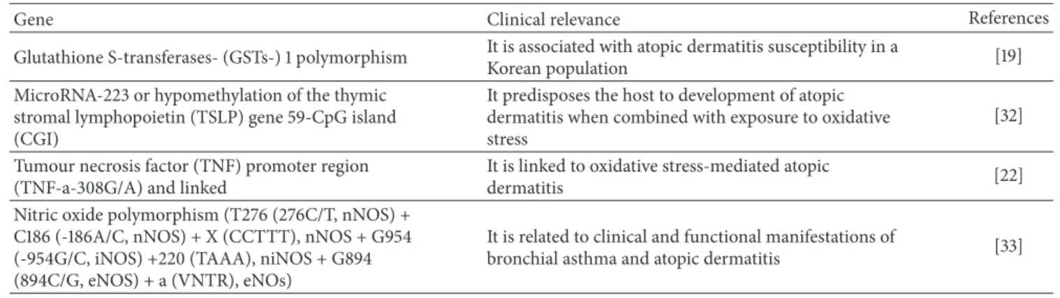
Table 3: Oxidative stress-inducible genes and atopic dermatitis.
Gene Clinical relevance References
Glutathione S-transferases- (GSTs-) 1 polymorphism It is associated with atopic dermatitis susceptibility in aKorean population [19] MicroRNA-223 or hypomethylation of the thymic
stromal lymphopoietin (TSLP) gene 59-CpG island (CGI)
It predisposes the host to development of atopic dermatitis when combined with exposure to oxidative stress
[32] Tumour necrosis factor (TNF) promoter region
(TNF-a-308G/A) and linked
It is linked to oxidative stress-mediated atopic
dermatitis [22]
Nitric oxide polymorphism (T276 (276C/T, nNOS) + C186 (-186A/C, nNOS) + X (CCTTT), nNOS + G954 (-954G/C, iNOS) +220 (TAAA), niNOS + G894 (894C/G, eNOS) + a (VNTR), eNOs)
It is related to clinical and functional manifestations of
bronchial asthma and atopic dermatitis [33]
and peripheral blood monocytes [40], are themselves a likely source of ROS production [62]. Confirming these findings, studies have shown higher H2O2, nitric oxide, and superoxide levels in exhaled gases from asthmatic patients than from control subjects [63–66]. A prooxidant status also induces a wide range of biological and molecular damage in the lung. Increased release of isoprostanes and ethane, both in epithelial and in endothelial cell membranes, as well as diminished activity of proteins, such as𝛼1-protease inhibitor, ascorbate,𝛼-tocopherol, and superoxide dismutase (SOD), has been reported [67].
Acting on other targets, such as airway smooth muscle, inducing acetylcholine-mediated contraction [68], mucin secretion [69], and nitric oxide- (NO-) mediated neurogenic inflammation [69], ROS can also impair broncho- and vasoregulation [70, 71].
Finally, large-scale genome-wide association studies (GWAS) have demonstrated that genetic susceptibility to allergic asthma is also determined by complex interactions between genes involved in OS, such as glutamate cysteine lig-ase (GCLM), glutathione peroxidlig-ase (GPX1), catallig-ase (CAT), myeloperoxidase (MPO), NADPH oxidase (CYBA, p22phox subunit), NAD(P)H, quinone oxidoreductase type 1 (NQO1), and microsomal epoxide hydrolase (EPHX1) [26] (Table 1).
As the primary cell of interface between internal and external environments, nasal mucosal epithelial cells are known to initiate the release of a cascade of proinflammatory mediators through redox pathways [20]. Moreover, these cells also exhibit the capacity to upregulate an effective antioxidant defence [20]. However, natural allergen exposure agents show the ability to interfere with oxidant/antioxidant balance, enhancing OS and upper airway inflammation [72].
Although it has been hypothesized that the role of OS in allergic rhinitis is similar to that of asthma, the exact underlying mechanism is still not understood. However, it has been reported that OS, playing a critical role in allergic asthma, can also contribute to the onset of allergic rhinitis and to enhancing the asthma-rhinitis link, as expression of united airways disease [73].
It has been widely assessed that the loss of antioxidant activities characterizes patients affected by allergic rhinitis. Studies reported that decreased activities of both antioxidant enzyme paraoxonase (PON1) [74] and reduced glutathione
[20] are inversely correlated to plasma total oxidant status and to severity of disease [20]. Consequently, increased nasal fraction of exhaled NO (FENO), 8-isoprostane, leukotriene-(LT-) B4, and PGE2 levels was detected in patients with allergic rhinitis [75]. An impaired function and distribution of superoxide anion, NADPH oxidase (NOX)1, and NOX4 in allergic nasal rhinitis has also been noted, as further confir-mation of the possible influence of OS on the development of allergic rhinitis [76] (Table 2).
The ability to interfere with the immune system allows ROS to induce and perpetuate skin injury, also in atopic dermatitis. In particular, authors reported that ROS, acting mainly on keratinocytes and partially on lymphocytes [77], induce oxidative protein damage in the stratum corneum, leading to the disruption of barrier functions and the exac-erbation of atopic dermatitis [78]. Therefore, in response to a variety of oxidant reactants, the skin upregulates transactivat-ing AP-1 components such as Fos and Jun, whereas it down-regulates anti-inflammatory components [79]. Precisely, it has been suggested that upregulation of AP-1 may be associ-ated with a defect in ceramide generation which could result in enhanced protein kinase-C activation, leading to exces-sive release of proinflammatory cytokines by keratinocytes [79]. Generally, peroxisome proliferator-activated receptors (PPARs), a member of the nuclear factor family, also influence the biological activity of keratinocytes. To be precise, PPAR isoform-𝛼 (PPAR-𝛼) counteracts the inflammatory response by inhibition of the expression of proinflammatory genes, as well as cytokines and metalloproteases. PPAR-𝛼 activation also induces antioxidant enzymes (catalase, SOD) which would reduce oxidative damage and inflammatory response [21].
The oxidant/antioxidant balance is also altered in atopic dermatitis. ROS reduce the physiological antioxidant levels of a number of compounds, such as 𝛼-tocopherol (VE), ubiquinol-10 (CoQH2-10), ascorbic acid (VC), and glu-tathione (GSH), in the epidermis and dermis and thus impair the cellular redox system [80]. Evidence of enhanced protein and lipid-oxidative damage was also found in atopic dermatitis patients, as demonstrated by the increase of carbonyl moieties both in lesional and in nonlesional skin, along with higher activity of SOD, an effective scavenger of ROS [81]. Recent experimental studies support a role for
oxidative/antioxidative imbalance also in the shift toward a Th2-skewed immune response, probably NO-mediated [38]. Accordingly, the administration of antioxidants to human T-cells culture downregulated Th2 polarization, with a decrease in the expression of IL-4 and IL-5, and simultaneous skewing toward a Th1- phenotype [38]. Finally, data suggest epigenetic changes linked to the development of atopic dermatitis through OS-mediated immune dysregulation [82] (Table 3).
5. Conclusions
To date, the exact underlying mechanisms of atopic disease are still not understood. Recently, more attention has been given to the critical role of OS-inducible genes in the pathogenesis of atopic diseases. However, in spite of much evidence linking atopic predisposition, inflammatory status, and maternal and neonatal OS, much more remains to be investigated. Moreover, a genomic approach would clarify the role of oxidant/antioxidant pathways, in order to better understand the pathogenesis of atopic diseases and identify innovative therapeutic strategies.
Competing Interests
The authors have declared no conflict of interests.
Authors’ Contributions
All authors had equally contributed to the manuscript.
References
[1] C. Anandan, U. Nurmatov, O. C. P. van Schayck, and A. Sheikh, “Is the prevalence of asthma declining? Systematic review of epidemiological studies,” Allergy, vol. 65, no. 2, pp. 152–167, 2010. [2] M. Olivieri, J. Heinrich, V. Schl¨unssen et al., “The risk of respiratory symptoms on allergen exposure increases with increasing specific IgE levels,” Allergy, vol. 71, no. 6, pp. 859– 868, 2016.
[3] I. Marenholz, J. Esparza-Gordillo, and F. R¨uschendorf, “Meta-analysis identifies seven susceptibility loci involved in the atopic march,” Nature Communications, vol. 6, no. 6, p. 8804, 2015. [4] T. A. Manolio, F. S. Collins, N. J. Cox et al., “Finding the missing
heritability of complex diseases,” Nature, vol. 461, no. 7265, pp. 747–753, 2009.
[5] M. Yamamoto, A. Singh, J. Hirota et al., “Immune-related gene expression profile in human airway epithelium are altered by co-exposure to diesel exhaust and allergen,” in Proceedings of the
American Thoracic Society 2014 International Conference, San
Diego, Calif, USA, 2014.
[6] Y. Okayama, “Oxidative stress in allergic and inflammatory skin diseases,” Current Drug Targets: Inflammation and Allergy, vol. 4, no. 4, pp. 517–519, 2005.
[7] J. G. O. ´Avila, I. Echeverri, C. A. de Plata, and A. Castillo, “Impact of oxidative stress during pregnancy on fetal epige-netic patterns and early origin of vascular diseases,” Nutrition
Reviews, vol. 73, no. 1, pp. 12–21, 2015.
[8] Y. J. Suzuki, H. J. Forman, and A. Sevanian, “Oxidants as stimulators of signal transduction,” Free Radical Biology and
Medicine, vol. 22, no. 1-2, pp. 269–285, 1996.
[9] M. S. Williams and J. Kwon, “T cell receptor stimulation, reactive oxygen species, and cell signaling,” Free Radical Biology
and Medicine, vol. 37, no. 8, pp. 1144–1151, 2004.
[10] M. R. King, A. S. Ismail, L. S. Davis, and D. R. Karp, “Oxidative stress promotes polarization of human T cell differentiation toward a T helper 2 phenotype,” The Journal of Immunology, vol. 176, no. 5, pp. 2765–2772, 2006.
[11] J. M. Morris, N. K. Gopaul, M. J. R. Endresen et al., “Circulating markers of oxidative stress are raised in normal pregnancy and pre-eclampsia,” British Journal of Obstetrics and Gynaecology, vol. 105, no. 11, pp. 1195–1199, 1998.
[12] L. Marseglia, G. D’Angelo, S. Manti et al., “Oxidative stress-mediated aging during the fetal and perinatal periods,”
Oxida-tive Medicine and Cellular Longevity, vol. 2014, Article ID
358375, 8 pages, 2014.
[13] E. Gitto, L. Marseglia, S. Manti et al., “Protective role of mela-tonin in neonatal diseases,” Oxidative Medicine and Cellular
Longevity, vol. 2013, Article ID 980374, 6 pages, 2013.
[14] L. Marseglia, G. D’Angelo, S. Manti, R. J. Reiter, and E. Gitto, “Potential utility of melatonin in preeclampsia, intrauterine fetal growth retardation, and perinatal asphyxia,” Reproductive
Sciences, 2015.
[15] L. Marseglia, S. Manti, G. D’Angelo et al., “Oxidative stress in obesity: a critical component in human diseases,” International
Journal of Molecular Sciences, vol. 16, no. 1, pp. 378–400, 2015.
[16] L. Marseglia, G. D’Angelo, S. Manti et al., “Oxidative stress-mediated damage in newborns with necrotizing enterocolitis: a possible role of melatonin,” American Journal of Perinatology, vol. 32, no. 10, pp. 905–909, 2015.
[17] R. P. Bowler and J. D. Crapo, “Oxidative stress in allergic res-piratory diseases,” Journal of Allergy and Clinical Immunology, vol. 110, no. 3, pp. 349–356, 2002.
[18] L. Marseglia, C. Cuppari, S. Manti et al., “Atopic dermatitis: melatonin as potential treatment,” Journal of Biological
Regula-tors and Homeostatic Agents, vol. 29, no. 2, supplement 1, pp.
142–149, 2015.
[19] F. Child, W. Lenney, S. Clayton et al., “The association of maternal but not paternal genetic variation in GSTP1 with asthma phenotypes in children,” Respiratory Medicine, vol. 97, no. 12, pp. 1247–1256, 2003.
[20] M. Celik, A. Tuncer, O. U. Soyer, C. Sac¸kesen, H. Tanju Besler, and O. Kalayci, “Oxidative stress in the airways of children with asthma and allergic rhinitis,” Pediatric Allergy and Immunology, vol. 23, no. 6, pp. 556–561, 2012.
[21] M. Y. Sheu, A. J. Fowler, J. Kao et al., “Topical peroxisome pro-liferator activated receptor-𝛼 activators reduce inflammation in irritant and allergic contact dermatitis models,” Journal of
Investigative Dermatology, vol. 118, no. 1, pp. 94–101, 2002.
[22] H. Batikhan, M. K. Gokcan, E. Beder, N. Akar, A. Ozturk, and M. Gerceker, “Association of the tumor necrosis factor-alpha−308 G/A polymorphism with nasal polyposis,” European
Archives of Oto-Rhino-Laryngology, vol. 267, no. 6, pp. 903–908,
2010.
[23] C.-C. Zou, L.-F. Tang, M.-Z. Jiang, Z.-Y. Zhao, T. Hirokazu, and M. Mitsufumi, “Methylenetetrahydrofolate reductase [correc-tion of reducatase] polymorphism and asthma,” Zhonghua Jie
He He Hu Xi Za Zhi, vol. 26, no. 3, pp. 161–164, 2003.
[24] M. A. R. Ferreira, A. F. McRae, S. E. Medland et al., “Association between ORMDL3, IL1RL1 and a deletion on chromosome 17q21 with asthma risk in Australia,” European Journal of Human
[25] M. Tamura and T. Shiroishi, “GSDM family genes meet autophagy,” Biochemical Journal, vol. 469, no. 2, pp. e5–e7, 2015. [26] A. V. Polonikov, V. P. Ivanov, A. D. Bogomazov, M. B. Freidin, T. Illig, and M. A. Solodilova, “Antioxidant defense enzyme genes and asthma susceptibility: gender-specific effects and heterogeneity in gene-gene interactions between pathogenetic variants of the disease,” BioMed Research International, vol. 2014, Article ID 708903, 17 pages, 2014.
[27] H.-Y. Cho and S. R. Kleeberger, “Noblesse oblige: NRF2 func-tions in the airways,” American Journal of Respiratory Cell and
Molecular Biology, vol. 50, no. 5, pp. 844–847, 2014.
[28] E. Fuertes, M. Brauer, E. MacIntyre et al., “Childhood allergic rhinitis, traffic-related air pollution, and variability in the
GSTP1, TNF, TLR2, and TLR4 genes: results from the TAG
Study,” Journal of Allergy and Clinical Immunology, vol. 132, no. 2, pp. 342–352.e2, 2013.
[29] T. Islam, R. McConnell, W. J. Gauderman, E. Avol, J. M. Peters, and F. D. Gilliland, “Ozone, oxidant defense genes, and risk of asthma during adolescence,” American Journal of Respiratory
and Critical Care Medicine, vol. 177, no. 4, pp. 388–395, 2008.
[30] M. Panek, T. Pietras, A. Fabijan et al., “Identification and association of the single nucleotide polymorphisms, C-509T, C+466T and T+869C, of the TGF-1𝛽 gene in patients with asthma and their influence on the mRNA expression level of TGF-𝛽1,” International Journal of Molecular Medicine, vol. 34, no. 4, pp. 975–986, 2014.
[31] M. T. Salam, T. Islam, W. J. Gauderman, and F. D. Gilliland, “Roles of arginase variants, atopy, and ozone in childhood asthma,” Journal of Allergy and Clinical Immunology, vol. 123, no. 3, pp. 596–602.e8, 2009.
[32] I.-J. Wang, S.-L. Chen, T.-P. Lu, E. Y. Chuang, and P.-C. Chen, “Prenatal smoke exposure, DNA methylation, and childhood atopic dermatitis,” Clinical and Experimental Allergy, vol. 43, no. 5, pp. 535–543, 2013.
[33] S. Hoffjan, I. Ostrovnaja, D. Nicolae et al., “Genetic variation in immunoregulatory pathways and atopic phenotypes in infancy,”
Journal of Allergy and Clinical Immunology, vol. 113, no. 3, pp.
511–518, 2004.
[34] E. Herrera and H. Ortega-Senovilla, “Lipid metabolism during pregnancy and its implications for fetal growth,” Current
Phar-maceutical Biotechnology, vol. 15, no. 1, pp. 24–31, 2014.
[35] R. M. Lewis, J. K. Cleal, and M. A. Hanson, “Review: placenta, evolution and lifelong health,” Placenta, vol. 33, supplement, pp. S28–S32, 2012.
[36] ¨U. Mutlu-T¨urkoglu, E. Ademoglu, L. Ibrahimoglu, G. Aykac¸-Toker, and M. Uysal, “Imbalance between lipid peroxidation and antioxidant status in preeclampsia,” Gynecologic and
Obstet-ric Investigation, vol. 46, no. 1, pp. 37–40, 1998.
[37] G. J. Burton and E. Jauniaux, “Oxidative stress,” Best Practice &
Research Clinical Obstetrics & Gynaecology, vol. 25, no. 3, pp.
287–299, 2011.
[38] S. B. Bennett and H. R. Griffiths, Regulation of T-Cell Functions
by Oxidative Stress, Springer, Berlin, Germany, 2012.
[39] F. B. Thor´en, ˚A. Betten, A. I. Romero, and K. Hellstrand, “Cut-ting edge: antioxidative properties of myeloid dendritic cells: protection of T cells and NK cells from oxygen radical-induced inactivation and apoptosis,” The Journal of Immunology, vol. 179, no. 1, pp. 21–25, 2007.
[40] M. Mittal, M. R. Siddiqui, K. Tran, S. P. Reddy, and A. B. Malik, “Reactive oxygen species in inflammation and tissue injury,”
Antioxidants and Redox Signaling, vol. 20, no. 7, pp. 1126–1167,
2014.
[41] H. Ogasawara, S. Yoshimura, and T. Kumoi, “Hydrogen perox-ide generation by eosinophils in allergic rhinitis,” Auris Nasus
Larynx, vol. 18, no. 2, pp. 133–143, 1991.
[42] I. Vachier, M. Damon, C. Le Doucen et al., “Increased oxygen species generation in blood monocytes of asthmatic patients,”
American Review of Respiratory Disease, vol. 146, no. 5, pp. 1161–
1166, 1992.
[43] S. Aversa, L. Marseglia, S. Manti et al., “Ventilation strategies for preventing oxidative stress-induced injury in preterm infants with respiratory disease: an update,” Paediatric Respiratory
Reviews, vol. 17, pp. 71–79, 2016.
[44] B. S. Polla, R. Alan Ezekowitz, and D. Y. M. Leung, “Monocytes from patients with atopic dermatitis are primed for superoxide production,” The Journal of Allergy and Clinical Immunology, vol. 89, no. 2, pp. 545–551, 1992.
[45] B. M. Willwerth, B. Schaub, K. G. Tantisira et al., “Prenatal, perinatal, and heritable influences on cord blood immune responses,” Annals of Allergy, Asthma and Immunology, vol. 96, no. 3, pp. 445–453, 2006.
[46] S. Perrone, R. Bracci, and G. Buonocore, “New biomarkers of fetal-neonatal hypoxic stress,” Acta Paediatrica, vol. 91, supplement 438, pp. 135–140, 2002.
[47] G. Devereux, A. Seaton, and R. N. Barker, “In utero priming of allergen-specific helper T cells,” Clinical and Experimental
Allergy, vol. 31, no. 11, pp. 1686–1695, 2001.
[48] Y. Kitagawa, J. B. Wing, and S. Sakaguchi, “Transcriptional and epigenetic control of regulatory T cell development,” in Progress
in Molecular Biology and Translational Science, vol. 136, chapter
1, pp. 1–33, Elsevier, 2015.
[49] J. Bousquet, W. Jacot, H. Yssel, A. M. Vignola, and M. Humbert, “Epigenetic inheritance of fetal genes in allergic asthma,”
Allergy, vol. 59, no. 2, pp. 138–147, 2004.
[50] P. C. J. Janson, M. E. Winerdal, and O. Winqvist, “At the cross-roads of T helper lineage commitment-Epigenetics points the way,” Biochimica et Biophysica Acta (BBA)—General Subjects, vol. 1790, no. 9, pp. 906–919, 2009.
[51] P. J. Vuillermin, A.-L. Ponsonby, R. Saffery et al., “Microbial exposure, interferon gamma gene demethylation in na¨ıve T-cells, and the risk of allergic disease,” Allergy, vol. 64, no. 3, pp. 348–353, 2009.
[52] C. E. West, D. J. Videky, and S. L. Prescott, “Role of diet in the development of immune tolerance in the context of allergic disease,” Current Opinion in Pediatrics, vol. 22, no. 5, pp. 635– 641, 2010.
[53] T. Arrigo, S. Leonardi, C. Cuppari et al., “Role of the diet as a link between oxidative stress and liver diseases,” World Journal
of Gastroenterology, vol. 21, no. 2, pp. 384–395, 2015.
[54] F. Perera, W.-Y. Tang, J. Herbstman et al., “Relation of DNA methylation of 5-CpG island of ACSL3 to transplacental exposure to airborne polycyclic aromatic hydrocarbons and childhood asthma,” PLoS ONE, vol. 4, no. 2, Article ID e4488, 2009.
[55] D. J. Martino and S. L. Prescott, “Silent mysteries: epigenetic paradigms could hold the key to conquering the epidemic of allergy and immune disease,” Allergy, vol. 65, no. 1, pp. 7–15, 2010.
[56] J. Liu, M. Ballaney, U. Al-Alem et al., “Combined inhaled diesel exhaust particles and allergen exposure alter methylation of T helper genes and Ige production in vivo,” Toxicological Sciences, vol. 102, no. 1, pp. 76–81, 2008.
[57] G. Buonocore and S. Perrone, “Biomarkers of oxidative stress in the fetus and newborn,” Hematology, vol. 2, no. 10, pp. 103–107, 2006.
[58] J. M. Gostner, K. Becker, D. Fuchs, and R. Sucher, “Redox regulation of the immune response,” Redox Report, vol. 18, no. 3, pp. 88–94, 2013.
[59] M. J. Jardim, “microRNAs: implications for air pollution research,” Mutation Research/Fundamental and Molecular
Mechanisms of Mutagenesis, vol. 717, no. 1-2, pp. 38–45, 2011.
[60] S. Corroyer, B. Maitre, V. Cazals, and A. Clement, “Altered regulation of G1 cyclins in oxidant-induced growth arrest of lung alveolar epithelial cells. Accumulation of inactive cyclin E-CDK2 complexes,” The Journal of Biological Chemistry, vol. 271, no. 41, pp. 25117–25125, 1996.
[61] W. J. Calhoun, H. E. Reed, D. R. Moest, and C. A. Stevens, “En-hanced superoxide production by alveolar macrophages and air-space cells, airway inflammation, and alveolar macrophage density changes after segmental antigen bronchoprovocation in allergic subjects,” The American Review of Respiratory Disease, vol. 145, no. 2, pp. 317–325, 1992.
[62] N. N. Jarjour and W. J. Calhoun, “Enhanced production of oxygen radicals in asthma,” Journal of Laboratory and Clinical
Medicine, vol. 123, no. 1, pp. 131–136, 1994.
[63] M. Ichinose, H. Sugiura, S. Yamagata, A. Koarai, and K. Shirato, “Increase in reactive nitrogen species production in chronic obstructive pulmonary disease airways,” American Journal of
Respiratory and Critical Care Medicine, vol. 162, no. 2, pp. 701–
706, 2000.
[64] K. Ganas, S. Loukides, G. Papatheodorou, P. Panagou, and N. Kalogeropoulos, “Total nitrite/nitrate in expired breath condensate of patients with asthma,” Respiratory Medicine, vol. 95, no. 8, pp. 649–654, 2001.
[65] P. Montuschi, M. Corradi, G. Ciabattoni, J. Nightingale, S. A. Kharitonov, and P. J. Barnes, “Increased 8-isoprostane, a marker of oxidative stress, in exhaled condensate of asthma patients,”
American Journal of Respiratory and Critical Care Medicine, vol.
160, no. 1, pp. 216–220, 1999.
[66] P. Paredi, S. A. Kharitonov, and P. J. Barnes, “Elevation of exhaled ethane concentration in asthma,” American Journal of
Respiratory and Critical Care Medicine, vol. 162, no. 4, pp. 1450–
1454, 2000.
[67] F. J. Kelly, I. Mudway, A. Blomberg, A. Frew, and T. Sandstrom, “Altered lung antioxidant status in patients with mild asthma,”
The Lancet, vol. 354, no. 9177, pp. 482–483, 1999.
[68] U. Katsumata, M. Miura, M. Ichinose et al., “Oxygen radicals produce airway constriction and hyperresponsiveness in anes-thetized cats,” American Review of Respiratory Disease, vol. 141, no. 5, pp. 1158–1161, 1990.
[69] D. T. Wright, B. M. Fischer, C. Li, L. G. Rochelle, N. J. Akley, and K. B. Adler, “Oxidant stress stimulates mucin secretion and PLC in airway epithelium via a nitric oxide-dependent mechanism,”
American Journal of Physiology—Lung Cellular and Molecular Physiology, vol. 271, no. 5, pp. L854–L861, 1996.
[70] C. M. Prado, M. A. Martins, and IF. Tib´erio, “Nitric oxide in asthma physiopathology,” ISRN Allergy, vol. 2011, Article ID 832560, 13 pages, 2011.
[71] H. Sugiura and M. Ichinose, “Nitrative stress in inflammatory lung diseases,” Nitric Oxide, vol. 25, no. 2, pp. 138–144, 2011. [72] C. Gratziou, N. Rovina, M. P. Makris, D. C. M. Simoes, A.
Papapetropoulos, and C. Roussos, “Breath markers of oxidative stress and airway inflammation in Seasonal Allergic Rhinitis,”
International Journal of Immunopathology and Pharmacology,
vol. 21, no. 4, pp. 949–957, 2008.
[73] M. Profita, P. Montuschi, A. Bonanno et al., “Novel perspectives in the detection of oral and nasal oxidative stress and inflamma-tion in pediatric united airway diseases,” Internainflamma-tional Journal
of Immunopathology and Pharmacology, vol. 23, no. 4, pp. 1211–
1219, 2010.
[74] E. Ozkaya, H. Akduman, U. Erenberk, A. Demir, and M. R. Dundaroz, “Plasma paraoxonase activity and oxidative stress and their relationship to disease severity in children with allergic rhinitis,” American Journal of Rhinology and Allergy, vol. 27, no. 1, pp. 13–17, 2013.
[75] K. Tanou, A. Koutsokera, T. S. Kiropoulos et al., “Inflammatory and oxidative stress biomarkers in allergic rhinitis: the effect of smoking,” Clinical and Experimental Allergy, vol. 39, no. 3, pp. 345–353, 2009.
[76] J. H. Moon, T. H. Kim, H. M. Lee et al., “Overexpression of the superoxide anion and NADPH oxidase isoforms 1 and 4 (NOX1 and NOX4) in allergic nasal mucosa,” American Journal
of Rhinology & Allergy, vol. 23, no. 4, pp. 370–376, 2009.
[77] C. Albanesi and S. Pastore, “Pathobiology of chronic inflamma-tory skin diseases: interplay between keratinocytes and immune cells as a target for anti-inflammatory drugs,” Current Drug
Metabolism, vol. 11, no. 3, pp. 210–227, 2010.
[78] Y. Niwa, H. Sumi, K. Kawahira, T. Terashima, T. Nakamura, and H. Akamatsu, “Protein oxidative damage in the stratum corneum: evidence for a link between environmental oxidants and the changing prevalence and nature of atopic dermatitis in Japan,” British Journal of Dermatology, vol. 149, no. 2, pp. 248– 254, 2003.
[79] ¨O. Uluc¸kan, J. Guinea-Viniegra, M. Jimenez, and E. F. Wagner, “Signalling in inflammatory skin disease by AP-1 (Fos/Jun),”
Clinical and Experimental Rheumatology, vol. 33, no. 4,
supple-ment 92, pp. S44–S49, 2015.
[80] Y. Yamamoto, “Role of active oxygen species and antioxidants in photoaging,” Journal of Dermatological Science, vol. 27, no. 1, pp. S1–S4, 2001.
[81] J. Fuchs, T. M. Zollner, R. Kaufmann, and M. Podda, “Redox-modulated pathways in inflammatory skin diseases,” Free
Radi-cal Biology and Medicine, vol. 30, no. 4, pp. 337–353, 2001.
[82] Early Genetics and Lifecourse Epidemiology (EAGLE) Eczema Consortium, “Australian Asthma Genetics Consortium(AAGC); Australian Asthma Genetics Consortium AAGC. Multi-ancestry genome-wide association study of 21,000 cases and 95,000 controls identifies new risk loci for atopic dermatitis,”
Submit your manuscripts at
http://www.hindawi.com
Stem Cells
International
Hindawi Publishing Corporationhttp://www.hindawi.com Volume 2014
Hindawi Publishing Corporation
http://www.hindawi.com Volume 2014
INFLAMMATION
Hindawi Publishing Corporation
http://www.hindawi.com Volume 2014
Behavioural
Neurology
Endocrinology
International Journal ofHindawi Publishing Corporation
http://www.hindawi.com Volume 2014
Hindawi Publishing Corporation
http://www.hindawi.com Volume 2014
Disease Markers
Hindawi Publishing Corporation
http://www.hindawi.com Volume 2014
BioMed
Research International
Oncology
Journal ofHindawi Publishing Corporation
http://www.hindawi.com Volume 2014
Hindawi Publishing Corporation
http://www.hindawi.com Volume 2014
Oxidative Medicine and Cellular Longevity
Hindawi Publishing Corporation
http://www.hindawi.com Volume 2014
PPAR Research
The Scientific
World Journal
Hindawi Publishing Corporation
http://www.hindawi.com Volume 2014
Immunology Research
Hindawi Publishing Corporation
http://www.hindawi.com Volume 2014
Journal of
Obesity
Journal ofHindawi Publishing Corporation
http://www.hindawi.com Volume 2014
Hindawi Publishing Corporation
http://www.hindawi.com Volume 2014
Computational and Mathematical Methods in Medicine
Ophthalmology
Journal ofHindawi Publishing Corporation
http://www.hindawi.com Volume 2014
Diabetes Research
Journal ofHindawi Publishing Corporation
http://www.hindawi.com Volume 2014
Hindawi Publishing Corporation
http://www.hindawi.com Volume 2014
Research and Treatment
AIDS
Hindawi Publishing Corporationhttp://www.hindawi.com Volume 2014
Gastroenterology Research and Practice
Hindawi Publishing Corporation
http://www.hindawi.com Volume 2014