Research Article
Role of Inhibitors of Apoptosis Proteins in Testicular
Function and Male Fertility: Effects of Polydeoxyribonucleotide
Administration in Experimental Varicocele
Letteria Minutoli,
1Salvatore Arena,
2Pietro Antonuccio,
2Carmelo Romeo,
2Alessandra Bitto,
1Carlo Magno,
3Mariagrazia Rinaldi,
1Antonio Micali,
4Natasha Irrera,
1Gabriele Pizzino,
1Federica Galfo,
1Francesco Squadrito,
1Domenica Altavilla,
2and Herbert Marini
11Department of Clinical and Experimental Medicine, University of Messina, AOU Policlinico “G. Martino”, Via Consolare Valeria, 98125 Messina, Italy
2Department of Paediatric, Gynaecological, Microbiological and Biomedical Sciences, University of Messina, AOU Policlinico “G. Martino”, Via Consolare Valeria, 98125 Messina, Italy
3Department of Human Pathology, University of Messina, AOU Policlinico “G. Martino”, Via Consolare Valeria, 98125 Messina, Italy 4Department of Biomedical Sciences and Morphofunctional Imaging, University of Messina, AOU Policlinico “G. Martino”,
Via Consolare Valeria, 98125 Messina, Italy
Correspondence should be addressed to Letteria Minutoli; [email protected] Received 23 February 2015; Revised 20 April 2015; Accepted 21 April 2015 Academic Editor: Joilson O. Martins
Copyright © 2015 Letteria Minutoli et al. This is an open access article distributed under the Creative Commons Attribution License, which permits unrestricted use, distribution, and reproduction in any medium, provided the original work is properly cited.
Neuronal apoptosis inhibitory protein (NAIP) and survivin might play an important role in testicular function. We investigated the effect of PDRN, an agonist of adenosine A2A receptor, on testicular NAIP and survivin expression in an experimental model of varicocele. After the creation of experimental varicocele (28 days), adolescent male Sprague-Dawley rats were randomized to one of the following treatments lasting 21 days: vehicle, PDRN (8 mg/kg i.p., daily), PDRN + 3,7-dimethyl-propargylxanthine (DMPX, a specific adenosine A2A-receptor antagonist, 0.1 mg/kg i.p., daily), varicocelectomy, and varicocelectomy + PDRN (8 mg/kg i.p., daily). Sham-operated animals were used as controls. Animals were then euthanized and testis expression of NAIP and survivin was evaluated through qRT-PCR, western blot, and immunohistochemical analysis. Spermatogenetic activity was also assessed. NAIP and survivin expressions were significantly reduced following varicocele induction when compared to sham animals whereas PDRN-treated rats showed an increase in NAIP and survivin levels. Immunohistochemistry revealed an enhanced expression of NAIP and survivin with a characteristic pattern of cellular localization following PDRN treatment. Moreover, administration of PDRN significantly restored spermatogenic function in varicocele rats. PDRN may represent a rational therapeutic option for accelerating recovery from depressed testicular function through a strategic modulation of apoptosis in experimental varicocele.
1. Introduction
Varicocele is the most common cause of infertility in men [1] and the exact pathophysiological mechanism by which it impairs fertility in affected men remains unknown [2,3]. Consequently, the early diagnosis of varicocele is necessary before testicular damage might occur and, as indicated by several clinical studies, the varicocele repair is possible
through surgical procedure [4–7]. Although many advances have occurred in the treatment of varicocele, it still represents an important and challenging aspect of basic research (male reproductive physiology and endocrinology, pathophysiol-ogy, and pharmacology of reproduction and fertility) and medical practice for urologists, pediatric surgeons, and gen-eral physicians, to date [4–7].
Volume 2015, Article ID 248976, 9 pages http://dx.doi.org/10.1155/2015/248976
The development of varicocele-related testis damage may be caused by disruption of homeostasis between cell proliferation and cell death [8–12]. This phenomenon is also related to different pathophysiological mechanisms (imbalance between reactive oxygen species and seminal antioxidants, lipid peroxidation, DNA fragmentation, and apoptosis) in testis following varicocele induction [13, 14]. Basically, apoptosis is a physiological process by which a sequence of intracellular events results in the programmed elimination of a cell from its environment [15,16]. Specifically, alterations in the apoptosis of germ cells may be crucial in varicocele-related human infertility [17] and, as a direct consequence, targeting apoptosis may represent an alterna-tive and rational therapeutic strategy in the treatment of varicocele complications [18–21].
Emerging contributors in this context are the inhibitors of apoptosis proteins (IAPs), which halt cell death in response to diverse stimuli [22]. IAPs family influences apoptosis by direct inhibition of caspases and modulation of the tran-scription factor nuclear factor-kB (NF-kB). Eight mammalian IAPs are known at present: X-chromosome-linked IAP (XIAP), cellular IAP1 and IAP2 (cIAP1 and cIAP2), neuronal apoptosis inhibitory protein (NAIP), survivin, BRUCE, livin, and testis-specific IAP (Ts-IAP).
NAIP was originally identified while searching for a gene on chromosome 5q13 responsible for childhood muscular atrophy and is also associated with spinal muscular dystrophy [23, 24]. Survivin too has a central role in the negative regulation of apoptosis; however the exact mechanism by which survivin controls programmed cell death has not yet been clarified [25].
It has been indicated that NAIP contrasts apoptosis by inhibition of the executioner caspase-3 and caspase-7 [26] while survivin has been shown to modulate the executioner caspase-3 [27]. However, recent findings suggest that IAPs have a much broader spectrum of action than promoting cell survival by caspase regulation; indeed, a crucial function of some IAPs consists in the regulation of inflammatory and innate immune signaling pathways, a function attributed to their E3 Ub-ligase activities [28].
Polydeoxyribonucleotide (PDRN) is the active fraction extracted from trout spermatozoa used for tissue repair [29] and, acting through stimulation of the adenosine A2A recep-tor (A2AR), is able to induce vascular endothelial growth factor (VEGF) production during pathologic conditions of low tissue perfusion [30]. This evidence prompted us to investigate the effect of PDRN on experimental varicocele; our previous published data indicated that A2AR stimulation could represent an interesting target to positively modulate the harmful pathophysiological signaling which characterizes the experimental varicocele [31,32]. Indeed, it has been also shown that PDRN improves the innate mechanism of neoan-giogenesis, through compensatory oxygen and metabolite supply to testis, thereby enhancing testicular function and restoring spermatogenic function [31,32].
In light of this background, we explored the effect of PDRN on testis neuronal apoptosis inhibitory protein (NAIP) and survivin expression in an experimental model of varico-cele.
2. Materials and Methods
2.1. Animals and Experimental Procedures. The protocol was
approved by the Committee of Animal Health and Care of University of Messina and all animal procedures were carried out according to Guide for the Care and Use of Laboratory Animals. A total of 42 male Sprague-Dawley adolescent rats aged 7 weeks and weighing 200 to 225 g were used [33]. During the experiments, the animals were maintained under controlled environmental conditions (12-hour light/dark cycle, temperature approximately 23∘C) and provided with standard laboratory food and water ad libitum. After induction of anesthesia with an intraperitoneal (i.p.) injection of sodium pentobarbital (50 mg/kg), varicocele was induced as previously described elsewhere [11, 31, 32, 34]. Briefly, varicocele was induced by an abdominal midline incision. The left renal vein, inferior vena cava, and left spermatic vein were identified and a clamp was passed behind the left renal vein just distal to the spermatic vein insertion. A 4-zero silk ligature was loosely placed around the left renal vein at this site and a rigid hydrophilic guide wire of 0.64 mm in diameter was placed on the left renal vein. The ligature was tied around the vein over the top of the guide wire. The guide wire was then withdrawn and the vein allowed to expand to the limits of the ligature causing the vein diameter to be decreased to approximately half of its original diameter. The renal vein and spermatic vein in each animal were dilated immediately. The midline incision was closed in 2 layers with 3-zero silk suture. Sham-operated rats underwent the same vertical midline incision, and the suture was also placed, but it was not tied. Animals were housed one per cage only during the first week following surgical procedures in all experimental groups to minimize the impact and time of social isolation [35].
Twenty-eight days after the creation of varicocele, animals were randomized to the following treatment: vehicle (0.9% NaCl solution 1 mL/kg i.p., daily), PDRN (8 mg/kg i.p., daily), PDRN + 3,7-dimethyl-propargylxanthine (DMPX, a specific adenosine A2A-receptor antagonist, 0.1 mg/kg i.p., daily), varicocelectomy, and varicocelectomy + PDRN (8 mg/kg i.p., daily).
Twenty-one days after randomization, all animals were euthanized by sodium pentobarbital overdose, and the left testis was harvested to evaluate NAIP and survivin expression by use of real-time quantitative polymerase chain reaction (qRT-PCR), western blot, and immunohistochemical analysis previously shown elsewhere [29–32]. Other molecular hall-marks of apoptosis (i.e., Bcl-2, BAX) and/or cell proliferation (i.e., VEGF) were previously examined in our research laboratory [31].
2.2. Real-Time Quantitative PCR (qRT-PCR) for NAIP and Survivin. For the gene expression study, the extraction of
total mRNA was performed from tissue testis using TRIZOL (Invitrogen, Milan, Italy) under sterile conditions, following the manufacturer’s protocol. For each sample, 5𝜇g of mRNA was reverse-transcribed into cDNA through cDNA Archive High-Capacity Reverse Transcription kit (Life Technologies, Foster City, CA, USA). cDNA was stored at −20∘C and it
was used to quantify by TaqMan Gene Expression Assay (Life Technologies) the amount of NAIP (Rn01757809 m1; Life Technologies, Foster City, CA, USA) and survivin (Rn00574012 m1; Life Technologies, Foster City, CA, USA), using the instrument SDS 7300 Real-Time PCR (Applied Biosystems, Foster City, CA, USA). The result was expressed using2−ΔΔCTmethod (normalized versus varicocele + vehi-cle).𝛽-actin (Rn00667869 m1; Life Technologies, Foster City, CA, USA) was used as endogenous control.
2.3. Isolation of Proteins and Determination of NAIP and Survivin by Western Blot Analysis. Samples from testis were
homogenized in lysis buffer (1% Triton; 20 mM Tris/HCl, pH 8.0; 137 mM NaCl; 10% glycerol; 5 mM EDTA; 1 mM phenylmethylsulfonyl fluoride; 1% aprotinin; 15𝜇g/mL leu-peptin) and centrifuged at 15000 rpm for 15 min at 4∘C, and the protein content was determined by using the DC protein assay (Biorad, Milan, Italy). Protein samples were denatured in 2X Laemmli sample buffer (Biorad, Milan, Italy) and separated by SDS-PAGE. The separated proteins were transferred onto a nitrocellulose membrane using a transfer buffer (39 mmol/L glycine, 48 mmol/L Tris, pH 8.3, and 20% methanol) at 200 mA for 1 hour. The membranes were stained with Ponceau S (0.005% in 1% acetic acid) to confirm equal amounts of protein and were blocked with 5% nonfat dry milk in TBS-0.1% Tween for 1 hour at room temperature, washed 3 times for 10 minutes each in TBS-0.1% Tween, and incubated with a primary antibody for NAIP (Abcam, Cambridge, MA, USA) and survivin (Cell Signaling, Beverly, MA, USA) in TBS-0.1% Tween overnight at 4∘C. After being washed 3 times for 10 minutes each in TBS-0.1% Tween, the membranes were incubated with a secondary antibody peroxidase-conjugated goat anti-rabbit immunoglobulin G (Pierce, Rockford, IL, USA) for 1 hour at room temperature. The protein signal was quantified by scanning densitometry using a bioimage analysis system ECL plus (Thermo Scientific, Waltham, MA, USA). The results were expressed as relative integrated intensity subtracting the respective backgrounds represented by𝛽-actin (Cell Signaling, Beverly, MA, USA) expression.
2.4. Immunohistochemistry. Excised rat testes were
longitu-dinally sectioned, formalin-fixed, and paraffin-embedded. Parallel serial sections (4𝜇m) were cut and antigen retrieval was performed using 0.01 M sodium citrate buffer heated for 30 minutes in microwave oven. Tissues were overnight incubated at 4∘C with polyclonal rabbit anti-NAIP (Abcam, Cambridge, MA, USA) and monoclonal rabbit anti-survivin (Cell Signaling, Beverly, MA, USA) antibodies, diluted 1 : 25 and 1 : 400, respectively.
Secondary antibody was provided by Innovex (Rich-mond, CA, USA) and the location of the reaction was visualized with 3,3-diaminobenzidine tetrahydrochloride (Sigma Aldrich, Milan, Italy). Slides were counterstained with hematoxylin and mounted with coverslips.
2.5. Spermatogenic Activity. Spermatogenesis was
quanti-fied in fifty tubular cross sections for each animal, using Johnsen’s score system. A score of 1–10 was given to each
seminiferous tubule, depending on the maturation rate of the germ cells: 10: full spermatogenesis; 9: slightly impaired spermatogenesis, many late spermatids, and disorganized epithelium; 8: less than five spermatozoa per tubule, few late spermatids; 7: no spermatozoa, no late spermatids, and many early spermatids; 6: no spermatozoa, no late spermatids, and few early spermatids; 5: no spermatozoa or spermatids, many spermatocytes; 4: no spermatozoa or spermatids, few spermatocytes; 3: spermatogonia only; 2: no germinal cells, Sertoli cells only; 1: no seminiferous epithelium [36].
2.6. Statistical Analysis. All data are expressed as mean ±
standard error of the mean (SEM). Data were analyzed by one-way analysis of variance (ANOVA) followed by Tukey’s post hoc evaluation.𝑃 < 0.05 was considered statistically significant.
2.7. Compounds. DMPX and 0.9% NaCl solution were
obtained from Sigma (Milan, Italy).
3. Results
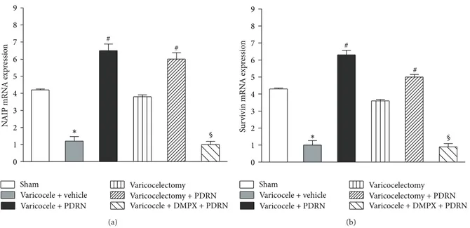
3.1. Testis NAIP and Survivin mRNA Levels. Testis from
sham animals had measurable levels of NAIP and survivin evaluated as mRNA level (Figures1(a)and1(b)). Testis from varicocele-injured rats showed a significant decrease in NAIP and survivin mRNA expression compared to sham animals (Figures1(a)and1(b)). The treatment with PDRN markedly increased NAIP and survivin mRNA expression. However, the effect of PDRN was abrogated by administration of DMPX. Varicocelectomy alone did not modify significantly the NAIP and survivin expression compared to sham animals while the concomitant administration of PDRN increased mRNA levels of these IAPs (Figures1(a)and1(b)).
3.2. NAIP and Survivin Expression by Western Blot.
Varico-cele produced a significant reduction in both testicular NAIP and survivin expression in comparison with sham animals (Figures2(a)and2(b)). Administration of PDRN markedly augmented both members of IAPs family. The concomitant administration of DMPX plus PDRN abrogated the effects of PDRN on the antiapoptotic proteins expression (Figures2(a)
and2(b)). Varicocelectomy alone did not modify significantly the NAIP and survivin expression whose levels are similar to sham animals. Varicocelectomy plus PDRN increased the NAIP and survivin expression but at significantly lower levels compared to those revealed in animals treated with PDRN alone (Figures2(a)and2(b)).
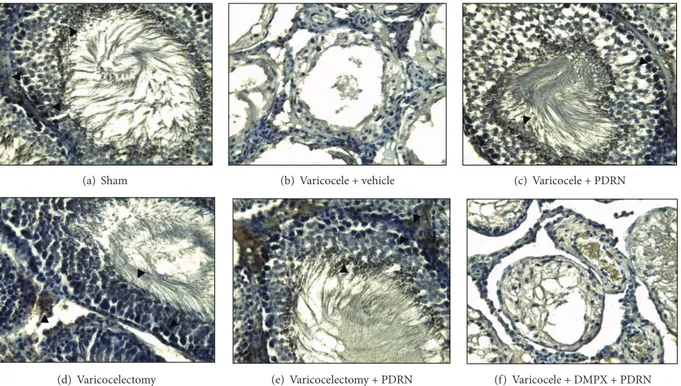
3.3. NAIP and Survivin Detection by Immunohistochemical Analysis. NAIP and survivin were also evaluated
immuno-histochemically. Specifically, NAIP immunostaining showed diffuse and strong positivity in spermatogonia, spermatozoa, and Leydig cells of the following groups: sham, varicocele + PDRN, and varicocelectomy + PDRN (Figures3(a),3(c), and 3(e)). Differently, just a focal immunoreaction was proved in sperm and Leydig cells of animals subjected to varicocelectomy alone (Figure 3(d)) and no immunostaining
0 1 2 3 4 5 6 7 8 9 Varicocele + vehicle Varicocele + PDRN N AIP mRN A exp re ssio n Sham Varicocelectomy Varicocelectomy + PDRN Varicocele + DMPX + PDRN # # ∗ § (a) Varicocele + vehicle Varicocele + PDRN Sham Varicocelectomy Varicocelectomy + PDRN Varicocele + DMPX + PDRN 0 1 2 3 4 5 6 7 8 9 Su rv iv in mRN A exp re ssio n # # § ∗ (b)
Figure 1: Expression of mRNA for NAIP (a) and survivin (b) (mean± SEM of seven animals) in specimens of left testis of sham-operated
rats and varicocele rats treated with vehicle (1 mL/kg, i.p.), PDRN (8 mg/kg, i.p.), DMPX (0.1 mg/kg, i.p.) plus PDRN (8 mg/kg, i.p.),
varicocelectomy alone, or varicocelectomy plus PDRN (8 mg/kg, i.p.).#𝑃 < 0.001 versus varicocele + vehicle;∗𝑃 < 0.001 versus sham;
§𝑃 < 0.001 versus varicocele + PDRN. 0 1 2 3 4 5 6 7 8 9 N AIP (in tegra te d in te n si ty) # # § ∗ Varicocele + vehicle Varicocele + PDRN Sham Varicocelectomy Varicocelectomy + PDRN Varicocele + DMPX + PDRN 𝛽-actin 160 kDa 45 kDa NAIP (a) 0 1 2 3 4 5 6 7 8 9 Su rv iv in (in te gra ted in te n si ty) # # § ∗ Varicocele + vehicle Varicocele + PDRN Sham Varicocelectomy Varicocelectomy + PDRN Varicocele + DMPX + PDRN 𝛽-actin 16 kDa 45 kDa Survivin (b)
Figure 2: Chemiluminescence image highlighting NAIP (a) and survivin (b) expression and relative quantitative data (mean± SEM of seven
animals) in specimens of left testis of sham-operated rats and varicocele rats treated with vehicle (1 mL/kg, i.p.), PDRN (8 mg/kg, i.p.), DMPX
(0.1 mg/kg, i.p.) plus PDRN (8 mg/kg, i.p.), varicocelectomy alone, or varicocelectomy plus PDRN (8 mg/kg, i.p.).#𝑃 < 0.001 versus varicocele
+ vehicle;∗𝑃 < 0.001 versus sham;§𝑃 < 0.001 versus varicocele + PDRN.
was detected in varicocele group (Figure 3(b)). Survivin immunostaining showed diffuse and strong positivity in spermatogonia of sham, varicocele + PDRN, and varicoc-electomy + PDRN groups (Figures4(a),4(c), and4(e)). By
contrast, slightly focal immunoreactivity was observed in germ cells of rats subjected to varicocelectomy (Figure 4(d)) and a negative reaction for survivin in varicocele group (Figure 4(b)). DMPX, antagonist of the A2AR, abrogated
(a) Sham (b) Varicocele + vehicle (c) Varicocele + PDRN
(d) Varicocelectomy (e) Varicocelectomy + PDRN (f) Varicocele + DMPX + PDRN
Figure 3: High-power-view light micrograph of a left testis harvested from sham-operated rats and varicocele rats administered with vehicle (1 mL/kg, i.p.), PDRN (8 mg/kg, i.p.), DMPX (0.1 mg/kg, i.p.) plus PDRN (8 mg/kg, i.p.), varicocelectomy alone, or varicocelectomy plus
PDRN (8 mg/kg, i.p.) at day 21 showing NAIP immunostaining (original magnification×200). Arrowheads show positive cells.
(a) Sham (b) Varicocele + vehicle (c) Varicocele + PDRN
(d) Varicocelectomy (e) Varicocelectomy + PDRN (f) Varicocele + DMPX + PDRN
Figure 4: High-power-view light micrograph of a left testis harvested from sham-operated rats and varicocele rats administered with vehicle (1 mL/kg, i.p.), PDRN (8 mg/kg, i.p.), DMPX (0.1 mg/kg, i.p.) plus PDRN (8 mg/kg, i.p.), varicocelectomy alone, or varicocelectomy plus
PDRN (8 mg/kg, i.p.) at day 21 showing survivin immunostaining (original magnification×200). Arrowheads show positive cells.
PDRN effects on both NAIP and survivin expression (Figures
3(f)and4(f)).
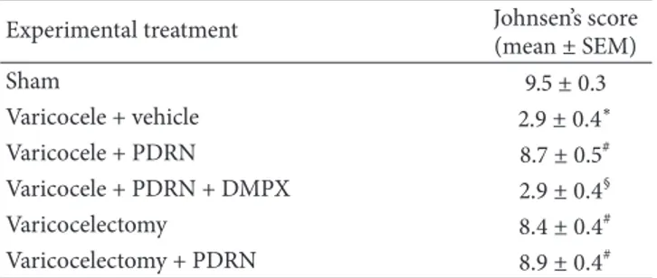
3.4. Spermatogenic Activity. Rats subjected to varicocele and
treated with vehicle showed a significant impairment in
spermatogenic activity after 21 days (Table 1). In contrast, administration of PDRN significantly increased Johnsen’s score while DMPX cotreatment did not improve the altered spermatogenesis, and it abated the beneficial effects of PDRN (Table 1). Varicocelectomy alone significantly reverted the
Table 1: Evaluation of spermatogenesis (Johnsen’s score) of left testis in different experimental groups.
Experimental treatment Johnsen’s score
(mean± SEM) Sham 9.5± 0.3 Varicocele + vehicle 2.9± 0.4∗ Varicocele + PDRN 8.7± 0.5# Varicocele + PDRN + DMPX 2.9± 0.4§ Varicocelectomy 8.4± 0.4# Varicocelectomy + PDRN 8.9± 0.4#
∗𝑃 < 0.001 versus sham;#𝑃 < 0.001 versus varicocele + vehicle;§𝑃 < 0.001
versus varicocele + PDRN.
altered spermatogenic activity while varicocelectomy with PDRN group did not observe any additional significant restorative effect of spermatogenic function (Table 1).
4. Discussion
We have previously shown that PDRN, acting as a selective agonist on adenosine A2AR, was effective in improving the experimental varicocele [31, 32]. Specifically, PDRN suc-ceeded in ameliorating the histologic damage and the poor spermatogenesis in the affected rats.
The present data represent novel and adjunctive findings distinct from those published previously [31, 32]. In fact, PDRN administration resulted in a specific and highly regu-lated action on apoptosis, particularly through the control of expression of NAIP and survivin. Indeed, it appears useful to point out that varicocele induction and subsequent postsur-gical isolation of rats could interfere with our results in this experimental model [35,37,38]. Consequently, since social isolation in rodents is associated with a range of behavioural and physiological changes, some of which indicate a stress response, we have minimized the impact and time of social isolation (only one week) in all experimental groups. This approach allowed us to promote adequate healing and post-operative recovery and prevent the spread of infections.
As a first step, our experimental findings suggest that NAIP and survivin could play a pivotal role in the pathogen-esis of varicocele. In fact, NAIP and survivin were markedly reduced following varicocele induction while PDRN admin-istration markedly augmented both members of IAPs fam-ily; moreover, the concomitant administration of DMPX, a specific adenosine A2AR antagonist, abrogated the effects of PDRN on the antiapoptotic proteins.
Indeed, our sham testis samples had measurable levels of NAIP in accordance with previous data obtained on human tissues [23]; this experimental finding, indicating the presence of NAIP within tissues that are not usually affected a neuron disease, would suggest that NAIP has functional/physiological implications in a broad spectrum of tissues, exerting both antiapoptotic and immunoendocrine modulatory functions.
As a matter of fact, qRT-PCR and western blot analysis revealed an enhanced expression of NAIP in testis samples
following PDRN administration compared to those vehicle-treated and/or after varicocelectomy, alone. This leads us to speculate that PDRN may reawake the depressed apop-totic machinery. In agreement with this latter observation, immunostaining for NAIP revealed that PDRN significantly causes diffuse and strong positivity in spermatogonia and spermatozoa, confirming the crucial role of the apoptotic signals in germ cells differentiation and testis endocrine and spermatogenic function [17]. This observation is intriguing in terms of infertility management in mammalian species because a previous work by Maier and coworkers indicated that (i) the expression pattern of NAIP in testis did not necessarily reflect the immunohistochemical data and (ii) the increase of NAIP might be also related to a generic compensatory mechanism involving inflammatory cells as macrophages in target tissues [23].
The difference between our results and those previously described could be easily due to the different species exam-ined (rat versus human), different microenvironment (patho-logical versus normal condition), and different therapeutic approach targeting specific molecular pathways.
So far, current knowledge indicates that, besides NAIP, survivin is highly expressed in adult human testis, especially in the nuclei of mature spermatocytes, and its downregulation is linked with spermatogenic failure [39–41]. Moreover, recent experimental evidences indicated that survivin could be associated with a direct control of cell survival: it acts as protein inhibitor of apoptosis by inhibiting caspases and antagonizing mitochondria-dependent apoptosis [42].
In our model of experimental varicocele we found an enhanced expression of survivin following PDRN admin-istration in agreement with previous experimental obser-vations by Bayomy and coworkers [43]. They showed that a left-sided varicocele causes bilateral testicular histolog-ical abnormalities, angiogenesis, and apoptosis, including a marked increase in the expression of caspase-3 and a marked decrease in the expression of survivin [43]. In the present research, we found that survivin immunostaining shows a more diffuse and strong positivity particularly in spermatogonia and Leydig cells of PDRN-treated rats; as expected, DMPX, blocking A2ARs, abrogated PDRN effects on survivin expression in varicocele-induced rats.
The positive modulation of IAPs molecular pathway obtained by PDRN treatment was also supported in our model by the evidence of a significant restorative effect of spermatogenic function. These effects are comparable to those obtained in rats subjected to varicocelectomy [4–7], suggesting that PDRN could represent a novelty in terms of valuable medical therapy of varicocele-induced infertility.
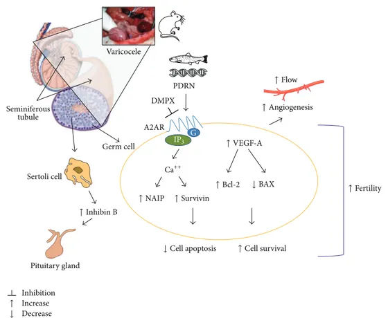
So far, the present data allow us to better clarify our previous experimental observations about the molecular pathways involved in testis following surgical induction of varicocele in rats. As summarized inFigure 5, the stasis and the consequent hypoxic-ischemic state following varicocele may severely damage the testicular function. Then, the acti-vation of A2AR by polynucleotides (PDRN) induces a further production of VEGF that, in turn, improves the angiogenic response in injured testis. At this stage, it is very likely that a pathophysiological cross-talk occurs between testicular flow,
A2AR PDRN DMPX Germ cell Seminiferous tubule Increase Decrease Inhibition Pituitary gland Sertoli cell Varicocele ↓ Cell apoptosis ↓ ↓ BAX Ca++ ↑ Inhibin B ↑NAIP ↑Survivin ↑Cell survival ↑Bcl-2 ↑Fertility ↑VEGF-A ↑Flow ↑Angiogenesis ↑ G IP3
Figure 5: Scheme showing some of the molecular pathways varicocele-induced and their strategical modulation by PDRN.
hormones, and cell homeostasis pathways and a plethora of signaling molecules including IAPs could have a great impact on angiogenesis, fertility, and testicular function [29–32,44]. Altogether, these findings point out the key role played by NAIP and survivin in the positive regulation of apop-tosis during varicocele, also confirming that, in the context of translational medicine, these members of IAPs family could represent an interesting and innovative target in testis homeostasis. Consequently, a specific approach with PDRN, a ligand of A2AR, could represent a valuable therapeutic strategy for varicocele management in subfertile patient; moreover, PDRN may also offer a structural model for the design of new analog compounds helpful for male infertility.
Conflict of Interests
The authors declare that there is no conflict of interests regarding the publication of this paper.
Acknowledgment
This study was supported by departmental funding. PDRN was a kind gift of Mastelli S.R.L. (Sanremo, Italy).
References
[1] N. G. Haddad, C. P. Houk, and P. A. Lee, “Varicocele: a dilemma in adolescent males,” Pediatric Endocrinology Reviews, vol. 11, supplement 2, pp. 274–283, 2014.
[2] J. G. Reyes, J. G. Farias, S. Henr´ıquez-Olavarrieta et al., “The hypoxic testicle: physiology and pathophysiology,” Oxidative
Medicine and Cellular Longevity, vol. 2012, Article ID 929285,
15 pages, 2012.
[3] K. Shiraishi, H. Matsuyama, and H. Takihara, “Pathophysiology of varicocele in male infertility in the era of assisted reproduc-tive technology,” International Journal of Urology, vol. 19, no. 6, pp. 538–550, 2012.
[4] E. C. Serefoglu, T. R. Saitz, J. A. La Nasa Jr., and W. J. Hellstrom, “Adolescent varicocoele management controversies,”
Androl-ogy, vol. 1, no. 1, pp. 109–115, 2013.
[5] G. Santoro and C. Romeo, “Morphological and surgical overview of adolescent testis affected by varicocele,” The
Scien-tific World Journal, vol. 2013, Article ID 469413, 8 pages, 2013.
[6] A. C. J. Kroese, N. M. de Lange, J. A. Collins, and J. L. H. Evers, “Varicocele surgery, new evidence,” Human Reproduction
Update, vol. 19, no. 4, article 317, 2013.
[7] N. Kwak and D. Siegel, “Imaging and interventional therapy for varicoceles,” Current Urology Reports, vol. 15, no. 4, article 399, 2014.
[8] G. Majno and I. Joris, “Apoptosis, oncosis, and necrosis: an overview of cell death,” The American Journal of Pathology, vol. 146, no. 1, pp. 3–15, 1995.
[9] I. Sukhotnik, A. Bernshteyn, and J. G. Mogilner, “The basic biology of apoptosis and its implications for pediatric surgery,”
European Journal of Pediatric Surgery, vol. 15, no. 4, pp. 229–235,
2005.
[10] M. G¨urdal, S. Kirec¸c¸i, E. Huri, I. Karaman, and L. T¨urkeri, “Correlation between duration of varicocele and apoptosis in
testicular tissue in an experimental model,” Urology, vol. 72, no. 4, pp. 933–936, 2008.
[11] J.-D. Lee, T.-H. Lee, W.-H. Cheng, and S.-Y. Jeng, “Involved intrinsic apoptotic pathway of testicular tissues in varicocele-induced rats,” World Journal of Urology, vol. 27, no. 4, pp. 527– 532, 2009.
[12] F.-W. Chang, G.-H. Sun, Y.-Y. Cheng, I.-C. Chen, H.-H. Chien, and G.-J. Wu, “Effects of varicocele upon the expression of apoptosis-related proteins,” Andrologia, vol. 42, no. 4, pp. 225– 230, 2010.
[13] Y. Sakamoto, T. Ishikawa, Y. Kondo, K. Yamaguchi, and M. Fujisawa, “The assessment of oxidative stress in infertile patients with varicocele,” BJU International, vol. 101, no. 12, pp. 1547– 1552, 2008.
[14] A. Agarwal, G. Virk, C. Ong, and S. S. du Plessis, “Effect of oxidative stress on male reproduction,” The World Journal of
Men’s Health, vol. 32, no. 1, pp. 1–17, 2014.
[15] I. Chowdhury, B. Tharakan, and G. K. Bhat, “Current concepts in apoptosis: the physiological suicide program revisited,”
Cel-lular and Molecular Biology Letters, vol. 11, no. 4, pp. 506–525,
2006.
[16] K. Sakamaki, “Physiological and pathological cell deaths in the reproductive organs,” Cell Structure and Function, vol. 28, no. 1, pp. 31–40, 2003.
[17] K. K. Shukla, A. A. Mahdi, and R. Singh, “Apoptosis, spermato-genesis and male infertility,” Frontiers in Bioscience—Elite, vol. 4, no. 2, pp. 746–754, 2012.
[18] M. Tek, S. C¸ ayan, N. Yilmaz, I. O˘guz, E. Erdem, and E.
Akbay, “The effect of vascular endothelial growth factor on spermatogenesis and apoptosis in experimentally varicocele-induced adolescent rats,” Fertility and Sterility, vol. 91, no. 5, supplement, pp. 2247–2252, 2009.
[19] H. Wang, Y. Sun, L. Wang et al., “Hypoxia-induced apoptosis in the bilateral testes of rats with left-sided varicocele: a new way to think about the varicocele,” Journal of Andrology, vol. 31, no. 3, pp. 299–305, 2010.
[20] D.-Y. Luo, G. Yang, J.-J. Liu, Y.-R. Yang, and Q. Dong, “Effects of varicocele on testosterone, apoptosis and expression of StAR mRNA in rat Leydig cells,” Asian Journal of Andrology, vol. 13, no. 2, pp. 287–291, 2011.
[21] T. Mostafa, L. Rashed, N. Nabil, and R. Amin, “Seminal BAX and BCL2 gene and protein expressions in infertile men with varicocele,” Urology, vol. 84, no. 3, pp. 590–595, 2014.
[22] C. S. Duckett, V. E. Nava, R. W. Gedrich et al., “A conserved family of cellular genes related to the baculovirus iap gene and encoding apoptosis inhibitors,” The EMBO Journal, vol. 15, no. 11, pp. 2685–2694, 1996.
[23] J. K. X. Maier, S. Balabanian, C. R. Coffill et al., “Distribution of neuronal apoptosis inhibitory protein in human tissues,”
Journal of Histochemistry and Cytochemistry, vol. 55, no. 9, pp.
911–923, 2007.
[24] S. Karimpour, J. Davoodi, and M.-H. Ghahremani, “Integrity of ATP binding site is essential for effective inhibition of the intrinsic apoptosis pathway by NAIP,” Biochemical and
Biophysical Research Communications, vol. 407, no. 1, pp. 158–
162, 2011.
[25] M. Pennati, M. Folini, and N. Zaffaroni, “Targeting survivin in cancer therapy,” Expert Opinion on Therapeutic Targets, vol. 12, no. 4, pp. 463–476, 2008.
[26] J. K. X. Maier, Z. Lahoua, N. H. Gendron et al., “The neuronal apoptosis inhibitory protein is a direct inhibitor of caspases 3
and 7,” Journal of Neuroscience, vol. 22, no. 6, pp. 2035–2043, 2002.
[27] J. Chen, W. Wu, S. K. Tahir et al., “Down-regulation of sur-vivin by antisense oligonucleotides increases apoptosis, inhibits cytokinesis and anchorage-independent growth,” Neoplasia, vol. 2, no. 3, pp. 235–241, 2000.
[28] Y. Estornes and M. J. M. Bertrand, “IAPs, regulators of innate immunity and inflammation,” Seminars in Cell and
Develop-mental Biology, 2014.
[29] D. Altavilla, F. Squadrito, F. Polito et al., “Activation of adenosine
A2𝐴receptors restores the altered cell-cycle machinery during
impaired wound healing in genetically diabetic mice,” Surgery, vol. 149, no. 2, pp. 253–261, 2011.
[30] D. Altavilla, A. Bitto, F. Polito et al., “Polydeoxyribonucleotide (PDRN): a safe approach to induce therapeutic angiogenesis in peripheral artery occlusive disease and in diabetic foot ulcers,”
Cardiovascular & Hematological Agents in Medicinal Chemistry,
vol. 7, no. 4, pp. 313–321, 2009.
[31] L. Minutoli, S. Arena, G. Bonvissuto et al., “Activation of adenosine A2A receptors by polydeoxyribonucleotide increases vascular endothelial growth factor and protects against testicu-lar damage induced by experimental varicocele in rats,” Fertility
and Sterility, vol. 95, no. 4, pp. 1510–1513, 2011.
[32] S. Arena, L. Minutoli, F. Arena et al., “Polydeoxyribonucleotide administration improves the intra-testicular vascularization in rat experimental varicocele,” Fertility and Sterility, vol. 97, no. 1, pp. 165–168, 2012.
[33] P. Sengupta, “The laboratory rat: relating its age with human’s,”
International Journal of Preventive Medicine, vol. 4, no. 6, pp.
624–630, 2013.
[34] F. Kilinc¸, F. Kayaselcuk, C. Aygun, S. Guvel, T. Egilmez, and H. Ozkardes, “Experimental varicocele induces hypoxia inducible factor-1𝛼, vascular endothelial growth factor expression and angiogenesis in the rat testis,” The Journal of Urology, vol. 172, no. 3, pp. 1188–1191, 2004.
[35] J. M. Koolhaas, “The laboratory rat,” in The UFAW Handbook
on the Care and Management of Laboratory and Other Research Animals, R. Hubrecht and J. Kirkwood, Eds., pp. 311–327,
Universities Federation for Animal Welfare, Wheathampstead, UK, 8th edition, 2010.
[36] S. E. Johnsen, “Testicular biopsy score count-a method for registration of spermatogenesis in human testes: normal values and results in 335 hypogonadal males,” Hormones, vol. 1, no. 1, pp. 2–25, 1970.
[37] S. Pietropaolo, P. Singer, J. Feldon, and B. K. Yee, “The postwean-ing social isolation in C57BL/6 mice: preferential vulnerability in the male sex,” Psychopharmacology, vol. 197, no. 4, pp. 613– 628, 2008.
[38] C. M. McCormick and I. Z. Mathews, “HPA function in adolescence: role of sex hormones in its regulation and the enduring consequences of exposure to stressors,” Pharmacology
Biochemistry and Behavior, vol. 86, no. 2, pp. 220–233, 2007.
[39] M. Al-Maghrebi, E. O. Kehinde, and J. T. Anim, “Survivin downregulation is associated with vasectomy-induced sper-matogenic damage and apoptosis,” Medical Principles and
Prac-tice, vol. 20, no. 5, pp. 449–454, 2011.
[40] S. Weikert, M. Schrader, F. Christoph et al., “Quantification of survivin mRNA in testes of infertile patients and in testicular germ cell tumours: high levels of expression associated with normal spermatogenesis,” International Journal of Andrology, vol. 28, no. 4, pp. 224–229, 2005.
[41] S. Weikert, M. Schrader, M. M¨uller, W. Schulze, H. Krause, and K. Miller, “Expression levels of the inhibitor of apoptosis survivin in testes of patients with normal spermatogenesis and spermatogenic failure,” Fertility and Sterility, vol. 83, no. 4, supplement, pp. 1100–1105, 2005.
[42] Z. Song, X. Yao, and M. Wu, “Direct interaction between Survivin and Smac/DIABLO is essential for the anti-apoptotic activity of Survivin during Taxol-induced apoptosis,” The
Jour-nal of Biological Chemistry, vol. 278, no. 25, pp. 23130–23140,
2003.
[43] N. A. Bayomy, N. I. Sarhan, and K. M. Abdel-Razek, “Effect of an experimental left varicocele on the bilateral testes of adult rats: a histological and immunohistochemical study,” Egyptian
Journal of Histology, vol. 35, no. 3, pp. 509–519, 2012.
[44] M. L. Eisenberg and L. I. Lipshultz, “Varicocele-induced infer-tility: newer insights into its pathophysiology,” Indian Journal of
Submit your manuscripts at
http://www.hindawi.com
Stem Cells
International
Hindawi Publishing Corporation
http://www.hindawi.com Volume 2014
Hindawi Publishing Corporation
http://www.hindawi.com Volume 2014 INFLAMMATION
Hindawi Publishing Corporation
http://www.hindawi.com Volume 2014
Behavioural
Neurology
Endocrinology
International Journal ofHindawi Publishing Corporation
http://www.hindawi.com Volume 2014
Hindawi Publishing Corporation
http://www.hindawi.com Volume 2014
Disease Markers
Hindawi Publishing Corporation
http://www.hindawi.com Volume 2014
BioMed
Research International
Oncology
Journal ofHindawi Publishing Corporation
http://www.hindawi.com Volume 2014
Hindawi Publishing Corporation
http://www.hindawi.com Volume 2014
Oxidative Medicine and Cellular Longevity
Hindawi Publishing Corporation
http://www.hindawi.com Volume 2014
PPAR Research
The Scientific
World Journal
Hindawi Publishing Corporation
http://www.hindawi.com Volume 2014
Immunology Research
Hindawi Publishing Corporation
http://www.hindawi.com Volume 2014
Journal of
Obesity
Journal ofHindawi Publishing Corporation
http://www.hindawi.com Volume 2014
Hindawi Publishing Corporation
http://www.hindawi.com Volume 2014
Computational and Mathematical Methods in Medicine
Ophthalmology
Journal of Hindawi Publishing Corporationhttp://www.hindawi.com Volume 2014
Diabetes Research
Journal ofHindawi Publishing Corporation
http://www.hindawi.com Volume 2014
Hindawi Publishing Corporation
http://www.hindawi.com Volume 2014
Research and Treatment
AIDS
Hindawi Publishing Corporation
http://www.hindawi.com Volume 2014
Gastroenterology Research and Practice
Hindawi Publishing Corporation
http://www.hindawi.com Volume 2014