Università degli Studi di Ferrara
DOTTORATO DI RICERCA IN
" STUDI UMANISTICI E SOCIALI"
CICLO XXIVCOORDINATORE Prof. Angela Andrisano
The development of motor activity:
observing spontaneous behavior in fetuses, preterm and term infants
Settore Scientifico Disciplinare 11/E2
Dottorando Tutore
Dott. Valente Angela Prof. Dondi Marco
1 Acknowledgments
First of all I would like to thank prof. Marco Dondi who introduce me in the wonderful world of scientific research. Thank you for your generous teachings and for believing in me.
Sincere thanks to my colleague and friend Tiziana, for sharing this adventure with me. Thank you for listening to me.
A special thanks to all the students I met on my course: thanks to Susy, Marica, Ilenia and Chiara for having learned with me.
Many many thanks to all the babies and all the mothers who took part in this research.
Thanks to all the researchers and the medical units staff involved in this study for their availability and kindness in data collection.
Thanks to Dr. Maria Teresa Gervasi, the obstetrician Maria Rosa Tran and all the staff of the Center for Prenatal Pregnancies at Risk, U. O. C. of Obstetrics and Gynecology, Hospital of Padua for supporting this research with their enthusiasm.
Thanks to Dr. Beatrice Dalla Barba and all the staff of the U. O. Pathology and Neonatal Intensive Care Unit of the Hospital of Padua.
Thanks to Professor Angela Costabile, to Dr. Flaviana Tenuta and to the U. O. team of Neonatology and Neonatal Intensive Care Unit of the Hospital of Cosenza.
Thanks to the Director of Neonatology and Neonatal Intensive Care Unit of the Hospital of Ferrara Prof. Giampaolo Garani and Dr. Elisa Ballardini.
A special thanks to prof. Maria Luisa Genta for her fond words. Thank you for having encouraged me.
Finally a special thanks to doctor Harriet Oster for having inspired me.
Ferrara, April, 2013 Angela Valente
2 TABLE OF CO TE TS
Introduction ... 4
Part I Origins and behavior ... 7
1. The origins of motor behavior ... 7
1.1. Historical background ... 7
1.2. Ontogeny of human behavior ... 11
1.3. The concept of neuro – behavior ... 13
1.4. The reasons for complexity ... 13
1.5. Continuity in development ... 14
2. Describing human fetal motor behavior ... 17
2.1. What is ultrasound technology? ... 17
2.2. Ultrasound 3D and 4D new technologies ... 18
2.3. Mapping the face: craniofacial development ... 19
2.4. Fetal abilities and skills ... 20
2.5. Behavioral states from fetus to neonates ... 23
3. Prematurity ... 26
3.1. Preterm newborn characteristic ... 29
3.2. Effects of preterm birth on the development of behavior ... 30
3.3. Behavioral states differences between term and preterm infants ... 31
3.4. The assessment of preterm and term infants ... 34
3.5. Neurological and neurobehavioral scale ... 37
Part II Experimental Section ... 39
4. Experiment I: Comparing fetuses and preterm infants at the same gestational age ... 39
4.1. Introduction ... 39
4.2. Method ... 40
4.2.1. Participants ... 40
4.2.2. Procedure ... 41
4.2.3. Coding spontaneous motor behavior in fetuses and preterm infants ... 41
4.2.4. Reliability... 42
4.2.5. Analysis of data ... 42
4.3. Results ... 42
4.4. Discussion ... 49
5. Experiment II: Observing spontaneous behavior in preterm infants before and after meal ... 54
5.1. Introduction ... 54
5.2. Method ... 57
5.2.1. Participants ... 57
5.2.2. Procedure ... 57
5.2.3. Coding spontaneous motor behavior ... 57
5.2.4. Reliability... 58
5.2.5. Data Analysis ... 58
5.3. Results ... 58
5.4. Discussion ... 68
6. Experiment III: Effects of birth-weight and gestational age on spontaneous motor behavior ... 71
6.1. Introduction ... 71
6.2. Method ... 72
6.2.1. Participants ... 72
6.2.2. Procedures ... 73
6.2.3. Coding spontaneous motor behavior in the preterm and term neonates... 73
6.2.4. Reliability... 74
3
6.3. Results ... 74
6.4. Discussion ... 80
APPENDIX ... 83
4
Introduction
Recently, the observation of spontaneous motor behavior from the beginning of prenatal life has been always more interesting for students and scientists. This is caused by the newborn’s skills and abilities that he proves to have just after birth, and it is assumed they emerge during prenatal life. In the Seventies the advent of the new ultrasound technologies made possible the observation of human fetal behavior and also the increasing of knowledge of embryo and fetus that has opened a new window to observe human development. Nowadays, ultrasound examination helps to promote the concept of continuity in human motor behavior from prenatal to postnatal life.
The main aim of this study is to observe spontaneous motor behavior from the beginning of movement in utero. Human motor behavior is related to the normal function of central nervous system and these observations can detect early, or even before birth, the integrity of central nervous system (Prechtl, 1997). It is also important to note that presently there is not a neurobehavioral scale universally accepted and recognized as valid for the observation of spontaneous fetal behavior yet (Di Pietro, 2005).
Spontaneous behavior is an activity endogenously generated by the central nervous system which reflects the state of neural development. As behavior is sensitive to changes in physiological and motivational functions, the study of spontaneous motor activity allows to understand the evolutionary trajectories of specific functions, and therefore, to identify atypical paths as well as newborn well-being. Therefore, it is possible to note that atypical motor patterns, characterized by monotonous, stereotyped and less fluent movements, may predict adverse neurological outcomes.
Over the past thirty years, even the way to observe and evaluate infant behavior has changed. Until the Sixties, newborn was considered a passive receptor unable to respond to environmental stimuli, and his assessment was predominantly made by observations of neurological reflexes. Presently, newborns prove to play an active role thanks also to their behavioral repertoire already showed after birth. The modern medicine challenge is especially in the field of developmental techniques of life support and these are allowing survival of fetuses aged even less than 28 weeks of gestational age. Since preterm newborn was not able to spend the last trimester of pregnancy in utero, he is called "premature" for his structural conditions of immaturity. Therefore, it could be interesting to study behavior in fetuses and their typical developmental trajectories to better understand how this development can occur in cases of premature births.
During our first experiment in order to describe behavior both before and after birth, we are going to code spontaneous motor patterns in a group of fetuses and to compare to a group of preterm infants, at the same gestational age. To specify each pattern we developed a new coding scale predominantly made on anatomical basis. We considered as a starting point the micro-analytic facial coding system for babies and children Baby FACS, written by Harriet Oster (2007). This new motor
5 patterns scale was designed for the detection of specific complex movements coming from the behavioral categories originally described by de Vries (1982), Prechtl (1985), Kurjak et al. (2003), Einspieler et al. (2008) and Wolff (1987), and observed in fetuses, preterm and term neonates. Therefore, we described in detail 22 complex motor patterns as much objective as possible, in order to ensure reliability regardless of the subjective interpretations of individual encoders. Our interest comes from the recent opportunity to observe spontaneous behavior in preterm infants at a very low gestational age, and it also comes from the opportunity to discover fetal behavior. The observation of spontaneous motor activity will help us to understand the ontogeny of human behavior, but it will also help us on the way to the evaluation of the activity of central nervous system and human well-being.
In order to describe spontaneous behavior both before and after birth, we are going to dedicate the first experiment to code spontaneous motor activity in a group of fetuses and preterm infants at the same gestational age.
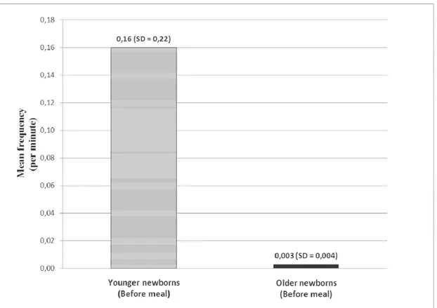
The theory of dynamical systems in the study of motor development emphasizes that changes in spontaneous motor pattern can be determined by changes in any factor subject to interaction, such as biomechanics growth, functional, motivational or cognitive changes (Kamm, Thelen& Jensen, 1990). According to this perspective, we can hypothesize that the fulfillment of a basic need, as a motivational factor, can influence the manifestation of behaviors. Therefore there could be a relationship between the onset of the condition of appetite and spontaneous behavior. In the second experiment we are going to test the same spontaneous motor activity coding scale already used before. Our intention in this second experiment is to test the sensitivity of this scale, coding two groups of preterm newborns video-recorded before and after meal. We wonder how the preterm behavior may be modulated by the necessity to satisfy the appetite basic need and what are the differences emerging from a comparison in spontaneous behavior observed before and after meal.
The second experiment is concerned with the observation of spontaneous motor behavior in premature infants in order to investigate how the primary condition of appetite can modulate the behavioral performance. We are going to observe preterm newborns behavior through the observation of video recordings of two groups of premature infantsrecorded before and after the meal.
The medical-technology advances led to an improvement of care and an increase of the indices of neonatal survival (Fava Vizziello, 1992). Today, they can ensure survival for infants at less than 400 grams and a gestational age of 24 weeks. Preterm birth is a quite common phenomenon. According to the latest OMS "Born too soon: the global action report on preterm birth," published on May 2012, every year 15 million children born premature, with a ratio of more than 1 in 10 preterm births, showing an alarming increase in cases in the last 20 years in almost all countries surveyed by OMS experts. Unequal cases were detected according to the different geographical areas analyzed, and it is found that more than 60% of premature births occurs in Africa and South Asia, and this variability also affects the countries of the North. In the United States of America there are 12% of infants born prematurely at a mean ratio of 9% of high-income countries, and 7% in Italy (World Health Organizations, Born too soon, the global Action Report On Preterm Birth, 2012 ). Despite the survival
6 of preterm infants, the quality of that survival is certainly improved thanks to the advances in care of preterm infants obtained in the last twenty years with the application of new knowledge of neonatal pathophysiology, technology monitoring of vital functions, the new possibilities for respiratory care, parenteral feeding and other modern diagnostic and therapeutic techniques.Among the survivors, the effects of premature birth can occur throughout the entire life and they can influence the neuro-functional development. The arrangement of the premature is different from a full-term newborn, where the focus is on the interaction with the family, relatives and friends. In case of preterm birth instead the focus is on health, on cardiac, respiratory, and brain functions. The premature baby is immediately accepted as a "different"child.
In the light of the last considerations in both previous studies, during the third and last experiment we are going to test the new motor coding scale for the observation of spontaneous behavior in a group of full-term newborn infants and to compare to a group of preterm newborn infants at the same gestational age. The last intention is to underline the sensitivity of this scale in revealing differences between preterm and full-term newborns. After the assessment of the actual sensitivity of the motor patterns scale specifically designed for the detection of spontaneous behavior, we are now wondering how the preterm newborn’s spontaneous behavior may differ from the full-term newborn behavior observed at the same gestational age.
The collected material was used in the respect to the privacy (in accordance to the Legislative Decree no. . 196 of June 30, 2003 "Code concerning the protection of personal data").
7
Part I Origins and behavior
1. The origins of motor behavior
“The actuality of being in power, as a being in power, is the
movement”
Aristotele, Fisica, III (G), 1, 201a10-11.
In the systematic thesis of movement and change (Phys. III 1-3) and the different types of them (De Gen. et corr. I 1-5), Aristotle takes up and develops themes that emerged in the survey on the principles. Nature is the principle of movement and change and do not know what movement is would, therefore, ignore what nature is, but, on the other hand, there is no movement outside of the things that are (pragmata) in motion, because there is nothing outside of these, then: to know what movement is you have to investigate what and how many ways they are and are said to be things that are.
Each of these things can then be in power or act (entelecheia).
The movement is the act of what is in potency itself. The generation and corruption of a substance, which is the coming to be of something that was not, and his cease to be while before it was, constitutes what may be called for Aristotle generation and corruption in an absolute sense, but this does not imply a generated by or corrupted in what absolutely is not.
Finally dismissing the old Eleatic aporia (according to Parmenides, in fact, being is and is not possible that it is not; non-being is not and it is necessary it is not), Aristotle says (De Gen. et corr. 3 I, 317 a 32-b 33) that even what is not, anyway is: it is in power, not in place. That 's why we can say that man is born from man, things are created by something that is in place (which is a substance), but that is in power the same that arises from it, nor it could be in power if it had not in place.
1.1. Historical background
The term “quickening” is the moment of pregnancy in which a woman first feels fetal movements and they have been attributed to the beginning of individual life. This is the criterion historically regarded to determine the starting point which it is conceded to the fetus the right to life.
8 Hippocrates (460-370 a. C.) had already suspected that fetal movements may set a few weeks earlier than expectant mother feels them, around 70-90 days after conception which correspond to a gestational age of 12-15 weeks.
Rebecca, Isaac’s wife, in the Bible is probably the first written account of human fetal movement (Luke 1:39-44)1 .
Actually, it is enough interesting to notice that charting the mothers’ perception of fetal motion is the oldest and simplest method to monitor fetal well-being during the second half of pregnancy.
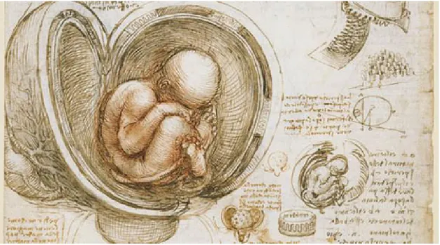
The modern age contribution to embryology comes from Leonardo Da Vinci, with his famous drawings of a fetus in a womb (1510-1512), where we can see the illustration of embryos and fetuses, according to the ability discovered for the first time the real measure and assess dimension and development related to the observation in all the different gestational ages. Leonardo’s anatomical drawings, discovered in 1900, should have been collected in a treaty never completed and left to his assistant Francesco Melzi, after the author’s death. In 1690 Charles II bought them and remained in the Royal Collection. At that time, dissection was not forbidden by the Church and the artist inspired by Avicenna and Galeno di Pergamo studied and the discoveries made in hospital and medical schools. during early dissections of human bodies. The beautiful studies of human skull and fetus give us the greatness of his ability and interest in neurology and represent the first and the most detailed testimony of human’s anatomy2.
Figure 1. Leonardo Da Vinci – Studies of the skull
1 “Now at this time Mary…entered house of Zacharias and greeted Elizabeth. And it came about that when Elizabeth heard Mary’s greeting, the baby leaped in her womb;…and she cried out with a loud voice…when the sound of your greeting reached my ear, the baby leaped in my womb for joy” (Luke 1:39-44).
9
Figure 2. Leonardo Da Vinci – Embryological Drawings of the fetus
In 1885 the English-German physiologist William T. Preyer (1841-1897) “heard” the fetal movement placing a stethoscope on a mother’s abdomen and noticed that movements were definitely present around 12 week of gestational age or even earlier. At that time scientists were convinced that these movements had to be evoked, but in spite speculation of that period, Preyer believed those early movements were spontaneously generated.
First non-invasive study of fetal behavior was conducted by Fels Longitudinal Study (the Fels research Institute was founded in 1929) originally designed to analyse the effects of the “Great Depression Time” child had development, posing the question: “what makes people different?”, derived from the study of individuals from prenatal life to adulthood. Then Sontag and Wallace (1934) were able to differentiate between slow squirming or writhing movements, which increased until 28 week of gestational age and then decreased. They assumed there is a great differnce among fetuses as well as s difference in fetus himself as he changes day by day (Sontag 1941), and realized first classification of fetal movements as kicking and punching, which decreased with the fetus growing and with the age, and rare, small rhythmic movements and possibly hiccups. This study also confirmed that maternal emotional stress is linked to fetal behavior as those infants remained irritable and hyperactive for weeks, cried a great deal and slept for short periods (Sontag 1941).
Through the perspective of the reflexology doctrine that considered the motility a merely response to exogeneous stimuli, we can classify other systematic studies on human fetal motility carried out after spontaneous miscarriages or after Cesarean sections. Examination of exteriorized embryos and fetuses were conducted by Hooker (1938, 1952) and Minkowsky (1928), and at the same time Coghill (1929), they applied tactile stimulations on amphibians embryos and fetuses, Barcroft et
10 al. (1940) on sheep and Windle and Becker (1940) on cats. According to these studies there were two different thesis: Coghill believed that reflex mass movements preceded individual reflex movements, while Windle (1940) suggested that complex coordinated movements develop by integration of local reflex circuits, according to the reflex-integration approach to reflexology of 1930s and 1940s.
It is due to Hooker Davenport the beginning of study on human fetal activity (in 1932 at the University of Pittsburgh) that he was influenced by previous studies conducted on animals and summarized by George E. Coghill. Although for those studies motility was considered to be evoked, it was common to observe movements without any evidence of prior stimulation and to classify them as “spontaneous reflex which the stimulus of was not known yet” (Hooker, 1952). It is also important to notice that these observations were conducted on dying fetuses, so presumably the nervous system was in a depressed state and activity almost ceased (de Vries et al. 1982; Prechtl 1989).
The use of B-mode linear scanner allowed the Viennese obstetrician Emil Reinold in 1971 to study fetal movements obtaining first sonar observation of cross-sections through uterus, although not yet simultaneously. He described two types of movements: a lively movement beginning with a short impulse and ending with a motionless phase and a slow and lazy one, generally followed by a pause of 1-5 minutes (Reinold, 1973). He stated fetal movements were not forceful enough to alter position of fetal body before 10th weeks of gestational age, and for the first time he was able to concluded that observed movements of the fetus were spontaneous rather than caused by external influences (Reinold, 1973). By these earliest reports, Birnholz was influenced and he made a categorization scheme of fetal motion with a particular attention to the extension of the head or limbs relative to the trunk, the rotation of the torso and to independent limbs movements, combined/repetitive and respiratory movements (Birnholz et al. 1978).
After the advent of new ultrasound scanners it has been possible to see simultaneously fetal movements in uterus. These new technologies allowed to determine correlation between fetal movements and the correspondent age. Having a great number of recorded images per second that can provide real-time recordings, some obstetricians such as Ianniruberto and Tajani in 1981, de Vries et al. in 1982, first of all have carried out examinations to evaluate the onset of motility and behavior in collaboration with neuropaediatrician Adriano Milano Comparetti and the developmental neurologist Heinz Prechtl. Real time observation opened a view to a new kind of studies in human behavior as well as to the possibility of neurological assessment in human fetus (de Vries et al 1982, 1985, 1988, Nijhuis et al. 1982).
High resolution in real scanning images became a new tool to observe movement patterns, underlying differences between quantitative and qualitative analysis, as a basis to understand condition and assessment of nervous system. It was due to the Groningen’s study founded by Prechtl, the beginning of investigation with few very important shared questions that we can summarize as follow: how should fetal movement patterns be classified? At what age do they appear at the first time? Do
11 they change in the course of intrauterine development? Are there any age-related preferences with regard to the fetal position? Are there any specific motor patterns which are responsible for changes in the fetal position? (Prechtl and Schleidt 1950, Prechtl 1953, 1958, 1977, Prechtl et al 1979). It was surprising to assume that all the observed patterns in uterus were repeated after birth in neonates (Prechtl 1984, 1985, 1989, 2001).
1.2. Ontogeny of human behavior
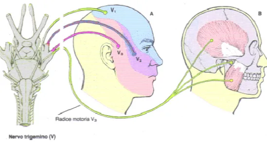
At the beginning, the study of human nervous system has been investigated in early human fetuses at the operating table. The reactions of many human fetuses older than 9 weeks of gestational age have been studied both by Minkowski (1928) and Hooker (1939). First reflexes are established by 8 ½ or 9 weeks, when the human nervous muscle system have attained degrees of maturity compatible with functioning before 8 weeks g.a. (Windle and Fitzgerald, 1937). In this study, James E. Fitzgerald and William F. Windle have had occasion to observe 15 fetuses of 7 weeks to a little over 8 weeks of gestation age under various operating conditions. They supposed that it was probable that the infrequent observation of early human fetal movements were related to the use of general anesthesia and narcotics as well as to the rapid onset of fetal anoxia during the usual surgical procedures. The three fetuses which did react were observed at operations performances under spinal cord anesthesia. No preoperative medication, other than 1|120 grains of atropine sulphate was given to the patients and no pituitrin was used and continuous motion picture records of the experiments performed at the operating table were made. Tapping upon the amniotic sac induced no movements of the fetus and no responses were obtained after touching and stroking the body and limbs with a needle passed through the amniotic sac. When the regions of the mouth and the nose were strongly stimulated with a needle, a contralateral flexion of the trunk appeared. When they tried to touch strongly the region of the entirely face, the whole of the trunk musculature contracted and the arms and legs moved with the body at their attachment. Homolateral responses predominated but contralateral movements were observed, too. At no time there were no movements elicited touching the limbs or the trunk; otherwise movements elicited touching the whole region of the face last for more than 3 minutes.
This lateral movement of the head and trunk, following stimulation of particular parts of the face supplied by the trigeminal nerve, principally maxillary division, has been reported by Hooker (1939) after removing from uterus.
These experimental observations were interpreted by the concept of a stage in development during the eight weeks of gestational age compatible with a limited degree of function These movements are essentially of reflex nature and although if we cannot say what receptors has been stimulated, it is possible to see that some receptors or synapses are more easily rendered non-functional than others: a stimulus applied to the face may induce a trunk flexion reflex or, if strong enough, a mass movement of trunk, arms and legs. So they were able to say that after afferent
12 connections involved in local responses cease functioning, some of the muscle units which were first activated can still be activated by adequate stimulation of the other afferent elements, as we can see for the face (James E. Fitzgerald and William F. Windle, 1942).
Human behavior has been studied since the early origin in prenatal life by Fitzgerald & Windle (1942) and Hooker (1952, 1954, 1958) thought the observation of the human embryo abilities of movement since the 7.5 weeks of menstrual age. They found that by the age of 14the weeks of menstrual age the fetus is able of a wide variety of reflex movements. Cinematographic pictures have been used by Hooker and have been widely used by psychologists and various authors even if for the most part of fetal reflexes were elicitated by stimulation of the cutaneous surfaces by touching or stroking with a needle or a plume.
The first reflexes observed by Hooker (1944, 1952, 1958) were obtained by stimulation of areas supplied by trigerminal nerve and were comparable to the total pattern movements described by Coghill (1929) in amphibians. He found that when the extremities developed they first moved in conjunction with the trunk and only later acted independently. The first response to sensory trigerminal stimulation in development of human fetal reflexes is the early total pattern type of activity. Hooker defined these reflexes as stereotyped and he found that repetition of the stimulus elicited the same response. He also found that local reflexes following trigerminal stimulation begin to appear by 9.5 weeks and as they increase in number the total pattern type of activity begins to disappear and is seen only as anoxia begins to suppress reflex activity (Hooker, 1952, 1958). Then he understood that as the movements become less stereotyped the same stimulus may produce a local reflexes, before anoxia appears. These combinations appear in sequences functional to postnatal life. After 13-14 weeks the movements become graceful and flowing (Hooker, 1952) and lose the more jerky appearance often seen earlier. In vertebrates we can see the connection between motor neurons and their target skeletal muscle fibers. Skeletal muscles allow animals voluntary movements that are controlled by the peripheral nervous actions. This system requires neuromuscolar conjuctions made by striated muscles and confined to a narrow endplate band in the central region of the muscle. At the moment, the process of neuromuscular synapse formation has been extensively studied and well described on a molecular level, but we don’t know what part of the nerve and muscle play in regulating this spatial arrangement. Of course we understand the importance of confining neuromuscular junctions to the central muscle region but the functional meaning of this activity is still unclear.
At the moment, one of the most important question is how this characteristic and stereotyped pattern of innervation is established and what extend motor neurons and muscles contribute to shaping this arrangement.
13
1.3. The concept of neuro – behavior
The concept of neuro-behavior reflects the idea that all human experiences have a double matrix: a biological matrix and a psychosocial one. These two systems dynamically influences each other and they are dependent on a neural response that determines the optimal synergic operation. Therefore the concept of neuro-behavior translates, a dynamic relationship between behavior and physiology.
A neuro-behavioral evaluation criterion should be sensitive to differences in behavior exhibited both in a typical and atypical development.
A tool for assessing the broad neuro-behavioral spectrum, observable from the earliest stages of the motor system development represents a possible allocation of evolutionary ontogenesis of motor activity.
The advent in the 70s of ultrasound technology made possible the observation of human fetal behavior, increased knowledge of embryo and fetus, and helped to promote the concept of continuity of fetal behavior from intrauterine life to the neonatal life (Almli, Ball & Wheeler, 2001; Stanojevic, Kurjak, Salihagic'- Kadic, Vasilj, Miskovic, Shaddad, Ahmed, & Tomasovic, 2011) .
1.4. The reasons for complexity
Many researchers observed a particular variation of the movement in the early stages of neuromuscular development. These studies were mainly related to the observations of infants during the first days after birth (Thelen, 1995). An atypical development of motor system, as well as in cases of cerebral palsy (c .p.), is associated with a lower variability of movements in the first months of the infant's life (Prechtl, 1997). The importance of the qualitative variability criterion is widely recognized in the observation of the motor system developmental trajectory, even during the earliest stages of development.
In 1978 Touwen, for example, argued that variability in movement was an important feature for newborn well-being, and he attributed the origins of this criterion to the properties of the nervous system. Therefore a brain damage was reflected in the form of a reduction in variability, due to the lack of "hardware", or available programs .
Even Prechtl (1990) noted that a reduced movements variability in subjects at risk was sloped in the repetition of monotonous and stereotyped patterns, that were not aimed at achieving a specific objective. These studies interpret the origin of the variability as a phenomenon determined by pre-programmed patterns already present at birth and, in the light of this theoretical perspective, they justify the less of variability in subjects at risk as a result of reduced availability of motor patterns caused by brain damage.
14 The theory of dynamical systems are strongly influenced by ecological perspective, which emphasizes the dynamic relationship between action and perception. Such a perspective sees in new forms of movement and behavior, emerging from the interaction between different factors, more than a particular predetermination of motor patterns chosen within the central nervous system. Thelen and Smith (1994) argued, in fact, that particular individual conditions are determined by the interaction of several factors of human behavior and specify that this approach is the "cornerstone of a dynamic theory of development".
From the point of view of dynamic systems theory variability would indicate a greater stability in behavior in general and not, as for the maturation approach, the index of an atypical development. In recent decades researches on the development of children's motor behavior abandoned the focus on developmental milestones and they focused on the concept of spontaneous behavior observed in early infancy. Therefore spontaneous movement has been recognized as an important precursor to the development of motor control. (Piek, 2002; Thelen, 1995).
Complexity in development means that dynamic systems interaction increases over time, that systems self-organize from subcomponents and context, and finally that development is not predetermined but emergent (Thelen & Smith, 1994). The notion of regulation also includes the concept of plasticity, according to the innate variability of regulating functions among the scenery of different contextual influences (Damasio, 1994). This approach emphasizes the interaction role of bottom-up component, such as physiological, emotional, attentional and self-regulatory functions, as well as the integrative component of brain stem, limbic and cortical system (Tucker et al., 2000). This model suggests that development is generally gradual and it is also constrained by the system’s state initial conditions of a previous point. The evolutionary perspective adapted by these interactive models is the theoretical comparability of ontogenesis and phylogenesis.
Although the concept of development is still argued in an open debate, it is defined as the change in a specific function over time, underlying the regulation mechanism applied to the multifaceted and multilevel constructions across time (Goldsmith, 1964).
1.5. Continuity in development
During the past many textbooks of developmental psychology ignored the functional significance of prenatal life in relation to development of postnatal behavior, and to the ontogeny of development. Researchers believed that prenatal life was an interlude of rapid physical growth, during which the fetus can experience a succession of spinal reflexes related to different gestational ages, as if the ten lunar months of pregnancy represent a limbo, or a state of suspended life, from which the fetus would have been released only after birth.
Around 1980, we can see a revival of observations and studies of prenatal behavior, especially thanks to the psychobiological and behavioral neurosciences (Smotherman & Robinson, 1988).
15 This renewed interest in prenatal life was preceded by the development of the new ultrasound examination technologies. The first important change was the birth of the subject’s point of view, thanks to the direct observation of fetal behavior in his intrauterine environment (Campbell, 2002). Direct observations of fetal life gave rise to an important line of research, with a particular attention to the study of individual motor patterns and human development trajectories from prenatal to postnatal life. Several following studies on development shared the fundamental assumption that neonates have a sophisticated repertoire of behavioral skills ready just a few minutes after birth, and that the mechanisms for the organization of behavior or appear ex novo after birth, or they take part in the trajectories of development during prenatal life (Kurjak et al., 2003).
According to the theory of the motor activity neural control, (Cioni et al., 1997; Birnholz et al.,, 1978), we assume that fetus has a simple control system that allows him to begin the path of action coordination, and the regulation of behavior. This simple system of coordination and regulation is responsible for the complexity in the specification and differentiation of behavior, which is of great relevance for the development of complexity in infant's behavior as well as in adult’s (Smotherman & Robinson, 1988).
Research on prenatal behavior produced some interesting methodological advantages. For example, we know that infant is strongly dependent on mother’s behavior, who represents a source of nutritional balance, as well as for adjustment of food, water and salt; moreover, this relation is the dyadic latest form of behavioral and physiological regulation of the very early postnatal period. Unlike the baby, the fetus in fact bases his nutrition on the connection of the umbilical cord and the placenta, that provide the transmission of nutritional and immunological properties, temperature control and protection. Therefore mother is indispensable to the satisfaction of fetal needs, physiological conditions, and her behavior is necessary and sufficient to provide the main elements of regulation during the entire period of prenatal life. The main advantage of the new technologies in the observation of fetal behavior is the chance to observe fetal life system independently from maternal narrative (Kurjak et al., 2002).
There is a long tradition in behavioral studies that interprets the phenomena of coordination and stereotyped behavior in species-specific motor activity as a model of a pre-existing plan or predetermined patterns in the central nervous system (Coghill, 1929). These interpretations explain the concept of fixed action patterns, or the concept of innate release mechanisms in the field of ethological classic studies. These interpretations were after declined in the modern neurobiological concepts of "power or generators of movement" (Grillner, 1985) or "command neurons." This paradigm may be useful to promote an adequate description of control in adults’ behavior but, it is not adequate to describe the development of control during the early stages of development, yet. For example Smotherman & Robinson, 1988 showed that some movement patterns are assembled both in real-time, and during the time of the future development. From the contingency point of view, we can observe a collection of continuous events precursor of movement that specify the organization of movement.
16 From the evolutionary point of view we can observe some behaviors occur at a specific age, and that other movement patterns appear later.
The amniotic fluid environment acts as a support or "scaffolding" to a series of physical movements later recognized as motor behavior. The most important conclusion of these assumptions is that systems that specify motor development are not only dependent on the actions (activity-dependent), but are also the result of experiences (experience-dependent). Consequently, we can assume that neural spontaneous activity and sensory experiences are indispensable for behavioral development. All these conditions contribute to the development of a set of motor experiences across time and they are functional to the life after birth.
17
2. Describing human fetal motor behavior
2.1. What is ultrasound technology?
Ultrasound technology is an electronic tool that allows clinicians to see organs of our body thanks to the high frequency sound waves (ultrasound). This system is composed by three parts: the electronic tool, the monitor and probes system. The electronic system sends the pulses of sound waves through the body, then the sound waves are partially reflected by the abdominal wall, the pelvic organs or by the fetus, and they creates echoes of return; finally, the electronic system reprocesses these echoes transforming them into images on monitor. Thanks to this system it is possible to observe the uterus, the ovaries and in pregnant women the fetus and placenta.
Ultrasound in pregnancy has different purposes depend on fetal gestational ages. The Ministerial Decree Guzzanti-Bindi of September the 10th in 1998 stated that in Italy three ultrasounds exams have to be performed (one for each trimester) during pregnancy. In some cases (for example in case of slow fetal growth), it could be necessary to perform a greater number of examinations. During the first trimester (up to 13 weeks of pregnancy) ultrasound allows to define the location of the pregnancy, the number of embryos or fetuses, to visualize cardiac activity and to assess whether the gestational age corresponds to the date of the last menstrual period (embryo is the product of conception up to the 10th week and fetus from the 11th week onwards), and finally to measure the thickness of the nuchal translucency. In the second trimester of pregnancy (from the 14th to the 26th week) we can observe fetal anatomy and assess whether fetal size (head, abdomen, thigh) corresponds to the reference gestation values. During this period, the exam will display the site of placental insertion and the amount of amniotic fluid. This examination is commonly called morphological ultrasound or ultrasound screening of fetal malformations. During the third and last trimester (from the 27th week until delivery) ultrasound is used to assess fetal growth, the amount of amniotic fluid and the insertion of the placent.
Therefore, ultrasound is tool to detect images that took a very important role in medicine in general and its role in the field of obstetrician is even central, as it is certainly a method not detrimental to the fetus (unlike X-rays, CT scans, etc..), and it allows to study the fetus in motion (unlike MRI). Thanks to ultrasound is now possible to study the "patient-fetus" (replacing the semiotic traditional observations, such as palpation, auscultation, etc.).
After 40 years of ultrasound in 2D, we wonder what the 2D ultrasound can reveal. We were able to investigate sufficiently certain development issues, such as birth, development of motor system and its continuity after birth. Ultrasound definitely improved our knowledge of neuromuscular development, but what are the limits of this technique?
18 This technology is very useful to describe fetal behavioral repertoire and topography of movement, but it can provide a narrow field of view which sometimes is not sufficient to characterize temporal and sequential patterns of fetal behavior (Smotherman & Robinson, 1988).
During the last twenty years researchers has focused on the characterization of behavior, as well as on the identification of the process, mechanisms and experiential factors of development (Kisilevsky & Low, 1998). Several studies on the so-called "fetal learning", including memory, habituation and intentionality, were mainly conducted by the vibro-acoustics-stimulation (VAS ) (James, 2010; Hepper, 1996; Hepper, 1991; Visser et al.,1989).
Recently, the use of ultrasound in 3D and 4D has connected the assessment of facial expressions and body movements with the assessment of fetal neurobehavioral development and fetal well-being (Hata et al.,2011).
2.2. Ultrasound 3D and 4D new technologies
The term 4D imaging was created by a craftsman to represent the addition of the time to the first static images in 3D.
Nowadays , the 4D scanning frame-rate is about 18/24 frames per second and it depends on the size of the region of interest and on the number of floors involved. However, there are considerable advantages in 3D and 4D.
Firstly, the impossibility to study fetal anatomy frame-by-frame has been removed, and secondly, the high rate of failure in 2D anatomical surface images is significantly reduced. However, it should be clearly stated that 3D/4D imaging is not an alternative to the 2D scan. Two-dimensional images of the interior of the fetal anatomy with rapid fetal biometry, will remain the cornerstone of prenatal display for the near future. However, according to Stuart Campbell (2002), the biggest benefit in terms of 4D scans are in two areas relatively unexplored: the parents behavior and fetal behavior. It is now recognized that the attachment relationship between mother and child is of critical importance to the future development of emotional and social development and parental attachment relationship begins during pregnancy, in response to fetal movements images.
The ultrasound images in early pregnancy can contribute to a greater sensitivity of attachment to the unborn child, as well as an improvement of maternal health behavior.
There are no empirical studies that show ultrasound examination improves the maternal-fetal bonding, but at least in the short term, and there are some evidences of added value with ultrasound in 3D (Sedgmen et al, 2006).
19
2.3. Mapping the face: craniofacial development
To understand and to explain variations and anomalous arrangements of facial muscles and the general patterns of development in the head and neck regions is useful to describe the morphogenesis and histogenesis of muscles innervated by the fifth and the seventh cranial facial nerve in man. This muscles are important in phonation, mastication, deglutition, audition and vision and they play an important role in postnatal life of man. After Hooker (1952, 1958) and Humphrey’s (1964) studies, we can see a new attention on prenatal life and a more specific knowledge of human fetal reflexes and their correlations with head and neck areas. These studies were conducted on aborted fetuses, segregated according to their state of development and sectioned in various planes.
The sections of facial pre-muscles masses were plotted and examined microscopically. Gasser R.’s article named “The development of facial muscles in man” ,published in 1967, presented five sequences stages of facial muscles development. Each stage is anticipated by some important developmental changes. When an individual muscle develops, it separates into a superficial and a deep group and only after this separation we can observe the development of the superficial muscles. The muscles innervated by the facial nerve are grouped according to their location (superficial or deep), and their common pre-muscle condensation (lamina, mesenchymal collection or complex). This study shows that the development of the peripheral branches of the facial nerve follows simultaneously the development of the facial muscle masses (Gasser, 1967). Since there is a close relationship between muscles and nerves, as the muscle masses are formed, the nerves supply to differentiate them and this process influences the histo-genesis and the morpho-genesis.
According to the ontogenetic perspective (Oster;1997; Oster et al.; 1992), that takes its starting point from the observation of provoked motor behavior, as a way to determine the origin of movement, we can look at the continuities and changes in human spontaneous movements repertoire as a way to understand both origins and meanings of human behavior. This ontogenetic perspective (Oster, 2005) suggests to interpret development of fetal abilities and skills as an important step in behavior, necessary to life after birth. Moreover according to challenge of the new epigenetic models, it seems to be necessary to ask how genes, uterine environment and maternal emotions can influence or act in development of emotional reactions, even in this very early development stage of prenatal life.
20
2.4. Fetal abilities and skills
The following description is based on longitudinal studies performed by (Prechtl et al., 1979, Prechtl et al., 1989; Prechtl et al., 1997) and his colleagues to understand fetal motility from the neurology point of view. First movement observed around 7.5/8 weeks of gestational age was the sideward bending of the head. At 9 weeks the so-called “complex and generalized movement patterns” and startles appeared (de Vries et al., 1982). Complex and generalized movement pattern is a slower, general and complex sequence that involves different part of the body and becomes extremely important for early diagnosis of brain dysfunction as well as prediction of later neurological outcome. The startle reflex is a quick and phasic movement that involves limbs, neck and trunk (Einspieler, et al., 2004). One week later, at 10 weeks, isolated movements of one arm and leg emerge, and they are more difficult than global activity of complex movements for a young nervous system. As we noted from Hooker (1952) in stimulations of fetal response, the ontogenesis of sensory system develops from cranial to caudal, isolated arm and leg movements occur at the same time, even if there is an higher frequency of arm movements than leg movements. At 10 weeks hiccup emerges and it is described as a short and repetitive contraction of diaphragm that can last for several minutes and it can be so forceful that the whole fetus can be passively moved in the amniotic cavity. At 11 weeks we can see head movements that can be differentiated in: head ante-flexions, head retro-flexions and rotations. We can also see the first hand-face contact but we can’t discuss the beginning of the intentionality yet, and they are still considered accidental. At 12 weeks of gestational age the episodically presence of breathing movements appear. This pattern made an exception despite other fetal movements as it depends on the maternal glucose level and it more easily seen after mother’s meal. At the same age (12 weeks) we can see stretches and yawns, described as complex patterns maintaining their aspect throughout the entire life without changing in form. During these weeks fetus starts to drink amniotic fluid with rhythmical movements called sucking and swallowing patterns. Head and trunk rotations, alternating leg movements and general movements are responsible of fetal position changes in utero. This is a very frequent event especially during the first half of pregnancy and may run up to 25 changes per hour. It is also very interesting to notice the functional meaning attributed to this pattern useful for the event of birth, underlying the ontogenetic adaptation of these motor patterns. Alternating leg movements can find correspondent patterns in postnatal life in newborn stepping phenomenon. At 20 weeks we can see slow eye movements and at 22 weeks a rapid eye movement (Birnholz et al. 1978, Nijhuis et al.,1982). Smiling movements are also present both in preterm fetuses (Kurjak et al., 2005).
The term “neuro-behavior” reflects the idea that all human experiences have both psychosocial and biological reasons and human behavior is a consequence of genetic and specific experiences made by infants during and prior birth. There is a dynamical interaction between biological and behavioral systems that influence each other. Starting observation of human fetal behavior Salisbury, Lester and
21 Salisbury and colleagues (2005) published a study on fetal motor patterns classification divider in an face view and a chest/upper body view.
Face view:
1. Fetal Eye Movement: Clear movement of the pupil or eyelid when a view of the eyes is obtained.
2. Suck/Rhythmic mouthing: Rhythmical bursts of regular jaw opening and closing at a rate of approximately one per second (or more). Regular and irregular sucking is coded.
3. Drinking and Mouthing Movements: Mouth opening and closing that is isolated or limited to less than 3 at one time, often with tongue protrusion. You may see swallowing of amniotic fluid.
4. Yawn: This movement is similar to the yawn observed after birth: prolonged wide opening of the jaws followed by quick closure often with flexion of the head and sometimes elevation of the arms.
5. Hand to Face: The hand slowly touches the face or mouth, the fingers frequently flex and extend.
6. Isolated Head Movement (IH), head rotation, extension : Movement of the head that is not accompanied by limb or trunk movement, either small jerky movements in the vertical plane, rotation from side to side, or extension.
Fetal behavior coding definitions: Chest/Upper Body View
1. Breathing Movements: Displacement of the diaphragm lasting less than 1s that is either small or large.
2. Isolated Limb Movement (IL): Movement of any limb or combination of limb movements that does not include trunk or head movement. Scored as smooth or jerky in quality.
3. General Body Movement (GB): An indiscriminate pattern of movement involving at least one limb, the trunk and the head. Scored as smooth or jerky in quality.
4. Startle: A quick, generalized movement, always initiated in the limbs and sometimes spreading to neck and trunk.
5. Back Arch: Extension of the trunk and maintenance in this position for greater than 1 second.
6. Stretch: Always carried out at a slow speed and consists of a forceful extension of the back, retroflexion of the head, and external rotation and elevation of the arms.
7. Hiccup: Consists of a jerky, repetitive contraction of the diaphragm.
22 Fetal motor patterns have been documented by ultrasound from 8–9 weeks of gestational age. Nadja Reissland and Brian Francis’contribution to the quality of the upper limb movements distinguished movements in healthy fetuses that are “performed strikingly similarly and fluently throughout prenatal life” with the exception of startles, hiccups, isolated twitches and head retro-flexion. The different quality of these fluent or smooth fetal movements are not only indicative of healthy development but they are positively correlated with postnatal behavior, too. There is continuity in motor behavior development from prenatal to postnatal life, as discussed by deVries and Hopkins for general movements that increase from 8 to 28 weeks, and then decline from 28 to 36 weeks. Other researchers suggest that there is continuity in movement patterns before and after birth starting from 8th week of postmenstrual age, around three months of postnatal age. The quality of fetal movements has been argued as a sensitive manifestation of central nervous integrity (Einspiele et al., 2012) and it predicts not only brain dysfunction but also points to specific lesion sites in the central nervous system of compromised fetuses. Reissland’s contribution starts assuming the idea that the quality of fetal movements provides a means of assessing fetal well-being. Since it is not clear yet whether the quality of fetal limb movements in terms of being jerky is related to fetal stress level or is a normal age-related phenomenon in the human fetus, the most important question that aimed this study is: are jerky limb movements predicted by the age, stress or both? Therefore, the different quality of upper limbs movements is declined as: jerky-movements, smooth-movements e no- movements.
Kurjak, and colleagues (2003) differentiated:
1. Hand to head: when hand movement ends at contact of fingers with the parieto-occipotolo-temporal region of the head;
2. Hand to mouth: when movement ends at contact of thumb or finger with mouth, lips or the immediate oral region;
3. Hand near mouth: when movement ends with fingers in fluid between nose and shoulders/nipples or between both shoulders. Hands must be below eyes and within the area defined by the ears, less than a hand away from the mouth;
4. Hand to face: when movement ends with hand in contact with the face (cheecks, chin, forehead);
5. Hand near face: when movement ends with finger in fluid in front of the face but not in mouth region;
6. Hand to eye: when movement ends with hand or palm or fingers in the eye region;
7. Hand to ear: when movement ends at hand contact with the ear.
23 1. Isolated blinking: consisted of eyelid movement;
2. Mouthing movements: consisted of a series of rhythmic movements involving the mandible and tongue, characterized by constant frequency and duration until disappearance;
3. Mouth and eyelid movement: simultaneous eyelid blinking and jaw movement;
4. Yawning: slow and prolonged wide opening of the jaws followed by quick closure with simultaneous retroflexion of the head and sometimes elevation of the arms of exoration;
5. Tongue expulsion: facial activity characterized by mouth opening with protruding of fetal tongue;
6. Pouting: full extension of the lips in a pout consisting of straightening the fibers of musculus orbicularis oris;
7. Smiling: the expression consists of the bilateral elevation of the mouth angle;
8. Scowling: the expression consists of bilateral contraction of eyebrows and mimic musculature between them.
2.5. Behavioral states from fetus to neonates
The behavioral state is a coordination of physiological parameters described for the first time in fetuses by Nijhuis and collegues in 1982. Behavioral states are a constellations of physiological variables, which are repeated over time, and are similar to those observed in premature infants by Prechtl and associates in 1979. As early as 36 weeks of gestational age, it is possible to observe a repetition of cycles of each variable observed in individual states; while starting from 38 weeks up to 40, we can observe a consistent synchronization of variables cycles in each behavioral state.
Fetal and neonatal behavioral states have been classified analogously, using four categories and excluding indeterminate sleep and crying state. Although intra-uterine and extra-uterine environment is profoundly different, their distribution is very similar at the same gestational age.
Fetal behavioral states are classified using heart rate pattern, eye movements and gross body movements and after 36 weeks of gestation they are well coordinated and almost indentical at the newborns correspondent age.
A particular attention is referred to fetal breathing movements, also defined as paradoxical. They start around 28 weeks of gestational age and from 36 weeks they seem to be reduced during quiet sleep, reflecting also development of quiet sleep state during late pregnancy. They are characterized by inward chest movements and outwards abdomen movements during inspiration, and occur also in neonates during REM sleep. This phenomenon is probably caused by a lack of intercostal muscle tone and a compensatory increase in diaphragmatic excursion.
24
Classification of fetal behavioral states
Behavioral state 1f:
Quiescence, which may be interrupted by sudden movements of the entire body, mostly startle. Absence of rapid eye movements.
Heartbeat stable. Occasionally there might be small accelerations of heart-related movements. This pattern of the heartbeat is called FHRP A.
Behavioral state 2f:
Frequent and regular movements of the whole body especially retroflection, bending and movements of the extremities.
Eye movements are always present.
FHRP B characterized by a wider oscillation with respect to FHRP A and a frequent acceleration during movements.
Behavioral state 3f:
Eye movements continuously present.
FHRP C heartbeat stable, but with an oscillation in the band wider than in FHRPA and devoid of accelerations.
Behavioral state 4f:
Continuing strong activities which also includes the trunk rotations. Eye movements are always present (where it is possible to observe them).
And FHRP beat is unstable, with large and sustained accelerations, which often result in a sustained tachycardia.
eonatal behavioral states
The states of sleep-wake affect every physiological function and neuro-behavioral. In newborn we can identify six behavioral organized pattern: two states of sleep (quiet and active), a state of drowsiness and three states of waking (quiet, active, crying) (Brazelton and Nugent, 1984):
- Quite Sleep: the child is completely relaxed, the breathing is regular, the eyelids are closed, and there areno eye movements;
25 - Active Sleep: breathing is irregular, movements of the body are made up of limited limbs movements and occasional body, arms and legs movements. In this state, child often contracts facial muscles, sometimes smiles or frowns. They are often recognizable sucking movements, and there are rapid eyes movements;
- Drowsiness: occurs in sleep-wake transition, the outlook is not bright, and the eyes are half-closed;
- Quite waking: the gaze is alert, the child is able to follow with the look and orienting to the sound, establishing interaction with the adult’s voice, face and attention;
- Active waking: the child moves briskly arms, legs, trunk and head, the eyes are kept open and active;
- Crying: the baby has vigorous physical activity with eyes open or closed and cries clearly.
In preterm infant sleep, wakefulness and periods are difficult to classify. Breathing tends to regularize progressively by gestational age. Davis & Thoman, 1967 defined three types of sleep in preterm infants: quiet, active and transitional. The increased frequency of active sleep in preterm infants has been associated with biochemical mechanism aimed to ensure the growth of central nervous system. Wakefulness state in premature are less defined than sleep, and may be better defined by behavioral observations: for example, lower gestational age prematures demonstrate almost exclusively drowsiness, while by age we can see the capacity to fix and follow (Dall'Antonio and Paludetto, 1987).
Several studies showed that (Dell'Antonio and Paludetto, 1987) when preterm infant is cared in a setting with a temperature equal to his body, he has more prolonged periods of quiet sleep; when premature infants of 29 weeks are subjected to tactile stimulation they show more prolonged periods of wakefulness.
26
3. Prematurity
Nowadays thanks to new the technologies, the number of infants that survive in spite of a very low birth weight keeps increasing. It is well known that preterm infants are at a great risk of developmental deficits and motor impairments, as well as of severe visual and hearing impairments. Although the most common deficit area of development is observed in cognitive domain, the 40% of 6-years-old children born with an extremely low birth weight have moderately to severely decreased IQs, while the 30% of children have mild cognitive impairments. there is also an important risk for the so-called “long-term” deficits, such as attentional, motor and visuo-spatial abilities. For these reasons it is very important an early identification of neurodevelopmental deficits. For example the quality assessment of General Movements (GMs) is a non-intrusiveness method for the assessment of fragile and physiologically unstable preterm infants.
During the last 15 years most researchers have proved the functional integrity of the young nervous system founding that abnormal GMs from preterm age onwards up to 3-4 months post term were associated not only with an high risk for cerebral palsy, genetic disorders, neurological deficits and behavioral problems but also lower intelligence.
Premature birth is a particular phenomenon that seems to prefigure the future development of children.
The World Health Organization (OMS) defines preterm a baby born before 37 weeks of gestation, that means 259 days from the first day of last menstruation. Pregnancy normally lasts 38-42 weeks, but if it stops before the 37th week, the baby is called premature. Nowadays statistically the vitality limit to ensure survival in preterm newborns is 23 weeks of gestation (OMS, 2012) .
A pregnancy that starts normally leads to a child’s birth whose average weight in our population is about 3000 - 3400 grams. The baby with low birth weight (Low Birth Weight, LBW) is a child with a body weight less than 2500 grams. This can happen for two main reasons:
1. the birth took place before the 38th week of pregnancy and the baby is premature;
2. during pregnancy, there was a situation of suffering and poor growth of the baby. The placenta and the bodies were not able to feed adequately the fetus.
In the first case we can assume that baby is premature, in the second case we can assume the child is small for gestational age. These two situations can be combined together, and so we would have a baby preterm small for gestational age (Pignotti, 2006).
These children have a higher risk of morbidity and mortality compared with term infants, although today with the modern techniques of intensive care the very real risk of death or chronic illness is
27 limited primarily to premature infants of gestational age less than 33 weeks and children extremely small for gestational age (Pignotti, 2006).
Within the large group of children born with low weight we can distinguish four groups:
• LBW - Low Birth Weight: low birth weight babies with a birth weight less than 2500 grams; • VLBW -Very Low Birth Weight: very low weight children with a birth weight less than 1500
grams;
• VVLBW-Very Very Low Birth Weight: weight children very, very low, less than 1000 grams; • ELBW Extremely Low Birth-Weight: children of extremely low weight with a birth weight of
less than 750 grams(MS Pignotti, 2006).
Considering the gestational age we can distinguish three groups of children:
• EXTREMELY preterm before 28 weeks; • VERY preterm ≥28 ≤ 32 weeks;
• MODERATE preterm ≥32 weeks.
Many of these children that are extremely premature (from 23-33 weeks of gestation) may be at increased risk of:
• perinatal mortality;
• diseases in the first days of life; • feeding problems;
• changes in intellectual, psychological, neuromotor development; • chronic diseases.
There are complex medical-care problems related to prematurity that require the intervention of specialized and very different prepared figures such as doctors, nurses, physiotherapists and specialists ready to ensure an immediate intervention for child and for parents (MS Pignotti, 2006). These children require prolonged hospitalization in neonatal intensive care unit, where there are all the modern systems for life support (Fava Vizziello, 1992), such as mechanical ventilation, nutrition and parental care.
One of the main medical problems is the temperature control, which is at birth the first problem to be addressed (MS Pignotti, 2006). In the last months of pregnancy baby lives in a small but comfortable, warm and soft place. The womb of the mother is a safe place away from the noise and light, allowing the child to prepare for his transition to extra-uterine life. During the months next term, the child is curled up on himself and he is able to touch and explore the boundaries of his environment by his hands and the body. After premature birth, the child is in an open space, he can feel the cold temperature, noises and sudden manipulations often painful. At birth the temperature control system is
28 not developed yet and for this reason preterm infants are placed in an incubator (incubator) or "caps Plexiglas”, in which it is possible to adjust temperature, humidity and oxygenation.
The small low-weight as a consequence of the low adipose tissue, of reduced mass muscles (which allows the contraction of muscles and the "creation" of chills to generate heat) and of the scarce resources of sugar don’t allow him to adjust his body temperature independently. In the absence of heating the child may experience acidosis, increase in acidity of blood and tissue, the decrease of sugar and oxygen in the blood, and loss of water and of weight. It is necessary to produce and control the heat by two probes: one located in the cradle which detects the temperature of the environment, and one placed on the skin indicating the body temperature. In cases of body lowered temperature, the thermostat of the cradle is raised to bring temperature to its normal state. Conversely, if the temperature is high, the thermostat works less to bring the temperature in the average values (MS Pignotti, 2006).
According to E. Verhagen (2005), the aim of the intensive care is not only to ensure the newborn survival, but also an acceptable quality of life. The advanced technologies and the development of clinical practice are increasingly deleting the limits of medical intervention: the survival of severely premature infants is a paradigmatic situation. For many years, the dominant approach has been to "let nature takes its course." During Sixties, coinciding with the spread of departments dedicated to neonatal intensive care, neonatal and perinatal mortality took a drastic reduction, as one of the real success in the history of modern medicine. However, the same techniques used to support the vital functions necessary to life can cause the onset of diseases in the future health conditions. In this situation it is not easy to decide (Pignotti, 2008) and even though the technologies of modern medicine significantly reduced the mortality rate, there is still a risk of neurological damages.
Nowadays in Italy and in the rest of developed world, there are structures at different levels that can accommodate preterm newborns and they can offer them as much support as possible including:
• to ensure the survival when necessary;
• to evaluate and provide neonatal care of healthy children;
• to stabilize and treat children born between the 35th and the 37th week of gestation (late preterms) that remain physiologically stable;
• stabilization of sick newborns and those born before 35 weeks of gestational ages until transfer to a facility that can provide an adequate level of service;
• to assist children weighing more than 1500 grams at birth but born after 32 weeks of gestation; care of convalescent children;