doi: 10.3389/fonc.2019.00670
Edited by:
Markus A. N. Hartl, University of Innsbruck, Austria
Reviewed by:
Ugo Moens, UiT the Arctic University of Norway, Norway Abhik Saha, Presidency University, India
*Correspondence: Mauro Tognon [email protected] Fernanda Martini [email protected] Specialty section:
This article was submitted to Molecular and Cellular Oncology, a section of the journal Frontiers in Oncology
Received: 25 March 2019 Accepted: 09 July 2019 Published: 25 July 2019 Citation:
Rotondo JC, Mazzoni E, Bononi I, Tognon M and Martini F (2019) Association Between Simian Virus 40 and Human Tumors. Front. Oncol. 9:670. doi: 10.3389/fonc.2019.00670
Association Between Simian Virus 40
and Human Tumors
John Charles Rotondo, Elisa Mazzoni, Ilaria Bononi, Mauro Tognon* and
Fernanda Martini*
Section of Pathology, Oncology and Experimental Biology, Department of Morphology, Surgery and Experimental Medicine, University of Ferrara, Ferrara, Italy
Simian virus 40 (SV40) is a small DNA tumor virus of monkey origin. This polyomavirus
was administered to human populations mainly through contaminated polio vaccines,
which were produced in naturally infected SV40 monkey cells. Previous molecular
biology and recent immunological assays have indicated that SV40 is spreading
in human populations, independently from earlier SV40-contaminated vaccines.
SV40 DNA sequences have been detected at a higher prevalence in specific
human cancer specimens, such as the brain and bone tumors, malignant pleural
mesotheliomas, and lymphoproliferative disorders, compared to the corresponding
normal tissues/specimens. However, other investigations, which reported negative data,
did not confirm an association between SV40 and human tumors. To circumvent
the controversies, which have arisen because of these molecular biology studies,
immunological researches with newly developed indirect ELISA tests were carried out in
serum samples from patients affected by the same kind of tumors as mentioned above.
These innovative indirect ELISAs employ synthetic peptides as mimotopes/specific
SV40 antigens. SV40 mimotopes do not cross-react with the homologous human
polyomaviruses, BKPyV, and JCPyV. Immunological data obtained from indirect ELISAs,
using SV40 mimotopes, employed to analyze serum samples from oncological patients,
have indicated that these sera had a higher prevalence of antibodies against SV40
compared to healthy subjects. The main data on (i) the biology and genetics of
SV40; (ii) the epidemiology of SV40 in the general population, (iii) the mechanisms of
SV40 transformation; (iv) the putative role of SV40 in the onset/progression of specific
human tumors, and (v) its association with other human diseases are reported in
this review.
Keywords: simian virus 40, polyomavirus, cancer, tumor, malignant pleura mesothelioma, osteosarcoma, healthy subjects, ELISA
INTRODUCTION
Simian virus 40 (SV40) is a monkey virus that was accidentally administered to human populations
through SV40-contaminated vaccines, mainly polio vaccines, between 1955 and 1963 (
1
). SV40 has
been assigned to the family of Polyomaviridae, Betapolyomavirus genus, which is closely related
to human JC (JCPyV) and BK (BKPyV) polyomaviruses (HPyVs) (
2
). Many studies have reported
on the transforming and tumorigenic properties of SV40, which have been experimentally proven
in cell cultures and animal models, respectively (
3
–
7
). These data have encouraged a significant
amount of new researches aimed developed aimed at verifying if an association between SV40 and
different human cancers exists.
This review provides a brief overview on the (i) biology
and genetics of SV40; (ii) the epidemiology of SV40 in the
general population, (iii) the mechanisms of SV40 transformation;
(iv) the putative role of SV40 in the onset/progression of
specific human tumors, and (v) its association with other
human diseases.
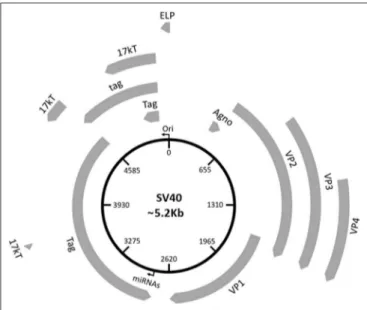
SV40 GENOMIC ORGANIZATION
The SV40 virion is formed by an unenveloped icosahedral
protein structure with a diameter of 45–50 nm and a density
of 1.34–1.35 g/cm
3(
8
). Its viral genome is a circular
double-stranded DNA molecule with ∼5.2 kb, depending on the SV40
strain (
9
). SV40 shares about 70–75% genome homology with
JCPyV (
10
–
12
) and BKPyV (
12
,
13
), whereas it has little
homology with other HPyVs, including HPyV 6 and 7 (
14
),
Malawi polyomavirus (MXPyV or HPyV 10) (
15
), Saint Louis
polyomavirus (STPyV or HPyV 11) (
16
), and Merkel cell
polyomavirus (MCPyV) (
17
–
19
).
Three main regions have been identified in the SV40 genome:
(i) a non-coding control region (NCCR), (ii) an early, and
(iii) late coding regions (Figure 1). NCCR includes the DNA
replication origin (ori) and a gene promoter, whose nucleotide
sequences are binding sites for transcription factors regulating
early and late gene expressions. The terms “early” and “late”
indicate the chronological order of gene transcriptions during
the viral life cycle in the host cell. Both early and late genes
are transcribed in opposing directions, i.e., anti-clockwise and
FIGURE 1 | Schematic representation of the SV40 genome. SV40 DNA is made up of three regions: the regulatory region and the early and late regions. The regulatory region contains the origin of replication (ori) and regulates the viral gene expression. The early region contains coding sequences for early genes, including the large tumor T antigen (Tag), the small tumor t antigen (tag), 17 kT, and the early leader protein (ELP). The late region contains coding sequences for late genes, including the major capsid protein VP 1, the VP 2, VP 3, VP 4, and the agnoprotein (Agno). The two miRNAs maps within the Tag gene sequences.
clockwise, respectively, in relation to NCCR. The early region
contains coding sequences for the large tumor antigen (Tag),
small tumor antigen (tag), 17 kT and the early leader protein
(Figure 1, Table 1). Both Tag and tag are transcribed with
alternative splicing. Tag and tag are viral oncoproteins, which
induce SV40 DNA replication, gene expression, as well as
S-phase entry and DNA synthesis in the host cell, thereby
triggering cycle progression (Figure 2) (
9
). In addition, these
two oncoproteins own transformation potential in vitro and
exert oncogenic activities in vivo (
9
). The 17 kT protein, which
shares most of its amino acid (aa) sequence with the Tag
N-terminal domain, promotes cell cycle progression in the presence
of tag, as well as presenting tumorigenic potential (
20
). The
early leader protein is a small protein of 23 aa whose function
is unclear (
21
). The late region contains genes transcribed into
two classes of late mRNAs: (i) 16S, which encodes the major
capsid protein VP 1; (ii) 19S, coding for the VP 2, VP 3, VP 4
polypeptides, and the agnoprotein (Figure 1, Table 1). VP
1-2-3 are structural proteins that enable viral DNA to be packaged
into the SV40 virion. A total of 360 VP 1 molecules form,
with 72 pentamers, the virion (
22
,
23
). The internal face of
each pentamer binds a single copy of VP 2 or VP 3 (
22
).
VP 4 seems to facilitate the lytic release of the SV40 virions
(
24
), but a recent study demonstrated that VP4 is not required
for this process (
25
). The agnoprotein controls the perinuclear
localization of VP 1 during virion formation, which then
triggers virion assembly (
26
). In total, SV40 translates for nine
proteins. Recently, two SV40-encoded microRNAs (miRNAs)
have been identified (Figure 1, Table 1) (
27
). More details are
reported on this topic in the section “SV40 microRNAs and viral
infection” (see below).
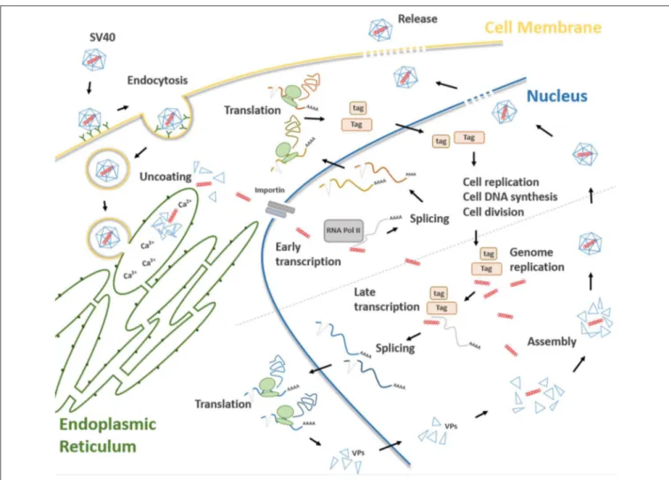
SV40 LIFE CYCLE
Attachment of the SV40 viral capsid to the target cell surface
is the first event to take place during the infection process
TABLE 1 | SV40 gene products.
SV40 expression products
Main function(s)
Early Large tumor T antigen (Tag) Cell cycle progression, viral DNA replication
Small tumor t antigen (tag) Cell cycle progression, viral DNA replication
17 KT Cell cycle progression Early leader protein Unclear function Early polarity SVmiRNA Tag regulation Late polarity SVmiRNA Tag
regulation
Late VP1 Capsid structure (external), viral attachment and entry VP2 Capsid structure (internal) VP3 Capsid structure (internal) VP4 Cell lysis, viral particles release Agnoprotein Virion assembly
FIGURE 2 | Main steps of the SV40 life cycle. The life cycle of SV40 starts with the attachment of the SV40 viral capsid to the target cell surface and proceeds through a lipid raft-mediated endocytosis. Then, the virion is transferred, by vesicular transport, toward the endoplasmic reticulum where it starts the uncoating process which continues in the cell cytosol. Uncoated SV40 genomes translocate inside the nucleus where the cellular RNA polymerase II mediates early viral transcription. Early transcription generates a precursor that is alternatively spliced into mRNAs, encoding the large T (Tag), and small t (tag) antigens. These mRNAs are translated in the cytosol into their corresponding proteins. Tag and tag migrate to the nucleus where they mediate several functions interfering with a number of host cellular pathways, thereby forcing the cells to proceed from the G1 to the S-phase. At the same time, Tag/tag starts the replication of the SV40 genome. The transition from early to late phase during the SV40 infection begins at the end of the viral DNA replication. After synthesis, late viral proteins are accumulated in the cytoplasm, migrates into the nucleus and then assemble with replicated viral DNA to form virions. Finally, a progeny virus is released through cell lysis.
in host cells. Binding is mediated by an interaction between
VP 1, the cell surface receptor ganglioside GM1 (
28
), and the
major histocompatibility complex (MHC) class I, that act as
co-receptors (
29
). Afterwards, SV40 capsid enters the target
cell via lipid raft-mediated endocytosis (
30
), which is triggered
by the interaction between VP 1 and cell surface ganglioside
GM1 (
31
) (Figure 2). Then, SV40 capsid is transferred, by
vesicular transport, in the endosomal compartment, toward the
endoplasmic reticulum (ER). In atypical circumstances, SV40
enters the cells via a caveolae-mediated endocytic pathway
by which the virus is directly translocated to the ER (
32
,
33
), bypassing the endosomal compartment. The uncoating
process begins in the ER, proceeds through ER absorption of
Ca
2+ions, thus inducing the loss of specific inter-pentamer
connections provided by invading VP 1 C-terminal arms in
the capsid. This process exposes the SV40 nuclear localization
signal, thereby inducing the translocation of the viral genome
in the nucleus via a mechanism mediated by the importin α
2/β
heterodimer and VP 3 (
8
,
34
,
35
) (Figure 2). Both early viral
transcription and DNA replication occur inside the nucleus.
Transcription is NCCR regulated (
36
), whereas DNA replication
starts from the ORI sequence contained in the same NCCR
region. SV40 DNA replication occurs soon after transcription
in the early region, whereas late region transcription initiates
after replication of viral DNA (
37
). Both early and late promoters
are recognized by cellular RNA polymerase II and host factors,
thereby inducing viral gene transcription. Early transcription
provides the generation of a precursor that is alternatively
spliced into two mRNAs encoding Tag and tag (
38
) (Figure 2).
In this phase, SV40 late genes are maintained silenced by
transcriptional repressors (
39
). In permissive cells, the role of
Tag is essential for DNA replication. Tag is a multifunctional
phosphoprotein that binds as a double hexamer to the SV40 viral
replication ORI, where it unwinds viral DNA. This molecular
process induces cellular protein recruitment required for viral
DNA replication, including DNA polymerase-α and replication
protein A (
40
,
41
). Tag is also responsible for an ATPase
activity that is required for viral DNA elongation (
42
). SV40
needs additional cellular co-factors for its DNA replication,
mainly expressed during the S phase. For this reason, Tag
is evolutionarily developed to modulate intracellular proteins
involved in crucial signal transduction pathways that control
cell cycle progression and apoptosis (
43
), such as hepatocyte
growth factor receptor (HGFR/Met) (
44
), insulin-like growth
factor 1 (IGF-1) (
45
), Notch-1 (
46
), and cdc2 (
47
). These
molecules force SV40-infected cells to proceed from the G1
to the S-phase (
48
). In this mechanism, tag seems to play
a cooperative role with Tag in both SV40 DNA replication
and S-phase progression (Figure 2) (
7
,
49
,
50
). The transition
from the early to late phase, during the SV40 infection, begins
at the end of the viral DNA replication. It seems that this
early-to-late transcriptional switch depends on changes on Tag
concentration. Initially, low Tag concentrations are sufficient for
an interaction between high-affinity NCCR Tag-binding motifs
and Tag and thus early transcription is positively regulated. Then,
high Tag concentrations enable this protein to interact with
low-affinity NCCR Tag-binding motifs. This interaction induces the
repressing early transcription by blocking the RNA polymerase
II complex. In addition, cellular repressors are titrated-off the
late promoter allowing the expression of late genes. Indeed,
since the number of SV40 genomes increases during viral DNA
replication, the concentration of repressors is reduced in the
late promoter. Tag, together with host transcription factors,
interacts with the late promoter, thereby inducing late gene
transcription (
51
). Late genes are transcribed in an opposite
direction to the early gene-encoding strand. Late proteins are
translated from two classes of late mRNAs: (i) 16S, which encodes
the major capsid protein VP 1; (ii) 19S, coding for VP 2, VP
3, VP 4 polypeptides, and agnoprotein (
52
) (Figure 2). After
synthesis, late viral proteins are accumulated inside the cell via
checkpoint kinase Chk1 activation by SV40, which negatively
regulates cell mitosis (
53
). Then, structural proteins assemble
with replicated viral DNA to form virions inside the nucleus
(Figure 2) (
26
). This mechanism is induced by the six tandem
GC-boxes within the SV40 genome, which represent the capsid
assembly signal. Viral assembly starts with GC-boxes interacting
with cellular transcriptional factor SP1 recruiting VP 2 and VP
3, which in turn bind to VP 1 pentamers (
54
). During this
process, the number of capsomers surrounding the viral DNA
increases until virion assembly has ended (
9
). At the same time,
the agnoprotein controls perinuclear VP 1 localization (
26
).
Then, the viral particle releasing (Figure 2) leads to cell lysis
and necrosis. However, the release of SV40 without displaying
a cytopathic effect (CPE) has been reported in specific cell
types, such as human mesothelial, epithelial, fibroblasts, and/or
embryonic kidney cells (HEK) (
44
,
55
–
57
).
SV40-MEDIATED CELL IMMORTALIZATION
AND TRANSFORMATION
Mammalian cells of different histotypes behave toward SV40
infection in different ways, depending on the ability of this
oncogenic polyomavirus (PyV) to complete the viral cycle and
produce a mature viral progeny. SV40-infected cells can be (i)
permissive, (ii) non-permissive, or (iii) semi-permissive (
56
,
58
–
60
). The main discriminant depends on viral DNA replication
potential expressed in permissive and semi-permissive cells. In
this case, viral progeny is produced, whereas SV40 infected
cells lyse and die. CV-1 and fibroblast-like COS cell lines,
both derived from monkey kidney tissue, are the prototype of
permissive cells (
58
,
61
). In non-permissive cells, no productive
viral cycle is established, whereas the infection occurs but is
abortive. Indeed, these cells are transformed/immortalized by
SV40. A typical example of non-permissive cells are rodent
cells, that carry SV40 DNA integrated in their genome, while
cells are transformed (
62
). Semi-permissive cells allow SV40
multiplication, but they produce a limited viral progeny (
63
).
The majority of cells lyse and die upon infection, but a fraction
of cells, which resist the SV40 infection, are transformed and
immortalized, while producing a viral progeny at low titer.
Several SV40 transformed/immortalized human fibroblasts have
been described in the literature (
64
,
65
). Cells differ in response to
SV40 infection depending on the ability of Tag to stimulate late
promoter transcription, which only occurs in
permissive/semi-permissive cells. Human cells support SV40 replication less
efficiently than monkey cells. Different in vitro cellular models
have been established to demonstrate the replicative potential of
SV40 in human cells (
66
). Early studies have shown that SV40
can replicate in human fetal neural cell lines (
4
), mesothelial cells
(
56
), and B-/T-lymphocytes (
64
,
65
,
67
). Although less efficiently,
this PyV can also replicate in human HEK lymphoblastoid
B-cell lines, as well as fibroblasts, such as WI-38 B-cells (
57
,
68
–
71
). In addition, in rare cases (<1/10
8cells), human fibroblasts
may become transformed due to the viral DNA integration in
the host cell genome (
72
). Early works have shown that one
out of seven human astrocytes could become transformed (
73
)
establishing continuous cell lines (
74
). In addition, several SV40
infected human cells produce a viral progeny at low titer without
displaying CPE (
44
,
56
,
57
). An example is provided by normal
human mesothelial cells (HMC), which seem to be persistently
infected by SV40 for a long period of time, while releasing
viral progeny (
44
,
56
,
57
). The molecular mechanism behind
the capacity of SV40 to enter into a true persistent/latent state
remains to be fully elucidated. It has been reported that SV40 is
able to establish a persistent infection in long-term immortalized
human fibroblasts, resulting in the production of infectious
viral progeny, which is able to infect both monkey and human
cells (
64
,
65
).
As for other DNA tumor viruses (
75
,
76
), several in
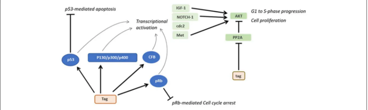
vitro cellular models have been developed to study SV40
transformation potentials. Tag and tag expression both cause
high cell transformation efficiently (
3
). Indeed, Tag blocks the
activities of many different cellular factors involved in cell
FIGURE 3 | Oncogenic activities of Tag and tag. During SV40 infection, Tag inhibits both the pRB and p53 tumor suppressor pathways. The main downstream effects of these interactions are the blocking of p53-mediated apoptosis and pRb-mediated cell cycle arrest. Furthermore, the interaction between Tag and pRB, p53 and other factors transactivates genes such as IGF-1 and Met, thereby triggering the transition from the G1 to the S-phase and the proliferation of SV40-infected cells. By inhibiting PP2A, tag activates pathways facilitating cell proliferation and transformation.
growth, differentiation and the cell cycle, such as p130, p300, and
p400 (Figure 3) (
9
). In addition, both Tag and tag can inhibit the
activities of p53 and pRb, which are two key tumor suppressor
proteins of animal and human cells (
9
). These interactions are
mandatory in order to achieve full human cell transformation
(
9
). Similarly, the transformation potential of Tag belonging to
another PyV, MCPyV, has been demonstrated. Indeed, MCPyV
Tag or LT has been detected to be overexpressed on
MCPyV-positive tumor tissues (
17
) and several in vitro studies evaluated
its transforming activity (
77
,
78
). However, it is important to
point out that many studies, while reporting the presence of SV40
sequences in human tumors, did not show the Tag expression. On
the other hand, in investigations based on IHC staining of human
tumor tissues, the Tag expression was revealed in SV40-positive
cells (
79
–
85
).
Interestingly, p53 was discovered for the first time when
detected bound to Tag in SV40-transformed cells during
immunoprecipitation experiments (
86
). Both Tag and tag can
interfere with many other host cellular pathways (
38
,
87
,
88
). For
instance, Tag has been found to be associated with many cellular
factors, such as Hsc70, Cul7, Bub1, TEF-1, Nbs1, and Fbw7
(
59
). It has also been reported that the Tag-p53 complexes bind
and activate IGF-1 promoter stimulating cell growth (
45
). The
expression of other growth factors may be potentially influenced
by Tag (
9
). Another cellular factor that interacts with Tag during
SV40 infection is the hepatocyte growth factor receptor (HGFR
or Met) (
44
). Furthermore, Tag is capable of inducing cell
immortalization and transformation by increasing CBP/p300 and
specific histone acetylation levels (
89
).
Upon interaction with the cellular genome and cell
factors, SV40 Tag induces different molecular changes in
the host cell. SV40 Tag possesses clastogenic and mutagenic
activities, which are shown by the appearance of chromosome
aberrations and point mutations in the host genome (
90
–
93
). These molecular/chromosome changes have not been
investigated/reported in human tumors presumably caused by
SV40. Specific genome alterations are characterized by numerical
and structural chromosomal aberrations, such as gaps, breaks,
dicentric and ring chromosomes, chromatid changes, deletions,
duplications, and translocations (
91
–
93
). SV40 Tag favors the
accumulation of point mutations, interfering with host DNA
repairs pathway (
90
,
94
,
95
). Indeed, SV40 Tag binds p53
protein thereby inhibiting p53-induced apoptosis and allowing
DNA-mutated cells to survive (
90
,
94
,
95
). It is well-established
that specific chromosomal aberrations can be detected in
human tumors of different histotypes, as reported in: (http://
atlasgeneticsoncology.org/Deep/CancerCytogenet2005ID20050.
html). In SV40-driven/positive human tumors, a similar
panel of chromosome alterations could be displayed (
90
–
93
).
Occasionally, SV40 DNA integrates into the host cellular
genome. This event can occur randomly in the cellular DNA,
while the viral DNA breaks randomly, as well (
96
). In cases
where SV40 DNA integration occurs, while maintaining Tag
and tag expression, these two viral oncoproteins support
the cell transformation phenotype (
9
). In addition, Liu and
collaborators demonstrated that the disruption of the human
chromosomal interval at 1q21.1 by SV40 integration, in the
human bronchial epithelial cell line, immortalized with a cloned
Ori– SV40, CRL-2504 cell line, can be an essential step for cellular
immortalization, altering the expression of genes involved in
senescence/apoptosis, which are located in the proximity of
viral integration sites (
97
). Integration of SV40 DNA within the
human genome has been reported in osteosarcoma samples,
thus suggesting that the viral DNA integration is involved
the tumorigenic process (
98
). Altogether these SV40-driven
alterations represent the genetic background, which drives
the phenotypical changes that lead to the SV40-induced
transformation process of the host cell.
The small tag viral oncoprotein interacts with the protein
phosphatase 2A (PP2A), thereby triggering several pathways
related to cellular transformation (Figure 3). The complex
tag-PP2A induces the entry on the cell cycle S-phase through
CDK inhibitor p27 degradation and cyclin A/CDK2 and cyclin
E/CDK2 promotion (
99
–
101
). Furthermore, tag-PP2A induces
MAPK cascade activation, thereby triggering the transforming
process in vitro (
102
). The third SV40 early protein, 17kT, also
transactivates the cyclin A/CDK2 (
100
). Although many factors
responsible for SV40 transformation have been discovered, the
total number of proteins involved in this activity is yet to
be determined.
An association between SV40 infection and DNA methylation
of different cellular genes has been reported. Indeed, improper
DNA methylation is involved in different diseases (
103
,
104
),
including cancer (
105
,
106
). DNA methylation is induced by
DNA tumor viruses in order to evade antiviral immunity,
which contributes to the immunosuppressive microenvironment
during cancer development (
107
). This process facilitates viral
multiplication/activity (
107
).
In early studies, SV40 transforming potential was largely
employed in developing different in vitro cellular models.
SV40-immortalized cells have been established and used to
study a large number of different molecular mechanisms,
including cell proliferation and transformation (
108
–
110
),
cytokine-production (
111
), and angiogenesis (
112
), as well
as mesenchymal stem cell differentiation (
113
) and neuronal
differentiation and neuroregeneration (
114
). Moreover, different
diseases, such as autoimmune disorders (
115
), male infertility
(
116
), fibrosarcoma (
108
), and corneal dystrophy (
117
) have
been studied employing SV40-immortalized cell models. Other
applications of SV40-immortalized cell in vitro models are
represented by cellular co-culturing (
118
) and suicide gene
therapy (
119
).
SV40 MICRORNAs AND VIRAL INFECTION
MicroRNAs (miRNAs) are small non-coding RNAs [18–22
nucleotides (nt)] that are involved in the post-transcriptional
negative regulation of gene expression in eukaryotes (
120
). These
small molecules and their regulatory effect have been described
in both eukaryote cells and viruses, including PyVs (
120
). In the
SV40 the early region maps a gene encoding for two miRNAs
(Figure 1), which are transcribed in opposite orientations. These
two viral miRNAs negatively regulate early mRNAs inhibiting
Tag translation through RNA-mediated interference (RNAi)
machinery, during the late phase of the SV40 life cycle. These
two miRNAs actively direct Tag mRNA cleavage at different
nucleotide positions (
27
,
121
). However, the silencing potential
of human genes by SV40 miRNAs cannot be ruled out (
27
,
121
).
These findings indicate that SV40 is able to use human RNAi
machinery to its own advantage. Since Tag is a target for host
cytotoxic T lymphocyte (CTL), the viral miRNA-mediated
down-regulation of Tag decreases the susceptibility of SV40-infected
cells to CTLs activity (
27
,
121
). Similarly, JCPyV and BKPyV
escape the innate and adaptive immune detection exploiting the
human RNAi machinery. Indeed, these two HPyVs code for
a miRNA identical in sequence between BKPyV and JCPyV,
which targets a member of the UL16 binding protein (ULBP)
family, the stress-induced ligand ULBP3 (
122
). Although a
recent study indicates that another member of this gene family,
ULBP1, is down-regulated following SV40 infection, it has been
demonstrated that SV40 miRNAs do not mediate this molecular
effect, thus suggesting the involvement of other mechanisms
behind the SV40 immune evasion ability (
123
). Another report
showed that the two SV40 miRNAs can negatively regulate
the degree of viral effects on B-cells as demonstrated using
SV40 miRNA-null mutants in experiments with infected
B-lymphocytes and myeloid cell lines (
124
).
SV40 ONCOGENICITY IN ANIMAL MODELS
SV40 is a high oncogenic small DNA tumor virus. However, this
PyV has not been reported in tumors of the rhesus macaque
(Macaca mulatta), which is its natural host. Indeed, SV40
infection in permissive monkey cells derived from kidney tissues
leads to cell lysis and death, without neoplastic transformation
(
58
,
61
). By contrast, SV40 experimentally inoculated in hamsters
induces tumors of different histotypes, depending of the route
of injection. Specifically, SV40 subcutaneously (s.c.) inoculated
in hamsters induces sarcomas and osteosarcomas; whereas
when injected intracerebrally (i.c.) it induces ependymomas and
choroid plexus papillomas (
5
,
38
). Hematological malignancies,
such as lymphocytic leukemia, histiocytic lymphomas, and B-cell
lymphomas are induced when SV40 is inoculated intravenously
(i.v.) (
5
,
6
). SV40 injected in the pleural space of hamsters induces
MPM in 100% of animals (
125
).
SV40 oncogenic activity is shown in transgenic mice, where
Tag/tag expression is under the control of its promoter (
126
)
or tissue-specific promoters (
127
–
145
). Exploiting the vast
SV40 oncogenicity in vivo, SV40 transgenic animals provided
good models for studying tumor initiation/progression and
innovative anti-cancer therapies (
146
). Transgenic animals
develop ependymomas, choroid plexus papillomas (
147
),
hepatocellular carcinomas (
139
,
144
), brain (
148
), bladder (
140
),
and bowel tumors (
149
), as well as eye tumors, including ocular
and/or lens tumors (
150
). The multistage progression of prostate
carcinoma has also been largely studied employing
SV40-transgenic mice (
128
–
130
,
132
,
134
–
136
,
151
,
152
). Interestingly,
transgenic mice generated with SV40 have also been employed
to study rare cancers, such as brown adipose tumor (
151
),
cardiac rhabdomyosarcoma (
142
) and adrenocortical carcinoma
(
151
). Lung cancers and MPM have also been studied in these
animal models (
127
,
137
,
141
). More recently, SV40-transgenic
mice have been developed to study chronic lymphocytic
leukemia (
143
). In addition, other SV40-transgenic mouse
models have also been developed to study fibrosarcoma (
108
)
and retinoblastoma (
153
) as well as breast (
154
,
155
) ovarian
(
131
,
156
,
157
), pancreatic (
158
), and liver cancers (
20
,
159
).
EPIDEMIOLOGY OF SV40 INFECTION IN
HUMAN POPULATIONS
SV40 infection in human populations has been widely reported
(
160
–
164
). Since SV40 is a monkey virus and the macaque is its
natural host, this viral infection in humans was considered a rare
event, being restricted to subjects in close contact with monkeys.
Indeed, inhabitants of Indian villages located near the jungle,
which is the natural environment for monkeys, and workers
attending to monkeys in zoos/animal facilities are prone to SV40
infection and develop antibodies against this PyV (
165
,
166
).
SV40 was inadvertently administered to humans between
1955 and 1963, when hundreds of millions of people in North
and South America, Canada, Europe, Asia, and Africa were
vaccinated with both inactivated and/or live polio vaccines, found
to be contaminated by SV40. This accident occurred because
these early polio vaccines were produced by growing polioviruses
in naturally SV40-infected monkey cell cultures (
167
–
169
). It
has been reported that in the former USSR, SV40-contaminated
polio vaccines were used until 1978 (
170
), whereas in Italy up to
1999, when the Italian Health Public Organization switched to
SV40-free anti-polio vaccines as indicated by the World Health
Organization (WHO) guidelines, following a note from the
British National Institute for Biological Standards and Control
(
9
,
74
,
171
). In other countries, the risk of SV40 contagion
through polio vaccines is still a problem, as these vaccines are
produced using SV40-positive monkey cells (
170
). The past
literature indicates that SV40 infection in different geographic
regions was influenced by the use of either SV40-contaminated
or non-contaminated vaccines, as well as the number of years
of vaccine administration. Sweet and colleagues quantified that
about 10–30% of polio vaccines were contaminated with SV40
(
1
). Furthermore, SV40 genotypes in polio vaccines overlap with
those detected in humans, thus suggesting that this oncogenic
PyV was introduced into the human population through
contaminated polio vaccines (
172
). It has also been reported that
shortly after SV40 infection, this PyV spread for weeks in the
stools of children vaccinated with SV40-contaminated vaccines
(
173
). This evidence indicates that SV40 replicates in some
gastrointestinal cells, thus suggesting that this virus could spread
in humans via horizontal infection, such as the fecal-oral route.
To a lesser extent, other vaccines against adenoviruses (
174
) and
hepatitis A (
175
), were SV40 contaminated. In addition,
SV40-contamination was detected in the respiratory syncytial virus
vaccine employed in infected-volunteers to whom the vaccine
was given to via the respiratory route (
176
). About two out
of three volunteers developed neutralizing antibodies against
SV40 (
176
). Altogether these data indicate that SV40 infects and
multiplies in humans.
Over the years, with the development of molecular biology
techniques (
12
,
18
,
76
,
177
,
178
), SV40 DNA sequences have been
investigated and detected in both normal and neoplastic tissues
from individuals vaccinated with polio vaccines contaminated
with SV40. DNA sequences from this PyV have been detected
in pituitary tissues (
179
) as well as in leukocytes from organ
(
180
) and blood (
181
) donors. Footprints from SV40 DNA
have also been reported in lymphoblastoid cells (
68
), as well as
blood sample specimens from normal individuals and oncologic
patients (
79
,
161
,
182
–
186
). In addition, SV40 DNA has been
detected in blood samples from healthy individuals exposed to
asbestos pollution (
187
). These data cumulatively demonstrate
that SV40 is circulating in the human population. It is also
possible that blood cells are the SV40 reservoir and vehicle of the
virus spreading in humans. Genomic sequences from this PyV
have also been found in stool samples and urine from children
and adults, suggesting that SV40 can potentially be transmitted
via different routes, such as sexual and fecal-oral routes which
are responsible for viral horizontal infection in humans (
164
,
183
,
188
). These additional sources of exposure may lead to
subclinical SV40 infections in the healthy population. However, it
has been reported that the SV40 transmission in monkeys seems
to occur in the environment rather than directly among animals
(
189
). It is plausible that in humans, a contaminated environment
or home setting is responsible for SV40 spreading, rather than
person to person transmission (
188
). The site of SV40 latent
infection in humans is yet to be elucidated. Since this PyV has
been detected in human kidney and urine samples (
183
,
184
) it
seems reasonable that kidneys might be the site of virus latency,
as in monkey it occurs to be the natural host (
1
,
189
).
SV40 primary infection occurs early in life and its
seroprevalence increases with age. Anti-SV40 antibodies in
the serum of immunized individuals and SV40 antigens have
been detected in normal subjects (
190
–
199
). Lusting et al.
reported a prevalence of serum anti SV40 antibodies in 7.6–14%
of Swedish children aged from 1 to 13 years old (
198
). In
early studies, a low prevalence, 11%, was detected in healthy
individuals from Africa (
200
) and the U.S (
201
). Similarly, a
more recent study detected anti SV40 antibodies in sera from
healthy adult blood donors with low rates, about 2% (
202
).
Altogether these serological data indicate that SV40 is present
in immunized healthy populations in the range of 1.3–15.6%,
suggesting that this PyV circulates in humans at low prevalence
(
200
,
201
,
203
–
206
). Interestingly, one study reported SV40
antibodies in human sera before the introduction of
SV40-contaminated polio vaccines, suggesting that SV40 was probably
circulating in humans independently from SV40-contaminated
vaccines (
207
). However, this study was conducted before the
identification of human PyVs, JCPyV, and BKPyV, which are
highly homologous to SV40 (
2
,
13
). Indeed, the high homology
among the three PyVs, BKPyV, JCPyV, and SV40, hampered
serological data due to antigenic cross-reactivity. For many
years, PyV cross-reactivity did not allow the verification of the
real SV40 prevalence in humans (
208
). Different immunological
assays based on the use of virions, soluble recombinant VP 1
protein and virus-like particles (VLPs), such as SV40 antigens,
always gave cross-reactivity with BKPyV/JCPyV (
208
).
In order to circumvent this technical issue, in recent years
specific and sensitive indirect ELISA tests with SV40 VPs/Tag
mimotopes as antigens were set up to investigate the presence in
healthy subjects of serum IgG/IgM antibodies reacting to SV40
VPs/Tag (
191
,
192
). Immunological data showed that healthy
blood donors carry IgG antibodies reacting to SV40 VPs and
Tag with a prevalence of 18 and 20%, respectively (
191
,
192
).
Furthermore, immunological data from children may suggest
that SV40 infection/seroconversion occurs early in life, i.e., at 6
months of age (
190
,
193
).
Antibodies against SV40 VPs in sera from multiple transfused
patients affected by thalassemia major had a higher prevalence
than healthy subjects of the same age (31 years old, both cohorts).
These data indicate that this PyV could have been transmitted
by blood transfusions, along with other natural sources (
209
).
Furthermore, the increased prevalence of SV40 antibodies was
significantly higher in the older age group of patients (41–50
years old) than in age-matched controls (38 vs. 20%).
In a recent investigation, SV40 neutralizing antibodies with
a prevalence in the range of 7 and 18% were revealed in sera
from women in Huston, Texas, employing a plaque reduction
SV40-neutralization assay. The authors identified ethnicity as
a significant factor associated with high seroprevalence SV40
neutralizing antibodies reported in Hispanic groups, including
subjects from Houston (
210
). It is worth recalling that
SV40-contaminated live polio vaccines, as candidate vaccines, were
tested during large field trials in some Latin American Countries,
due to their potential for being naïve vaccines.
Overall, immunological data indicate that SV40 is circulating
in humans inducing IgM, IgG including neutralizing antibodies,
which can be detected in sera with a mean prevalence of ∼20%
in healthy individuals. Recent immunological data, despite being
obtained with specific SV40 mimotopes, do not rule out the
hypothesis that another polyomavirus, still unknown, closely
related to SV40 is circulating in humans.
ASSOCIATION OF SV40 WITH HUMAN
TUMORS
The hypothesis that SV40 might be associated to human
malignancies has been investigated with a large number
of molecular, immunological, and epidemiological studies
(Table 2). This oncogenic PyV was previously associated with
a broad range of tumor types including, malignant pleural
mesothelioma (MPM) (
80
,
81
,
227
–
233
,
235
,
238
,
240
–
243
,
256
–
258
), bone (
98
,
215
,
224
), brain (
212
–
214
,
217
,
219
–
221
,
259
,
260
), lung (
227
,
234
), thyroid (
82
,
244
), pituitary
(
179
), and urothelial (
245
) tumors, pleomorphic adenomas of
parotid glands (
83
), choroid plexus tumors, and ependymomas
in children (
160
). In addition, footprints from SV40 DNA
have been detected in breast (
84
) and colon cancer specimens
(
222
). Interestingly, DNA sequences belonging to SV40 have
also been found in an AIDS patient with a cerebral lesion
(
216
). More recently, a study conducted with an innovative
analysis, known as RNA sequencing (RNA-seq), identified SV40
mRNAs, in tumor samples from low-grade glioma affected
patients (
219
). Different lympho-proliferative disorders (
85
,
239
,
254
,
255
), including non-Hodgkin’s lymphoma (
79
,
247
–
251
,
260
) have also been associated with SV40 infection.
It has been reported that the homologous and autologous
implantation of SV40-transformed cells in humans caused the
growth of nodules, thus suggesting that this PyV presents
oncogenic capacity in humans (
261
). These studies support
an association of human cancers with SV40. These results,
obtained mainly by PCR, Southern blot hybridization, DNA
sequencing, and immunohistochemistry (IHC) were confirmed
by a multi-institutional study (
80
), but confuted by another
group of investigators (
262
). Among SV40-positive tumor types,
MPM has been detected as SV40-positive in many investigations
(
80
,
81
,
227
–
233
,
235
,
238
,
240
–
243
,
256
–
258
,
263
), while
the mechanisms of SV40 oncogenesis have also been studied
(
9
,
44
,
258
,
264
,
265
). Interestingly, when normal HMC were
exposed to both SV40 and asbestos fibers, the transformation
rate increased significantly compared to the controls (
9
). High
exposition to asbestos fibers alone can cause progressive fibrosis
(i.e., asbestosis) and in the worse cases, lung cancer (
266
) and
MPM (
9
). Many studies reported the synergistic activities of
SV40 and asbestos on MPM development in geographical areas
with high levels of asbestos exposure and SV40-contaminated
polio vaccines (
9
,
81
,
187
,
232
,
236
,
239
). High prevalence of
SV40 DNA sequences in MPM tissues reflects that relation to
SV40-contaminated polio vaccines (
9
,
81
,
187
,
232
,
236
,
239
).
The majority of human cancers mentioned above correspond to
tumors, which develop in rodents inoculated with SV40 and in
SV40-transgenic mice.
It appears that in the same kind of tumor, prevalence
of SV40 sequences differs in distinct geographical areas. For
example, it has been reported that in the U.S. and Europe
20–83% of MPM tested SV40-positive (
80
,
228
,
230
,
235
,
236
,
249
), while sequences of this PyV in MPM from Turkey and
Austria have never been detected (
235
,
267
), or detected with
low frequency, as in Sweden (
228
). Similarly, the prevalence
SV40 DNA detected in bone tumors is different for example
in Hungary (74%) and Germany (24%) (
226
). In addition,
SV40-positive MPMs were found in two different studies in
Japan with a prevalence ranging between 6 and 44% (
241
,
268
), whereas another investigation conducted in a cohort
of Vietnamese MPM patients detected 20% SV40-positive
tumors (
81
).
The association between SV40 and human tumors is based
on results obtained by many investigators (
80
,
98
,
213
,
218
,
220
,
225
,
227
,
230
,
234
,
255
,
256
). Most of these studies
detected SV40 DNA sequences, using qualitative PCR techniques,
in tumor specimens. However, these assays do not provide
the quantification of SV40 DNA as a copy number, nor
investigated the physical state of the viral DNA, i.e., integrated
and/or episomal (
96
,
98
). In other studies SV40 mRNA
and/or Tag/tag oncoproteins were analyzed in tumors (
9
,
68
,
81
,
83
,
160
,
219
), or quantified the amount of SV40 DNA
sequences via qPCR methods (
240
,
268
,
269
). The majority of
these qualitative/quantitative assays (Table 2) were carried out
targeting different SV40 Tag sequences. Together with these
sequences, other SV40 regions were PCR analyzed, including
the control region and late gene sequences (
181
,
220
). These
data indicate that SV40 DNA regions detected in human tumors
were not due to PCR contaminations with recombinant plasmids
carrying SV40 Tag sequences. In addition, in studies based on
IHC staining, the localization of the SV40 Tag in the
SV40-infected cell is shown (
79
–
85
). Studies carried out on SV40
Tag-positive cells demonstrated that the cell transformation is
related to the activation of specific autocrine/paracrine loops. In
this context, different growth factors and their receptors were
analyzed, such as HGF and its receptor (HGFR or Met) (
44
),
the vascular endothelial growth factor (VEGF) and its receptor
(VEGFR) (
270
,
271
) as well as (IGF-1) and its receptor, in
T-antigen-mediated growth (
272
,
273
). Concordant data were
reported on the ability of the SV40 Tag to induce growth factor
receptor/growth factor loops, which in turn stimulates
cell-cycle progression into the S phase. These results suggest that
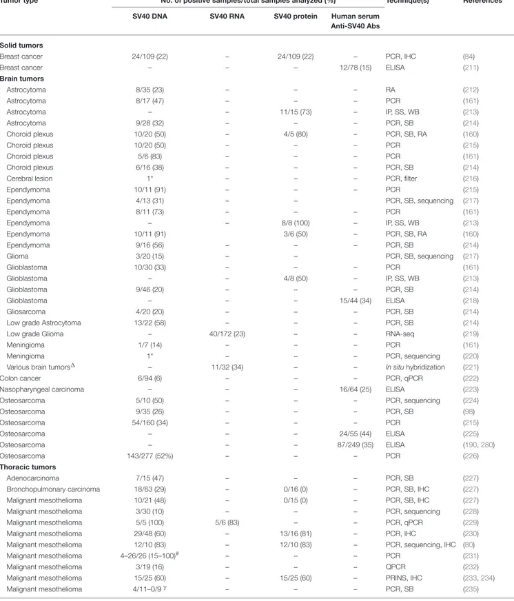
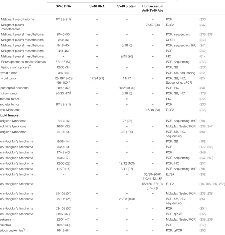
TABLE 2 | SV40-positive human tumors.
Tumor type No. of positive samples/total samples analyzed (%) Technique(s) References SV40 DNA SV40 RNA SV40 protein Human serum
Anti-SV40 Abs
Solid tumors
Breast cancer 24/109 (22) – 24/109 (22) – PCR, IHC (84)
Breast cancer – – – 12/78 (15) ELISA (211)
Brain tumors Astrocytoma 8/35 (23) – – – RA (212) Astrocytoma 8/17 (47) – – – PCR (161) Astrocytoma – – 11/15 (73) – IP, SS, WB (213) Astrocytoma 9/28 (32) – – – PCR, SB (214) Choroid plexus 10/20 (50) – 4/5 (80) – PCR, SB, RA (160) Choroid plexus 10/20 (50) – – – PCR (215) Choroid plexus 5/6 (83) – – – PCR (161) Choroid plexus 6/16 (38) – – – PCR, SB (214)
Cerebral lesion 1* – – – PCR, filter (216)
Ependymoma 10/11 (91) – – – PCR (215) Ependymoma 4/13 (31) – – PCR, SB, sequencing (217) Ependymoma 8/11 (73) – – – PCR (161) Ependymoma – – 8/8 (100) – IP, SS, WB (213) Ependymoma 10/11 (91) 3/6 (50) – PCR, SB, RA (160) Ependymoma 9/16 (56) – – – PCR, SB (214) Glioma 3/20 (15) – – PCR, SB, sequencing (217) Glioblastoma 10/30 (33) – – – PCR (161) Glioblastoma – – 4/8 (50) – IP, SS, WB (213) Glioblastoma 9/46 (20) – – – PCR, SB (214) Glioblastoma – – – 15/44 (34) ELISA (218) Gliosarcoma 4/20 (20) – – – PCR, SB (214)
Low grade Astrocytoma 13/22 (58) – – – PCR, SB (214)
Low grade Glioma – 40/172 (23) – – RNA-seq (219)
Meningioma 1/7 (14) – – – PCR (161)
Meningioma 1* – – – PCR, sequencing (220)
Various brain tumors1 – 11/32 (34) – – In situhybridization (221)
Colon cancer 6/94 (6) – – – PCR, qPCR (222)
Nasopharyngeal carcinoma – – – 16/64 (25) ELISA (223)
Osteosarcoma 5/10 (50) – – – PCR, sequencing (224) Osteosarcoma 9/35 (26) – – – PCR, SB (98) Osteosarcoma 54/160 (34) – – – PCR (215) Osteosarcoma – – – 24/55 (44) ELISA (225) Osteosarcoma – – – 87/249 (35) ELISA (190,280) Osteosarcoma 143/277 (52%) – – – PCR (226) Thoracic tumors Adenocarcinoma 7/15 (47) – – – PCR, SB (227)
Bronchopulmonary carcinoma 18/63 (29) – 0/16 (0) – PCR, SB, IHC (227)
Malignant mesothelioma 10/21 (48) – 0/15 (0) – PCR, SB, IHC (227)
Malignant mesothelioma 3/30 (10) – – – PCR, sequencing (228)
Malignant mesothelioma 5/5 (100) 5/6 (83) – – PCR, qPCR (229)
Malignant mesothelioma 29/48 (60) – 13/16 (81) – PCR, IHC (230)
Malignant mesothelioma 12/10 (83) – 12/10 (83) – PCR, sequencing, IHC (80)
Malignant mesothelioma 4–26/26 (15–100)# – – – PCR (231)
Malignant mesothelioma 3/19 (16) – – – QPCR (232)
Malignant mesothelioma 15/25 (60) – 15/25 (60) – PRINS, IHC (233,234)
Malignant mesothelioma 4/11–0/9γ – – – PCR, SB (235)
TABLE 2 | Continued
Tumor type No. of positive samples/total samples analyzed (%) Technique(s) References SV40 DNA SV40 RNA SV40 protein Human serum
Anti-SV40 Abs
Malignant mesothelioma 8/19 (42.1) – – – PCR (236)
Malignant pleural mesothelioma
– – – 25/97 (26) ELISA (237)
Malignant pleural mesothelioma 20/40 (50) – – – PCR, sequencing (238,239)
Malignant pleural mesothelioma 2/35 (6) – – – QPCR (240)
Malignant pleural mesothelioma 8/18 (45) – 0/18 (0) – PCR, sequencing, IHC (241)
Malignant pleural mesothelioma 4/9 (45) – – – PCR (242)
Malignant pleural mesothelioma – – 9/45 (20) – IHC (81)
Pleural/peritoneal mesotheliomas 67/118 (57) – – – PCR, sequencing (243)
Various lung cancers6 12/35 (34) – – – PCR, SB (227)
Thyroid tumor 3/69 (4) – – – PCR, SB, sequencing (244)
Thyroid tumor 12–19/19–29
(66–100)#
17/24 (71) 11/17 – PCR, SB, IHC,
Sequencing, qPCR (82)
Pleomorphic adenoma 28/45 (62) – 26/28 (93%) – PCR, IHC (83)
Pituitary tumor 26/30 (87)§ – 0/18 (0) – PCR, SB, IHC (179)
Urothelial tumor – – 1* IHC (245)
Urothelial tumor 6/18 (42.1) – – – PCR (236)
Uveal Melanoma – – – 16/48 (33) ELISA (246)
Liquid tumors
Hodgkin’s lymphoma 7/43 (16) – 2/7 (28) – PCR, sequencing, IHC (79)
Hodgkin’s lymphoma 16/54 (30) – – – Multiplex Nested PCR (238,247)
Hodgkin’s lymphoma 2/19 (10) – 2/2 (100) – PCR, SB, IHC,
sequencing
(85)
Non-Hodgkin’s lymphoma 8/58 (14) – – – PCR, SB (182)
Non-Hodgkin’s lymphoma 3/29 (10) – – – PCR (172,248)
Non-Hodgkin’s lymphoma 17/42 (40) – – – PCR (249)
Non-Hodgkin’s lymphoma 6/36 (17) – – – PCR, sequencing (247,250)
Non-Hodgkin’s lymphoma 12/55 (22) – 12/12 (100) – PCR, IHC (251)
Non-Hodgkin’s lymphoma 11/79 (14) – 3/11 (27) – PCR, sequencing, IHC (79)
Non-Hodgkin’s lymphoma – – – 36/89–26/61 (40,41,42,43)γ ELISA (252) Non-Hodgkin’s lymphoma – – – 55/150–37/104 (37–36)γ ELISA (18,195,197,253)
Non-Hodgkin’s lymphoma 85/158 (54) – – – Multiplex Nested PCR (238,239)
Non-Hodgkin’s lymphoma 28/106 (26) – 28/28 (100) – PCR, SB, IHC,
sequencing
(85)
Non-Hodgkin’s lymphoma 63/108 (56) – – – PCR (254)
Non-Hodgkin’s lymphoma 38/60 (63) – – – PCR, qPCR (255)
Leukemia 22/54 (41) – – – Multiplex Nested PCR (238,239)
Leukemia 16/48 (30) – – – PCR (249)
Various LeukemiasU 16/19 (84) – – – PCR, qPCR (255)
*Case reports;∆Angiofibroma, astrocytoma, metastatic brain tumors, meningiomas, neurinomas, oligodendrogliomas;#Different primer set;γTwo different cohorts;ΣPleomorphic carcinoma, Neuroendocrine carcinoma, Squamous cell carcinoma, others not specified§Polyomaviral primers that hybridized to SV40 and BKPyV internal probes;UBcell acute lymphoblastic leukemia, B-cell precursor acute lymphoblastic leukemia, T-cell acute lymphoblastic leukemia. IP, Immunoprecipitation; SS, Silver staining; WB, Western blot; PCR, Polymerase chain reaction; PRINS, Primed in situ assay (DNA detection); qPCR, real time quantitative PCR; IHC, Immunohistochemistry; RA, restriction analysis.