The Role of Visfatin in Pregnancy, Complications
and Procreation
Lucia Marseglia
1Sara Manti
1Gabriella D
’Angelo
1Caterina Cuppari
1Vincenzo Salpietro
1Martina Filippelli
1Valeria Chirico
1Eloisa Gitto
1Carmelo Salpietro
1Teresa Arrigo
11Department of Pediatrics, University of Messina, Messina, Italy
J Pediatr Biochem 2015;5:2–7.
Address for correspondence Sara Manti, MD, Department of
Pediatrics, University of Messina, Via Consolare Valeria, 98125 Messina, Italy (e-mail: [email protected]).
Introduction
Adipose tissue is considered as an endocrine organ secreting numerous neuroendocrine and peripheral peptides, called adipokines (adiponectin, leptin, resistin,1vaspin, apelin, vis-fatin, tumor necrosis factor-α [TNF-α], acylation-stimulating protein [ASP], interleukin-6 [IL-6], plasminogen activator inhibitor-1 [PAI-1], and transforming growth factor-β [TGF-β]2,3). They act as inflammatory molecules, playing a critical role in both short- and long-time energy homeostasis, meta-bolic processes, and body fat regulation.4
Moreover, these peptides are considered as a new link between obesity, insulin resistance,5cardiovascular disease, hypertension, as well as hyperlipidemia.6
This review aims to emphasize the molecular and endo-crine mechanisms of visfatin and its role in fetal development.
Review
Nicotinamide phosphoribosyltransferase (Nampt) has been identified originally as pre–B-cell colony-enhancing factor (PBEF), capable of promoting the maturation of B-cell pre-cursors together with IL-7 and stem cell factor (SCF).7It was rediscovered as an adipocytokine, also known as“visfatin,”
found in visceral adipose tissue, playing a role in glucose homeostasis.8,9 Nampt has also been identified as nicotin-amide adenine dinucleotide (NAD) biosynthetic enzyme. However, some of its functions are controversial and are still being discussed. Nampt exists in two different types: intra-cellular (iNampt) and extraintra-cellular (eNampt) forms.
iNampt
In 1957,10 iNampt was identified as NAD biosynthetic en-zyme.11In mammals, tryptophan, nicotinic acid, and nicotin-amide are the major precursors for NAD, but nicotinnicotin-amide is mainly used to synthesize it.12
Nampt promotes the transfer of a phosphoribosyl group from 5-phosphoribosyl-1-pyrophosphate (PRPP) to nicotin-amide, forming nicotinamide mononucleotide (NMN) and pyrophosphate (PPi).13 NMN is then converted to NAD by nicotinamide mononucleotide adenylyltransferase. NAD plays a critical role in cellular redox reactions. Furthermore, NAD is useful for deacetylase activity of proteins and synthe-sis of transcriptional regulation factor (e.g., silent information regulator [SIR]).14,15 SIR, NAD-dependent enzymes called sirtuins, mediates life span extension cell (mouse fibroblasts, human vascular smooth muscle cells, cardiac myocytes, and pancreatic B cells) caused by variety of stresses as well as
Keywords
►
visfatin
►
newborns
►
pregnancy
►
gestational diabetes
mellitus
►
preeclampsia
►
breast-feeding
Abstract
Adipose tissue is considered an endocrine organ secreting numerous neuroendocrine
and peripheral peptides, also known as
“adipokines.” Although, the role of adipokines,
precisely visfatin, is still controversial, it was recently discovered their involvement
in different mechanisms, including metabolism, in
flammation, and
endocrine-immunologic system. A literature search of electronic databases was undertaken for
the major studies published from 1957 to present. The databases searched were:
PubMed, EMBASE, Orphanet, Midline, and Cochrane Library. This review aims to
emphasize the molecular and endocrine mechanisms of visfatin and its role in fetal
development. This review also reviews the role of adipocytokine in the pathogenesis of
inflammatory-endocrine disorders. Further research will bring new insight into linkage
between visfatin and humans, during pregnancy and perinatal period.
Copyright © 2015 by Georg Thieme Verlag KG, Stuttgart · New York
DOI http://dx.doi.org/ 10.1055/s-0035-1554783. ISSN 1879-5390.
nutritional restriction.16 In fact, pancreatic B-cell–specific Sirt2-overexpressing (BESTO) mice show strongly enhanced glucose-stimulated insulin secretion (GSIS) and enhanced glucose tolerance. Thesefindings suggest a further Nampt-mediated role in metabolic balance.17,18
eNampt
The molecular mechanism of secretion (through passive and/ or active secretory pathway)18,19and the role of eNampt are not still clear.20Three different functions have been assigned to eNampt: insulin-mimetic hormone (adipocytokine named “visfatin”), cytokine, and aNAD biosynthetic enzyme.21
Visfatin has a molecular weight of 52 kDa. The coding region of the gene encodes for 491 amino acids.22 It is synthesized by the bone marrow, liver, lungs, skeletal muscle, brain, heart, pancreas, and peripheral blood lymphocytes.23 In particular, it is mostly expressed in human visceral fat.24 In a case–control study conducted on prepubertal obese children, it was demonstrated that obese patients have higher concentrations than normal-weight people. Elevated serum visfatin levels were also positively correlated with body fat mass; in fact visfatin concentration decreases when weight loss occurs.25
Friebe and colleagues, in an experimental study, revealed that its expression is higher in adipocytes compared with preadipocytes,9 suggesting that visfatin is a differentiated adipocytes-specific molecule and its production is influenced by serum lipid levels.
The role of visfatin in glucose metabolism is still unclear. Several case–control studies assessed that visfatin was related to several conditions such as insulin resistance, obe-sity, dyslipidemia, and metabolic syndrome.26–29
In fact, Tascilar and colleagues noted that visfatin is correlated with homeostatic model assessment-insulin resis-tance (HOMA-IR) in children.27 However, in comparative study, Berndt and colleagues noted that plasma visfatin correlates significantly with percent body fat, body mass index (BMI), and visfatin messenger RNA (mRNA) level in visceral adipose tissue, but not with visceral fat mass or waist-to-hip ratio.28 Furthermore, no relationship was ob-served between serum visfatin levels and fasting plasma insulin, fasting glucose and insulin sensitivity in nondiabetic subjects. In two recent studies, plasma visfatin was higher in patients with type 2 diabetes mellitus (T2DM) than in control group.29
Probably, visfatin promotes a glucose uptake (through 3T3l1 on adipocytes and L6 on myocytes and glycolysis, regardless insulin).30In fact, visfatin binds the same insulin receptor through a distinct epitope.24
Visfatin, stimulated phosphorylation of insulin receptor substrate (IRS)-1/2 and activate phosphatidyl inositol 3-kinase (PI3K), induced glucose uptake, glucose transporter (GLUT)-1 protein expression, production of profibrotic fac-tors (including type I collagenase, TGF-β 1, PAI-1), increased metalloproteinase-9 activity in THP-1 cells (monocyte-like cell line derived from a patient with acute monocytic leuke-mia), and production of TNF-α and IL-8 in peripheral blood mononuclear cells.31
Friebe and colleagues assessed that Nampt influenced leucocyte count and this latter is also correlated to insulin resistance and obesity.32
In fact, eNampt, proinflammatory transcription factor nuclear factor-κB (NF-κB) in a reactive oxygen species (ROS)–dependent manner, induces the adhesion of leuko-cytes to endothelial cells, by activating intercellular adhesion molecule (ICAM)-1 and vascular cell adhesion molecule (VCAM)-1.
These evidences suggest underlying low-grade vascular inflammation in obesity and T2DM.33
Visfatin and In
flammation
In experimental research, Moschen and colleagues demon-strated inflammatory activities of visfatin. In fact, its serum levels were higher in patients with inflammatory disease than in healthy controls20,30(►Table 1). Visfatin seemed to induce human leukocytes and pro- and anti-inflammatory cytokine production: IL-1b, IL-1Ra, IL-6 (in synovial34 and amniotic epithelial cells), IL-8 (in neutrophils35), IL-10, and TNF-α (in monocytes,31 macrophages,36 and neutro-phils).20,30 Furthermore, visfatin increased the surface ex-pression of costimulatory molecules CD54, CD40, and CD80 (►Fig. 1). These effects involved p38 as well as MEK1 path-ways as determined by inhibition with MAPK inhibitors and activation of NF-κB. In addition, macrophages, dendritic, and epithelial cells might be a source of visfatin. These data confirmed a proinflammatory role of this adipocytokine.30
Visfatin and Pregnancy
During pregnancy, changes in maternal metabolism and redistribution of maternal adipose tissue occur in response to growing and metabolic needs of fetus and placenta. In particular, during fetal development, visfatin is expressed in exocrine and endocrine tissue. iNampt is highly released by fetal membranes, amnion, myometrium, placenta, and adi-pose tissue.
The amnion and deciduae at term contained higher levels of PBEF mRNA.37
However, the release and regulatory mechanisms of visfa-tin in the fetus and the neonate remain unclear. Probably, it may be regulated by glucose and insulin,38and it increases with progressive B-cell deterioration, insulin resistance, and maternal weight.29,39
Serum visfatin levelsfluctuate with advancing gestation. Mastorakos and colleagues recorded that median concentra-tion of visfatin were higher in the second (24–26 weeks) and third trimesters (34–36 weeks).40
Nampt seems to confer protection from apoptosis41and to increase infection-induced response. It also promotes the release of inflammatory cytokines, such as IL-6 and IL-8, in amnion-like epithelial cells in sepsis patients.20 Therefore, Cekmez and colleagues, in case–control study (►Table 1), proposed that visfatin could be used as a diagnostic marker similar to C-reactive protein (CRP), procalcitonin, and IL-6 in neonatal sepsis.42
After birth, Nampt expression decreases in exocrine tissue but remains in endocrine cells, predominatelyβ cells.23
Unlike term newborns, a preterm infant frequently shows glucose homeostasis imbalance.43After hypoglycemic phase, because of limited glycogen and fat stores, hyperglycemia occurs for a condition of insulin resistance, relative insulin deficiency, and increased ratio of the glucose transporters (Glut)-1/Glut-2 in fetal tissues.44Several studies have con-firmed that alterations in circulating adipokines can be associated with adaptations to gestation, as well as in com-plications of pregnancy.45 Precisely, hypoadiponectinemia has been shown to be associated with insulin resistance,46 leptin may influence the “programming” of satiety,47,48and visfatin can increase in parallel with hyperglycemia for insu-lin-like effect.49
In fact, several studies demonstrated that higher maternal plasma visfatin concentration is associated with insulin resis-tance, gestational diabetes mellitus (GDM), patients with small-for-gestational-age (SGA),50,51 and neonates with in-trauterine growth restriction (IUGR).52
Visfatin and Gestational Diabetes Mellitus
It has been reported increased serum visfatin levels during pregnancy, in T1DM or T2DM, and in obese patients.39Insulin
resistance is accompanied by increased visfatin production and/or secretion, it reflect a compensatory mechanism favor-ing insulin deficiency. In fact, a gradually increasing insulin resistance during 1st and 2nd trimester of pregnancy may be compensated for by a sustained increase of visfatin, an insulinomimetic molecule.8,53 However, visfatin did not change in a similar way during all 3 trimesters. This condition can be attributable to an increase of visfatin production by an additional source other than adipose tissue, the placenta. Therefore, the increased serum concentration observed in gestational diabetes mellitus (GDM) may result from placen-tal oversecretion.54Insulin resistance is physiologic in wom-en with normal pregnancies and it is directly correlated with gestational age. Women affected by GDM showed hypergly-cemia and hyperinsulinemia. Insulin response can decrease by up to 40% in late pregnancy.40Other studies, however, have revealed opposite effects in gestational diabetes55and obesi-ty.56 Zhaoxia and colleagues evaluated visfatin levels in normal pregnancy and in women with GDM. They found that visfatin levels increased following oral glucose in normal pregnancy, and this directly correlated with glycemia, cho-lesterol, and insulin resistance. Otherwise, serum visfatin levels were lower in women with GDM57(►Fig. 1).
Visfatin in IUGR and SGA Newborns
Recently, a relationship between circulating maternal visfatin and fetal growth has been proposed. In the third trimester of pregnancy, Fasshauer and colleagues reported that women with fetal growth restriction (FGR) presented higher plasma maternal visfatin than with control group.58 It has been hypothesized that visceral adipose tissue is the major source of visfatin,8and data suggest that low-birth-weight (LBW) and IUGR newborns may have increased visceral fat stores. Therefore, Malamitsi-Puchner and colleagues prospectively considered that visfatin might be considered as biomarker for metabolic syndrome in IUGR.52 However, Harrington and colleagues did not note differences in fat distribution between newborns with IUGR and control group.59 Other authors noted that cord blood visfatin concentrations did not differ between SGA and adequate-for-gestational-age (AGA) newborns. Some studies showed that cord serum visfatin is closely associated with indices of fetal size in infants from smoking mothers, but not control group.60 Otherwise, other authors achieved opposite resultsfinding higher visfatin levels in large-for-gestational-age (LGA) new-borns. In cross-sectional study Mazaki-Tovi and colleagues investigated the linkage between visfatin and risk of develop-ment of GDM and fetal size, precisely in LGA neonate.45In response to maternal hyperglycemia, fetus develops hyper-glycemia and hyperinsulinemia due to pancreatic islet cells stimulation. In LGA neonates, hyperinsulinemia in utero leads to fetal macrosomia. LGA, SGA, and LBW newborns have an increased risk for developing metabolic syndrome in adulthood.49
Recently, however, it has been reported that preterm newborn, whether SGA or AGA, with normal BMI, did not show insulin resistance in childhood. There were no detected
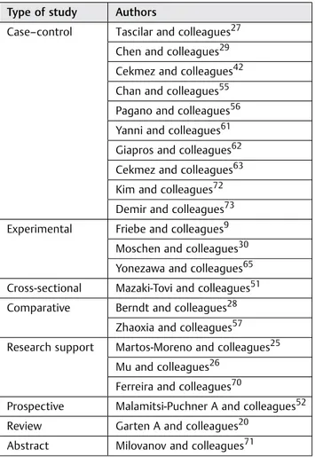
Table 1 Descriptive table of the most important studies conducted on Visfatin function
Type of study Authors
Case–control Tascilar and colleagues27 Chen and colleagues29 Cekmez and colleagues42 Chan and colleagues55 Pagano and colleagues56 Yanni and colleagues61 Giapros and colleagues62 Cekmez and colleagues63 Kim and colleagues72 Demir and colleagues73 Experimental Friebe and colleagues9
Moschen and colleagues30 Yonezawa and colleagues65 Cross-sectional Mazaki-Tovi and colleagues51 Comparative Berndt and colleagues28
Zhaoxia and colleagues57 Research support Martos-Moreno and colleagues25
Mu and colleagues26 Ferreira and colleagues70
Prospective Malamitsi-Puchner A and colleagues52 Review Garten A and colleagues20
Abstract Milovanov and colleagues71
significant differences in adipocytokine levels between the preterm SGA and AGA groups.61,62
However, in a control study, it has been suggested that, independent of being SGA or LBW, visfatin might be an early indicator of insulin resistance.63 It was also noted lower adipopectic and higher visfatin levels in LGA and SGA than AGA neonates.52Consistently, an increased release of visfatin was demonstrated in the amnion of twins and triplets. It can be assumed maternal plasma visfatin also derived from the stretched fetal membranes of women with LGA fetuses.64 These data suggest that visfatin is a possible link between maternal and fetal environment.
Visfatin and Breast-Feeding
In thefirst 6 months of lactation, Yonezawa and colleagues demonstrated, in animal/experimental study, the presence of mRNA-visfatin in cloned bovine mammary gland and human breast cancer cell line.65 Moreover, milk visfatin levels are directly related to preconceptional maternal BMI. In fact, it has been found higher breast milk visfatin concentrations for a BMI greater than 28 to 30 kg/m2. Visfatin seems to limit a weight loss in newborns.66
Visfatin and Preeclampsia
Preeclampsia (PE) is a multisystemic disorder of pregnancy, characterized by onset hypertension, endothelial dysfunc-tion, and proteinuria that develop after 20 weeks of gestation in previously normotensive women.67The precise cause of PE is still unclear, but it is believed to be likely multifactorial. The
role of visfatin in PE was investigated by several studies. Circulating visfatin concentrations are increased in PE in some studies,68whereas other investigators show similar52 or even decreased concentrations.69Ferreira and colleagues hypothesized that visfatin develops PE, promoting an im-paired placental vascularization, due to increased serum vascular endothelial growth factor (VEGF) levels70(►Fig. 1). Conversely, Milovanov and colleagues noted decreased ex-pression of VEGF as well as visfatin in pregnancies complicat-ed by PE.71During the third trimester of pregnancy, in women affected by PE, Kim and colleagues further showed decreased placental visfatin levels.72Although the severity of PE did not influence serum visfatin values, authors investigated rela-tionship between PE, gestational age (SGA), and maternal circulating visfatin concentrations. These were higher in women who delivered SGA newborns than normal pregnancy and PE groups.73However, further studies did not confirm these data.51
In conclusions, though the primary purpose of adipose tissue is energy storage, it has been also identified as an active endocrine-immune organ releasing many cytokines (adipo-cytokines) and hormones. In response to an autocrine and paracrine manner to impair adipocyte function, it has been reported that phenotypic changes of adipocytes occur into inflammatory response and promote a variety of diseases. Here, we have focused on visfatin expression, a recently identified adipocytokine. Although its precise function re-mains to be established, this review aims to emphasize the molecular and endocrine mechanisms of visfatin and to clarify its role in the pathophysiology of in flammatory-endo-crine disorders, also during normal and pathologic pregnancy
Fig. 1 Visfatin secretion and function. HDL, high-density lipoprotein; IL-6, interleukin-6; TNF-α, tumor necrosis factor-α.
(e.g., PE, GDM, and alteration of fetal growth). Moreover, for its potential diagnostic and/or prognostic role in the predic-tion of any associate disease or condipredic-tion, visfatin could also be considered as a new biomarker, indicating specific disor-ders in the neonatal metabolic profile, determining the interconnection of the different processes, and defining disease severity. In addition, several diseases could be treated by normalization and/or regulation of the proinflammatory cytokine/adipokine profile. For these novel concepts, visfatin might be promising candidates for future pharmacologic treatment strategies. Further research will bring new insight into linkage between visfatin and humans, during pregnancy and perinatal period.
Authors’ Contributions
All authors read and approved thefinal manuscript.
Conflict of Interest
None.
References
1 Mamì C, Marseglia L, Manganaro R, et al. Serum levels of resistin and its correlation with adiponectin and insulin in healthy full term neonates. Early Hum Dev 2009;85(1):37–40
2 Rajala MW, Scherer PE. Minireview: the adipocyte—at the cross-roads of energy homeostasis, inflammation, and atherosclerosis. Endocrinology 2003;144(9):3765–3773
3 Arrigo T, Chirico V, Salpietro V, et al. High-mobility group protein B1: a new biomarker of metabolic syndrome in obese children. Eur J Endocrinol 2013;168(4):631–638
4 Chirico V, Cannavò S, Lacquaniti A, et al. Prolactin in obese children: a bridge between inflammation and metabolic-endo-crine dysfunction. Clin Endocrinol (Oxf) 2013;79(4):537–544 5 d’Annunzio G, Vanelli M, Pistorio A, et al; Diabetes Study Group of
the Italian Society for Pediatric Endocrinology and Diabetes. Insulin resistance and secretion indexes in healthy Italian children and adolescents: a multicentre study. Acta Biomed 2009;80(1):21–28 6 Kralisch S, Bluher M, Paschke R, Stumvoll M, Fasshauer M.
Adipo-kines and adipocyte targets in the future management of obesity and the metabolic syndrome. Mini Rev Med Chem 2007;7(1): 39–45
7 Samal B, Sun Y, Stearns G, Xie C, Suggs S, McNiece I. Cloning and characterization of the cDNA encoding a novel human pre-B-cell colony-enhancing factor. Mol Cell Biol 1994;14(2):1431–1437 8 Fukuhara A, Matsuda M, Nishizawa M, et al. Visfatin: a protein
secreted by visceral fat that mimics the effects of insulin. Science 2005;307(5708):426–430
9 Friebe D, Löffler D, Schönberg M, et al. Impact of metabolic regulators on the expression of the obesity associated genes FTO and NAMPT in human preadipocytes and adipocytes. PLoS ONE 2011;6(6):e19526
10 Preiss J, Handler P. Enzymatic synthesis of nicotinamide mononu-cleotide. J Biol Chem 1957;225(2):759–770
11 Revollo JR, Grimm AA, Imai S. The NAD biosynthesis pathway mediated by nicotinamide phosphoribosyltransferase regulates Sir2 activity in mammalian cells. J Biol Chem 2004;279(49): 50754–50763
12 Rongvaux A, Andris F, Van Gool F, Leo O. Reconstructing eukaryotic NAD metabolism. BioEssays 2003;25(7):683–690
13 Revollo JR, Grimm AA, Imai S. The regulation of nicotinamide adenine dinucleotide biosynthesis by Nampt/PBEF/visfatin in mammals. Curr Opin Gastroenterol 2007;23(2):164–170
14 Lin SJ, Guarente L. Nicotinamide adenine dinucleotide, a metabolic regulator of transcription, longevity and disease. Curr Opin Cell Biol 2003;15(2):241–246
15 Imai S, Armstrong CM, Kaeberlein M, Guarente L. Transcriptional silencing and longevity protein Sir2 is an NAD-dependent histone deacetylase. Nature 2000;403(6771):795–800
16 van der Veer E, Nong Z, O’Neil C, Urquhart B, Freeman D, Pickering JG. Pre-B-cell colony-enhancing factor regulates NADþ-depen-dent protein deacetylase activity and promotes vascular smooth muscle cell maturation. Circ Res 2005;97(1):25–34
17 Moynihan KA, Grimm AA, Plueger MM, et al. Increased dosage of mammalian Sir2 in pancreatic beta cells enhances glucose-stimu-lated insulin secretion in mice. Cell Metab 2005;2(2):105–117 18 Revollo JR, Körner A, Mills KF, et al. Nampt/PBEF/Visfatin regulates
insulin secretion in beta cells as a systemic NAD biosynthetic enzyme. Cell Metab 2007;6(5):363–375
19 Hug C, Lodish HF. Medicine. Visfatin: a new adipokine. Science 2005;307(5708):366–367
20 Garten A, Petzold S, Körner A, Imai S, Kiess W. Nampt: linking NAD biology, metabolism and cancer. Trends Endocrinol Metab 2009; 20(3):130–138
21 Sethi JK. Is PBEF/visfatin/Nampt an authentic adipokine relevant to the metabolic syndrome? Curr Hypertens Rep 2007;9(1):33–38 22 Körner A, Blüher S, Kapellen T, et al. Obesity in childhood and
adolescence: a review in the interface between adipocyte physi-ology and clinical challenges. Hormones (Athens) 2005;4(4): 189–199
23 Kover K, Tong PY, Watkins D, et al. Expression and regulation of Nampt in human islets. PLoS ONE 2013;8(3):e58767
24 Bełtowski J. Apelin and visfatin: unique “beneficial” adipokines upregulated in obesity? Med Sci Monit 2006;12(6):RA112–RA119 25 Martos-Moreno GA, Kratzsch J, Körner A, et al. Serum visfatin and vaspin levels in prepubertal children: effect of obesity and weight loss after behavior modifications on their secretion and relation-ship with glucose metabolism. Int J Obes 2011;35(10):1355–1362 26 Mu J, Feng B, Ye Z, et al. Visfatin is related to lipid dysregulation, endothelial dysfunction and atherosclerosis in patients with chronic kidney disease. J Nephrol 2011;24(2):177–184
27 Taşçilar ME, Cekmez F, Meral C, et al. Evaluation of adipocytokines in obese children with insulin resistance. Turk J Pediatr 2011; 53(3):269–273
28 Berndt J, Klöting N, Kralisch S, et al. Plasma visfatin concentrations and fat depot-specific mRNA expression in humans. Diabetes 2005;54(10):2911–2916
29 Chen MP, Chung FM, Chang DM, et al. Elevated plasma level of visfatin/pre-B cell colony-enhancing factor in patients with type 2 diabetes mellitus. J Clin Endocrinol Metab 2006;91(1):295–299 30 Moschen AR, Kaser A, Enrich B, et al. Visfatin, an adipocytokine
with proinflammatory and immunomodulating properties. J Im-munol 2007;178(3):1748–1758
31 Dahl TB, Yndestad A, Skjelland M, et al. Increased expression of visfatin in macrophages of human unstable carotid and coronary atherosclerosis: possible role in inflammation and plaque desta-bilization. Circulation 2007;115(8):972–980
32 Friebe D, Neef M, Kratzsch J, et al. Leucocytes are a major source of circulating nicotinamide phosphoribosyltransferase (NAMPT)/ pre-B cell colony (PBEF)/visfatin linking obesity and inflammation in humans. Diabetologia 2011;54(5):1200–1211
33 Adya R, Tan BK, Chen J, Randeva HS. Nuclear factor-kappaB induc-tion by visfatin in human vascular endothelial cells: its role in MMP-2/9 production and activation. Diabetes Care 2008;31(4):758–760 34 Nowell MA, Richards PJ, Fielding CA, et al. Regulation of pre-B cell colony-enhancing factor by STAT-3-dependent interleukin-6 trans-signaling: implications in the pathogenesis of rheumatoid arthritis. Arthritis Rheum 2006;54(7):2084–2095
35 Jia SH, Li Y, Parodo J, et al. Pre-B cell colony-enhancing factor inhibits neutrophil apoptosis in experimental inflammation and clinical sepsis. J Clin Invest 2004;113(9):1318–1327
36 Iqbal J, Zaidi M. TNF regulates cellular NADþ metabolism in primary macrophages. Biochem Biophys Res Commun 2006; 342(4):1312–1318
37 Ognjanovic S, Bryant-Greenwood GD. Pre-B-cell colony-enhanc-ing factor, a novel cytokine of human fetal membranes. Am J Obstet Gynecol 2002;187(4):1051–1058
38 Haider DG, Schaller G, Kapiotis S, Maier C, Luger A, Wolzt M. The release of the adipocytokine visfatin is regulated by glucose and insulin. Diabetologia 2006;49(8):1909–1914
39 López-Bermejo A, Chico-Julià B, Fernàndez-Balsells M, et al. Serum visfatin increases with progressive beta-cell deterioration. Diabe-tes 2006;55(10):2871–2875
40 Mastorakos G, Valsamakis G, Papatheodorou DC, et al. The role of adipocytokines in insulin resistance in normal pregnancy: visfatin concentrations in early pregnancy predict insulin sensitivity. Clin Chem 2007;53(8):1477–1483
41 Kendal-Wright CE, Hubbard D, Bryant-Greenwood GD. Chronic stretching of amniotic epithelial cells increases pre-B cell colony-enhancing factor (PBEF/visfatin) expression and protects them from apoptosis. Placenta 2008;29(3):255–265
42 Cekmez F, Canpolat FE, Cetinkaya M, et al. Diagnostic value of resistin and visfatin, in comparison with C-reactive protein, pro-calcitonin and interleukin-6 in neonatal sepsis. Eur Cytokine Netw 2011;22(2):113–117
43 van Aerde JE, Wilke MS, Feldman M, Clandinin MT. Chapter 42, Accretion of Lipid in the Fetus and Newborn. In: Polin RA, Fox WW, Abman SH, eds. Fetal and Neonatal Physiology. 4th ed. Philadelphia, PA: Elsevier Saunders; 2011:454–469
44 Blanco CL, Baillargeon JG, Morrison RL, Gong AK. Hyperglycemia in extremely low birth weight infants in a predominantly Hispanic population and related morbidities. J Perinatol 2006;26(12): 737–741
45 Mazaki-Tovi S, Romero R, Kusanovic JP, et al. Visfatin in human pregnancy: maternal gestational diabetes vis-à-vis neonatal birth-weight. J Perinat Med 2009;37(3):218–231
46 Lihn AS, Pedersen SB, Richelsen B. Adiponectin: action, regulation and association to insulin sensitivity. Obes Rev 2005;6(1):13– 21
47 Mamì C, Manganaro R, Marseglia L, Saitta G, Gemelli M, Martino F. Plasma leptin, insulin, and neuropeptide Y response to feeding in newborn infants. Arch Dis Child Fetal Neonatal Ed 2006;91(5): F389
48 Mamì C, Manganaro R, Saitta G, Marseglia L, Martino F, Gemelli M. Plasma leptin, insulin, and neuropeptide Y concentrations in infants. Arch Dis Child Fetal Neonatal Ed 2005;90(1):F86–F87 49 Meral C, Cekmez F, Pirgon O, et al. The relationship between
serum visfatin, adiponectin, and insulin sensitivity markers in neonates after birth. J Matern Fetal Neonatal Med 2011;24(1): 166–170
50 Krzyzanowska K, Krugluger W, Mittermayer F, et al. Increased visfatin concentrations in women with gestational diabetes mel-litus. Clin Sci (Lond) 2006;110(5):605–609
51 Mazaki-Tovi S, Romero R, Kim SK, et al. Could alterations in maternal plasma visfatin concentration participate in the pheno-type definition of preeclampsia and SGA? J Matern Fetal Neonatal Med 2010;23(8):857–868
52 Malamitsi-Puchner A, Briana DD, Boutsikou M, Kouskouni E, Has-siakos D, Gourgiotis D. Perinatal circulating visfatin levels in intra-uterine growth restriction. Pediatrics 2007;119(6):e1314–e1318 53 Sethi JK, Vidal-Puig A. Visfatin: the missing link between
intra-abdominal obesity and diabetes? Trends Mol Med 2005;11(8): 344–347
54 Ferreira AF, Rezende JC, Vaikousi E, Akolekar R, Nicolaides KH. Maternal serum visfatin at 11-13 weeks of gestation in gestational diabetes mellitus. Clin Chem 2011;57(4):609–613
55 Chan TF, Chen YL, Lee CH, et al. Decreased plasma visfatin con-centrations in women with gestational diabetes mellitus. J Soc Gynecol Investig 2006;13(5):364–367
56 Pagano C, Pilon C, Olivieri M, et al. Reduced plasma visfatin/pre-B cell colony-enhancing factor in obesity is not related to insulin resistance in humans. J Clin Endocrinol Metab 2006;91(8): 3165–3170
57 Zhaoxia L, Ying W, Danqing C. Changes in visfatin levels after oral glucose tolerance test in women with gestational diabetes melli-tus. Diabetes Res Clin Pract 2012;96(3):e76–e79
58 Fasshauer M, Blüher M, Stumvoll M, Tönessen P, Faber R, Stepan H. Differential regulation of visfatin and adiponectin in pregnancies with normal and abnormal placental function. Clin Endocrinol (Oxf) 2007;66(3):434–439
59 Harrington TA, Thomas EL, Frost G, Modi N, Bell JD. Distribution of adipose tissue in the newborn. Pediatr Res 2004;55(3):437–441 60 López-Bermejo A, de Zegher F, Díaz-Silva M, Vicente MP, Valls C,
Ibáñez L. Cord serum visfatin at term birth: maternal smoking unmasks the relation to foetal growth. Clin Endocrinol (Oxf) 2008; 68(1):77–81
61 Yanni D, Darendeliler F, Bas F, Kucukemre Aydin B, Coban A, Ince Z. The role of leptin, soluble leptin receptor, adiponectin and visfatin in insulin sensitivity in preterm born children in prepubertal ages. Cytokine 2013;64(1):448–453
62 Giapros VI, Kiortsis DN, Evagelidou EN, et al. Visfatin levels in prepubertal children born small or large for gestational age. Horm Metab Res 2012;44(2):135–139
63 Cekmez F, Canpolat FE, Pirgon O, et al. Adiponectin and visfatin levels in extremely low birth weight infants; they are also at risk for insulin resistance. Eur Rev Med Pharmacol Sci 2013;17(4):501–506 64 Ma Y, Cheng Y, Wang J, Cheng H, Zhou S, Li X. The changes of visfatin in serum and its expression in fat and placental tissue in pregnant women with gestational diabetes. Diabetes Res Clin Pract 2010;90(1):60–65
65 Yonezawa T, Haga S, Kobayashi Y, Takahashi T, Obara Y. Visfatin is present in bovine mammary epithelial cells, lactating mammary gland and milk, and its expression is regulated by cAMP pathway. FEBS Lett 2006;580(28-29):6635–6643
66 Bienertová-Vašků J, Bienert P, Zlámal F, et al. Visfatin is secreted into the breast milk and is correlated with weight changes of the infant after the birth. Diabetes Res Clin Pract 2012;96(3):355–361 67 Sibai B, Dekker G, Kupferminc M. Pre-eclampsia. Lancet 2005;
365(9461):785–799
68 Fasshauer M, Waldeyer T, Seeger J, et al. Serum levels of the adipokine visfatin are increased in pre-eclampsia. Clin Endocrinol (Oxf) 2008;69(1):69–73
69 Hu W, Wang Z, Wang H, Huang H, Dong M. Serum visfatin levels in late pregnancy and pre-eclampsia. Acta Obstet Gynecol Scand 2008;87(4):413–418
70 Ferreira AF, Rezende JC, de Cassia C Oliveira R, Akolekar R, Nicolaides KH. Maternal serum visfatin at 11–13 weeks’ gestation in preeclampsia. J Hum Hypertens 2013;27(4):261–264 71 Milovanov AP, Sidorova IS, Solonitsyn AN, Borovkova EI.
Immuno-histochemical evaluation of the distribution of vascular endothe-lial growth factor in the placenta, placental bed in normal pregnancy and in women with preeclampsia [in Russian]. Arkh Patol 2008;70(3):12–15
72 Kim SC, Park MJ, Joo BS, Joo JK, Suh DS, Lee KS. Decreased expressions of vascular endothelial growth factor and visfatin in the placental bed of pregnancies complicated by preeclampsia. J Obstet Gynaecol Res 2012;38(4):665–673
73 Demir BC, Atalay MA, Ozerkan K, Doster Y, Ocakoglu G, Kucukko-murcu S. Maternal adiponectin and visfatin concentrations in normal and complicated pregnancies. Clin Exp Obstet Gynecol 2013;40(2):261–267