MYC protein expression scoring and its impact on
the prognosis of aggressive B-cell lymphoma
patients
This study examined the reproducibility of MYC and BCL-2 immunohistochemical scoring as well as the impact of higher expression of both proteins (double expressor status, DE) on survival and progression in a large retrospective cohort of aggressive B-cell lymphoma patients treated with rituximab plus cyclophosphamide, doxorubicin, vincristine and prednisone (CHOP) or R-CHOP-like regimens with a median follow up of 67 months (range 0-138). We also investigated possible MYC protein expression cut offs with the highest repro-ducibility among pathologists and predictability of gene translocation. We showed that immunohistochemistry (IHC) for MYC and BCL-2 is highly reproducible when cut-off values of >70% for MYC and >50% for BCL-2 are used. This threshold not only predicts the presence of rearrangements (with respect to MYC), but is also clini-cally valuable. In fact, it identifies a subset of patients who are poor responders and who may benefit from alternate therapeutic strategies.
Several investigators have demonstrated that diffuse large B-cell lymphomas (DLBCL) with MYC and BCL-2 double expression (DE-LBCL) have adverse prognosis when treated by conventional immunochemotherapies.1 Therefore, evaluation of MYC and BCL-2 protein expres-sion by IHC is an important tool in the prognostic strati-fication of patients.2Although expression of MYC-IHC in ≥40% neoplastic cells and the BCL-2 in ≥50% have been indicated as prognostically significant cut offs in many reports, among hematopathologists, some disagreement about these thresholds remains.3,4 Standardizing cut offs to define positivity for MYC-IHC and BCL-2-IHC with higher reproducibility and prognostic impact is thus high-ly desirable in order to optimize patient management.5,6
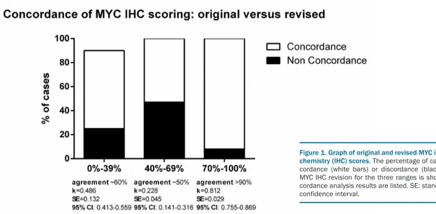
We analyzed 753 aggressive B-cell lymphoma patients. These included: Burkitt lymphomas (BL, n=223); DLBCL not otherwise specified (DLBCL-NOS, n=456); high grade B-cell lymphomas (HGBCL) with MYC and BCL-2 and/or BCL-6 rearrangement [HG double hit (HGDH), n=51, including 8 blastoid, 16 diffuse monomorphic and 27 intermediate morphology]; and HGBCL, NOS (n=23).7-9 All cases belonging to these categories, except for BL, will be referred to hereon as non-BL. The follow-ing clinical data were collected for non-BL: sex, age at diagnosis, ECOG performance status, stage (Ann Arbor), lactate dehydrogenase (LDH) level, International Prognostic Index (IPI), bulky disease, extranodal versus nodal, bone marrow involvement. Original MYC-IHC and BCL-2-IHC slides were re-evaluated by 3 trained pathologists blinded as to the original scores. Although aware that immunohistochemical interpretation may be affected by technical issues, we intentionally decided not to re-stain all slides in only one laboratory, but instead to confront original slides from different laboratories which apply different unmasking procedures and use different clones on a “real-life” basis (Table 1). Scores were report-ed in 5% intervals roundreport-ed up to the nearest 10%. Any discrepancies in MYC-IHC and BCL-2-IHC scoring was resolved by consensus on a multi-head microscope until an agreement of >95% concordance was reached. Discrepancy was defined as 10% deviation. Our findings confirm that BCL-2-IHC scoring is highly reproducible across the different institutions and that a ≥50% cut off is reliably assessable [agreement >90%; k=0.97; standard error (SE):=0.018; 95% confidence interval (CI): 0.765-0.901]. On the contrary, scoring MYC-IHC staining was confirmed to be critical, mainly because of the variability of staining intensity, percentage of positivity, presence of necrosis and crush artifacts. Concordance was high only for MYC-IHC positivity >70%, while a larger discrepan-cy was observed in the range 40-69%10 (Figure 1). The lat-ter can have a crucial impact on clinical decision making
haematologica 2018; 103:e25
L
ETTERS TO THE
E
DITOR
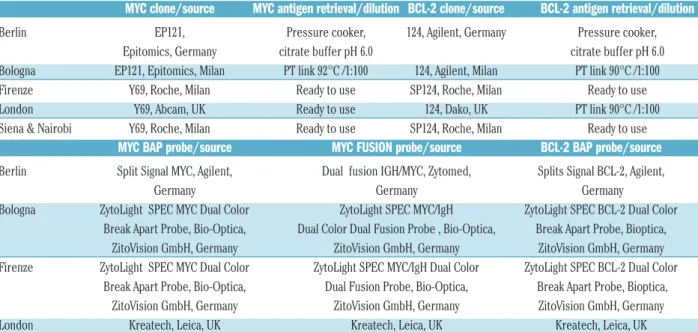
Table 1. Immunohistochemical and FISH analysis of MYC* and BCL-2* proteins and genes in the different institutions.
MYC clone/source
MYC antigen retrieval/dilution BCL-2 clone/source
BCL-2 antigen retrieval/dilution
Berlin EP121, Pressure cooker, 124, Agilent, Germany Pressure cooker,
Epitomics, Germany citrate buffer pH 6.0 citrate buffer pH 6.0
Bologna EP121, Epitomics, Milan PT link 92°C /1:100 124, Agilent, Milan PT link 90°C /1:100
Firenze Y69, Roche, Milan Ready to use SP124, Roche, Milan Ready to use
London Y69, Abcam, UK Ready to use 124, Dako, UK PT link 90°C /1:100
Siena & Nairobi Y69, Roche, Milan Ready to use SP124, Roche, Milan Ready to use
MYC BAP probe/source
MYC FUSION probe/source
BCL-2 BAP probe/source
Berlin Split Signal MYC, Agilent, Dual fusion IGH/MYC, Zytomed, Splits Signal BCL-2, Agilent,Germany Germany Germany
Bologna ZytoLight SPEC MYC Dual Color ZytoLight SPEC MYC/IgH ZytoLight SPEC BCL-2 Dual Color Break Apart Probe, Bio-Optica, Dual Color Dual Fusion Probe , Bio-Optica, Break Apart Probe, Bioptica,
ZitoVision GmbH, Germany ZitoVision GmbH, Germany ZitoVision GmbH, Germany
Firenze ZytoLight SPEC MYC Dual Color ZytoLight SPEC MYC/IgH Dual Color ZytoLight SPEC BCL-2 Dual Color Break Apart Probe, Bio-Optica, Dual Fusion Probe, Bio-Optica, Break Apart Probe, Bioptica,
ZitoVision GmbH, Germany ZitoVision GmbH, Germany ZitoVision GmbH, Germany
London Kreatech, Leica, UK Kreatech, Leica, UK Kreatech, Leica, UK
Siena & NairobiZytoLight SPEC MYC Dual Color Break Apart Probe, Optica, ZitoVision GmbH, Germany ZytoLight SPEC MYC/IgH Dual Color Dual Fusion Probe, Bio-Optica, ZitoVision GmbH, GermanyZytoLight SPEC BCL-2 Dual Color Break Apart Probe, Bioptica, ZitoVision GmbH, Germany. *On 4 m thick formalin-fixed paraffin-embed-ded sections; BAP: break-apart probe.
and patient care, as well as on the comparison between different studies. Therefore, we propose the threshold of MYC-IHC 70-100% to define non-BL cases with high expression of MYC and 0-39% to classify samples with low expression of MYC protein. Samples with MYC-IHC scores between 40% and 69% could not be categorized as MYC protein negative or positive with acceptable accuracy in routine practice and should be termed as cases with intermediate MYC expression. Moreover, we suggest scoring should preferably be defined on moder-ately to strongly stained nuclei and in hot spot areas, if present. After revision, in the non-BL series, prevalence of DE-LBCL as defined by the cut offs proposed by the literature (MYC-IHC
≥
40% and BCL-2-IHC ≥50%) was 32% (n=176/530);7 of these 100 showed MYC-IHC ≥70% (DE 70% in the following).A second end point of this study was to investigate the relation between MYC-IHC and MYC gene alterations, trying to identify parameters potentially applicable in a value-for-money approach. When using the threshold of MYC-IHC ≥80% in BL and ≥70% in non-BL, around 99% BL and 88% non-BL cases with MYC rearrangements can be predicted. Therefore, considering the high correlation between MYC gene rearrangement and MYC protein expression in BL, FISH analysis could theoretically be limited to cases with atypical morphology and/or immunophenotype and MYC-IHC <80%. In non-BL, a cut off of MYC-IHC ≥70% would identify cases at higher probability of MYC rearrangement. However, it should be noted that roughly 10% of HGDH have a lower expression of MYC; in these cases, FISH should be rec-ommended.11
The third end point was to perform a survival analysis in cases of HGDH, DE, nonDH/nonDE patients for whom overall survival (OS) and progression-free survival (PFS) data were available. HGDH lymphomas are known to be very aggressive neoplasms that do not respond to standard therapy and they have a dismal prognosis.12 However, it has recently been reported that the adverse prognosis may be mitigated by other factors such as lack of BCL-2 protein, and absence/low MYC protein expres-sion.5The clinical significance of MYC-IHC and BCL-2-IHC expression has been extensively studied but the results have not been consistent; hence, there is still no
clear use for protein expression evaluation in the clin-ic.13,14
Univariate Cox analysis showed that the factors signif-icantly associated with OS and PFS were: age [Odds Ratio (OR): 4.8% each year; confidence interval (CI): 1.03-1.066; P<0.001], LDH >150 UI/L (OR: 187%, CI: 1.756-4.715; P<0.001), IPI (OR: 71%, CI: 1.422-2.056; P<0.001), bone marrow involvement (OR: 86.2%, CI: 1.102-3.145; P=0.02), MYC translocation (OR: 71%, CI: 1.281-2.297; P<0.001), BCL-2 translocation (OR: 97%, CI: 1.389-2.795; P=0.002), MYC-IHC ≥70% (OR: 65%, CI: 0.22-0.539; P<0.001), DH status (OR: 310%, CI: 2.529-6.662; P<0.001), DE 70% status (OR: 61.5%, CI: 1.976-2.67; P=0.04). Stepwise multivariate analysis demonstrated that IPI (OR: 62.2%, CI: 1.225-2.146; P=0.001), MYC-IHC ≥70% (OR: 224.8%, CI: 1.714-6.155; P<0.001), DH status (OR: 131.7%, CI: 1.003-5.350; P=0.049), DE 70% status (OR: 177.5%, CI: 1.020-1.080; P=0.006) were independent prognostic factors that influence patients' outcome.
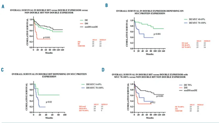
A comparison of HGDH, DE-LBCL [according to World Health Organization (WHO) criteria, i.e. MYC-IHC
≥
40% and BCL-2-IHC ≥50%] and nonDH/nonDE sub-sets of non-BL in terms of OS and PFS confirmed that HGDH patients showed significantly worse survival than the other two groups (P<0.001; χ239.24 for OS and 13.13for PFS) (Figure 2A and Online Supplementary Figure S1A). By stratifying DE-LBCL according to MYC protein expression (70-100%, 40-69%,) the prognosis turned out to be poorer in the former (P<0.001, χ213.152 for OS and
10.723 for PFS) (Figure 2B and Online Supplementary Figure S1B).
Dividing HGDH cases according to MYC protein posi-tivity revealed that those with MYC-IHC ≥70% had a worse prognosis than cases with MYC-IHC ≤69% (P<0.001, χ248.215 for OS and 35.549 for PFS) (Figure 2C
and Online Supplementary Figure S1C). This supports prior observations that patients with MYC rearrange-ments or DH lymphomas devoid of MYC-IHC expres-sion might exhibit less aggressive clinical behaviors.13
Given that the highest concordance and reproducibility of MYC-IHC was >70%, we compared OS and PFS of DE-LBCL showing MYC-IHC >70% and BCL-2-IHC >50% with HGDH cases; we found no significant differ-haematologica 2018; 103:e26
L
ETTERS TO THE
E
DITOR
Figure 1. Graph of original and revised MYC immunohisto-chemistry (IHC) scores. The percentage of cases with con-cordance (white bars) or discon-cordance (black bars) after MYC IHC revision for the three ranges is shown. The con-cordance analysis results are listed. SE: standard error; CI: confidence interval.
ences in prognosis (P=0.2, χ20.777 for OS and 3.441 for
PFS) (Figure 2D and Online Supplementary Figure S1D). Interestingly, the OS and the PFS of HGBCL, NOS strictly depended on MYC expression being 30 and 28 months in HGBCL, NOS with MYC 40-69%, 10 and 14 months in those with MYC 70-100%.
These findings again highlight the need to establish more widely reproducible cut-off values for IHC assays with higher predictive value, to safely stratify high-risk patients. Moreover, they may also, at least in part, explain the above mentioned inconsistencies in pub-lished data.
Finally, since the cell-of-origin (COO) classification of LBCL exerts a prognostic impact, with non-GCB lym-phomas showing a poorer prognosis than GCB ones, we stratified HGDH and DE-LBCL according to COO defined by Hans' algorithm.15 A poorer prognosis was demonstrated only in patients with MYC-IHC
≥
70%, with no differences according to the COO. However, no conclusions can be drawn due to both the limited num-ber of cases examined per group and to the IHC assess-ment of COO which is known to misclassify a subset of samples.Different molecular mechanisms may be responsible for the same disease entity and neoplastic phenotype. FISH currently represents the gold standard for identify-ing rearrangements, but it is not capable of detectidentify-ing genetic deregulation affecting gene expression at
tran-scriptional and post-trantran-scriptional levels that might result in protein overexpression and neoplastic transfor-mation.4 Our results support the role of MYC protein as the active trigger of the MYC-mediated oncogenic effects since protein expression levels likely represent a more direct measure of the activity of a particular gene16 and MYC-IHC should be undertaken in all cases. However, the last WHO criteria assessed that MYC staining is not reliable enough to select cases for FISH analysis.7 According to our results, the cut off of MYC-IHC ≥70%, based both on reproducibility among pathologists and clinical impact, is a good indicator of underlying MYC gene rearrangement. Nonetheless, our promising data need further validation in an independent cohort of patients. In addition, given the highest scoring efficiency, the cut offs of MYC-IHC ≥70% and BCL-2-IHC
≥
50% could be applied when FISH is not available (due to issues of practicability, cost, short turn-around-time, quality control) to select a subset of patients who are poor responders and who may benefit from alternate thera-peutic strategies.Maria R. Ambrosio,1Stefano Lazzi,1Giuseppe Lo Bello,1
Raffaella Santi,2Leonardo Del Porro,1Maria M. de Santi,3
Raffaella Guazzo,1Lucia Mundo,1Luigi Rigacci,4
Sofia Kovalchuck,4Noel Onyango,5Alberto Fabbri,6
Emanuele Cencini,6Pier Luigi Zinzani,7Francesco Zaja,8
Francesco Angrilli,9Caterina Stelitano,10Maria G. Cabras,11
Giuseppe Spataro,12Roshanak Bob,13Thomas Menter,14,15
haematologica 2018; 103:e27
L
ETTERS TO THE
E
DITOR
Figure 2. Kaplan-Meier curves of overall survival (OS) in the different patient groups. (A) Kaplan-Meier curve of OS in double hit (DH) versus double expressor (DE) versus non-double hit/non-double expressor (nonDH/nonDE) lymphomas. There was a significant difference in OS between the DH and the other two groups (log rank test, P<0.001). DH lymphomas were defined by having both MYC and BCL-2 rearrangements; DE were characterized by showing MYC-immuno-histochemistry (IHC) ≥40% and BCL-2-IHC ≥50%; nonDH/nonDE included cases without double hits or with one only single hit, and with none or only one protein expression between MYC and BCL-2. (B) Kaplan-Meier curve of OS in DE depending on MYC protein expression. OS was significantly lower in the group showing MYC-IHC ≥70% than in the group with MYC-IHC 40-69% (log rank test, P<0.001) indicating that a MYC-IHC ≥70% cut off defines a clinically relevant subgroup of DE cases. (C) Kaplan-Meier curve of OS in DH depending on MYC protein expression. There was a significant difference in OS between the two groups (MYC-IHC 0-69% and BCL-2-(MYC-IHC ≥50%, and MYC-(MYC-IHC ≥70% and BCL-2-(MYC-IHC ≥50%) with MYC ≥70% cases having the lower survival rate (log rank test, P<0.001). Noteworthy, among HGDH with MYC-IHC 0-69%, the 3 cases showing MYC-IHC <40% had a mean OS of 44 months (data not shown). These findings indicate that adverse survival in HGDH is highly impacted by high MYC-IHC expression (≥70%). (D) Kaplan-Meier curve of OS in DH versus DE as defined by MYC-IHC ≥70% and BCL-2-IHC ≥50% versus nonDH/nonDE. OS overlaps in DH and DE with MYC-IHC ≥70% and BCL-2-IHC ≥50% (log rank test, P=0.2 for DH versus DE with MYC ≥70% and IHC ≥50%;), this is worse than the OS of the nonDH/nonDE (log rank test, P<0.001 for either DH or DE with MYC ≥70% and BCL-2-IHC ≥50% versus nonDH/nonDE).
A
C
D
Massimo Granai,1Gabriele Cevenini,1Kikkeri N. Naresh,15
Harald Stein,13Elena Sabattini16and Lorenzo Leoncini1 1Department of Medical Biotechnologies, University of Siena, Italy; 2Division of Pathological Anatomy, University of Florence, Italy; 3Unit of
Pathological Anatomy, AOU Siena, Italy; 4Hematology Division,
AOU Careggi, University of Firenze, Italy; 5Department of Clinical
Medicine and Therapeutics, Unit of Medical Oncology, University of Nairobi, Kenya; 6Haematology Unit, Azienda Ospedaliera
Universitaria Senese, Siena, Italy; 7Institute of Hematology "L. e A.
Seràgnoli", University of Bologna, Italy; 8Clinica Ematologica ed Unità
di Terapie Cellulari "Carlo Melzi", DAME, University of Udine, Italy; 9Ematologia Ospedale Civile dello Spirito Santo, Pescara, Italy; 10Ematologia, Ospedale Bianchi Melacrino Morelli, Reggio Calabria; 11Hematology, Cagliari Hospital, Italy; 12Post Graduate School of
Public Health, University of Siena, Italy; 13Pathodiagnostik Lab. Berlin,
Germany; 14Institute of Pathology and Medical Genetics, University
Hospital of Basel, Switzerland; 15Department of Cellular and
Molecular Pathology, Hammersmith Hospital Campus, Imperial College Healthcare NHS Trust, London, UK and 16Unit of
Haemolymphopathology, Department of Hematology and Oncology, University Hospital of Bologna, Italy
Acknowledgments: the Authors would like to thank additional hematological centers that contributed with clinical data: Di Raimondo F. - Hematology, Catania University, Italy, Cimino G. and Centra N. Hematology, Latina Hospital, Galieni P. – Hematology, Ascoli Piceno Hospital, Martelli M. and Di Rocco A. – Hematology, Roma University “La Sapienza”, Italy, Vigna E. – Hematology, Cosenza Hospital, Italy, Taratini G. – Hematology, Barletta Hospital, Italy, Scalone R. – Hematology, casa di Cura La Maddalena, Palermo, Italy, Mannina D. – Hematology, Papardo Hospital, Messina, Italy
Correspondence: [email protected] doi:10.3324/haematol.2018.195958
Information on authorship, contributions, and financial & other disclo-sures was provided by the authors and is available with the online version of this article at www.haematologica.org.
References
1. Sarkozy C, Traverse-Glehen A, Coiffier B. Double-hit and double-protein-expression lymphomas: aggressive and refractory lym-phomas. Lancet Oncol. 2015;16(15):e555-567.
2. Miura K, Takahashi H, Nakagawa M, et al. Clinical significance of co-expression of MYC and BCL-2 protein in aggressive B-cell lym-phomas treated with a second line immunochemotherapy. Leuk.
Lymphoma. 2016;57(6):1335-1341.
3. Kluk MJ, Ho C, Yu H, et al. MYC Immunohistochemistry to Identify MYC-Driven B-Cell Lymphomas in Clinical Practice. Am. J. Clin. Pathol. 2016;145(2):166-179.
4. Mahmoud AZ, George TI, Czuchlewski DR, et al. Scoring of MYC protein expression in diffuse large B-cell lymphomas: concordance rate among hematopathologists. Mod. Pathol. 2015;28(4):545-551. 5. Kawashima I, Inamoto Y, Maeshima AM, et al. Double-Expressor
Lymphoma Is Associated with Poor Outcomes after Allogeneic Hematopoietic Cell Transplantation. Biol Blood Marrow Transplant. 2018;24(2):294-300.
6. Rosenthal A, Younes A. High grade B-cell lymphoma with rearrange-ments of MYC and BCL-2 and/or BCL6: Double hit and triple hit lymphomas and double expressing lymphoma. Blood Rev. 2017; 31(2):37-42.
7. Swerdlow S.H, Campo E, Harris NL, et al. WHO classification of
Tumours of Haematopoietic and Lymphoid, 4thed. Lyon, France,
IARC Press, 2017.
8. Naresh KN, Hazem AHI, Lazzi S, et al. Diagnosis of Burkitt Lymphoma using an algorithmic approach applicable in both resource-poor and resource-rich countries. Br J Haematol. 2011; 154(6):770-776.
9. Menter T, Medani H, Ahmad R, Flora R, Trivedi P, Reid A, Naresh KN. MYC and BCL2 evaluation in routine diagnostics of aggressive B-cell lymphomas - presentation of a work-flow and the experience with 248 cases. Br J Haematol. 2017;179(4):667-688.
10. Landis JR, Koch GG. The measurement of observer agreement for categorical data. Biometrics. 1977;33(1):159-174.
11. Scott, DW, King RL, Staiger AM, et al. High-grade B-cell lymphoma with MYC and BCL2 and/or BCL6 rearrangements with diffuse large B-cell lymphoma morphology. Blood. 2018;131(18):2060-2064. 12. Friedberg JW. How I treat “Double Hit” lymphoma. Blood. 2017;
130(5):590-596.
13. Herrera AF, Mei M, Low L, et al. Relapsed or Refractory Double-Expressor and Double-Hit Lymphomas Have Inferior Progression-Free Survival After Autologous Stem-Cell Transplantation. J Clin Oncol. 2017;35(1):24-31.
14. Green TM, Young KH, Visco C, et al. Immunohistochemical double-hit score is a strong predictor of outcome in patients with diffuse large B-cell lymphoma treated with rituximab plus cyclophos-phamide, doxorubicin, vincristine, and prednisone. J Clin Oncol. 2012;30(28):3460-3467.
15. Staiger AM, Ziepert M, Horn H, et al. Clinical Impact of the Cell-of-Origin Classification and the MYC/ BCL-2 Dual Expresser Status in Diffuse Large B-Cell Lymphoma Treated Within Prospective Clinical Trials of the German High-Grade Non-Hodgkin's Lymphoma Study Group. J Clin Oncol. 2017;35(22):2515-2526.
16. Ott G, Rosenwald A, aCampo E. Understanding MYC-driven aggressive B-cell lymphomas: pathogenesis and classification. Blood. 2013; 122(24):3884-3891.
haematologica 2018; 103:e28