1
ACKNOWLEDGEMENTS
Firstly, I desire to express my thankfulness to my supervisor Dr. Lidietta Giorno for her support, motivation, and immense knowledge in the field of membrane science. Thanks for encouraging my research and for allowing me to grow as a research scientist. I would also like to thank Dr. Teresa Poerio who helped me and supported me throughout these years. Thanks also to Dr. Rosalinda Mazzei for her support. A special thanks to my colleagues and friends of the “third floor”. Finally, I would like to mention my family, and in particular Mattia who always encourage and support me.
2
Index
List of figures
6List of tables
10Sommario
11Contenuti della tesi 14
Summary
15Dissertation outlines 17
Chapter 1
18Introduction to bio-functionalized membranes
1.1 Membranes and membrane processes 19
1.2 Bio-functional membranes basic concepts 20
1.3 Applications of bio-functionalized membranes 21
1.3.1 Biocatalysis: biocatalytic membrane reactors 21
1.3.2 Separation: affinity membranes 22
1.3.3 Analysis: biosensors 23
1.4 Preparation of bio-functional polymeric membranes 23
1.4.1 Membrane surface functionalization/activation 24
1.4.1.1 Coating 25
1.4.1.2 Wet chemical 26
1.4.1.3 Self-assembly 29
1.4.1.4 Plasma treatment 30
1.4.1.5 UV irradiation 30
1.4.1.6 Surface Graft Polymerization 30
1.4.2 Biomolecules immobilization 31
1.5 Factors affecting biomolecules immobilization 34
1.5.1 Surface properties 35
1.5.2 Protein properties 36
3
1.6 Concluding remarks and advancements 38
Chapter 2
48Membranes for biosensors development
2.1 Biosensors basic concepts 49
2.2 Membranes for biosensing 52
2.2.1 Membrane properties and functions 53
2.2.2 Membrane materials 54
2.2.3 Membranes integration with miniaturized systems 57
2.3 Orientation of the bioreceptor 58
2.4 Transduction mechanisms and systems 60
2.4.1 Electrochemical detection 61
2.4.2 Optical detection 65
2.5 Applications of membranes in biosensors 66
2.6 General considerations and advancements 69
Chapter 3
79Development of bio-functionalized membranes: study of the relationship between proteins properties and membrane surface coverage
3.1 Materials and chemicals 81
3.2 Experimental methods 82
3.2.1 Membrane surface functionalization strategies 82
3.2.2 Functionalized membranes characterization 84
3.2.2.1 Infrared analysis 84
3.2.2.2 Ninhydrin test 84
3.2.2.3 Water permeability measurement 85
3.2.3 Biomolecules characterization in solution 85
3.2.3.1 Dynamic light scattering 86
3.2.3.2 Native gel Electrophoresis 86
3.2.4 Biomolecules immobilization on functionalized membrane surface 87
3.2.4.1 Reduction step optimization 87
3.2.4.2 Effect of proteins concentration and contact time 87
3.2.4.3 Determination of the amount of immobilized biomolecules 88
4
3.2.6 Performances of free and immobilized proteins 88
3.3 Results and discussion 90
3.3.1 Functionalized membranes characterization 90
3.3.2 Biomolecules characterization 94
3.3.3 Biomolecules immobilization 98
3.3.3.1 Adsorption tests 98
3.3.3.2 Reduction step optimization 99 3.3.3.3 Biomolecules immobilization trend 100
3.3.4 Kinetics of immobilization 105
3.3.5 Bio-functionalized membranes surface morphology 107
3.3.6 Performances of free and immobilized proteins 111
3.4 Conclusions 114
Chapter 4
119
Development of immuno-selective membranes for targeted interleukin-6 antigen capture, concentration and detection 4.1 Materials and chemicals 121
4.2 Experimental procedures 122
4.2.1 Protein G immobilization 122
4.2.1.1 Binding capacity of immobilized protein G 122
4.2.1.2 Analysis of the equilibrium adsorption data 123
4.2.2 Strategies for anti IL-6 antibody bio-affinity immobilization 124
4.2.2.1 Quenching step optimization 124
4.2.3 Evaluation of IL-6 capturing by the immuno-affinity membranes 126
4.2.3.1 ELISA tests 126
4.2.3.2 Confocal analysis 127
4.2.4 Desorption tests and membrane reuse 127
4.2.5 IL-6 pre-concentration and detection 128
4.2.5.1 Method for IL-6 pre-concentration 128
4.2.5.2 Organic Electrochemical Transistor (OECT) for IL-6 detection 130
4.3 Results and discussion 133
4.3.1 Antibody binding capacity of protein G-coupled membrane 133
5
4.3.2.1 Quenching step optimization 136
4.3.2.2 Quantification of IL-6 capturing 137
4.3.2.3 Visualization of IL-6 capturing 139
4.3.2.4 Desorption tests and membrane reuse 141
4.3.3 Preliminary results on IL-6 detection 145
4.4 Conclusions and general considerations 146
Overall conclusions
152APPENDIX
155Pubblications 155
6
List of figures
Figure 1.1 Concept of bio-functional membrane preparation
Figure 1.2 Schematic representation of membrane surface coating by functional layers
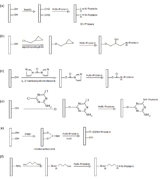
Figure 1.3 Schematic representation of some well-known membrane wet chemical activation reactions: (a) periodate oxidation, (b) epoxy activation, (c) carbonyldiimidazole activation, (d) triazine activation, (e) cyanogen bromide activation, (f) glutaraldehyde activation.
Figure 1.4 Schematic representation of layer-by-layer deposition process starting i.e. with a positively charged membrane
Figure 1.5 Schematic representation of biomolecules immobilization methods
Figure 2.1 A schematic representation of a biosensor and working principle
Scheme 2.1 Schematic of biosensors classification
Figure 2.2 Chemical structure of commonly used polymeric membrane materials
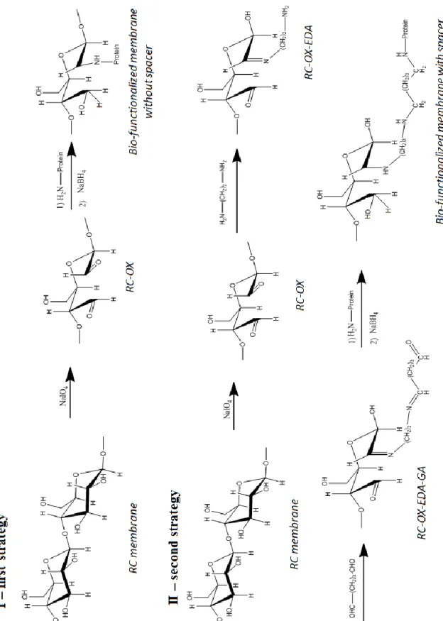
Figure 3.1 SEM image of RC membrane (reprinted from www.merckmillipore.com) Schematization of membranes bio/chemical functionalization without (I) and with the spacer (I)
Figure 3.3 Calibration curve of Ninhydrin test obtained by using different contents (µmol) of glycine as standard
Figure 3.4 Schematic representation of the biphasic lipase-immobilized membrane bioreactor
Figure 3.5 ATR-FTIR spectra of pristine (violet) and oxidized membranes with NaIO4 0.02 wt% (blue) and 0.2 wt % (red). A magnification for the marked circle is depicted on the right representing the range from 1750 to 1720 cm-1
Figure 3.6 ATR-FTIR spectra of oxidized (red) and aminated (violet) membranes
Figure 3.7 Ninhydrin test of RC-OX-EDA (left) and RC-OX-EDA-GA (right) membranes
Figure 3.8 Membrane water permeance after each step of functionalization
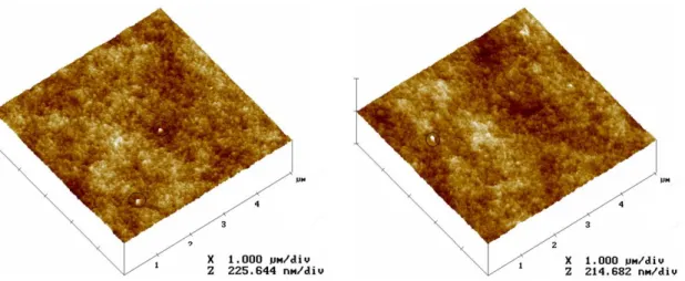
Figure 3.9 3D AFM images of pristine RC membrane (left) and RC-OX-EDA-GA functionalized membrane (right).
Figure 3.10 Particle size distribution (A) and Electrophoresis in native conditions (B) of BSA
7
protein G
Figure 3.12 Particle size distribution (A) and Electrophoresis in native conditions (B) of lipase
Figure 3.13 Particle size distribution (A) and Electrophoresis in native conditions of phosphotriesterase (B)
Figure 3.14 AFM images of lipase (I) and BSA (II) adsorbed on RC virgin membrane Figure 3.15 Absorbance value as a function of reaction time with NaBH4 to optimize the
reduction step (λ= 562 nm)
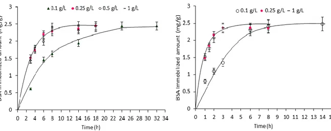
Figure 3.16 Effect of contact time and initial protein concentration on BSA immobilization onto RC-OX (left) and RC-OX-EDA-GA (right)
Figure 3.17 Effect of contact time and initial protein concentration on protein G immobilization onto RC-OX-EDA-GA
Figure 3.18 Irreversible binding isotherm
Figure 3.19 Effect of contact time and initial protein concentration on lipase immobilization onto RC-OX (left) and RC-OX-EDA-GA (right)
Figure 3.20 Effect of contact time on phosphotriesterase immobilization onto RC-OX-EDA-GA
Figure 3.21 Linear plot of pseudo-second order rate for BSA (1 g/L concentration) immobilization on RC-OX-EDA-GA
Figure 3.22 AFM image of RC-OX-EDA-GA membranes with covalently immobilized BSA after 4 h of incubation. The bearing histograms for the marked squares (a and b) are depicted respectively. (a) Some cluster structures with a depth of around 50 nm and (b) protein filament depth of around 15 nm.
Figure 3.23 AFM image of RC-OX-EDA-GA membranes with covalently immobilized protein G after 4 h of incubation. The bearing histograms of the marked squares (a and b) are depicted respectively. Protein filaments depth ranges from 6 to 9 nm for the thinnest filaments (b) and from 10 to 20 nm for the thickest ones (a).
Figure 3.24 AFM images of RC-OX-EDA-GA membranes with covalently immobilized lipase after 1 h (A) and 20 h (B) of incubation. The bearing histograms for the marked squares are depicted for both A and B. Small lipase globular aggregates can be detected after 1 h of incubation with a depth at around 14 nm (A), while large aggregates are detected after 20 h of incubation with a
8
maximum peak to valley height of around 60 nm.
Figure 3.25 Immobilized lipase stability
Figure 3.26 Size distribution of lipase (1 g/l concentration) before (red) and after disaggregation (green).
Figure 3.27 Effect of multilayers formation on the specific activity of immobilized phosphotriesterase
Figure 4.1 Schematic representation of the two strategies used for anti IL-6 antibody bioaffinity immobilization without chemical cross-link I) and with glutaraldehyde cross-link II)
Figure 4.2 Schematic representation of the membrane used as blank to evaluate the specificity of IL-6 binding
Figure 4.3 Schematic representation of the method used for IL-6 pre-concentration by using the immuno-affinity membrane
Figure 4.4 (a) Device structure and electrical circuit of a PEDOT:PSS organic electrochemical transistor (OECT); (b) and (c) OECT working principle [26]
Figure 4.5 Components of the OECT used for the electrical measurements
Figure 4.6 Schematic of the procedure used for electrochemical IL-6 detection by using the antibody-functionalized gate electrode and the IL-6 concentrated solution
Figure 4.7 Adsorption isotherm for commercial human IgG onto Protein G-coupled membrane
Figure 4.8 Linear Langmuir isotherm for hIgG adsorption on protein G-coupled membrane
Figure 4.9 Scatchard plot
Figure 4.10 Quenching step optimization. Influence of glycine quenching time on the amount of IL-6 (pg/cm2) non-specifically bounded on the membrane surface (RC-PG) at different IL-6 initial concentrations (pg/ml).
Figure 4.11 Amount of IL-6 captured by the RC-PG-Ab and RC-PG-Ab-(GA) membranes as a function of initial IL-6 concentration
Figure 4.12 Capture efficiency of RC-PG-Ab and RC-PG-Ab-(GA) membranes at different IL-6 initial concentrations.
9
membrane (a), RC-PG-Ab-(GA) membrane (b) and RC-PG-Ab-(GA)-Ag membranes incubated with IL-6 at 31 pg/ml (c), 250 pg/ml (d) and 500 pg/ml (e), all treated with anti IL-6 antibody FITC-conjugated
Figure 4.14 SDS-PAGE under reducing condition of the samples after the first cycle of acid desorption. 1 – wide range molecular weight marker (1:20 dilution). 2 – anti IL-6 antibody used as standard (65 ng). 3 – mouse IL-6 used as standard (13 ng). 4 – RC-PG-Ab-(GA)-Ag desorption. 5 – RC-PG-Ab-Ag desorption
Figure 4.15 Amount of IL-6 captured by the RC-PG-Ab-(GA) membrane for repeated use as a function of initial IL-6 concentration
Figure 4.16 Capture efficiency of RC-PG-Ab-(GA) membrane for repeated use, at different IL-6 initial concentration.
Figure 4.17 SDS-PAGE under reducing condition of the samples after BSA non-specific adsorption tests on RC-PG-Ab and RC-PG-Ab-(GA) membranes. 1 – wide range molecular weight marker (1:20 dilution). 2 – BSA initial solution. 3 – BSA final solution after incubation with RC-PG-Ab membrane. 4 – BSA final solution after incubation with RC-PG-Ab-(GA) membrane. 5 – RC-PG-Ab_BSA desorption. 6 – RC-PG-Ab-(GA)_BSA desorption
Figure 4.18 OECT response. Comparison between OECT modulation (ΔI/I0), as a function of gate voltage (Vgs), using bare Au-gate electrode (black), Ab-gate electrode (red) and Ab-Ab-gate electrode after IL-6 binding (blue).
10
List of tables
Table 2.1 Some applications of membrane biosensors
Table 3.1 Physicochemical properties of the biomolecules used in this study
Table 3.2 Summary of results obtained from DLS and Electrophoresis measurements for all concentrations tested of each protein
Table 3.3 Kinetic parameters for immobilization of BSA and protein G molecules on RC-OX and RC-OX-EDA-GA
Table 3.4 Hydrolytic activity for free and immobilized lipase
Table 3.5 Comparison between lipase activity before and after filtration
Table 4.1 Summary of mass balance results obtained from ELISA quantification experiments
11
Sommario
Negli ultimi 50 anni, biomolecole quali proteine, enzimi e anticorpi, sono state immobilizzate su superfici solide per un‘ampia gamma di applicazioni, per esempio, in diagnostica, nei processi di separazione e nei bio-processi in generale. Questa combinazione ‗ibrida‘ tra una entità biologica e un materiale di supporto conferisce funzionalità ‗biologiche‘ innovative al materiale sintetico, allo stesso tempo, permette di stabizzare e di riutilizzare la molecola biologica, offrendo anche la possibilità di modulare le proprietà della superficie solida.
Oggi, un grande interesse è rivolto all‘uso di materiali nanostrutturati come supporti solidi. Tra questi materiali, le membrane assumono sempre più maggiore rilievo grazie alle loro proprietà peculiari. Infatti, la loro elevata superficie di scambio per unità di volume e la possibilità di combinare il processo di separazione con il riconoscimento molecolare, hanno permesso di sviluppare sistemi bio-ibridi con molteplici funzionalità, selettività e stabilità per svariate applicazioni.
Questo lavoro di ricerca ha riguardato lo sviluppo di membrane polimeriche bio-ibride allo scopo di i) investigare e promuovere avanzamenti nella comprensione delle proprietà delle proteine che influenzano le nanostrutture di membrane nano-ingegnerizzate mediante l‘immobilizzazione di tali proteine e ii) progettare e sviluppare un sistema con proprietà di bio-riconoscimento e bio-separazione che si basa su interazioni di affinità.
Per la progettazione di una superficie bio-ibrida è necessario considerare diversi parametri. Infatti, l‘immobilizzazione di una biomolecola a le relativa attività biologica e stabilità dipendono da diversi fattori, ovvero, il metodo usato per l‘immobilizzazione, le proprietà della superficie di supporto, le proprietà della proteina e le condizioni in cui viene condotta l‘immobilizzazione. A tal riguardo, in questo lavoro di ricerca inizialmente l‘attività è stata focalizzata sullo studio sperimentale delle relazioni tra le proprietà di bulk delle proteine durante l‘immobilizzazione covalente e la risultante superficie bio-ibrida. Per lo studio, sono state utilizzate membrane in cellulosa rigenerata funzionalizzate mediante diverse procedure. Le membrane sono state attivate chimicamente secondo due strategie e sono state utilizzate quattro proteine modello (aventi diverse proprietà e diverso comportamento, ovvero, l‘albumina di siero bovino, la proteina G, l‘enzima lipasi da C. Rugosa e l‘enzima fosfotriesterasi) per studiare il processo di immobilizzazione e per produrre membrane bio-ibride. È stato studiato l‘effetto delle proprietà native delle proteine in soluzione (ovvero la concentrazione, la carica, la dimensione e i fenomeni di aggregazione) su i) la cinetica di
12
legame, ii) il rivestimento superficiale della membrana, iii) il riarrangiamento strutturale delle proteine, ed è stata testata l‘attività delle proteine dopo l‘immobilizzazione. La caratterizzazione rigorosa delle proteine in soluzione ha permesso di determinare la dimensione effettiva delle proteine e i fenomeni di aggregazione. Il loro comportamento durante l‘immobilizzazione è stato studiato sia in funzione della concentrazione iniziale delle proteine che del tipo di funzionalizzazione di membrana. I risultati hanno dimostrato che l‘aggregazione di una proteina in soluzione ha una influenza significativa sulla formazione dello strato di proteina sulla superficie della membrana. Inoltre, sono state realizzati diversi tipi di membrane bio-ibride con diverse proprietà funzionali (in base al tipo di proteina immobilizzata) che hanno mostrato buone performances con potenziali applicazioni nei bio-processi (bio-separazioni, bio-catalisi e bio-sensori). Questo studio è stato ulteriormente esteso allo scopo di progettare sistemi bio-ibridi capaci di riconoscere in modo selettivo molecole target di interesse diagnostico, sulla base di interazioni di immuno-affinità. In particolare, la membrana bio-funzionalizzata con la proteina G è stata utilizzata per sviluppare membrane di immuno-affinità altamente selettive, specifiche, stabili e riutilizzabili per la cattura, il riconoscimento e il rilevamento di molecole coinvolte in processi infiammatori, ovvero, l‘interleuchina-6 (IL-6). Sono state sviluppate due diverse strategie in cui la membrana con la proteina G legata è stata utilizzata per l‘immobilizzazione sito-specifica ed orientata dell‘anticorpo per l‘IL-6. Nella prima strategia, dopo l‘immobilizzazione dell‘anticorpo, la membrana di immuno-affinità è stata usata direttamente per la cattura ed il riconoscimento dell‘IL-6. Nella seconda strategia, l‘anticorpo per l‘IL-6 è stato stabilizzato mediante un cross-link chimico prima di effettuare la cattura ed il riconoscimento dell‘IL-6. In altri termini, nel primo caso l‘anticorpo è immobilizzato mediante interazioni di affinità con la proteina G, nel secondo il legame dell‘anticorpo è stabilizzato mediante legame covalente. Sono stati studiati ed ottimizzati diversi aspetti e parametri: i) la capacità della proteina G di legare l‘anticorpo;
ii) le proprietà di bio-riconoscimento di entrambe le membrane di immuno-affinità, ovvero,
la capacità delle membrane di catturare l‘IL-6 e l‘efficienza di cattura; iii) la stabilità e la selettività dell‘anticorpo e la possibilità di riutilizzarlo; iv) il miglioramento della specificità del sistema di riconoscimento mediante la riduzione delle interazioni non specifiche. Entrambe le strategie hanno condotto allo sviluppo di sistemi bio-ibridi altamente efficienti, che hanno dimostrato la possibilità di una applicazione pratica per il riconoscimento ed il rilevamento di molecole target nel campo dei bio-sensori. Tuttavia, solo il legame mediante cross-linking ha permesso di stabilizzare e riutilizzare il sistema di
13
immuno-affinità. In questo caso, la membrana di immuno-affinità è stata successivamente integrata con un sistema di rilevamento elettrochimico ed è stata utilizzata per concentrare l‘IL-6 allo scopo di aumentare la sensibilità di rilevamento del sistema.
14 Contenuti della tesi
La tesi è stata suddivisa in 4 capitoli:
o Il capitolo 1 tratta l‘analisi dello stato dell‘arte attuale relativo alle membrane polimeriche bio-ibride. Il capitolo include la discussione di aspetti importanti nello sviluppo delle membrane bio-funzionalizzate e le relative applicazioni adottando una visione critica dei diversi fattori che incidono sul processo di immobilizzazione. Inoltre, sono stati evidenziati gli avanzamenti proposti in quest‘area di ricerca.
o Nel capitolo 2 sono stati presentati diversi aspetti relativi all‘uso delle membrane nel campo dei bio-sensori, come: le proprietà delle membrane, le loro funzioni, i vantaggi e le applicazioni. Ciò ha condotto ad una considerazione globale ed alla progettazione del lavoro che è stato svolto in questo specifico ambito di ricerca.
o Nel capitolo 3 è riporta l‘attività sperimentale relativa allo sviluppo di membrane bio-ibride multifunzionali di cellulosa rigenerata mediante l‘immobilizzazione covalente di diverse biomolecole, e lo studio approfondito dell‘influenza delle proprietà delle proteine in soluzione sul processo di immobilizzazione nonché sulla struttura delle membrane bioibride. La discussione include: la funzionalizzazione e la caratterizzazione delle membrane; la caratterizzazione delle proteine in soluzione e la correlazione tra le loro proprietà ed il loro comportamento durante l‘immobilizzazione; lo studio dell‘attività delle biomolecole dopo l‘immobilizzazione e la valutazione delle potenziali applicazioni delle membrane bio-ibride sviluppate.
o Nel capitolo 4 è riportata l‘attività sperimentale basata sullo sviluppo di membrane di cellulosa rigenerata altamente selettive aventi proprietà di bio-riconoscimento e bio-separazione e la loro relativa applicazione per il rilevamento di molecole target. La discussione riguarda: la progettazione e lo sviluppo di membrane di immuno-affinità; la valutazione delle abilità delle membrane di riconoscere molecole target; lo studio e l‘ottimizzazione di diversi aspetti e parametri quali la selettività, la stabilità e la possibilità di riutilizzare le membrane; l‘applicazione pratica nella concentrazione e rilevamento di molecole target.
15
Summary
Over the last 50 years, biomolecules (including proteins, enzymes, antibodies) have been immobilized on solid support surfaces for a wide range of diagnostic, separation and bioprocess applications. This ‗hybrid‘ combination of a biological entity and a support material confers advanced ‗biological‘ functionality to the support material and, at the same time, promotes the stabilization and permits the reuse of the costly biological molecule, giving also the possibility to tailor the properties of the solid surface.
Today, a great interest is devoted to nanostructured materials as solid supports, among which membranes are increasingly attractive because of their unique properties. Indeed, their high surface area per unit volume and the possibility to combine separation with molecular recognition permitted the development of bio-hybrid systems with multifunctionality, selectivity and stability for different applications.
This research work has been directed towards the development of bio-hybrid polymeric membranes with the aim i) to investigate and promote advances in understanding properties affecting nanoengineered protein-bounded membranes ii) to design a system with bio-recognition and bio-separation properties based on affinity interactions.
The design of a bio-hybrid surface needs consideration of various parameters. Indeed, the immobilization of a biomolecule and the related bio-activity and stability are dependent on several factors including the method used for the bio-conjugation, the properties of the surface, the properties of the protein and the immobilization conditions. In this respect, this research work was focused on the experimental study of the relationship between the bulk proteins properties during covalent immobilization and the obtained bio-hybrid surface. Different functionalized regenerated cellulose (RC) membranes as solid supports were used. RC membranes were chemically activated into two different ways and, four model proteins (with different properties and behavior, namely, bovine serum albumin, protein G, the enzyme lipase form Candida rugosa and the enzyme phosphotriesterase) were used to study the immobilization process and to produce bio-hybrid membranes. The effect of the proteins bulk properties, including concentration, charge, size and aggregation phenomena, on the i) kinetics of binding, ii) surface coverage, iii) structural rearrangement and the proteins bio-activity after immobilization have been studied.
The in-depth characterization of the proteins in solution allowed the determination of the effective proteins size and aggregation phenomena. The immobilization behavior was investigated as a function of the initial proteins concentration and membrane type and it
16
was correlated to the proteins properties in solution. The results demonstrated that the aggregation behavior of a protein has a significant influence on the bio-layer formation on the membrane surface (including surface coverage, protein distribution and rearrangement and protein bio-activity). Moreover, different types of bio-hybrid membranes have been obtained with different functional properties (since different proteins have been used) showing good performances and potential applications in bioprocesses including bio-separation, bio-catalysis and bio-sensing. This study has been further extended to specifically design bio-hybrid systems able to selectively recognize target molecules of diagnostic interest, on the basis of immuno-affinity interactions. The bio-functional membrane with covalently immobilized protein G was employed to develop highly selective, specific, stable and reusable immuno-affinity membranes for the capture, recognition and detection of molecules involved in inflammatory processes, namely, interleukin-6 (IL-6). Two different strategies have been developed in which the protein G-coupled membrane was used for the site-specific and oriented immobilization of the antibody to IL-6. In the first strategy, after the antibody immobilization, the immuno-affinity membrane was directly used for the IL-6 capturing and recognition. In the second strategy, the anti IL-6 antibody was stabilized by chemical cross-linking before IL-6 capture and recognition. Several aspects and parameters have been studied and optimized:
i) the ability of protein G to bind the antibody; ii) the bio-recognition properties of both
immuno-affinity membranes, including the IL-6 capture ability and efficiency; iii) the stability and selectivity of the antibody and the possibility of reuse; iv) the improvement of the specificity of the system by minimizing non-specific interactions. Both strategies permitted the development of highly efficient bio-hybrid systems showing the possibility of a practical application for target molecules recognition and detection in bio-sensing field, while, only the second strategy permitted the stabilization and reuse of the immuno-affinity system. The latter was further integrated with an electrochemical detection system and was used for IL-6 concentration with the aim to increase the detection sensitivity.
17
1The content of this chapter is part of a book chapter accepted by Pan Stanford Publishing 2
Part of this chapter has been published in Colloids and Surface B: Biointerfaces, 143, 309-317
3Part of this chapter has been published in Biosensors and Bioelectronics, 92, 54-60
Dissertation outlines
The thesis has been organized in four main chapters:
o Chapter 1 contains the analysis of the current state of the art related to polymeric bio-hybrid membranes. It includes the discussion of several aspects on the development of bio-functionalized membranes and related applications taking a critical view of different factors affecting the immobilization process. Moreover, the proposed advancements in this research area are highlighted.
o In Chapter 2, different aspects of the use of membranes in bio-sensing field, such as the membrane properties, functions, advantages and applications were assessed, leading to a general consideration and design of the work carried out in this research topic1.
o Chapter 3 reports the experimental activity related to the development of multifunctional bio-hybrid regenerated cellulose (RC) membranes by means of the covalent immobilization of different biomolecules and the detailed study of the influence of the proteins bulk properties on the immobilization process. It includes the chemical functionalization and characterization of the membranes; the characterization of the proteins in solution and the correlation of their bulk properties with their behavior during immobilization; the study of the activity of the biomolecules after immobilization and the evaluation of the potential applications of the developed bio-hybrid membranes2.
o Chapter 4 reports the experimental activity related to the development of highly selective RC membranes with bio-recognition and bio-separation properties and their application on the detection of target molecules. It includes the design and development of immuno-affinity membranes; the evaluation of the ability of the membranes to recognize target molecules; the study and optimization of different aspects and parameters such as the selectivity, stability and reusability of the membranes; the practical application on target molecules concentration and detection3.
18
Chapter 1
19
This chapter summarizes the current state of the art related to polymeric bio-functional membranes. The literature analysis was focused on several aspects related to the development of bio-functional membranes concluding with a general consideration and the proposed advancements in this research area.
1.1 Membranes and membrane processes
A membrane is an interphase between two adjacent phases acting as a selective barrier, regulating the transport of substances under the influence of a certain driving force. The driving force is generally due to the difference in chemical or electrical potential in two sides of the membrane, and is expressed in terms of pressure (ultrafiltration, microfiltration, pervaporation), concentration (osmosis), temperature (membrane distillation) or electrical (electro dialysis) potential gradients [1]. Membranes can be made of polymers, metals, inorganic compounds, carbon, ceramic and liquids. They can be homogenous or heterogeneous, symmetric or asymmetric in structure, solid or liquid,can carry a positive or negative charge, or can be neutral or bipolar. Although there is a continuous research and development on new materials for membrane preparation, the most common starting materials used for membranes in industrial applications are cellulosic materials, nylon, polyether sulfone (PES), polysulfone (PS), polypropylene (PP), polyethylene (PE) and polyvinylidene fluoride (PVDF) [2]. Thus, even though ceramic, metal and liquid membranes are gaining more attentions, polymeric membranes are very important as they allow the design of membranes with a wide variability of barrier structures and properties and their low cost.
The commercial application of membranes started in late 1960s. One of the discoveries that decisively influenced the development in the field of membranes and their applications was that of Loeb and Sourirajan [3]. They developed the first asymmetric integrally skinned reverse osmosis membrane. They were the first to introduce the concept of a thin skin layer of separating barrier on the top of a highly porous polymer support. This configuration was associated with a much lower pressure drop than symmetric membranes. After that, membrane based separation techniques gained even more popularity both in industrial processes and academic. Membranes have been used extensively in water desalination, waste water treatment, biotechnology, biomedical industries, pharmaceutical industries, food industries and gas separation [4 – 7]. Membrane based techniques are
20
numerous and the most common include reverse osmosis (RO), pervaporation (PV), nanofiltration (NF), ultrafiltration (UF), microfiltration (MF) and electrodialysis (ED). Earlier, membranes were considered to be a separation media based solely on size (UF, MF) or solubility (RO, PV) of species. However, with the advancement of functionalization chemistry and availability of new materials and technologies, it was realized that membrane has more potential than just being used as a separation media. Then, the concept of functionalized membranes was conceived and researchers started modifying membranes to incorporate various kinds of functionality, including biomolecules, to use them in different fields of research including the developing area of biocatalysis and sensing. This research work involves the experimental studies of membranes functionalized with biomolecules, known as bio-functional membranes.
1.2 Bio-functional membranes basic concepts
According to a clear definition given by Butterfield [8], bio-functional membranes are entities in which a biomolecule/collection of biomolecules (such as enzymes, antibodies, etc.), or cells are immobilized onto or into polymeric matrices, cast in the form of porous membranes, to give these relatively chemically inert matrices biological properties. Bio-functional membranes combine the process properties of traditional polymeric membranes and the selectivity, through molecular recognition, of biological membranes. For example, by introducing an enzyme to a membrane, the resulting bio-functional membrane can combine the functions of separation and catalysis together. Besides to the advantages of combining different functionality in a unique operation unit, several benefits can be imparted to the immobilized bio-active molecules. They are confined in a certain defined region of space with a retention of their bio-activity and they can be used repeatedly and continuously [9]. Moreover, an enhancement of their stability under both storage and operational conditions is proven [10]. This permits their application in continuous operationmaking them more attractive for different applications both in large and in down scale operations.
Bio-functional membranes have been used in catalysis (i.e., membrane-based enzyme bioreactors), separations (i.e., affinity membranes), analysis (i.e., biosensors, metal ion-specific separations), and artificial organs. These uses of bio-functional membranes take advantage of molecular recognition chemistry, prominent in biological membranes. The
21
application of bio-functional membranes in biocatalysis, separation and analysis are discussed below.
1.3 Applications of bio-functional membranes 1.3.1 Biocatalysis: biocatalytic membrane reactors
Biocatalytic reactions involving enzymes are becoming increasingly important in different research areas. In particular, the increasing demands of even more environmentally friendly technologies and sustainable production methodologies have increased the use of enzyme in industrial processes. Enzymes are natural biocatalysts that offer substantial advantages over chemical catalysts in that they are derived from renewable resources, are biodegradable, work under mild conditions of temperature and pH, and offer improved selectivity in both reactant and product stereochemistry. These attributes have resulted in myriad applications including food, pharmaceutical and biotechnological industries. The main problems of using enzymes in industrial processes are related to their lack of stability (under harsh conditions), the impossibility of reuse in multiple reactions and sometimes the enzyme inactivation and the difficulty of products purification. Some of these issues can be overcome by the immobilization of the enzymes [11]. Using a membrane as support for enzyme immobilization can simultaneously realize the function of catalysis and separation. Moreover, the high surface area-to-volume ratio of membranes ensures a higher amount of immobilized catalyst resulting in an increased surface reaction. The resulting biocatalytic membrane is integrated in reactors known as biocatalytic membrane reactors (BMRs) which can be used for production, processing and treatment operations. An important feature made possible by BMRs is process intensification [12]. The continuous removal of the products can also increase the productivity of product-inhibited enzymes.Moreover, this continuous removal can shift the equilibrium of a reaction towards the product side and thereby increasing the productivity of the whole process [12]. A detailed review article has been published by Giorno and Drioli [13] describing many aspects and applications of biocatalytic membrane reactors. They are used in different industrial sectors such as in agro-food (for reducing the viscosity of juices by hydrolysing pectins; for reducing the lactose content in milk and whey by its conversion into digestible sugar;forthe treatment of musts and wines by the conversion of polyphenolic compounds and anthocyanes; for the removal of peroxides from dairy
22
products), for pharmaceuticals production (such as amino acids, antibiotics, anti-inflammatories, vitamins, etc.), in waste-water treatment and so on.
Despite all these advantages, only a small part of industrial biocatalytic processes utilize immobilized enzymes in one of their processes [14]. To enhance the acceptability of immobilized enzymes in industrial processes, the major challenge is to find the immobilization techniques and operational conditions that suit the ground requirements [12].
1.3.2 Separation: affinity membranes
The rapid growth in biotechnology and the wide potential of proteins for applications in different areas, such as in medicine, food and pharmaceutical industries, result on an increasing demand for efficient and reliable tools for separation and purification of proteins from mixtures as well as for techniques that can be easily scaled up from laboratories to industrial level [15]. Among different separation technologies, affinity chromatography is the most widely used. This kind of chromatography is associated with highest level of selectivity as bio-specific (biological) interactions are generally used to achieve a fine purification of proteins; it is the only technique that permits the purification of proteins based on biological functions rather than individual physical or chemical properties [16,17]. In this technique, a ligand, that has specific interaction with the target protein, is permanently bounded onto an inert matrix and specifically recognizes the molecule of interest that can be separated. When mixture of proteins percolates through the column, only the target protein is captured. In next step, the target protein is eluted by changing the operational parameters (i.e., pH, ionic strength, etc.). A detailed review article has been published by Varilova and co-workers [18] describing various kinds of solid matrix used for affinity chromatography of proteins, including membranes. The integration of membranes and affinity chromatography provides a number of advantages over traditional affinity chromatography with porous-bead packed columns [19]. Affinity membranes combine the outstanding selectivity of affinity interactions with the high productivity associated with filtration membranes. The distinct benefit of membrane chromatography is the shorter diffusion times than those obtained in column chromatography, as the interactions between biomolecules and ligands on the membrane occur in convective through-pores, rather than in diffusion inside the pores of an adsorbent resin. As a result of the convective flow of the solution through the pores, the mass transfer
23
resistance is reduced. This results in rapid processing, which greatly improves the adsorption, washing, elution, and regeneration steps. Due to the macroporous structure of the membrane support, membrane chromatography has a lower pressure drop, higher flow-rate, and higher productivity than column chromatography [19].
Affinity membranes have been used for several different applications such as purification of biomolecules, removal of unwanted substances from biological fluids and also for small scale analytical separations. The most common application is the separation and purification of proteins for large scale production [20].
1.3.3 Analysis: biosensors
Biosensors are highly selective chemical sensors which involve biological elements in their sensing layer [21]. They are able to selectively recognize a target analyte in a complex mixture, leading to a physical or chemical signal. This signal is converted by the transducer into an electrical output which can be correlated to the concentration of the target species. In designing the bioactive layer of a biosensor system, membranes play an important role. Indeed, bio-functional membranes incorporating enzymes or affinity species are considered the cornerstones of most biosensors [22]. In several cases, the membrane not only serves as a matrix for supporting the biological element but also ensure to a certain extent additional selectivity to the whole system [23]. They have been used in several detection systems, such as for glucose detection, nucleic acid detection, bacteria detection [24]. Since the second part of this research work was focused on the development of bio-functionalized membranes for bio-sensing, a more detailed analysis of membrane application in biosensors in addressed in chapter 2.
1.4 Preparation of bio-functional polymeric membranes
Membranes are required to have some specific properties for a specific application. The type of polymer used for membrane preparation strongly influences the characteristics of a polymeric membrane. Polymeric membrane can be made of synthetic polymers, such as nylon, polysulfone, poly(methyl-methacrylate), polyethylene, polyamide, polypropylene, polyvinylidene fluoride polyaniline, or natural polymers, such as cellulose and its derivatives, chitosan, etc. All the mentioned polymeric materials have been used to prepare membrane for different applications, including biocatalytic membrane reactors, biosensors, affinity separations, as they have excellent physical-chemical bulk properties
24
[25-31]. However, in most cases, the resulting membrane does not possess the appropriate surface properties required for a particular purpose, like proteins immobilization. Indeed, the immobilization of biomolecules is only possible on a reactive surface provided of functional sites (reactive functional groups), that are regions where the membrane is able to interact with the protein. Because of the inert nature of most commercial polymers, these binding regions are not always present on the native membrane, then a surface modification of the membrane is sometimes necessary. Therefore, prior to attachment of a bioactive compound, membranes generally need to be undergo surface modification/activation [32].
While the end use of a bio-functional membrane may vary with each application, the overall concept of bio-functional membrane preparation can be the same, as illustrated in figure 1. Therefore, the first step is to fabricate or to select the proper polymeric membrane with bulk structure matching the needs of the end use. The second step is to perform and optimize surface functionalization techniques in order to introduce the desired type and quantity of reactive functional groups and the third step is then to attach the bio-active molecule.
Figure 1.1 Concept of bio-functional membrane preparation
The different available techniques for membranes functionalization and the proteins immobilization are discussed below.
1.4.1 Membrane surface functionalization/activation
Membrane surface modification techniques aim to introduce functional groups on the membrane surface and thereby improving its surface properties without actually affecting the bulk structure of the base membrane. The main advantage of functionalization is the
25
versatility of the active groups. The membrane functionalization can be tailor-made to impart the desired functionality from a pool of active groups/ moieties, depending on the type of functionalization method, immobilization and final application.Moreover, an ideal surface for proteins immobilization should possess further requirements, such as hydrophilicity, inertness toward proteins ease of derivatization, biocompatibility andresistance to microbial attack [11]. Surface modification methodologies can also have additional advantages in terms of improving these properties.
In order to achieve these improvements several physical or chemical approaches have been proposed, e.g. coating, chemical treatment, self-assembly, plasma treatment, UV irradiation and surface graft polymerization. Most of these techniques allow the introduction on the membrane surface of specific functional groups able to react with the proteins.
1.4.1.1 Coating
Coating is a physical functionalization method based on a very simple principle and operation [33]. It consists on the physical deposition of hydrophilic and/or biocompatible materials (such as dextran, chitosan, poly ethylene glycol (PEG), poly vinyl alcohol (PVA), etc.) on the top of the base membrane (figure 1.2) via one or more of the following mechanisms:
o adsorption-adhesion, the functional layer is only physically attached/fixed on the base polymer membrane, for example by casting method; the binding strength can be increased via multiple interactions between functional groups in the macromolecular layer and on the membrane surface.
o Interpenetration via mixing between the added functional material and the base polymer in an interphase. For the modification of the membrane, physically assisted methods such as plasma polymerization, chemical vapor deposition (CVD) are often applied. For example, when using plasma-assisted methods, interphase layers between the base polymer and the added polymer are involved.
o Mechanical interpenetration of an added material layer and the pore structure of the membrane. The in situ crosslinking copolymerization of hydrophilic acrylate monomers in macroporous hydrophobic membranes is one of the most representative and important example as it leads to a permanent hydrophilization of the pore surface by a thin polymer layer.
26 Figure 1.2 Schematic representation of membrane surface coating by functional layers
By coating these functional materials on the membrane, the surface property of the membrane could mutate from hydrophobic or non-biocompatible to hydrophilic and biocompatible. Some examples include the modification of hydrophobic membranes, such as polysulfone [34], polyethersulfone [35], polyvinylidene difluoride (PVDF) and nylon [36], with hydrophilic polymer coating such as chitosan, dextran, etc. These modified membranes have been used for example for the development of affinity membranes for antibody purification, showing improved fouling resistance and biocompatibility [36]. However, this method cannot gain a stable surface as the materials absorbed on the membrane surface run away easily due to the relatively weak interactions [37].Moreover, the use of this modification method is sometimes limited to improving surface properties like hydrophilicity, biocompatibility, fouling resistance, without introducing specific functional groups for proteins immobilization.
1.4.1.2 Wet chemical
Wet chemical treatment is one of the most common methods for membrane functionalization/activation and the modified surface is relatively stable. Usually the modifications allow retaining mechanical properties of the membrane while changing the interfacial properties. In wet chemical surface modification, the membrane is treated with liquid reagents to generate reactive functional groups on the surface, such as amino, epoxy, azido, carboxyl, etc. The introduction of such functional groups generally affects the hydrophilicity of the membrane surface and can be used for biomolecules immobilization. This classical approach to surface modification does not require specialized equipment and thus can be conducted in most laboratories. It is also more capable of penetrating porous three-dimensional substrates than plasma and other energy source surface modification techniques [38]. Chemical treatments include oxidation, addition, substitution and hydrolysis. Oxidative treatment is the dominant means for chemically modified reactions. Wet chemical oxidation involves the use of nitric acid, sulfuric acid, phosphoric acid, alone
27
or in combination with hydrogen peroxide, sodium hypochlorite, permanganate, chromate or dichromate of potassium, transition metal nitrates, etc. [32]. The other reactions are also very useful for introducing functional groups to membranes, such as the hydrolysis of nitrile groups by sodium hydroxide or amine to generate carboxyl, acrylamide or amide groups. The activation processes are generally designed to generate electrophilic groups on the base membrane which, in the immobilization step, can react with the strong nucleophiles groups on the proteins to form covalent bonds.
Some well-known membrane wet chemical activation techniques are described below and the different chemical reactions are schematized in figure 1.3.
o Periodate oxidation: Membranes containing vicinal cis-hydroxyl groups (such as cellulose) can be selectively activated by oxidizing with sodium periodate to form dialdehyde groups. This aldehyde activated membrane can then react with any primary amine containing molecule, following Schiff‘s base reaction [39], [40] o Epoxide activation: Membranes containing free hydroxyl groups or primary amine
(such as cellulose, poly-(ethylenevinyl alcohol), nylon) can be activated with epoxide group, by reacting with epichlorohydrin or 1, 4-butanediol diglycidol ether. The resultant epoxide group can react further with any amine containing molecule, such as protein [41], [42].
o Carbonyldiimidazole activation: Membranes containing hydroxyl and primary amine can again be activated by reacting with 1, 1‘-carbonyldiimidazole, which releases its first imidazole group in this reaction. The attached active group further participates in nucleophilic substitution with any primary amine containing molecule, such as protein, to release its second imidazole group and form very stable amide linkage [43].
o Triazine activation: Another approach of activating membranes containing hydroxyl and primary amine is known as triazine activation. In this technique, 2, 4, 6-trichlorotriazine (cyanuric chloride) is reacted with the membrane to attach it in the solid matrix by releasing highly reactive first chlorine. Since, the second and third chlorines are now attached with the membrane and available for substitution, this forms a highly reactive system [44].
o Cyanogen bromide activation: Membranes containing hydroxyl groups can be again activated by reacting with cyanogen bromide to form cyanate esters or
28
imidocarbonates that react readily with primary amines of protein molecules, under mild conditions [45].
o Glutaraldehyde activation: Glutaraldehyde is a bi-functional reagent that can be used for the activation of membranes containing amino groups. Glutaraldehyde may then react with different protein moieties, mainly involving primary amino groups of proteins by formation of Schiff bases, although it may eventually react with other groups (thiols, phenols, and imidazoles) [46].
Figure 1.3 Schematic representation of some well-known membrane wet chemical
29
activation, (d) triazine activation, (e) cyanogen bromide activation, (f) glutaraldehyde activation.
1.4.1.3 Self-assembly
Self-assembly is a common phenomenon in nature, indeed, many super-molecular structures and complex systems are formed by self-assembly. It is a relatively new technique for membrane surface modification. The technique involves self-assembled monolayer (SAMs) and layer by layer assembly (LbL). SAMs are ordered molecular assemblies formed by the adsorption of an active surfactant on a solid surface [47], [48]. The order in the two-dimensional systems is produced by a spontaneous chemical synthesis at the interface, as the system approaches equilibrium leading to a closely packed, well-ordered, and stable configuration on the surface [49]. The LbL is a related method developed for film assembly on solid surfaces by means of alternate adsorption of linear polycations and polyanions, or bipolar amphiphiles [50] (figure 1.4). The LbL process is simple and can be performed in mild conditions using environmentally friendly reagent and can be adopted to almost any surface as long as surface charges are present. It involves repeated dipping of a charged substrate, in this case a charged membrane, into polyelectrolyte solutions.Oppositely charged polyelectrolytes are capable of co-assembly, directed by multivalent ionic interactions, resulting in supramolecular architectures that can be built up with precise control simply by varying the nature of the polyelectrolyte solutions.Thickness, surface charge, and composition can be controlled through changing the type of polyelectrolytes, the number of dip/wash cycles and the reaction conditions [51]. A vast range of functional groups can be incorporated within the structure of the film. The structure exhibits negative and positive charges, which may allow the binding of charged molecules, such as proteins, for example by electrostatic adsorption [52].
Traditional layer-by-layer assemblies are synthesized using alternate polyanion/polycation deposition on the membrane external surface. A more recent alternative is to grow the multilayer assemblies inside the membrane pores, as the internal membrane area is much higher, to increase the reactive surface area for proteins immobilization. The polyelectrolytes are transported through the membrane under convective conditions, and the film grows perpendicular to the direction of solvent flux until the desired pore coverage is achieved [52].
30 Figure 1.4 Schematic representation of layer-by-layer deposition process starting i.e. with
a positively charged membrane
1.4.1.4 Plasma Treatment
Plasma is a high energy state of matter, in which a gas is partially ionized into charged particles, electrons, and neutral molecules [53]. Plasma can provide modification of the top nanometer of a polymer surface without using solvents. The type of functionalization imparted can be varied by selection of plasma gas (Ar, N2, O2, H2O, CO2, NH3) and operating parameters (pressure, power, time, gas flow rate) [54]. Thus, plasma surface modifications exhibit multifunctional chemistries (oxidation, degradation, changes in the carbon/hydrogen ratio). The unique advantage of plasma modification is that the surface properties and biocompatibility can be enhanced selectively while the bulk attributes of the membrane remain unchanged as these chemistriesoccurs only at the contact point between plasma and surface of the solid materials. Plasma treatment for the surface modification of membranes includes plasma sputtering, etching, implantation, and spraying [32].
1.4.1.5 UV irradiation
When exposed to UV light, polymer surfaces generate reactive sites which can become functional groups upon exposure to gas or can be used to initiate UV-induced graft polymerization. UV irradiation has been used to introduce carboxylic acid functionality to PMMA [55], as well as to activate PS surfaces for enzyme immobilization [56]. These techniques require only simple equipment, but are only able to modify a shallow region near the membrane surface.
1.4.1.6 Surface Graft Polymerization
Grafting generally involves the chemical attachment of hydrophilic compounds to the membrane surface, in view of increasing its hydrophilicity [57]. Grafting methods are usually classified into ‗grafting-to‘ and ‗grafting-from‘ processes [32]. ‗Grafting-to‘ is
31
performed by coupling preformed polymer chains to the membrane surface by one of the following strategies: 1) direct coupling on reactive side groups or end groups of the membrane material; 2)primary functionalization of the membrane (introduction of amino, aldehyde, epoxide, carboxyl or other reactive groups on the surface) and subsequent coupling; 3) adsorption on the membrane surface and subsequent physically activated coupling. While, during ‗grafting-from‘, monomers are polymerized using an initiation at the membrane surface. The techniques require the prior introduction of functional groups on the membrane surface. The reactive sites can be generated in many ways including chemical treatment, UV irradiation , plasma treatment. For example, UV initiated photopolymerization was used to coat a polyurethane (meth)acrylate and GMA blend on a polypropylene membrane to have a better surface hydrophilicity, biocompatibility, strength and chemical stability. This membrane was further used for lipase immobilization [58]. Compared with the physical modification methods such as coating, an important advantage is the long-term chemical stability of the modified surface ensured by the covalent attachment of polymer chains onto the membrane surface which avoids detaching.
1.4.2 Biomolecules immobilization
Once the membrane is functionalized/activated, the next step for the development of a bio-functional membrane is the immobilization of the bio-active molecule. The selection of the immobilization method may depend on different factors, such as the type of reactive groups/moieties introduced, the impact of the immobilization on the protein bio-activity, stability and then, the final application of the bio-functional membrane. Different strategies can be adopted for biomolecules immobilization: adsorption, cross-linking, entrapment, ionic binding, covalent binding, site-directed. A schematic representation and description of such methods is presented below.
32 Figure 1.5 Schematic representation of biomolecules immobilization methods
Adsorption: The physical adsorption is the most simple and inexpensive method for immobilizing biomolecules. Proteins adsorb on the membrane surface via weak intermolecular forces including hydrogen bonds, hydrophobic, Van der Waals interactions or a combination of them.Itcan be also performed on unmodified membranes resulting in a more economic process. The method is very simple as it consists on the static incubation of the protein solution with the membrane or the continuous flow of the solution over the membrane surface. However, the resulting protein layer is generally heterogeneous and randomly oriented which sometimes may result in low reproducibility and reduction of protein activity. Indeed, proteins can form multiple contacts, in different orientations, with
33
the surface, resulting, in some cases, in conformational changes and reduction of accessibility to the functional binding sites [59]. Another important disadvantage of this immobilization method is the poor stability of the protein layer. Since the interactions between the membrane and the biomolecule are relatively week, the leaching of the adsorbed protein generally occurs by simply changing the reaction conditions (e.g., pH, ionic strength, temperature, or polarity of the solvent).
Cross-link: Sometimes, to increase the stability of adsorbed proteins, adsorption is followed by intermolecular crosslinking of the biomolecules with bifunctional reagents (i.e., glutaraldehyde). This technique is sometimes associated with loss of active properties of immobilized biomolecules, while increasing its stability.
Entrapment: The entrapment is a physical immobilization method based on the passive trapping of the biomolecule into the membrane pores, involving some advantages such as the ability to entrap large amounts of biomolecules with a relatively enhanced thermal and chemical stability and the easy of preparation. The main problems of this method are the possibility of occlusion of the membrane pores, mass transfer limitations and the possible leakage of the biomolecules.
Ionic binding: This method consists on the electrostatic interaction between the membrane charged surface and a biomolecule that has a net opposite charge. A protein molecule becomes positively charged at any pH below its isoelectric point (PI) and negatively charged at any pH above its PI. This characteristic can be exploited to immobilize biomolecules to any charged surface. The method is simple but, in general, it is difficult to find conditions under which the biomolecule remains both strongly bound and fully active. As in the case of adsorption, the ionic binding can be reversed by relatively low ionic strengths or by mild changes in pH.
Covalent binding: The covalent binding is the most widely used immobilization method.
The main advantage of this method is that, because of the highly stable nature of the bond formed between the protein and the membrane, the biomolecule is not released into the solution upon use.The binding is generally formed between the reactive functional groups, already present or properly introduced, of the membrane surface and the functional groups of exterior amino acid residues of proteins, resulting in an irreversible and stable binding. The reactive groups of amino acids that can be involved are primary amine (N-terminus
34
and ε-amino group of Lysine), thiol (Cysteine), carboxylic acid (C-terminus, Glutamic acid and Aspartic acid), hydroxyl (Serine and Threonine), phenol (Tyrosine), imidazole (Histidine). Based on the type of functional group involved, several bioconjugation techniques have been developed for the covalent immobilization of biomolecules [60]. The most commonly used are the Amine Chemistry, Thiol Chemistry, Epoxy Chemistry and
Carboxyl Chemistry. Moreover, the development of numerous bi-functional reagents (or
cross-linkers) has expanded the array of usable conjugation chemistries [38]. Cross-linkers can link the bioactive molecule directly to the functionalized membrane (zero-length cross-linkers), or they can introduce a spacer of several angstroms with the additional advantage of increasing protein mobility. One disadvantage of this method is the possibility of reduction of protein activity, mostly due to the linkage with amino acid residues located on the active site of the protein and the multiple binding involving simultaneously many amino acid residues that may restrict the conformational flexibility of the protein. However, in some cases, this multipoint covalent immobilization has demonstrated to contribute to protein stabilization [60].
Site-directed: This immobilization technique allows to retain the activity of the biomolecule by means of a selective interaction and proper orientation on the membrane surface. It is based on the affinity binding between the biomolecule and a complementary affinity ligand or the modification of the biomolecule with specific groups on a specific location. The procedure often requires the prior covalent binding of an affinity ligand to the membrane.
1.5 Factors affecting proteins immobilization
In the previous section the different steps that are involved on the preparation of a bio-functional membrane have been described. One of the key requirements for a reliable application of a functional membrane is to bind proteins without losing their bio-activity. It is well known that the activity and the stability of a protein depend on both the immobilization method and the type of polymeric matrix. A lot of detailed review articles have been published describing recent techniques on proteins immobilization on different surfaces [62], [63]. The interactions of biomolecules with solid surfaces and the related bio-activity may be influenced by several factors. As the number of applications employing biomolecules immobilized to solid surfaces has increased, so too has the understanding of the factors that impact activity, as well as strategies to manipulate the
35
material–protein interface [64]. Along with the method used for proteins immobilization, the main parameters that influence the immobilization process and the protein performances include the surface properties, the protein properties and the immobilization conditions (the medium).
1.5.1 Surface properties
The surface properties of the membrane are of primary importance on the performances of immobilized bio-active molecules. They have a great impact on protein conformation, re-orientation and structural rearrangement on the surface. The choice of the membrane support for proteins immobilization is governed by several factors, among which is the relative hydrophilicity/hydrophobicity of the polymer from which the membrane is formed. Proteins are amphiphilic in nature, having both hydrophilic and hydrophobic amino acids capable of interactions at a solvent-material interface. The structure of a water-soluble protein has a hydrophobic core (non-polar amino acid residues that preferentially enter the interior of the molecule to shield themselves from the water) in which side chains are buried from water, which stabilizes the folded state. Charged and polar side chains are situated on the solvent-exposed surface where they interact with surrounding water molecules. In general, protein molecules change their conformations to a large extent on hydrophobic surfaces than on hydrophilic surfaces. This is because the hydrophobic part of the protein and the hydrophobic part of the surface strongly interact together resulting in a structural rearrangement and a less compact protein conformation [65]. Thus, the general trend is to minimize the hydrophobicity of the membrane, which can often lead to non-specific protein binding to the polymer surface, for example lack of binding specificity and enhanced probability for protein denaturation can result [66]. Despite the general assumption, retention of immobilized proteins activity is not just a simple matter of rendering a material hydrophilic. Indeed, enzymes have been shown to lose activity on both hydrophilic and hydrophobic surfaces [64]. The so called ―soft proteins‖, have shown to change conformation also on hydrophilic surfaces, thus the conformational stability of a protein is also important [65]. Other important factors, related to the surface, to be considered when immobilizing a bio-molecule are the type of functional groups and the type and density of surface charges.
When the immobilization is performed by chemical coupling, the functional groups of the surface react with amino acid residues of the protein that can be located near or on the