PhD in co-tutoring between University of Naples “Federico II” and Aix-Marseille University in the frame Italy-France Vinci
Project
PhD in Chemical Sciences (XXXI Cycle) at University of Naples “Federico II” PhD in Structural Biochemistry (LXII Cycle) at Aix Marseille University.
Understanding the innovative viral glycosylation
machinery using a combination of chemical and
structural methodologies
Supervisor Cristina De Castro
Co-Tutor PhD Student Chantal Abergel Anna Notaro
INDEX
Abbreviations……… pag. I Sugars structures………pag.III
Abstracts
Abstract……… pag. V Resumé……… pag. VII
Riassunto………. pag. X
Section I: Introduction
Chapter 1
Discovery of the giant DNA viruses: a new concept of viruses……….. pag.1 1.1 The first giant DNA virus: Mimivirus……… ……….. pag.2
1.1.1 Mimivirus virion morphology………... ……….. pag.4 1.1.2 Mimivirus genome………... ……….. pag.9 1.1.3 Mimivirus replication cycle………. ……….. pag.10 1.2 The Mimiviridae family………. ……….. pag.14 1.2.1 Acanthamoeba polyphaga Moumouvirus and Moumouvirus australensis…… pag.18 1.2.2 Megavirus chilensis…...…… pag.19
1.2.3 Tupanvirus……… pag.20
1.3 The discovery of distinct families of Giant DNA viruses infecting amoeba…………pag.22
Chapter 2
Viral glycosylation………..pag. 25
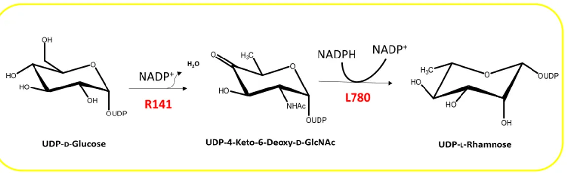
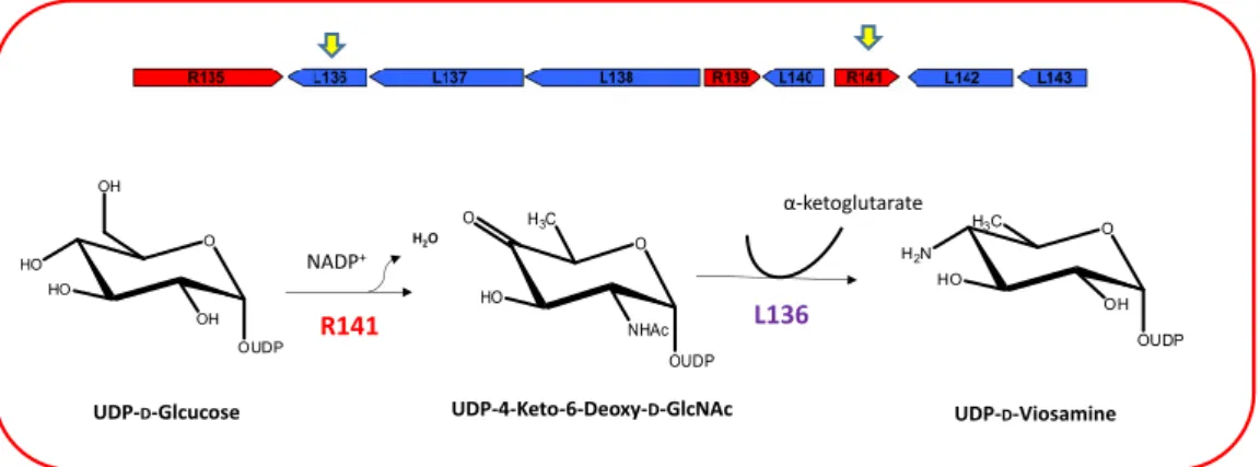
2.1 The giant DNA viruses encode an autonomous glycosylation machinery………….. pag. 27 2.1.1 The Mimivirus UDP-L-Rhamnose pathway………... pag. 28 2.1.2 The Mimivirus UDP-D-Viosamine pathway………. pag. 30 2.1.3 The Mimivirus biosynthesis of UDP-D-N-Acetylglucosamine…………... pag. 31
2
2.1.4 The Megavirus chilensis UDP-L-Rhamnosamine pathway……….. pag. 33
2.1.5 Glycosyltransferases……….. pag. 34
Thesis objective
The understanding of the innovative glycosylation system of the giant DNA
viruses……….pag. 35
Section II: Results and discussion
Chapter 3
Characterization of the glycans associated to the fibrils of Mimivirus, Megavirus chilensis and Moumouvirus australensis ………. pag. 38 3.1 The fibrils of the giant DNA virus Mimivirus capsid are heavily glycosylated………pag. 38
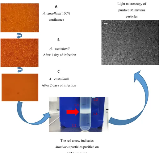
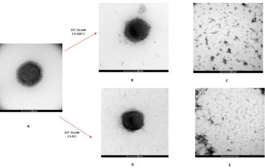
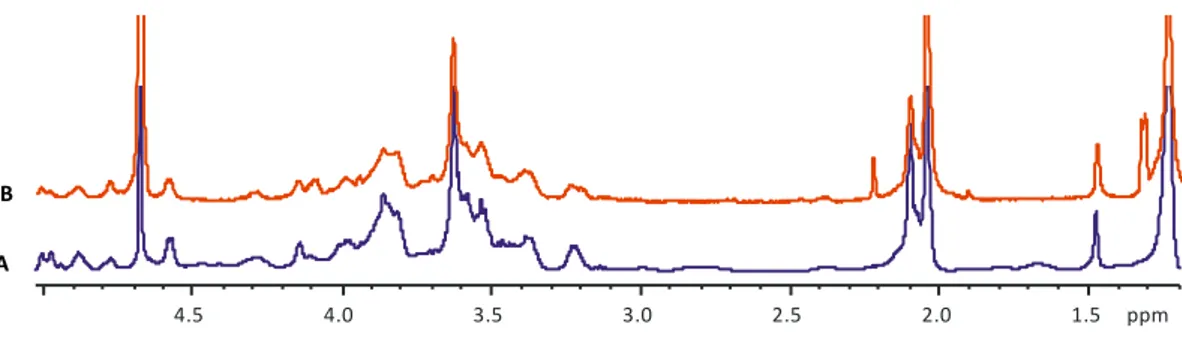
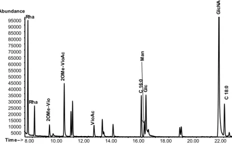
3.1.1 Production and purification of Mimivirus……….. pag. 38 3.1.2 Isolation of the Mimivirus glycans………pag. 40 3.1.3 Charachterization of Mimivirus glycans structure……….. pag. 42 3.1.4 Separation of Mimivirus polysaccharides by anion exchange chromatography…. pag. 51 3.1.5 Separation of Mimivirus polysaccharides by Reverse Phase HPLC……….. pag. 54 3.1.6 NMR analysis and molecular weight determination of the Mimivirus
purified polysaccharaides………. pag. 55 3.1.7 Identification of the protein/s attached with the two polysaccharides……… pag. 58 3.1.8 Conclusions………. pag. 61 3.2 The fibrils of the giant DNA virus Megavirus chilensis exhibit a different
glycosylation pattern compared to Mimivirus……… pag. 64 3.2.1 Production and purification of Megavirus chilensis virions ……… pag. 64 3.2.2 Isolation of Megavirus chilensis glycans………pag. 64 3.2.3 Characterization of Megavirus chilensis glycans structure……….pag. 65 3.2.4 Megavirus fibrils glycans purification……….... pag. 67 3.2.5 De-polimerization of Megavirus fibrils glycans ……… pag. 71 3.2.6 NMR analysis on the RP- HPLC products……… pag. 74 3.2.7 Conclusions ……… pag. 80 3.3 Glycans composition of Moumouvirus australensis fibrils ……… pag. 82
3
3.3.1 Production and purification of Moumouvirus australensis virions………... pag. 82 3.3.2 Isolation of the glycans fibrils……… pag. 82 3.3.3 Sugar composition of Moumouvirus australensis fibrils glycans……….. pag. 83 3.3.4 Conclusions ………pag. 85 3.4 Collective discussion of Mimivirus, Megavirus and Moumouvirus results
for fibril glycosylation……… pag. 86
Chapter 4
Lineage specificities of the genes involved in the Megavirinae fibrils
formation………. pag. 90
4.1 Extension of the nine-gene cluster of Mimivirus……….. pag. 91 4.2 Moumouvirus australensis UDP-D-diNAcBac pathway………pag. 96 4.3 Identification of a fourteen-gene cluster in Moumouvirus australensis……….…pag. 101 4.4 The N-acetylglucosamine is conserved along all the Mimivirinae
family and Tupanvirus………. pag. 104 4.5 Conclusion and discussion ………... pag. 107
Chapter 5
Validation in vitro of Mimivirus L142 function……….. pag. 109 5.1 The Rare Sugar N-acetylated Viosamine is a Major Component
of Mimivirus Fibers………... pag. 109 5.2 Preliminary study of the C-terminal domain of L142………pag. 110 5.2.1 Cloning of the the gene corresponding to the C-terminal domain of L142……...pag. 111 5.2.2 Expression and purification of the C-terminal domain of L142 ……….pag. 112 5.2.3 Gel filtration of the C-terminal domain of L142 ……….pag. 113 5.3 Prespectives ……….. pag. 115
Section III: Materials and methods
Chapter 6
6.1 Production and purification of viruses……….. pag. 117 6.2 Fibrils isolation………. pag. 117 6.3 Transmission electron microscopy……… pag. 118
4
6.4 Sugar composition of the fibrils……… pag. 119 6.5 NMR analysis of fibrils glycans……… pag. 119 6.6 Separation of Mimivirus polysaccharides and Megavirus chilensis fibrils
glycans by ion exchange chromatography……….... pag. 120 6.7 Separation of Mimivirus polysaccharides by RP-HPLC of untreated fibrils………… pag. 122 6.8 Molecular weight determination of Mimivirus fibrils polysaccharides……… pag. 123 6.9 Protein Electrophoresis of Mimivirus………pag. 123 6.10 Comparative proteomic analysis of Mimivirus ……… pag. 124 6.11 Depolymerization of the fibrils glycans of Megavirus chilensis ………pag. 125 6.12 Reverse phase HPLC on the carbohydrate material obtained
after TFMS treatment on Megavirus chilensis viral particles……… pag. 125 6.13 Multiple alignment of sequences……… pag. 126 6.14 Heat map………. pag. 128 6.15 Cloning of the gene corresponding to the C-terminal of L142………pag. 128 6.16 Expression and purification of C-terminal of L142……… pag. 129 6.17 Gel filtration of the C-terminal of L142……….. pag. 130
References………pag. 132
Appendix A
The rare sugar N-acetylated viosamine is a major component of Mimivirus fibers……....pag. ii
Appendix B
Abbreviations
AMG: acetylated methyl glycosides COSY: correlation spectroscopy diNAcBac: N,N’-di-acetylbacillosamine DTT: dithiothreitol
EI-MS: Electron ionization-mass spectrometry GC-MS: gas-chromatography-mass spectrometry GlcNAc: N-acetylglucosamine
HMBC: Heteronuclear multiple-bond correlation spectroscopy HPLC: High performance Liquid Cromatography
HSQC: Heteronuclear single-quantum correlation spectroscopy NMR: nucelear magnetic resonance spectroscopy
NOESY: nuclear Overhauser effect spectroscopy PMAA: partially methylated alditol acetates Qui2NAc: N-acetylquinovosamine
Rha: rhamnose
RhaNAc: N-acetylrhamnosamine
RP-HPLC: Reverse Phase- High Performance liquid Cromatography TFMS: trifluoromethanesulfonic acid, also known as triflic acid TOCSY: Total correlation spectroscopy
UDP: uridine diphosphate
UDP-diBacNAC: Uridine diphosphate-N,N’-di--acetylbacillosamine UDP-GlcNAc: Uridine diphosphate-N-acetylglucosamine
UDP-Rha: Uridine diphosphate-rhamnose
II
UDP-Vio. Uridine diphosphate-viosamine VioNAc: N-acetylviosamine
III
Sugars structures
UDP-D-GlcNAc UDP-L-Rha UDP-D-Vio UDP-L-RhaN O HO HO OUDP NHAc OH O OUDP OH H3C HO HO O H2N HO OUDP OH H3C O H2N HO OUDP OH H3CIV UDP-L-QuiN UDP-D-N,N’-di-acetylbacillosamine O OUDP H3C HO HO NHAc O AcHN HO OUDP NHAc H3C
V
Abstract
The aim of this thesis is the study of the innovative glycosylation machinery of the
Mimiviridae family, using Mimivirus, Moumouvirus australensis and Megavirus chilensis as prototypes of lineages A, B and C, respectively.
In 2003 the discovery of Mimivirus, the first giant DNA virus infecting amoeba, challenged the traditional view of viruses. Mimiviruses are giant viruses due to the size of their virions, easily visible by light microscopy, with a diameter of 700 nm against 200 nm for “traditional virus”. Their genomes encode 1000 proteins and count up to 1.2 Mbp, so they are as complex as the smallest free-living bacteria.
Mimiviruses exhibit heavily glycosylated fibrils surrounding their capsid that differ
in length depending on the lineages. Surprisingly, it was evidenced that they encode the proteins involved in their fibrils glycosylation.
The glycosylation of the fibrils was confirmed by the analysis of their sugar content, revealing that the major saccharide components were rhamnose, acetylglucosamine, and viosamine for Mimivirus and acetylglucosamine and N-acetylrhamnosamine for Megavirus chilensis. Until now, we lack information on the sugar composition of fibrils from members of the B lineage.
In this thesis, the innovative glycosylation machinery of these giant DNA viruses was investigated combining three different strategies: carbohydrate chemistry, bioinformatic and biochemical methodologies.
The carbohydrate chemistry methodologies allowed to elucidate the structures/composition of the glycans associated to the giant DNA viruses fibrils.
Mimivirus fibrils are decorated with two distinct polysaccharides, called poly_1 and
poly_2. Poly_1 is characterized by a linear disaccharide repeating unit made of 3)--L-Rha-(1→3)--D-GlcNAc-(1→, with a pyruvic acid branched at position 4,6 of GlcNAc. Poly_2 has a branched repeating unit with the sequence 2)--L -Rha-(1→3)--D-GlcNAc-(1→ in the linear backbone and rhamnose further branched at
VI
position 3 by viosamine methylated at position 2 and acetylated at position 4. Regarding the novelty of the identified structures, they have no equivalent in eukaryotes, while some components were reported in bacteria. Megavirus chilensis has a different sugar composition of its shorter fibrils, with acetylglucosamine, N-acetylrhamnosamine and N-acetylquinovosamine as major components. Purification results suggested that Megavirus fibrils were decorated by more than one polysaccharides/oligosaccharide species, one having this trisaccharide: -L -4OMe-RhaNAc-(1→3)--L-RhaNAc-(1→3)--L-RhaNAc-(1→. A preliminary analysis revealed that Moumouvirus australensis fibrils were decorated with glucosamine and quinovosamine in addition to the rare sugar, bacillosamine.
Starting from this experimental data, it was possible to identify new genes involved in glycosylation. As a result, the published nine-gene cluster of Mimivirus was extended to thirteen genes. A different cluster of fourteen genes was identified in
Moumouvirus australensis, representing the first glycosylation gene cluster
identified for the B lineage. A comparison of the glycosylation genes in the
Mimiviridae family reinforced our finding that fibrils glycosylation was lineage
specific. However, Moumouvirus australensis is an exception as it exhibits a cluster of glycosylation genes that is missing in other member of the B lineage.
Among the genes with the glycosylation cluster, the function of L142 was investigated in vitro, demonstrating that it is a N-acetyltransferase that acetylates the 4 amino group of viosamine. L142 represents the first virally encoded N-acetyltransferase.
To conclude, the fibrils of Mimiviridae are heavily glycosylated and the type of sugars and their organization depends on their lineage. The majority of the genes responsible for sugar production, sugar modification and glycosyltransferases were identified, strongly suggesting that Mimiviridae are autonomous for their fibrils glycosylation.
VII
Résumé
The sujet de cette these portait sur la caractérisation de la machinerie originale utilisée par les Mimiviridae pour glycosyler les fibrilles entourant leurs capsides en travaillant sur les prototypes des 3 lignées connues, Mimivirus (A), Megavirus
chilensis (B) et Moumouvirus australensis (C).
La découverte en 2003 de Mimivirus, le premier virus géant ADN infectant l’amibe, a bouleversé notre vision traditionnelle des virus. Les Mimiviridae sont des virus géants, visibles au microscope optique en raison des tailles de leurs particules (700nm contre 200nm plus les virus "classiques"). Leurs génomes de ~1.2 Mb codent pour un millier de protéines et sont aussi complexes que les plus petites bactéries non parasites. Les Mimiviridae infectant les amibes présentent des capsides couronnées de fibrilles très glycosylées de longueurs variables en fonction des lignées virales. De manière surprenante, ce sont des protéines virales qui sont responsables de la glycosylation des fibrilles. L’analyse de ces fibrilles a révélé que les sucres majoritaires étaient du Rha, GlcNAc et VioNAc pour Mimivirus, and GlcNAc and RhaNAc pour Megavirus chilensis. A ce jour, on ne connaissait pas la composition en sucres des fibrilles des virus de la lignée B.
Au cours de cette thèse, nous avons étudié la machinerie de glycosylation de ces virus géants en combinant différentes approches : la chimie des carbohydrate, la bioinformatique et la biochimie des protéines.
La chimie des carbohydrate a permis de déterminer la composition en glycans associés aux fibrilles virales et d’en résoudre les structures. Les fibrilles de Mimivirus sont décorées par 2 polysaccharides différents, poly-1 et poly-2. Poly-1 est caractérisé par la répétition d’un disaccaride linéaire fait de 3)--L -Rha-(1→3)--D-GlcNAc-(1→, avec un pyruvate branché en position 4,6 du GlcNAc. Poly-2 présente une unité répétée branchée de séquence 2)--L-Rha-(1→3)--D -GlcNAc-(1→ pour le squelette linéaire et du rhamnose branché en position 3 par de la
VIII
viosamine méthylée en position 2 et acétylée en position 4. Ces structures originales n’existent pas dans le monde eucaryote, tandis que certains de leurs composants ont déjà été identifiés dans le monde bactérien.
Megavirus chilensis présente une composition en sucres différente. Ses fibrilles plus
courtes sont composées en majorité de N-acetylglucosamine,
N-acetylrhamnosamine and N-acetylquinovosamine. Après purification, il semble que les fibrilles de Megavirus sont fait de plus d’un type de polysaccharide, l’un ayant présentant un trisaccharide de RhaNAc : -L-4OMe-RhaNAc-(1→3)--L -RhaNAc-(1→3)--L-RhaNAc-(1→. L’analyse préliminaire des fibrilles de Moumouvirus révèle la présence de glucosamine et quinovosamine qui décorent les fibrilles et un sucre rare, la bacillosamine.
A partir de ces données expérimentales il devenait possible de rechercher de nouveaux gènes responsables de ces glycosylations spécifiques. Ainsi, le cluster de 9 gènes déjà publié de Mimivirus a pu être étendu à 13 gènes. Un cluster de 14 gènes a été d’autre part identifié dans le génome de Moumouvirus australensis, le premier cluster de gènes de la glycosylation identifié dans la lignée B. La comparaison des gènes de glycosylation des Mimiviridae renforce le fait que la glycosylation des fibrilles soit lignée spécifiques. Cependant, Moumouvirus australensis reste une exception avec un cluster de gènes de glycosylation absent des autres membres de la lignée B.
Parmi les gènes de glycosylation, l’analyse fonctionnelle in vitro de la protéine L142 a permis de démontrer qu’il s’agit d’une N-acétyltransferase qui acétyle le groupement amino 4 de la viosamine, le sucre rare composant les fibrilles de Mimivirus. N-L142 est ainsi la première N-acétyltransferase virale identifiée. En conclusion, les fibrilles des Mimiviridae sont lourdement glycosylées and le type de sucres et leur organisation dépend de la lignée considérée. La majorité des gènes responsables de la production de ces sucres, de leur modification ainsi que les glycosyltrasnférases impliquées ont pu être identifiées, argument fort en faveur de
IX
l’autonomie de ces virus par rapport à leur hôte cellulaire dans la glycosylation de leurs fibrilles.
X
Riassunto
Lo scopo di questa tesi è lo studio dell’innovativo macchinario di glicosilazione della famiglia Mimiviridae, usando Mimivirus, Moumouvirus australensis e Megavirus
chilensis come prototipi delle cladi A, B and C, rispettivamente.
Nel 2003 la scoperta di Mimivirus, il primo virus gigante a DNA, ha cambiato la tradizionale visione sui virus. Mimiviruses sono virus giganti a causa delle dimensioni dei loro virioni, infatti sono visibili facilmente al microscopio ottico, con un diametro di 700 nm contro i 200 nm per un “virus classico”. I loro genomi di 1.2 Mbp, codificano 1000 proteine e risultano più complessi dei più piccoli batteri non parassiti. Mimiviruses esibiscono fibrille altamente glicosilate intorno al loro capside, che differiscono in lunghezza a seconda della clade a cui appartengono. Sorprendentemente, è stato evidenziato che questi virus posseggono le proteine deputate alla glicosilazione delle loro fibrille.
La glicosilazione delle fibrille è stata poi confermata dalla lora analisi degli zuccheri, rivelando che i maggiori componenti saccaridici erano ramnosio, acetilglucosammina, e viosammina per Mimivirus and acetilglucosammina e N-acetilramonosammina per Megavirus chilensis. Fino ad oggi, non ci sono informazioni circa la composizione degli zuccheri delle fibrille per la clade B. In questa tesi, il macchinario innovativo di glicosilazione di questi virus gianti a DNA è stato investigato integrando tre diverse strategie, dalla chimica dei carboidrati alla bioinformatica e biochimica strutturale.
Le metodologie della chimica dei carboidrati hanno permesso di elucidare le strutture o la composizione dei glicani connessi con le fibrille di questi virus giganti. Le fibrille di Mimivirus sono decorate con due dstinti polisaccaridi, chiamati poly_1 e poly_2. Poly_1 è caratterizzato da un’unità ripetitiva di 3)--L-Rha-(1→3)--D -GlcNAc-(1→, e la GlcNAc lega in 4 e 6 l’acido piruvico. Poly_2 ha un’unità ripetitiva ramificata con 2)--L-Rha-(1→3)--D-GlcNAc-(1→ nello scheletro
XI
lineare; il ramnosio è ulterirormente ramificato in posizione O-3 con la viosamina che è metilata in posizione 2 e acetilata in posizione 4.Queste strutture non trovano riscontri nel mondo eucariotico, mentre alcuni component sono stati già identificati nel mondo batterico. Le fibrille di Megavirus chilensis, più corte di quelle di Mimivirus, hanno una diversa composizione in zuccheri con N-acetilglucosammina, N-acetilramnosammina e N-acetilchinovosammina come principali costituenti. La purificazione dei glicani connessi alle fibrille di Megavirus has suggerito che le fibrille erano decorate da più di una specie polisaccaridica o oligosaccaridica, una delle quali presentava questo trisaccaride: -L-4OMe-RhaNAc-(1→3)--L -RhaNAc-(1→3)--L-RhaNAc-(1→. Un’analisi preliminare ha mostrato che le fibrille di Moumouvirus australensis erano decorate con un altro zucchero raro, la bacillosammina, in aggiunta alla glucosammina e chinovosammina.
A partire da questi dati sperimentali, è sato possible identificare nuvi geni coinvolti nella glicosilazione. Di conseguenza, il cluster di nove geni di Mimivirus, già pubblicato, è stato esteso a tredici geni. Un cluster di quattordici geni è stato identificato in Moumouvirus australensis, rappresentando il primo cluster di glicosilazione ad essere stato identificato per la clade B. Un’analisi comparativa dei geni della glicosilazione nella famiglia Mimiviridae ha consolidato i dati sperimentali, confermando che la glicosilazione delle fibrille è clade specifica. Inoltre, Moumouvirus australensis costituisce un’eccezione, in quanto il suo cluster genico di glicosilazione non è condiviso dagli altri membri della clade B.
Tra i geni della glicosilazione, la funzione di L142 è stata investigata in vitro, dimostrando che è un’ N-acetiltransferasi che va ad acetilare la funzione amminica in posizione 4 della viosammina. N-L142 rappresenta la prima N-acetiltransferasi codificata da virus.
In conclusione, le fibrille della famiglia Mimiviridae sono altamente glicosilate e il tipo di zuccheri e la loro organizzazione dipende dalla clade a cui appartengono. La maggior parte dei geni responsabili della produzione degli zuccheri, delle loro
XII
modifiche e le glicosiltransferasi sono state identificate, suggerendo fortemente che la famiglia Mimiviridae è autonoma per la glicosilazione delle fibrille.
Section I
- 1 -
Chapter 1
Discovery of the giant DNA viruses: a new concept of viruses
What is a virus? Are viruses organisms? Are viruses alive? Scientists have tried to answer these questions since the discovery of the first virus, the causative agent of Tobacco Mosaic disease (Beijerinck, 1898; Ivanovski, 1892) in the XIX century. This signed the birth of virology (1886-1898) and we had to wait for 60 years before André Lwoff established the concept of virus (Lwoff, 1957). Interestingly, the viruses were only defined by negative properties that excluded them for the cellular world:
✓ Viruses are not retained by the Chamberland filter
✓ Viruses have only one type of nucleic acid (DNA or RNA); ✓ Viruses are unable to grow and divide;
✓ Viruses do not produce the enzymes for their energy production; ✓ Viruses have no protein translation apparatus.
In addition, Lwoff clearly stated that viruses were not organisms and not alive by concluding his article by the following statement: “viruses are viruses”. One century after this first definition of “virus”, these criteria are still accepted by the scientific community. For instance, many authors consider viruses as not living organisms, because they lack autonomy and metabolism (Moreira and Brochier-Armanet, 2008). In 2003 the discovery of Mimivirus (Scola, 2003) , the first giant DNA virus infecting amoeba from the Acanthamoeba genus, challenged the traditional view of viruses. Mimivirus was a dogma breaker, shaking the foundation of virology. “We are now entering a new era where the most basic concepts about viruses are revisited” (Claverie and Abergel, 2010). The same old questions were raised again by its discovery: What is a giant DNA virus? What are the boundaries between viruses and
- 2 -
cellular organisms? What is viruses origin? (Abergel et al., 2015; Forterre, 2017; Sharma et al., 2016)
The objective of this chapter is to describe the giant DNA viruses infecting amoebae, stressing their differences with “traditional virus”. A detailed dissertation will be done on Mimivirus and the Mimiviridae family that are the focus of this thesis.
1.1 The first giant DNA virus: Mimivirus
Acanthamoeba polyphaga Mimivirus, more commonly known as Mimivirus,
represents the first giant DNA virus infecting amoeba. In 1992, it was isolated from an amoebal co-culture from a water sample of the cooling tower of the hospital of Bradford, collected after a pneumonia outbreak. It was
misidentified as a bacterium, due to the gram-positive staining of the spherical particles visible by light microscopy and was named Bradfordcoccus (Fig.1.1). Subsequently, the team of Rowbotham involved in the investigation of the outbreak, and then the Rickettesia Unit at the School of Medicine (URMITE, Marseille, France), tried different approaches to characterize this microorganism, including the 16S ribosomal gene DNA amplification which all failed. In order to shed light on the nature of this microorganism, it was visualized by electron
microscopy in infected cells in 2003. The presence of icosahedral particles in the amoeba, finally, revealed its viral nature (Fig. 1.2)(Scola, 2003). Unexpectedly, the particles reached a diameter of 700 nm. Finally, the sequencing of its dsDNA genome revealed it was as complex as the smallest free-living bacteria with of 1.2 Mbp genome encoding 1000 proteins.
According to these studies, Bradfordcoccus was renamed Mimivirus for “Mimicking microbe virus”. Mimivirus was the first virus large enough to be visualized by light microscopy. It presented much bigger viral particles than any other known virus at
Fig.1.1. Mimivirus (arrows)
in cytocentrifuged A.
polyphaga as Gram-positive particles. Reproduced from La Scola et al., 2003.
- 3 -
the time of its discovery (usually 0.2-0.3µm), reaching the size of some bacteria such as Mycoplasma and Rickettsia (300-500 nm), and was also bigger than Nanoarchaeum equitans, the smallest known archaea (400 nm): Mimivirus is definitively a giant due to the size of its virions.
The viral particle size has broken one dogma of the virology: viruses were supposed to be smaller than bacteria. It has led to reconsider the strategy for the isolation of viruses from different environments, in order to discover new giant viruses.
Mimivirus is a giant virus not only due to the diameter of its particles, but also by the
dimension of its genome (1.2 Mbp). Usually viral genomes are measured in thousands of base pairs which correspond to a handful of genes. Prior to Mimivirus discovery, viral genomes bigger than the ones of small bacteria were already identified, such as Paramecium bursaria chlorella virus 1 (PBCV-1) with a genome up to 560 Kb (Van Etten et al., 1982).
Mimiviruses is so different from the other viruses, that it started a new viral family,
the Mimiviridae (Scola, 2003). This family was included in the group of the Nucleocytoplasmic Large DNA Viruses (NLCDVs), which consists of five families of viruses infecting eukaryotes: Poxviridae, Asfarviridae, Phycodnaviridae,
Ascoviridae and Iridoviridae (Iyer et al., 2001; Yutin et al., 2009). The NLCDVs are
dsDNA viruses that replicate partly or entirely in the cytoplasm of eukaryotic cells. They share a set of conserved genes. From the preliminary analysis of Mimivirus genome it shares 21 of these core genes (Iyer et al., 2001). However, the phylogenetic tree suggests an early divergence from the other viruses’ family.
- 4 -
1.1.1 Mimivirus virion morphology Overview
Mimivirus viral particles from the outside to the
inside, exhibit (Fig. 1.2):
- A layer of fibrils of 150 nm (Xiao et al., 2009); - An icosahedral capsid (Kuznetsov et al., 2010; Scola, 2003; Xiao et al., 2009); the perfect symmetry is broken by the stargate (Zauberman et al., 2008) structure;
- Nucleoid (Kuznetsov et al., 2010; Seibert et al., 2011) containing the dsDNA genome.
Mimivirus fibrils
A dense layer of 150 nm fibrils covers Mimivirus mature particle, with the exception of the “stargate”(Xiao et al., 2009). Fibrils have also been observed in other NLCDVs, such as PBCV-1 (Cherrier et al., 2009; Kuznetsov et al., 2005; Zhang et al., 2011), CIV(Yan et al., 2009) and PbV01(Yan et al., 2005). However, Mimivirus fibrils are unique in terms of length, abundance, and complexity.
The atomic force microscopy (AFM) studies of Mimivirus fibrils detached from the virus, revealed some specific features (Fig. 1.3). The fibrils length is uniform, ranging from 125 to 140 nm, suggesting that the fibrils are either precisely “cut to measure” from longer precursor fibrils, or that they are assembled from small precursors until the linear aggregate reaches some predetermined length (Kuznetsov et al., 2010). The fibrils are probably composed of protein/s and should be of helical construction. It was observed that there are globular heads proteins attached to the fibrils shafts (Fig. 1.3). Concerning the fibrils precise anchoring, there are two different hypotheses: Stargate Fibrils Icosahedral capsid Nucleoid Membrane dsDNA
Fig. 1.2 Electron microscopy of
Mimivirus in the vacuole of the amoeba cell. Copyright IGS.
- 5 -
1) The fibrils could be attached to extended polypeptide loops of the major capsid protein (MCP)(Kuznetsov et al., 2010; Xiao et al., 2009);
2) The fibrils could be anchored to a layer of protein positioned immediately above the capsid (Kuznetsov et al., 2010; Xiao et al., 2009).
Until now, it was not possible to discriminate between these hypotheses, but the evidence of multiple fibrils attached to one anchor supported the first hypothesis. Although the number of anchors was estimated to be the same as the number of depressions on the surface of the capsid (~3000), the total number of fibrils remains unknown, because it was not possible estimated the number of fibrils attached for one anchor.
Fig.1.3 In (a) is a mass of surface fibers, most still attached to their head groups, shed from Mimivirus by a
treatment with lysozyme and bromelain. A cluster of fibers still attached to the anchoring polypeptide at their proximal ends is also visible. In (b) the fibers are seen at higher magnification. Single fibers are identified due to their more or less uniform lengths and for the ellipsoidal protein heads. In (c) are two individual fibers, one of which is surrounded by a penumbra of some diffuse material. Reproduced from Kuznestov et al 2010.
Of particular interest is the evidence that the fibrils are heavily glycosylated. In the fig. 1.3 C there is a visible shadow surrounding the fibrils along their lengths, and later experiments suggested that it originated from poly-oligosaccharides decorations.
Indeed, first evidences came from the Gram-positive staining of the viral particles, suggesting the presence of peptidoglycan-like structures (Scola, 2003). However, this hypothesis was later disproved because it appeared that Mimivirus fibrils were retaining any stain.
- 6 -
The glycosylation of the fibrils was next evidenced by the analysis of Mimivirus particles sugar content, which revealed that the major saccharide components were Rha, GlcNAc, Vio. These data have excluded the presence of a true peptidoglycan (Piacente et al., 2012).
The in-depth investigation of Mimivirus, and other Mimiviridae, fibrils glycosylation is the subject of this thesis and it will be discuss in the following section.
The fibrils function is still under investigation. The current hypothesis is that the presence/absence of fibrils does not impair the viral replication in laboratory controlled condition, while they seem to play an important role in the adhesion process to the host cells (Rodrigues et al., 2015). These fibrils may favor the phagocytosis by the amoebal host and could be essential in the complex natural environment.
Mimivirus capsid organization
Mimivirus capsid consists of 20 large triangular
faces joined at their edges to produce the required 12 five-fold vertices (Xiao et al., 2005).
The gene L425 encodes the Mimivirus major capsid protein, which is homolog to the Chlorella virus PBCV-1major capsid protein Vp54. Their N-terminal domains (position 1-287) share 46% identity and the C-terminal regions 40% identity at the protein level (positions 483-593). This homology suggested that Mimivirus, like other NCLDVs, had hexameric capsomers consisting of double jelly-rolls protein. A model of the L425 MCP was proposed based on the structure of Vp54(Azza et al., 2009; Nandhagopal et al., 2002) (Fig. 1.4).
Fig. 1.4 Model of L425 Mimivirus capsid
protein based on Vp54, the major capsid protein of Chlorella virus PBCV-1 (in green). In contrast with Vp54, Mimivirus MCP presented an insertion of ~190 amino acids into the DE loop (in pink). Reproduced from Azza et al., 2009
- 7 -
In contrast with Vp54, Mimivirus MCP presented an insertion of ~190 amino acids into the DE loop (in pink in Fig. 1.4) of the second jelly-roll fold (the -strands along the polypeptide of each jelly-roll are named A to E), predicted to be made of 10 -strands (Azza et al., 2009; Xiao et al., 2009).
In order to elucidate the Mimivirus capsid structure, structural studies were performed using Mimivirus untreated and enzymatically treated virions to remove the fibrils. The combination of AFM and cryo-EM reconstruction revealed that the capsid surface is composed of trimeric MCP (three monomers of the MCP constitute one capsomer), arranged in a very open honeycomb hexagonal array. These capsomers are organized in p6 plane group instead of the conventional p3 for the others icosahedral NCLDVs. The center-to-center distance between capsomers is 14 nm, and the triangulation number (T), in the capsid architecture, remains uncertain. The perfect icosahedral symmetry is broken by a five-pronged star structure, called the ‘stargate’, at one unique vertex of the capsids (Zauberman et al., 2008). This structure is a unique feature of Mimivirus, and has been proved to be present on each virion by of sectioned Mimivirus-infecting acanthamoeba cells (Zauberman et al., 2008) and by cryo-EM and AFM on fibreless Mimivirus particles (Kuznetsov et al., 2010; Xiao et al., 2009) . The stargate arms have a width of about 500 Å, a thickness of about 400 Å and a length of about 2,000 Å. (Fig. 1.5).
Fig. 1.5 (A–C) Surface-shaded rendering of cryoEM reconstruction of untreated Mimivirus. (A) Looking down
the starfish-shaped feature associated vertex, (B) looking from one side, and (C) looking from the opposite side of the “starfish”-associated vertex. Reproduced from Xia et al., 2009.
- 8 -
The arms are inserted between two neighboring triangular faces causing a deviation of about five degrees from the geometry of a perfect icosahedron for these five faces. In addition, it was demonstrated that the stargate represents the only point of the
Mimivirus virion which is not covered by fibrils (Xiao et al., 2009; Zauberman et al.,
2008). This feature suggested that the stargate could be involved in the delivery of the dsDNA genome in the cytoplasm of the host cell. The role of the stargate as the genome delivery portal was revealed by the seminal work of Zauberman et al. (2008) followed by the one of Kuznestov et al. (2010). Between the stargate and the genomic nucleoid, there is a space that could be filled by the enzymes required to digest the structural component of the host, to initiate the infectious cycle. The existence of a unique portal for genome delivery reminds tailed bacteriophages.
Mimivirus nucleoid
A membrane surrounded structure, initially called “Seed” corresponds to the nucleoid which encloses the dsDNA genome of Mimivirus (Claverie and Abergel, 2010) as well as all the proteins necessary to initiate transcription. The estimated diameter of the nucleoid is about 340 nm and the dsDNA contained inside is 1.2x10⁶
bp, which gives a packing density of 0.06 nm3/bp. The DNA density in the nucleoid
is relatively low and it has been suggested that the DNA could be associated with proteins (Kuznetsov et al., 2010). The nucleoid has a defined shape and a fixed position relative to the external capsid (Seibert et al., 2011).
Of particular interest is the presence of 300-500Å gap between the nucleoid and the outer capsid (Xiao et al., 2009). Except for PBCV-1, this space is absent in other viral particles for which the genome is closely surrounded by the capsid. The nucleoid plays an important role during the infection cycle, as discussed later.
- 9 -
Even more surprisingly, fibers with a 7 nm periodicity along their lengths (Fig. 1.6) were evidenced and probably localized in the space between the nucleoid and the outer capsid(Kuznetsov et al., 2010). These fibers are not composed by RNA or DNA. This type of structure was never observed in viral particles before, they are mechanically flexible
and resistant to breakage. They could be cushion for the nucleoid. Another hypothesis is that these fibers might be the precursors for the fibrils surrounding the
Mimivirus capsid. Additional studies are required to
elucidate the function of these fibers (Kuznetsov and McPherson, 2011).
1.1.2. Mimivirus genome
Mimivirus has a double stranded DNA genome of 1.18 Mbp encoding more than
1000 proteins, blurring the boundary between viruses and bacteria (Arslan et al., 2011; Legendre et al., 2010; Raoult, 2004; Renesto et al., 2006). Mimivirus genome is rich in adenine and thymine (A+T 72%). Codon and amino acid usage is thus dissimilar to the one of its amoebal host and is correlated with the high AT content of the Mimivirus genome (Colson et al., 2013a).
Although Mimivirus shares several core genes with the NLCDVs, its genome also exhibits genes encountered for the first time in a viral genome, raising the question on its possible origin. The most impressive feature is the presence of several genes related to the translation machinery, a process thought to be exclusively restricted to the cellular world. Some tRNA-like genes have been identified in other dsDNA viruses, such as chlorella virus (Van Etten and Meints, 1999), but Mimivirus presents an extended repertoire of genes involved in this process. For instance, it encodes several translation initiation, elongation and termination factors and four
aminoacyl-Fig. 1.6 It is a low magnification
AFM image of large tangles of the fibers, which appear to have very long lengths. Reproduced from Kuzneston and McPherson, 2011.
- 10 -
tRNA synthetase (aaRS): ArgRS, CysRS, MetRS and TyrRS. The aaRS are responsible of the attachment of the appropriate amino acid on its cognate tRNA and it has been demonstrated that these enzymes were all active and specific (Abergel et al., 2007).
In addition to a complete transcription machinery, Mimivirus encodes several types of DNA repair enzymes, including a remote homologue of the E. coli MutS, that can be used as marker in environmental studies (Wilson et al., 2014). Another unique feature of Mimivirus is the presence of three different topoisomerases of types IA, IB and II. The type IA was never found in viruses before (Raoult, 2004).
Although the Mimivirus genes are not interrupted by spliceosomal introns, some include inteins. Additionally, two type I introns
were identified in the major capsid protein gene (Azza et al., 2009).
Once again, Mimivirus invades the cellular world with the presence of genes encoding glycosylation machinery components. The hypothesis is that some of these genes could be
involved in Mimivirus fibrils glycosylation. The investigation of these genes is part of this thesis and it will be discussed in the Chapter 2 and Section II.
Most of the genes with predicted functions (ORFs) have homologs in bacteria, archaea, eukaryotes or viruses. In addition, it is important to underline that 2/3 of
Mimivirus genes are ORFans genes encoding unique proteins, without homologues
in the cellular or viral world (Fig. 1.7).
This complex mosaic of genes raised again the question on Mimivirus origin, still under investigation.
Fig. 1.7 Predicted proteins encoded in the
genome of Mimivirus. Reproduced from Abergel
- 11 - 1.1.3 Mimivirus replication cycle
The viral world is highly heterogeneous and complex, and different general replication strategies have been reported. Until recently, it was assumed that dsDNA viruses replicate in the host nucleus, while RNA viruses replicate in the cytoplasm. The first exception to this “rule” came from Vaccina virus, a dsDNA virus belonging to the Poxviridae family, for which it has been demonstrated that its replication cycle occurs entirely in the host cytoplasm (Carter et al., 2005). Mimivirus, as poxviruses, has a cytoplasmic replicative cycle (Mutsafi et al., 2010, 2014). In addition, as all the NLCDVs, Mimivirus builds a viral factory in the infected cell cytoplasm, a distinct “organelle”, in which transcription and genome replication occurs (Netherton and Wileman, 2011; Suzan-Monti et al., 2007).
The first step of the infection is the adhesion of the viral particle to the surface of its amoebal host. It was suggested early on that this interaction was glycan mediated (Rodrigues et al., 2015). There are evidences that the viral particles are able to interact with the N-acetylglucosamine, which is an adhesion factor on the
Acanthamoeba surface. In addition it was demonstrated that a Mimivirus mutant, M4
(Boyer et al., 2011), without fibrils, has a significantly lower level of attachment to the Achanthamoeba surface (Rodrigues et al., 2015) . Accordingly, it was clear that the Mimivirus heavily glycosylated fibrils could play an important role in the host adhesion-process. A complete elucidation of Mimivirus glycan nature could shed light on this interaction process.
The second step is the entry of Mimivirus into the amoeba through phagocytosis. This entry mechanism is related to the heterotrophic nature of Acanthamoeba, which feeds on bacteria. Mimivirus, mimicking a microbe due to its particle size and “sweet taste”, can be phagocytized by Acanthamoeba. It was reported that phagocytosis can be triggered by individual particles larger than 0.6 µm (Korn and Weisman, 1967). Particle size of 750 nm could thus provide an evolutionary advantage to Mimivirus to be internalized by its amoebal host.
- 12 -
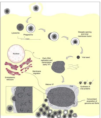
After internalization there is the opening of the stargate and the delivery of the nucleoid, containing the viral DNA and all the proteins needed to initiate the early genes expression, into the cytoplasm (Fig. 1.8 A to I). The biochemical signal responsible for the stargate opening is still unknown, but a recent study of Andrade (Andrade et al., 2017) and colleagues revealed that an important role in Mimivirus uncoating could be play by the phagosome acidification.
Fig. 1.8 Transmission electron microscopy images of Mimivirus particles entering cells by phagocytosis (A to C), the phagosome-lysosome fusion (D to G), stargate opening (H), and release of the viral seed (I). Reproduced
from Andrade et al., 2017.
After delivery of the nucleoid, the transcription and DNA replication takes place in the early viral factory (VF) (Mutsafi et al., 2010). The assembly of new viral particles takes place in the periphery of the mature VF.
A model for the capsid formation has been proposed through the combination of different imaging techniques, including scanning transmission electron microscopy
A B C G F E D H I
- 13 -
tomography (Mutsafi et al., 2013) and atomic force microscopy (Kuznetsov et al., 2013a) . It was evidenced that, 7.5 h post infection, host cisternae, from which 70 nm vesicles bud out, are recruited to the viral factory. These vesicles probably originate from the host cell endoplasmic reticulum and act as scaffolding for the assembly of the neo-synthesized capsids. In addition it appears that the L425 MCP acts as a scaffolding protein (Mutsafi et al., 2013). The morphogenesis of the capsid starts from the stargate, followed by the thickening of the protein layer in its vicinity (Kuznetsov et al., 2013a). Then the capsid is filled with the DNA through a transient aperture in the center of an icosahedral face, which is distinct from the stargate used for genome delivery: this is a unique characteristic of Mimivirus.
The final stage corresponds to the acquisition of the external fibrils, by an unknown process still investigated. To date, two hypotheses have been proposed:
1) In 2013 Kuzestov et al. proposed that the surface fibrils were acquired by viral capsids (Fig. 1.9.i) when they passed
sequentially through a membrane embedded with a protein sheet, called in this study, integument protein (ii) and then through a protein sheet containing the fibrils (iii and iv) (Fig.1.9).
According to this model, layers of integument and fibrils are acquired as an
envelope around the capsid (Fig. 1.9). In addition, this study suggested that the integument protein layer is acquired near the viral factory, while the fibrils are acquired near the cell periphery (Kuznetsov et al., 2013b).
2) In the 2017, Andrade et al., used transmission electron microscopy, to reveal at the periphery of the viral factory a less-electron dense region made of fibrils, which was called the fibril acquisition area (FAA). They suggested that the newly formed capsids were acquiring the fibrils layer by passing through this
Fig. 1.9 Model for the acquisition fibrils layer.
- 14 -
region. This hypothesis is strengthened by the observation of a reduction of the FAA, after the release of the viral particles from the viral factory (Fig. 1.10) (Andrade et al., 2017).
After the acquisition of the fibrils layer, the Mimivirus particles are mature. The replication cycle ends with host cell lysis and the release of approximately 1000 mature particles.
- 15 -
1.2. Mimiviridae family
The Mimiviridae family was created in 2003 with the discovery of Acanthamoeba
polyphaga Mimivirus (Scola, 2003), and it was included in the large monophyletic
group of the Nucleo-Cytoplasm Large DNA viruses (NCLDVs; proposed order Megavirales (Colson et al., 2013b)). The Mimiviridae family is in constant growth and encompasses not only giant DNA viruses infecting amoeba, but also giant viruses infecting unicellular eukaryotes (Claverie and Abergel, 2018) (Fig. 1.11). This family was originally constituted by at list four subfamilies.
The Mimiviridae group I (also reported as Megavirinae subfamily (Gallot-Lavallée et al., 2017), this term will be used in this thesis) includes only the giant DNA viruses isolated by culturing on amoeba from water, soil, insect and human samples. Based on phylogenetic data, this group is divided in three lineages (Diesend et al., 2017):
✓ Lineage A – The prototype is Acanthamoeba polyphaga Mimivirus and it counts 18 members;
✓ Lineage B – The prototype is Acanthamoeba polyphaga Moumouvirus with a total of five members;
✓ Lineage C – The prototype is Megavirus chilensis and it includes 12 members.
The sizes of the icosahedral virions from the three lineages range between 400 and 600 nm diameter. They enclose a double stranded DNA genome ranging from 1.02 to 1.26 Mb, rich in A+T, encoding 930-1120 putative proteins. Their replication cycles are the same than the one described for Mimivirus.
- 16 -
The Mimiviridae group II consists of Cafeteria roenbergensis virus (CroV), infecting the marine phagotrophic flagellate C. roenbergensis (Fischer et al., 2010). In contrast with Mimivirus, CroV exhibits a smaller icosahedral capsid, about 300 nm, not decorated by a layer of fibrils, but it features 30 Å-long surface protrusions that are formed by the loops of the major capsid protein (Xiao et al., 2017). CroV has a dsDNA genome of 730 kbp, encoding 544 putative proteins. As for Mimivirus, it encodes translation factors, 22 tRNAs, DNA repair enzymes such as MutS and inteins (Fischer et al., 2010). The inclusion of CroV in the Mimiviridae family represents the first demonstration that Mimiviruses relatives could infect a wide spectrum of host.
The Extended Mimiviridae family (or Mesomimivirine family (Gallot-Lavallée et al., 2017)) is another subfamily gathering giant DNA viruses infecting unicellular algae.
The complete genome sequences of Phaeocystis globosa virus (PgV)(Santini et al., 2013), of Aureococcus anophagefferens virus (Moniruzzaman et al., 2014) and
Chysochromulina ericina virus (CeV) (Gallot-Lavallée et al., 2017) clearly confirms
a common origin with the Megavirinae. Most recently, it has been included in this family the TetV virus infecting Tetraselmis (Schvarcz and Steward, 2018), extending the spectrum of algae-infecting Mimiviridae.
The Klosneuvirinae is a new subfamily that encompass four linages: Kloseneuvirus, Catovirus, Hokovirus, Indivirus (Schulz et al., 2017). These giant DNA viruses have
only been identified throughout metagenomics studies. In absence of physical isolated particles, their morphology and replication cycle are still unknown. The Kloseneuvirus’s genome is 1.57 Mb, and thus more complex than the fully sequenced genomes of the previously isolated Mimiviruses.
- 17 -
A new subfamily could be represented by the two Tupanviruses strains (Tupanvirus
soda lake and Tupanvirus deep ocean), which is considered as a sister group of the Mimiviruses (Abrahão et al., 2018).
Fig. 1.11 Phylogeny of the Mimiviridae based on MutS7. MutS sequences from Epsilon proteobacteria (from
thegenus Arcobacter, and Sulfurospirillum) are used as root, as they are most likely the source of the
Mimiviridae MutS7 gene (MutS homologs are present throughout the epsilon division). The tree suggests with great confidence that the mitochondrial MutS present in octocorals (in grey) and that present in all Mimiviridae have a common origin. Reproduced from Claverie et Abergel, 2018, Viruses.
In conclusion, the Mimiviridae family is in constant expansion, including Mimivirus relatives infecting hosts other than amoeba. With the discovery of new Mimiviruses, the picture become more and more complex, opening the question on their origin. An in depth analysis of these viruses could play a key role to establish the criteria
- 18 -
for the classification of the new members that will be isolated in the future (Claverie and Abergel, 2018).
It is important to precise that to date there is no official classification including the recently identified subfamilies in the Mimiviridae family.
In this thesis, I focused my attention on Mimivirus (Lineage A), Megavirus chilensis (Lineage C) and Moumouvirus australensis (Lineage B). In addition, I took in account the newly discovered Tupanviruses to perform some comparative analyses of the glycosylation pathways.
1.2.1 Acanthamoeba polyphaga moumouvirus and moumouvirus australensis
Acanthamoeba polyphaga moumouvirus represents the prototype of the lineage B of
the Mimiviridae infecting Acanthamoeba. It was isolated in 2008 by inoculating A.
polyphaga with water from an industrial cooling tower (France)(La Scola et al.,
2010). A. polyphaga moumouvirus presents the same morphology as Mimivirus and
Megavirus chilensis virions, except for its smaller capsid size (420 nm, instead of
500-520 nm) and fibrils lengths (100 nm instead of 125-175 nm for Mimivirus and
Megavirus, respectively). Its replication cycle is similar to that of Mimivirus. Moumouvirus has a dsDNA genome of 1 Mbp, smaller than the ones of Mimivirus
(1.18 Mb) and Megavirus (1.26 Mbp). Based on the phylogenetic data Moumouvirus is closer to Megavirus.
The lineage B includes four strains: Moumouvirus, Moumuvirus monve (La Scola et al., 2010), Moumouvirus goulette(Boughalmi et al., 2013) and Moumouvirus Saudi (Bajrai et al., 2016).
In addition to these four strains, the IGS laboratory in Marseille identified two new members, named Moumouvirus australensis and Moumouvirus maliensis (unpublished).
- 19 - Moumouvirus australensis (Fig. 1.12) was isolated from a sample
of muddy water collected in the Ross river mangrove near Townsville. Its morphology and replication cycle are similar to that of the other Mimiviruses. The genome of Moumouvirus australensis was assembled in one counting of 1,098,002 bp in length with 25% G+C content. It has been attributed to the lineage B, although it is clearly divergent from the other members (Jeudy et al., submitted). As for Mimivirus, all the four strains present genes involved in protein glycosylation (Section II).
Moumouvirus maliensis was isolated from Mali and the analysis of its genome
revealed that is very close to Moumouvirus australensis (unpublished).
The B lineage seems to be more divergent from the other two lineages, suggesting that it underwent a relaxed selection and faster evolution.
1.2.2 Megavirus chilensis
Megavirus chilensis is the prototype of the lineage C. It was isolated in 2011 from a
water sample collected in the Pacific ocean, off the coast of central Chile, and was able to replicate in the fresh water Acanthamoeba castellanii (Arslan et al., 2011).
Megavirus chilensis is between the Megavirinae (Gallot-Lavallée et al., 2017) the
most distant from Mimivirus. Although, the morphology of Megavirus resembles that of Mimivirus, it is possible to distinguish it based on some specific features (Fig. 1.13).
Fig. 1.12 Electron
microscopy of M.
australensis particle.
- 20 -
Fig. 1.13 Electron microscopy image of Megavirus compared to Mimivirus. (A) Mimivirus (Right) and
Megavirus (Left) particles are in a same vacuole (coinfection). (Insert) Cowlicks (arrow) are often seen in the Megavirus fibrils layer. (Scale bars200 nm). Reproduced from Arslan et al., 2011.
The fibrils layer of Megavirus is different in length (fibrils of 75 ± 5 instead of 120 ± 5 nm) and thickness (440 ± 10 nm against 390 ± 10 nm). In addition, Megavirus mature particles exhibit one or two patches of slightly longer and denser fibrils, coined “cowlicks” (Fig. 1.13, insert).
The major difference between Megavirus and
Mimivirus is their genomes (Fig. 1.14). In the
first place, the Megavirus genome is larger than
Mimivirus (1.26 Mb against 1.18 Mb) and it
was the largest until the two Tupanviruses were discovered in 2018 (see next paragraph). Its genome encodes 1120 putative proteins (instead of 979 for Mimivirus), of which the
23% have no homolog in Mimivirus. Megavirus and Mimivirus share 50% of their genes with an average sequence similarity of 50% identical residues at the protein level (Fig. 1.14).
As Mimivirus, it presents several cell-like genes, including the ones predicted to encode the transcription apparatus. Megavirus has three additional aaRS: TrpRS, IleRS and AsnRS, which is the first viral type II aaRS , demonstrating that the viral aaRS are not confined to class I (Arslan et al., 2011) . Megavirus, as Mimivirus, presents several genes involved in the glycosylation of its fibrils. Part of these genes will be presented in the next chapter. The differences in the glycosylation between
Megavirus and Mimivirus are the subject of this thesis (Section II).
Fig. 1.14 Comparison of Mimivirus and
Megavirus gene contents. Reproduced from Arslan et al., 2011.
- 21 -
It has been hypothesized from Arslan et colleagues that the differences in the genome content of Megavirus and Mimivirus could be related to: linage specific gains and losses of genes; lineage specific gene family expansion or deletion; insertion/migration of mobile elements (intron, intein).
1.2.3 Tupanviruses
In 2018, the long tailed Mimiviruses infecting amoeba were discovered: Tupanvirus soda lake and Tupanvirus deep ocean (Fig. 1.15)(Abrahão et al., 2018). Tupanvirus soda lake, as suggested by its name, was isolated from Soda lakes, which are considered the most extreme aquatic environments (high salinity and pH). Similarly, Tupanvirus deep
ocean was isolated from ocean sediments
collected at a depth of 300 m in Brazil.
Regarding their morphology, they exhibit a capsid of 450 nm also decorated with fibrils. In contrast with the others Mimiviruses, they present a large cylindrical tail also decorated with fibrils (550 nm extension; 450 nm diameter including fibrils) (Fig. 1.15). The tail of
Tupanvirus represents the longest one in the virosphere.
The mature viral particles have a length of 1.2 µm, but some can reach up to 2.3 µm due to a variation in the tail lengths.
Tupanvirus soda lake and Tupanvirus deep ocean present the most complex genomes
in the Mimiviridae family, with sizes of 1.43 Mb and 1.51 Mb, respectively. Their
Fig. 1.15 Tupanvirus soda lake particles. (a) Optical
microscopy of Tupanvirus particles after haemacolour staining (1000 ×). Scale bar, 2 µm. (b)
Super particle (>1000 nm) observed by
transmission electron microscopy (TEM). Scale bar, 500 nm. (c, d) Scanning electron microscopy (SEM) of Tupanvirus particles. Scale bars 250 nm and 1 µm, respectively. Reproduced from Abrahão
- 22 -
most surprising feature is the presence of the most complex translation machinery in the virosphere, with 70 tRNA, 20 aaRS (against 4 for Mimivirus and 7 for Megavirus
chilensis), 11 factors for all the translational phases, and factor related to
tRNA/mRNA maturation and ribosome protein modification. Only the ribosomal genes are lacking from this complete translation machinery.
Are Tupanvirus fibrils glycosylated? Does Tupanvirus encodes its glycosylation machinery? A glyco-related genes prediction was conducted in Section II.
1.3The discovery of distinct families of large and giant DNA viruses infecting amoeba
The Mimiviridae family is not the only family of DNA viruses infecting Acanthamoeba. Starting from 2003 until now, three distinct families have been described: the Pandoraviridae, Pithoviridae and Mollivirus.
Pandoraviridae (2013)
Ten years after the discovery of Mimivirus, the Pandoraviridae family, a new family of giant DNA viruses infecting acanthamoeba, was discovered. Their virions present an amphora-shaped morphology completely different from that of Mimiviridae (Fig. 1.16), and unique genomic characteristics. The first two strains, named P. salinus and P. dulcis, were isolated in 2013 by co-colturing Acanthamoeba castellanii with samples recovered from sediments collected on the coast of central Chile and from mud collected from a freshwater pond close to Melburne (Philippe et al., 2013). Subsequently, in 2015, a previously misidentified
Fig. 1.16 Electron
microscopy image of
Pandoravirus particle.
Reproduced from
- 23 -
parasite (Germany) was sequenced and named P. inopinatum (Antwerpen et al., 2015). Most recently, three new strains have been isolated by the IGS laboratory:
Pandoravirus quercus, isolated from ground soil in Marseille (France); Pandoravirus neocaledonia, isolated from the brackish water of a mangrove near
Noumea airport (New Caledonia); Pandoravirus macleodensis, isolated from a freshwater pond near Melbourne (Australia) (Legendre et al., 2018). Three additional strains were isolated from Brazil, P. massiliensis, P. braziliensis, and P. pampulha (Aherfi et al., 2018).
They all share amphora-shaped particles of about 0.8-1.2 µm in length and 0.5 µm in diameter (Fig.19). Instead of the “stargate” corresponding to the apex of the
Megavirinae (Gallot-Lavallée et al., 2017), there present an apical pore closed by a
diffuse plug. There are no noticeable differences between the different members. The Pandoraviruses have dsDNA genomes ranging from 1.84 to 2.45 Mb, rich in G+C (60%). The numbers of ORFs is ranging from 1552 (P. inopinatum) up to 2500 (P. salinus) (Legendre et al., 2018). In contrast with the other giant DNA viruses, there is recognizable gene encoding for a major capsid protein. The replication cycle shares with Mimivirus only the phagocytosis-dependent entry mechanism.
Genes involved in the glycosylation seem to be present but need to be investigated. Eighty percent of their genes correspond to ORFans. Based on phylogenetic analysis, it appears that there are two clades within the Pandoraviridae family: clade A (P.
salinus, P. dulcis, P. quercus, P. inopinatum) and clade B (P. neocaledonia, P. macleodensis).
Pithoviridae (2014)
One year after the discovery of the Pandoraviridae, another giant DNA virus infecting amoeba was isolated that was the first member of the proposed Pithoviridae family. The founding member, P. sibericum, was isolated from a 30000-year-old sample of Siberian permafrost. The family is currently made of at least four clades, one enclosing Pithovirus sibericum (Legendre et al., 2014) and its modern relative
- 24 - Pithovirus massiliensis (Levasseur et al., 2016), isolated from a sewage sample,
another enclosing Cedratvirus(Andreani et al., 2016), isolated in A. castellanii from an Algerian environmental sample and the last one enclosing Orpheovirus (Andreani et al., 2018), isolated from a rat stool sample using Vermamoeba vermiformis as host cell.
The morphology of Pithovirus (Fig. 1.17) resembles that of Pandoraviruses, but with larger ovoid viral particles. Recently, cryo-EM images
revealed that Pithovirus virions exhibit a variability in length (major axis of the particle) and in width (minor axis of the particle), ranging from1,350 to 1,650 nm in length and from 750 to 850 nm in width (Okamoto et al., 2017). In contrast with Pandoraviruses, the apical pore is plugged by a “cork”, composed of highly regular hexagonal honeycomb-like grid. The Cedratvirus’ viral particle has an ovoid-shaped, but in
contrast with the two Pithoviruses, exhibits two crocks at each extremity of the virion. Their replication cycle is as for the Mimivirinae, namely cytoplasmic.
Pithovirus’ genome (600 Kbp) is smaller than Pandoraviruses and Mimiviruses
genomes. It is rich in A+T and encodes about 450 proteins. Pithoviruses also seem to present genes involved in glycosylation.
Mollivirus (2015)
Mollivirus sibericum represents a new giant DNA virus
infecting amoeba, which was isolated from the same 30,000-years-old Siberian permafrost sample as P.
sibericum (Legendre et al., 2015). Its morphology (Fig.
1.18) is very different in comparison with those of previously isolated viruses, with a spherical particle of 500-600 nm, covered by 2-3 layers of fibrils.
Fig. 1.18 Electron microscopy
image of Mollivirus particle. Copyright IGS.
Fig. 1.17 Electron microscopy
image of Pithovirus particle. Copyright IGS.
- 25 - Mollivirus has a 651 Kbp G+C rich dsDNA genome (60%), encoding 523 putative
proteins out of which 65% are ORFans.
Are Mollivirus fibrils glycosylated? Does Mollivirus have genes involved in glycosylation? These questions still need to be addressed.
- 26 -
Chapter 2
Viral glycosylation
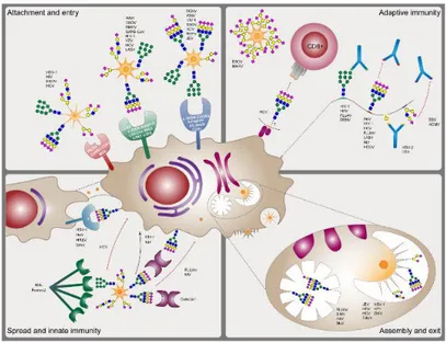
Glycosylation is one of the most abundant modification of proteins and it is classified as N-, O- and C-glycosylation, based on the type of amino acid to which the sugar moiety is attached. Glycosylation influences the protein stability, solubility, rigidity, interactivity, transport and signaling. This proteins’ modification occurs not only in all domains of the life (Eukarya, Bacteria, Archaea) but also in viruses. It has been reported in literature that eukaryotic viruses use the ER and Golgi apparatus of the host cell to add and to remove sugar to their nascent glycoproteins located on the envelope/surface. Viral proteins can be glycosylated at relatively few sites or can be densely glycosylated with N-linked or O-linked glycans. For instance, Ebola virus glycoprotein is modified by a very high content O-linked glycans (Jeffers et al., 2002), while the HIV-1 glycoprotein gp160 presents multiple N-linked glycans (Pabst et al., 2012).
Viral glycosylation play a key role during the virus infection (Bagdonaite and Wandall, 2018) (Fig. 2.1):
✓ Attachment and entry: viruses, such as Ebola virus, SARS coronavirus, use the N-linked glycans as a bite to interact with cellular lectins or mannose receptors, which later promote their uptake in the cells. Less representative are the cases in which the O-linked glycans participate in the host-virus interaction;
✓ Assembly and exit: glycosylation is involved in viral particle formation and release;
✓ Immunity: glycans shield immunodominant epitopes from immune recognition of the host cell.
- 27 -
Fig. 2.1 Roles of glycosylation in the biology of enveloped viruses. Reproduced from Bagdonaite et al., 2018.
Due to the importance of the glycosylation in virus biology, viruses have developed several strategies to exploit the host glycosylation machinery. For instance, several viruses are able to change the expression level or activity of some host glycosylation elements, in order to modify the final glycan structure. In addition, several viruses to increase their survival and propagation include in their genome glycosyltransferase genes. The bacteriophages T2, T4 and T6 of E. coli encode a glycosyltransferase that transfer glucose residue to the hydroxymethylcytosines (HMC) of their DNA, protecting it against specific restriction endonucleases encoded by the chromosome of the host or its plasmid (Markine-Goriaynoff et al., 2004) . Some baculoviruses have developed a different strategy to improve their propagation. Baculoviruses infecting Lepidoptera larvae express a glucosyltransferase able to attach glucose residues to the hormone ecdysone, inactivating the hormone and subsequently the pupation of the host. This strategy allows these viruses to complete their infection cycle (Markine-Goriaynoff et al., 2004).
- 28 -
2.1 The giant DNA viruses encode an autonomous glycosylation system. In the last years, it has become increasingly evident that, in contrast to other viruses, some members of Nucleo-Cytoplasmic Large DNA Viruses (NCLDVs) group encode most, if not all, the machinery required for the glycosylation of their structural proteins. The concept of autonomous viral glycosylation system emerged from Paramecium bursaria chlorella virus 1 (PBCV-1) which encodes the glycosylation machinery for its major capsid protein (Vp54), whose glycans structure was disclosed only recently(De Castro et al., 2013) . Vp54 presents four N-linked glycosylation sites. The structure of the N-N-linked glycans revealed unique feature:
✓ The glycan is bound to asparagine (Asn) via -glucose, instead of N-acetylglucosamine or N-acetylgalactosamine typical of N-linked glycoproteins;
✓ The presence of L-Fucose fully substituted (it was observed only in the phosphoglycan epitope of Trypanosoma cruzi) and D-Rhamnose (a very rare sugar);
✓ The Asn acceptor is not in the classical consensus sequence Asn-X-Ser/Thr of N-glycosylation both in eukaryotes and in prokaryote;
✓ The Vp54 N-linked glycan structure is unique, does not resembles any structure found in the three domains of life.
PBCV-1 not only encodes for several glycosyltransferases presumably involved in the glycan structure of Vp54, but also has the enzymes to produce the nucleotide-sugars used as substrate for glycan formation. PBCV-1 has a functional pathway for GDP-L-Fucose and D-Rhamnose, which represents the first example of nucleotide-sugar metabolism described in viruses (Tonetti et al., 2003).
Starting from PBCV-1, enzymes for glycan biosynthesis have been identified in the giant DNA viruses of amoeba, in particular in members of the Mimiviridae family,