0
SAPIENZA UNIVERSITY OF ROME
SCHOOL OF BIOLOGY AND MOLECULAR MEDICINE
PhD THESIS
Functional studies of human K
+channels KCNH1 and KCNK4
and their role in human pathogenesis
Human Biology and Medical Genetics PhD course
Medical Genetics curriculum
XXXII Cycle
Coordinator: Prof. Antonio Pizzuti
Tutor: Prof.ssa Viviana Caputo
Candidate: Dr. Giulia Napoli
1
INDEX
ABSTRACT...2
1. INTRODUCTION...4
1.1. Human K+ channels: cell functions...4
1.2. Potassium channels in human diseases...5
1.2.1 Potassium voltage-gated channel subfamily H member 1 - KCNH1...6
1.2.2 Potassium channel subfamily K member 4 - KCNK4...12
2 AIM OF THE WORK...17
3 MATERIALS AND METHODS………...18
3.1 Cell culture...18
3.2 Constructs preparation and mutagenesis...18
3.3 Immunoelectron microscopy...18
3.4 Sub-cellular localization...19
3.5 Cilia formation and Immunofluorescence...19
3.6 Fibroblasts growth curve……….………….20
3.7 Protein extraction, SDS-PAGE and western blotting...20
3.8 Real-Time Quantitative PCR………20
4 RESULTS...22
4.1 Characterization of the effect of KCNH1 and KCNK4 mutations on cell cycle...22
4.2 Characterization of the effect of KCNH1 and KCNK4 mutations on ciliogenesis...22
4.3 KCNH1 and KCNK4 variants affect SHH Pathway...23
4.4 Characterization of the effect of KCNH1 mutations on KCNH1 sub-cellular localization...24
4.5 Characterization of the effects of KCNH1 mutations on protein localization...26
4.6 Characterization of the effect of KCNH1 mutations on primary cilium morphology and function...28
4.7 KCNK4 sub-cellular localization...32
4.8 Localization of KCNK4 during cell cycle...33
5 DISCUSSION...35
2
ABSTRACT
Potassium (K+) channels constitute the most diversified class of ion channels with regard to structure and gating characteristics. They contribute to the maintenance and stabilization of the resting potential and are key players in regulating cell excitability and functions in response to multiple signals (Tian et al., 2014). In recent years, the aberrant function of some of these channels has been documented to affect development and underlie syndromic disorders (Bauer et al., 2018).
KCNH1 (MIM 603305) is a member of the EAG (ether-à-go-go) family of voltage-gated K+ channels (Cázares-Ordoñez and Pardo, 2017). Recent studies have demonstrated that gain-of-function mutations in KCHN1 are implicated in Zimmermann-Laband syndromes (ZLS; MIM 135500;Kortüm et al., 2015) and other forms of developmental deficits that include mental retardation and epilepsy (Simons et al., 2015;Bramswig et al., 2015; Mégarbané et al., 2016; Fukai et al., 2016). These findings suggest that KCNH1 might be important for cognitive development in human. Recently, gain-of-function mutations in
KCNK4 (MIM 605720), encoding a two-pore-domain K+ channel (K2P), have been reported in subjects with a phenotype of facial dysmorphism, hypertrichosis, epilepsy, developmental delay/ID, and gingival overgrowth (FHEIG syndrome, MIM 618381; Bauer et al., 2018). The clinical features of the KCNK4-related condition are reminiscent of ZLS (Kortüm et al., 2015), providing evidence for a channelopathy caused by hyperactivation of K+ channels, including KCNH1 and KCNK4.
Some ion channels are bifunctional proteins contributing to several cell functions (Hegle et al., 2006). To date, only electrophysiology experiments were performed to explore the mechanism involving KCNH1 and KCNK4 in ZLS and FHEIG related developmental processes (Kortüm et al., 2015; Simons et al.,2015; Bauer et al., 2018).
Based on these considerations, major aims of my PhD project were to better characterize the cell role of KCNH1 and KCNK4 channels and to explore the functional impact of the KCNH1-ZLS and KCNK4-FHEIG mutations using cellular and molecular biology approaches, including Immunofluorescence, Western Blot and Real-Time Quantitative PCR. To this aim, we used cell lines, control fibroblasts and primary skin fibroblasts derived from ZLS-patients carrying mutations in KCNH1 (p.Arg330Gln and p.Leu352Val) and from FHEIG-patients carrying mutations in KCNK4 (p.Ala172Glu and p.Ala244Pro). We demonstrated impaired proliferation of KCNH1 and KCNK4 mutant fibroblasts, confirming the role of KCNH1 as a regulator of cell cycle of non-transformed cells and highlight a new function for KCNK4 in regulation of cell proliferation. We also found a significant increase of cilia number and a cilium-related pathway, i.e. SHH pathway, activation in all the mutant fibroblasts, thus suggesting functional role of KCNH1 and KCNK4 in cilia regulation. Moreover, confocal microscopy analysis revealed defects in cilia morphology in fibroblasts from ZLS patients. Confocal analysis refined also the reported KCNH1 localization in the cilium (Sánchez et al., 2016) to be concentrated at the centrosome and ciliary pocket regions for both wild-type and mutant fibroblasts. Finally, immunofluorescence analysis conducted in
3 wild-type fibroblasts and cells lines disclosed a nucleolar localization of KCNK4 channels, possibly indicating unrevealed roles of KCNK4.
In summary, we demonstrated that KCNH1 and KCNK4 may have specific ion channel-independent function converging on some share cellular pathways whose alteration might affect development processes. Overall, these findings highlight the importance to characterize the intracellular role of K+ to shed light on the mechanisms of pathogenesis of ZLS and ZLS related developmental processes.
4
1. INTRODUCTION
1.1 Human K+ channels: cell functions
Ion channels are transmembrane proteins that regulate the flow of ions across biological membranes and are central regulators of the distribution of potassium (K+), sodium (Na+), calcium (Ca2+), and chloride (Cl-) ions for cellular ionic homeostasis. K+ channels are the most diverse group of ion channels and are classified into four superfamilies, based on their domain structure and activation mechanisms, including voltage-gated K+ (Kv) channels, Ca2+-activated K+ (KCa) channels, inwardly rectifying K+ (Kir) channels, and two-pore domain K+ (K2P) channels (Xi Huang et al., 2014).
The human genome contains almost 80 genes encoding K+ channels, which constitute the most diversified class of ion channels with regard to structure and gating features. Such diversity allows K+ channels to participate to many cell processes of both excitable and non-excitable cells. They control
the K+ flux through the cell membrane in response to multiple signals to modulate membrane potential and other cell functions. K+ channels contribute to the maintenance and stabilization of the resting membrane potential, help to repolarize action potentials and mediate hyperpolarization. They are also key players in regulating cell excitability, cell volume, proliferation, apoptosis and cell cycle (Bauer et al., 2018). They also control neuronal information, muscle contraction and are important regulators of Ca2+-triggered secretion of neurotransmitters and hormones. Furthermore, K+ channels provide the driving force required for Ca2+ to enter the cell by keeping the membrane potential at hyperpolarized values. Ca2+ is an important mediator of intracellular signals implicated in the control of proliferation and other main cell processes (Urrego et al., 2014). Although different studies have shown that the expression and function of K+ channels differ along cell cycle, the mechanisms controlling ion channels
expression during the whole cell cycle remain elusive.
In proliferating cells, K+ permeability determines the periodic changes of the resting membrane
potential associated with each phase of the cycle. K+ channels also play a role in cell volume changes
required for the completion of mitosis and might influence signal transduction elicited by cell cycle checkpoints through protein-protein interactions or phospholipid clustering that modulates mitogenic signaling (Urrego et al., 2016).
Several studies showed that some voltage-gated ion channels are bifunctional proteins because they can also contribute to transcriptional regulation, protein scaffolding, cell adhesion and intracellular signaling, using mechanism independent of ion conduction (Hegle et al., 2006).
5 Figure1. K+ channels in cell-cycle.
K+ channels influence cell-cycle progression through cell volume regulation, modulation of membrane potential, generation of driving for Ca2+ and protein–protein interactions. K+ channel expression or function can be in turn regulated by progression through the cell cycle (Urrego et al., 2014).
1.2 Potassium channels in human diseases
K+ channels play important roles in a range of cell physiological processes and their aberrant activation or inhibition has been causally linked to altered neurotransmission, cardiac arrhythmias and endocrine and kidney dysfunction (Tian et al., 2014). Remarkably, the aberrant function of some of these channels has been documented to affect development and underlie syndromic disorders (Bauer et al., 2018).
K+ channels are critical to neurotransmission in the nervous system and alterations in their function lead to remarkable perturbations in membrane excitability and neuronal function. Many neuronal disorders, such as episodic ataxia, benign familial neonatal convulsions, Alzheimer's disease, are related to mutations in K+ channels (Shieh et al., 2000). Moreover, recent studies on K+ channel gene expression in the basal ganglia, indicate that dysfunctions of several K+ channels may be involved in the pathogenesis of Parkinson's disease (Tian et al., 2014).
There are many types of K+ channels in mammalian cardiac cells, whose expression varies greatly throughout the heart, from atria to ventricles, from epi‐ to endocardium, and from apex to base. The critical role of K+ channel dysfunction in cardiac arrhythmias is particularly evident in congenital channelopathies such as long‐QT and short‐QT syndromes, Brugada syndrome, and familial atrial
6 fibrillation, all associated with an increased likelihood of tachy-arrhythmias (Chiamvimonvat et al., 2017).
Besides neurologic and cardiac diseases, some diseases in other systems have been associated to K+ channels dysfunctions. Permanent neonatal diabetes mellitus and type 2 diabetes mellitus are disorders associated with mutations in KCNJ11, encoding the Kir6.2 subunit of the KATP channel, and KCNJ15, encoding the inwardly rectifying K+ channel, subfamily J, member 15, both involved in insulin release. Moreover, Bartter's disease, a familial salt‐wasting nephropathy, is caused by mutations in the Kir1.1 channel, which mediates K+ secretion and regulates NaCl reabsorption in kidney contributing to
maintain plasma K+ levels in the normal range (Tian et al., 2014).
There is increasing evidence showing that alterations in the expression of ion channels are involved in the progression and physiopathology of diverse human cancers. The roles of ion channels in cancer are differentiated and include regulation of cell proliferation, cell migration, cell volume control and apoptosis (Cázares-Ordoñez and Pardo, 2017). K+ channels regulate cancer cell behaviors such as proliferation and migration through both canonical ion permeation–dependent and non-canonical ion permeation–independent functions. Highly proliferative cells are more depolarized than differentiated or quiescent cells. However, transient hyperpolarization is needed for progression during the first stages of the cell cycle. Therefore, a change in the membrane potential must occur for cell cycle progression, as well as during cell migration and adhesion and cytokine production against the tumor. K+ channels would provide the driving force required for the Ca2+ influx that is necessary for cell cycle progression. At the same time, they may be involved in G1/S hyperpolarization, avoiding a loss of intracellular K+ and preventing the cells from entering the apoptotic pathway. In addition to changes in Ca2+ homeostasis, apoptosis is characterized by voltage membrane decay and cell shrinkage, both related to the activity of K+ channels (Serrano-Novillo et al., 2019).
1.2.1 Potassium voltage-gated channel subfamily H member 1 - KCNH1
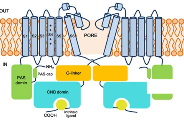
KCNH1 (Kv10.1, Eag1, OMIM 603305) encodes the KCNH1 ion channel, a member of the Eag
(ether-à-go-go) family of voltage-gated K+ channels. In humans, KCNH1 gene is located on the long arm of chromosome 1 (1q32.2). Alternative splicing of this gene results in two transcript variants (NM_172362.2 and NM_002238.3) coding respectively for two proteins of 989 (NP_758872.1) and 962 (NP_002229.1) amino acids, differing for an extracellular loop between the transmembrane helices 3 and 4 and encoding the same domains: the aminoterminal region, containing an eag domain, which is comprised of a Per-Arnt-Sim (PAS) domain and a PAS-cap domain, while the carboxy-terminal region, containing a cyclic nucleotide binding homology domain (CNBHD), and six transmembrane helices (S1-S6). The S4 helix works as a voltage sensor domain, the S5 and S6 helices are connected by the P-loop and together constitute the pore. Both the N- and the C-terminal domains are cytoplasmic and are necessary for the gating of the channel (Haitin et al., 2013). Additional splice variants were recently
7 identified, failing to form functional ion channels, but able to alter the glycosylation and expression patterns of canonical isoforms (Ramos Gomes et al., 2015).
Figure 2. Schematic view of the KCNH1 channel showing the major structural domains.
The opposite two subunits are shown. Transmembrane domains are in light blue, the N-terminal PAS domains are in green, the C-linkers are in orange, and the CNB domains are in blue green. The intrinsic ligand motifs are highlighted in yellow (Haitin et al., 2013).
KCNH1 is highly expressed in adult central nervous system and in most human tumours, at low levels
in placenta, testis, adrenal gland and transiently in myoblasts (Wang et al., 2017). It is aberrant expressed in cancer cells in contrast to its restricted distribution in healthy tissues. As its transforming activity and ectopic expression occurs in 70% of human cancers, KCNH1 is considered a clinically relevant ion channel and a marker and therapeutic target of several tumours. Cancer cells overexpressing KCNH1 acquire selective advantages that promote cancer progression, such as chronic cell proliferation and migration, interference with oxygen homeostasis, regulation of cell cycle and modulation of ciliogenesis (Cázares-Ordoñez and Pardo, 2017).
KCNH1 is considered a bifunctional protein that, not only regulate K+ flux, but also intracellular signalling pathways independent from its channel function. Non-cancer cells overexpressing KCNH1 show increased p38-MAP kinase (p-38MAPK) activity and p-38 MAPK inhibition abolishes the effect of the channel on cell proliferation (Hegle et al., 2006).
8 Cell functions of KCNH1 include the regulation of the resting membrane potential in both excitable and non‐excitable cells, the modulation of cell volume and the regulation of proliferation and cell cycle (Urrego et al., 2014). Ion permeation does not seem to be a necessary condition for KCNH1 mediated proliferation and tumourigenesis, as non-conducting mutants retain the ability to influence both processes. In contrast, voltage-dependent conformations may be crucial for KCNH1 to support proliferation, as the non-conducting mutants in open conformation fail to influence proliferation. Channel blockers could reduce proliferation not only by inhibiting permeation but also by trapping the channel in a particular conformation (Urrego et al., 2014).
KCNH1 plays an active role in cell cycle progression in both cancer and non-transformed cells. In HeLa cells and mouse embryonic fibroblasts, KCNH1 depletion leads to delayed G2/M progression, indicating that channel expression at the end of the cell cycle facilitates G2/M completion. Functional studies performed in breast cancer cells also demonstrated that KCNH1 regulates the progression of cell cycle from G1 to S phase. Cells synchronized in G0/G1 shows increased KCNH1 mRNA levels accompanied by membrane hyperpolarization towards the phase and defective checkpoint control between G1 and S-phase. The expression of KCNH1 is coupled to cell cycle not only in cancer but also in normal cells, in which is temporally restricted to a time period immediately prior to mitosis. Moreover, KCNH1 promoter contains an E2F1-responsive element that during the G2/M phase is directly regulated by the activation of E2F1 pathway promoting cell cycle progression (Urrego et al., 2016).
The peak of KCNH1 expression during G2/M phase, which coincides with the time of disassembly of primary cilia, and its interaction with proteins that participate in ciliary regulation, such as Rabaptin 5 and cortactin, allowed to hypothesize a role for the channel in ciliogenesis (Sánchez et al., 2016).
KCNH1 is important for the neuronal excitability and is expressed in different brain regions in both rodents and humans but its physiological function remains poorly understood. Morphological and behavioral analysis were performed in KCNH1 knock-out mice to investigate the functional role of the channel in the brain and indicated that deletion of KCNH1 does not produce functional alterations in brain or severe cognitive or behavioral effects (Ufartes et al., 2013).
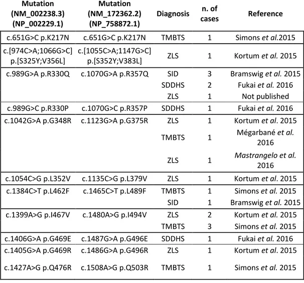
Mutations in KCNH1 have been recently associated to Zimmermann-Laband syndrome (ZLS; MIM 135500; Kortüm et al., 2015) and also reported in Temple-Baraitser syndrome (TMBTS; Simons et al.,
2015; Mégarbané et al., 2016), in unrelated individuals with intellectual disability (ID, Bramswig et al., 2015) and in individuals with syndromic developmental delay, hypotonia and seizures (SDDHS, Fukai et al., 2016), suggesting that KCNH1 might be important for human brain physiology and cognitive development.
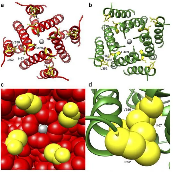
KCNH1 channels are homotetramers, with each subunit composed of six transmembrane helices. In ZLS cases, five of six KCNH1 mutations are located in the two helices (S5 and S6) involved in the opening of the channel and one mutation maps in the voltage-sensor helix (S4). All variants are single-nucleotide substitutions causing missense mutations, with a germline origin. A homology model for the S5 and S6 helices of KCNH1 was used to study the structural impact of p.Gly469Arg, p.Ile467Val, p.Leu352Val and
9 p.Val356Leu mutations. The p.Gly469Arg change is predicted to impair tetramer formation or favor the open state with a lower conductance by inserting four cationic residues close to each other in the closed state of the homotetramer. The p.Ile467Val, p.Leu352Val and p.Val356Leu substitutions are predicted to affect the closed-open transition. These residues form a tight hydrophobic cluster in the open structure which rearranges in the closed conformation (Kortüm et al., 2015; Figure 3).
Figure 3. Structural impact of ZLS-associated KCNH1
Helices S5 and S6 of KCNH1 in their closed (A) and open (B) states are shown. The pore-closing region is enlarged in the closed state (C) and in the open state the hydrophobic cluster formed by Leu352, Val356 and Ile467 is enlarged (D), (Kortum et al. 2015).
The functional consequences of KCNH1 mutations identified in ZLS patients (p.Gly348Arg, p.Leu352Val, p.Ile467Val, p.Gly469Arg and p.Ser352Tyr/Val356Leu) were assessed by patch-clamp experiments on Chinese hamster ovary (CHO) cells expressing wild-type or mutant KCNH1 channels. An effect of gain of function for all disease-associated KCNH1 mutants was observed, showing a shift in the activation threshold to more negative potentials. The mutants channels exhibited accelerated channel activation
10 and slower deactivation, producing dramatic increases in whole cell K+ conductance, compared to wild-type KCNH1 (Kortüm et al., 2015; Figure 4).
Figure4. Voltage-dependent activation of human wild-type (WT) and mutant KCNH1 channelsexpressed in CHO cells.
Families of whole-cell currents elicited with 2-s variable test pulses (A). KCNH1 current amplitudes as a function of the test pulse potential(B). Whole-cell conductance as a function of the test pulse potential (C). Potential of half-maximal KCNH1 channel activation (V0.5 activation),(Kortum et al. 2015) (D).
Simons et al. studied the effect of TMBTS associated-KCNH1 mutations (p.Ile467Val, p.Gln476Arg, p.Leu462Phe and p. Lys217Asn) in Xenopus oocytes and human HEK293T cells expressing mutant human channels and analyzed their activities using two-electrode voltage-clamp or patch-clamp electrophysiology. As for mutations in KCNH1 associated with ZLS, all TMBTS associated mutants showed enhanced activity and slower deactivation, compared to the wild-type channel (Simons et al., 2015; Figure 5).
A
B C
11 Figure 5. Mutant KCNH1 channels show a decreased activation threshold and delayed deactivation.
(A) Whole-cell currents in HEK293T cells expressing wild-type (WT) and mutant KCNH1. (B) Current density versus test voltage for wild-type and TBS-associated mutant KCNH1 channels expressed in HEK293T cells. (C) Activation current-voltage relationship for wild-type, Lys217Asn, Leu489Phe, Ile494Val and Gln503Arg channels expressed in HEK293T cells. (D) Fast and slow time constants (τ) of deactivation for wild-type and mutant KCNH1 channels expressed in HEK293T cells (Simons et al., 2015).
The identical mutations in the voltage-sensing domain of KCNH1(c.989G>A, p.Arg330Gln; Bramswig et al., 2015) was found in three individuals of a cohort of intellectual disability. Moreover, a fourth individual was identified with a previously TMBTS described mutation (Simons et al., 2015) in the channel pore domain (c.1384C>T, p.Leu462Phe; Bramswig et al., 2015).In addition, Fukai et al. identified two novel
KCNH1 (1406G>A, p.Gly469Glu and c.989G>C,p.Arg330Pro) and a previously reported (c.989G>A,
p.Arg330Gln; Bramswig et al., 2015) mutations in patients with SHDDH. In at least five cases, the same mutation has been reported in association with different clinical diagnosis (e.g. c.1399A>F, p.Ile467Val associated with ZLS and TMBTS, Kortum et al. 2015; Simons et al., 2015)(Table 1).
Despite these identified KCNH1 mutated patients could not be assigned to the same syndrome, a comparison of them, allowed to disclose the presence of some shared clinical features, like intellectual disability, craniofacial dysmorphism, seizures and/or epilepsy, and hypoplastic nails.
A
12 Mutation (NM_002238.3) (NP_002229.1) Mutation (NM_172362.2) (NP_758872.1) Diagnosis n. of cases Reference c.651G>C p.K217N c.651G>C p.K217N TMBTS 1 Simons et al.2015 c.[974C>A;1066G>C] p.[S325Y;V356L] c.[1055C>A;1147G>C]
p.[S352Y;V383L] ZLS 1 Kortum et al. 2015 c.989G>A p.R330Q c.1070G>A p.R357Q SID 3 Bramswig et al. 2015
SDDHS 2 Fukai et al. 2016
ZLS 1 Not published
c.989G>C p.R330P c.1070G>C p.R357P SDDHS 1 Fukai et al. 2016 c.1042G>A p.G348R c.1123G>A p.G375R ZLS 1 Kortum et al. 2015
TMBTS 1 Mégarbané et al. 2016 ZLS 1 Mastrangelo et al. 2016 c.1054C>G p.L352V c.1135C>G p.L379V ZLS 1 Kortum et al. 2015 c.1384C>T p.L462F c.1465C>T p.L489F TMBTS 1 Simons et al. 2015
SID 1 Bramswig et al. 2015
c.1399A>G p.I467V c.1480A>G p.I494V ZLS 2 Kortum et al. 2015
TMBTS 3 Simons et al. 2015
c.1406G>A p.G469E c.1487G>A p.G496E SDDHS 1 Fukai et al. 2016 c.1405G>A p.G469R c.1486G>A p.G496R ZLS 1 Kortum et al. 2015 c.1427A>G p.Q476R c.1508A>G p.Q503R TMBTS 1 Simons et al. 2015
Table 1. A summary of mutations identified in KCNH1 patients.
TMBTS= Temple–Baraitser Syndrome; ZLS= Zimmermann-Laband syndrome; SID= Syndromic Intellectual Disability; delay; SDDHD= Syndromic Developmental Delay Hypotonia and Seizures.
Epilepsy is a key manifestation in KCNH1-associated disorders but the mechanism by which KCNH1 mutations cause epilepsy remains to be elucidated. A possible mechanism could be that gain of channel function results in increased K+ conductance with subsequent inhibition of Na+ and Ca2+ currents, two major effectors during epilepsy, and perturbation of neurotransmitter release. However, there is not a clear relationship among KCNH1 mutations and severity, age of onset, response to antiepileptic drugs and type of seizures, that include focal motor seizures and generalized tonic-clonic seizures with seizure control (Mastrangelo et al., 2016).
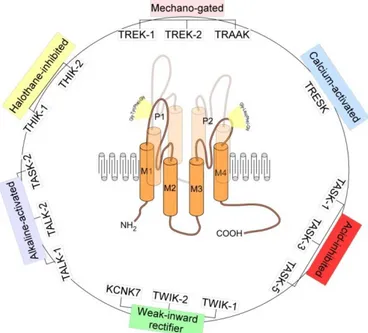
1.2.2 Potassium channel subfamily K member 4 - KCNK4
KCNK4 (OMIM 605720), also known as TRAAK (TWIK-related arachidonic acid-stimulated K+ channel), is one of three lipids and mechano-sensitive K2P channels constituting the TREK subfamily. Two-pore-domain (K2P) K+ channels are dimeric proteins with a unique structure of four transmembrane domains and two pore-forming domains. The 15 mammalian members have been grouped into six subfamilies on
13 the bases of function and structure (Figure 6). In addition to physical factors such as pH, temperature and membrane stretch, K2P channels are regulated by neurotransmitters acting through G protein-coupled receptors, hormone, polyunsaturated fatty acids and by several medicinal agents, including volatile anaesthetics and respiratory stimulants, anti-psychotics, anti-depressants and neuroprotective agents (Noël et al., 2011).
Figure 6. Structure and classification of K2P channels.
K2P channels are two-pore domain potassium channels and assemble as dimers of four transmembrane segments (M1–M4) and two-pore domain (P1 and P2). They have an extended M1-P1 extracellular loop and cytosolic N- and C-termini and a unique pore signature sequence Tyr(Phe)-Gly in the 1st pore (P1) and Gly-Leu(Phe)-Gly in the 2nd pore (P2) (Djillani et al., 2019).
KCNK4 gene is located on the long arm of chromosome 11 (11q13.1) and encodes for a 393
amino-acid long protein (NM_001317090.1, NP_001304019). KCNK4 is mainly expressed in the human and the rodent brains where it contributes to the background K+ currents and regulate cell viability. In the mouse brain, KCNK4 mRNA is expressed in the whole hippocampus neurons. Moreover, it can be found in the spinal cord and retina (Fink et al., 1998).
Most studies on KCNK4 have focused on the functions and mechanism of the channel, such as its roles in CNS degenerative disease processes, synaptic transmission, the maintenance of membrane resting potential and cell volume regulation. In degenerative processes of ischemia-induced neurons, KCNK4 channel demonstrates the capability to downregulate neuronal excitability that leads to lower cellular metabolic rate (Noël et al., 2011).
All members of the TREK channel family, including KCNK4, are expressed in human retinal pigmented epithelial cells (RPE-1) and their activation protect cells against oxidative stress, which is important in the onset and development of retinal degeneration diseases. They increase the survival rates by enhancing the levels of intracellular protective factors, such as Bcl-2 and αB-crystallin and decrease
14 apoptosis by enhancing factors, such as Bax and cleaved-caspase-3. Moreover, under oxidative stress these channels protect the integrity of the RPE cell structure (Huang et al., 2018). However, the specific function of each member of these channels in regulating apoptosis and oxidative stress has not yet been characterized.
It is known that actin cytoskeleton inhibits mechanosensation of another member of TREK subfamily, KCNK2, and that this channel influences the assembly of the actin network and the formation of filopodia-like structures independently of its channel activity. However, the molecular mechanism of the cross-talk between the channel and actin is still unknown as well as the potential involvement of the other members of the family in modulating cytoskeletal structures like primary cilium (Noël et al., 2011). K2P channels are dimeric proteins, with each subunit contributing two pore domains to the channel. Two different de novo missense mutations in KCNK4 (c.515C>A, p.Ala172Glu and c.730G>C ,p.Ala244Pro), have been reported in three subjects (FHEIG syndrome, MIM 618381), highlighting the role of K+ channels in developmental processes (Bauer et al., 2018). Subjects with FHEIG syndrome had significant generalized hypertrichosis and a similar facial gestalt, including hypotonic faces, bitemporal narrowing, micrognathia, deep-set eyes, bushy eyebrows and long eyelashes, low-set ears, short deep philtrum, gingival overgrowth, prominent upper and lower vermilion, and everted upper lip. Other more variable features included nystagmus with optic hypoplasia, hypotonia, hyperreflexia and DD/ID. Two of three patients showed also early-onset focal and generalized seizures (Bauer et al., 2018).
KCNK4 channel is a dimeric protein. Each subunit comprises an extracellular cap covering the pore, four transmembrane helices (M1 to M4) and two pore domains (P1 and P2) constituting the selectivity filter. Missense pathogenic mutations in KCNK4 mapped in regions of the channel possibly involved in KCNK4 activation: Ala244 is located at the center of helix M4 while Ala172 is located in M3 helix which interacts with M2 (Figure 7A). Channel opening is mediated by the transition of M4 helix: when the M4 helix is in the down state, lipid moieties insert in the lateral fenestration blocking K+ flux; when the M4 is in the up conformation state, fenestration are closed, lipid insertion is prevented and the channel is open (Figure 7B). Molecular dynamics stimulations showed that M4 helixes of wild-type and of p.Ala172Glu mutant were in down state in absence of activating stimuli, in contrast to up conformation of M4 helixes of p. Ala244Pro mutant channel. Moreover, the lateral fenestration of both mutants were less open compared to wild-type, suggesting basal activation in mutants. p.Ala172Glu substitution induced the formation of an inter-domain salt bridge between Glu172 and Arg147 (helix M2) which might be responsible for the closing of fenestration, observed even though both helices were in down conformation (Figure 7C).
15 Figure 7. The schematic structure of KCNK4 monomer and location and structural consequences of disease causing mutations
(A) Schematic and crystallographic structure of KCNK4 channel. Each subunit of homodimer comprises an extracellular cap covering the pore, four transmembrane helices (M1 to M4), and two pore domains (P1 and P2) constituting the selectivity filter. (B) Structure of the KCNK4 channel in the two conformational states with open and closed lateral fenestrations. (C) Structural impact of the p.Ala244Pro and p.Ala172Glu amino acid substitutions, as observed in the molecular dynamics simulations. Residues Leu125, Phe246, and Leu250 involved in the hydrophobic cluster occluding the fenestration (Bauer et al., 2018).
Patch-clamp experiments were performed in order to explore the structural impact of p.Ala172Glu and p.Ala244Pro mutationsin transfected CHO cells (Figure 8). Both disease-associated amino acid substitutions have shown activating effect. The mutant channels p.Ala244Pro and p.Ala172Glu showed a higher maximal basal activity compared to the wild-type and could not be stimulated further by mechanical stimuli. In addition to their mechanosensitivity, KCNK4 is activated by arachidonic acid and polyunsaturated fatty acids. Both KCNK4 mutants were found to lack the channel activation in response to arachidonic acid, which likely reflects their basal maximal activation. Accordingly, the two disease-causing KCNK4 mutations resulted in again of function effect, indicated by a dramatically increased basal K+ conductance (Bauer et al., 2018).
16 Figure 8. Whole-cell current recordings from CHO cells expressing wild-type or mutant KCNK4 channels
(A) Current recordings in cells expressing the wild-type (WT) or mutant (p.Ala244Pro or p.Ala172Glu) KCNK4 channels. (B) The I/V-plot shows current amplitudes recorded with the voltage step protocol. (C) I/V-plots of representative KCNK4 current recordings elicited with the ramp protocol shown as inset. Cells were previously transfected or co-transfected with cDNA coding for WT and mutant KCNK4. (D) Current amplitude at 0 mV ramp potential (Bauer et al., 2018).
Recently, dominantly acting missense mutations in KCNN3, coding for one of three members of the small-conductance Ca2+-activated K channels (SK channels), were identified in three subjects with the major clinical features of ZLS and significant clinical overlap with KCNH1 ZLS- and KCNK4 FHEIG -related disorders (Bauer et al., 2019). With the identification of gain-of-function mutations in KCNN3, it has been proposed to include the phenotypes associated with mutations in KCNH1, KCNK4, and KCNN3 in a new subgroup of neurological potassium channelopathies, characterized by developmental delay/ intellectual disability (DD/ID), coarse facial features, and gingival hyperplasia as common findings and epilepsy, hypertrichosis, and nail aplasia or hypoplasia as variable manifestations, associated with an increase in K+ conductance, especially evident at negative membrane potentials (Bauer et al., 2019).
17
2. AIM OF THE WORK
Aberrant functionof K+ channels have recently been documented to affect development and underlie syndromic disorders, including ZLS and FHEIG syndrome (Bauer et al., 2018).
The aims of this project were to gain insight on the specific cell functions of KCNH1 and KCNK4 channels and explore the functional impact of pathogenic KCNH1-ZLS and KCNK4-FHEIG mutations using cell models. To this aims, we obtained primary skin fibroblasts from ZLS and FHEIG patients, to be analyzed through molecular and cellular approaches.
Overall, our study contributed to highlight ion channel-independent function of KCNH1 and KCNK4 converging on some share cellular pathways whose alteration might affect development processes. Our understanding of the intracellular role of K+ channels could contribute to shed light on the mechanisms of pathogenesis of ZLS related developmental processes.
18
3. MATERIALS AND METHODS
3.1 Cell Culture
Functional characterization of KCNH1 and KCNK4 wild-type and mutant proteins was carried out on Human Dermal Fibroblasts (HDF) obtained from ATCC and cultured skin fibroblasts obtained from subcutaneous biopsies of ZLS and FHEIG syndrome patients, as well as in human TERT-immortalized retinal pigment epithelial cell line (hTERT RPE-1) obtained from ATCC. We obtained primary skin fibroblasts of patients with KCNH1 mutations causing ZLS (c.1054C>G, p.Leu352Val and c.989G>A, p.Arg330Gln; Kortüm et al., 2015) and with KCNK4 mutations causing FHEIG syndrome (c.515C>A, p.Ala172Glu and c.730G>C ,p.Ala244Pro; Bauer et al., 2018). The p.Arg330Gln mutation in KCNH1 causing ZLS was also reported in two individuals with ID (Bramswig et al., 2015) and in three individuals with SDDHS (Fukai et al., 2016).
Fibroblasts and RPE1 cell lines were cultured in Dulbecco’s modified Eagle’s medium (DMEM)and in DMEM/F-12, respectively, both supplemented with 10% heat-inactivated fetal bovine serum (FBS, EuroClone) and 1% penicillin-streptomycin, at 37 °C with 5% CO2.
3.2 Constructs preparation and mutagenesis
The human KCNH1 coding sequence corresponding to the short isoform (NM_002238.3, NP_002229.1, 962 amino acids) was amplified from human fetal cDNA using the following primers: KCNH1 Forward 5’-GTTTCCTGCTGTCGTAAGAAGC-3’; KCNH1 Reverse 5’-TGTTGGTCATGTGGACATATGTG-3’. The PCR products were gel-purified using the Wizard SV Gel and PCR Clean-Up System kit (Promega) and cloned in pSC-A-amp/kan vector using the Strataclone TA Cloning Kit (Agilent) according to the manufacturer’s protocol. Positive colonies were amplified, and plasmidic DNA purified with WizardPlus SV Minipreps DNA Purification Systemkit (Promega) was subjected to Sanger sequencing.
Then, the fragment corresponding to the KCNH1 isoform was cloned from pSC-A-amp/kan vector to the eukariotyc expression vector pCMV-TAG2A using the restriction enzymes sites BamHI and SalI. Missense mutations associated to ZLS (p.L352V; Kortüm et al., 2015) and ZLS, SID and SDDHS phenotypes (p.R330QBramswig et al., 2015; Fukai et al., 2016) were introduced by site-directed mutagenesis, using PickMutant Site-directed Mutagenesis kit (Canvax) in accordance with the manufacturer's protocol. All constructs were checked by direct sequencing.
3.3 Immunoelectron microscopy
hTERT RPE-1 cells were fixed in a mixture of 0.1% glutaraldehyde-2% paraformaldehyde, in 0.1 M phosphate buffer, pH 7.4, for 2 h at 25 °C, scraped, centrifuged, embedded into 10% gelatin (Sigma-Aldrich) in 0.1 M PBS, pH 7.4 and solidified on ice. After infusion in 2.3 M sucrose on at 4 °C, cell blocks
19 were mounted on aluminium pins and frozen in liquid nitrogen. Ultrathin cryosections (60 nm) were cut at -120°C using an Ultracut EM FC6 cryo ultramicrotome (Leica Microsystems), collected with 1% methylcellulose in 1.15 M sucrose in carbon coated grids and single- or double-immunolabelled with primary antibodies. Bound antibodies were visualized using goat anti-rabbit or anti-mouse antibodies conjugated with 10- or 15-nm colloidal gold (British BioCell International). Immunolabeling was performed with the primary polyclonal antibodies (KCNH1), grids were finally stained with 2% methylcellulose -0.4% uranyl acetate and analysed by a Philips CM10 and/or Morgagni transmission electron microscope.
3.4 Sub-cellular localization
For immunofluorescence analysis, 10x103 of wild-type and mutated KCNH1 (p.Leu352Val and p.Arg330Gln;) and KCNK4 (p.Ala172Glu and p.Ala244Pro) fibroblasts and 25x103 of hTERT RPE-1cell lines were seeded in 24-well cluster plates onto 12-mm cover glasses and maintained in culture complete medium for 48h. hTERT RPE-1 cells were transfected with vectors expressing wild-type or mutants Flag-tagged KCNH1, respectively, using Lipofectamine 3000. After 48h of transfection (for hTERT RPE-1) or culture (for fibroblasts) cells were fixed with 4% paraformaldehyde (20 min at RT). After permeabilization with 0.1% Triton X-100 (10 min at room temperature), non-transfected cells were stained with selected primary antibodies anti-KCNH1 (sigma) and KCNK4 (Sigma), whereas transfected hTERT RPE-1 were stained with anti-Flag (SIGMA). To investigate the specific sub-cellular localization of the two K+ channels, co-localization analysis was performed using specific cellular organelles markers: Golgin (Sigma) for the Golgi apparatus, RAB5 (Santa Cruz Biotecnology) for early endosomes, acetylated α-Tubulin (Santa Cruz Biotecnology) for primary cilium assoneme, CEP170 and CEP164 (Santa Cruz Biotecnology) for cilium basal body, EHD (Santa Cruz Biotecnology) for ciliary pocket, Lamin A/C (Sigma) for nuclear membrane, Fibrillarin (Santa Cruz Biotecnology) for nucleolus, and Ki-67 (Santa Cruz Biotecnology) for nucleus. The appropriate secondary antibodies were used (Invitrogen) followed by nuclei staining with Dapi (Sigma). Coverslips were extensively rinsed and mounted onto microscope slides using mounting medium (Sigma). 3D images were sequentially acquired by Olympus’ PLAPON60XOSC2 super-corrected objective confocal apparatus.
3.5 Cilia formation and Immunofluorescence
To explore the role of KCNH1 mutations in cilium assembly/disassembly, dynamics immunofluorescence analyses were performed. To this aim, HDF and hTERT RPE-1 cells were plated onto coverslips and cultured in complete medium for 24h. The day after, cells were serum starved in serum free media to induce ciliogenesis and after 48h serum was reintroduced for 1, 2 and 4h to induce cilia disassembly. At the end of incubation period, cells were washed with Phosphate Buffered Saline (PBS), fixed using 4% paraformaldeyde and permeabilized with 0.1% Triton X‐100. Primary cilia were
20 visualized by acetylated α-tubulin (Santa Cruz Biotecnology, axoneme), Arl13b (Proteintech, axoneme), IFT172 (Santa Cruz Biotecnology, anterograde transport) and CEP170 and CEP164 (Santa Cruz Biotecnology, basal body) staining. Images were captured by confocal microscopy on Olympus FV1000.
For quantification of primary cilia, both acetylated α-tubulin and CEP170 labelling were used to define cilia. The proportion of ciliated cells in a given field was determined by counting the number of cilia and the number of nuclei present and was expressed as percentage of total cell population. Cilium prevalence was measured for 6 fields in three separate experiments. Sequential 0.5 µm thick z-stacked sections were imaged through the entire cell profile using a 60X objective lens and were used to create maximum intensity projections (MIPs). ImageJ analysis software was used to measure the morphology of cilia and the co-localization in MIPs. Statistical significance was determined by unpaired t-test.
3.6 Fibroblasts growth curve
Once the fibroblasts reached 80 to 90% of confluence, they were plated at a density of 1x10⁴ cells per well in a 24 well plate and incubated at 37°C/5% CO2 on DMEM with 10% fetal bovine serum, for 24,
48, 72 and 96 hours. A combination of Trypsin/Ethylene Diamine Tetra Acetic acid (EDTA) solution was used to detach cells from tissue culture dishes, and detachment was monitored by means of light microscopy. Trypsin/EDTA action was neutralized with media containing 10% FBS. The density of cells was assessed by counting cells in a Burker chamber. The cell concentration and growth rate were recorded in triplicate at defined time points (0, 24, 48, 72 and 96 h after plating).
3.7 Protein extraction, SDS-PAGE and western blotting
For western blot analyses, cell pellets were washed twice in PBS and lysed in RIPA buffer (20 min, 4 °C). with 1 × Protease and Phosphatase Inhibitor Cocktail. Lysates were kept for 30 min on ice and then centrifuged for 10 min at 14 000 × g at 4 °C. Supernatants were collected and assayed for protein quantification by the Bradford method (Serva). Analysis of protein components was performed by polyacrylamide gel electrophoresis separation (Bio-Rad) and transferred onto nitrocellulose membranes (Amersham Biosciences). After saturation, blots were incubated overnight at 4 °C, with anti-Bcl2 (Santa Cruz Biotecnology) antibody, and finally incubated for 1 h with HRP-conjugated secondary antibodies and detected on X-ray film (Aurogene) using the ECL Advance Western Blotting Detection Kit (Amersham Biosciences). Quantifications were performed by ChemiDoc (Biorad). GAPDH (Sigma) was used for protein normalization. Densitometric analyses were performed with the ImageJ software.
3.8 Real-Time Quantitative PCR
Total RNA was extracted from wild-type and mutated KCNH1 (p.Leu352Val and p.Arg330Gln) and KCNK4 (p.Ala172Glu and p.Ala244Pro) fibroblasts with TRIzol Reagent(Invitrogen) according to the manufacturer’s instructions. Briefly, fibroblasts were homogenized in TRIzol and chloroform was added
21 at 1:5 of the initial volume of TRIzol. Sample were incubated at room temperature for 15 min and then centrifugated at 12,000 × g for 15 min at 4 °C. After centrifugation, aqueous phase was transferred to a new tube, mixed with 0.5 ml of cold isopropyl alcohol, placed at 4 °C for 20 min, and then centrifuged at 12,000 × g for 10 min at 4 °C. The RNA pellet was washed with 75% ethanol, dried by air, and dissolved in 0.01% diethyl pyrocarbonate water. The purity and amounts of the RNA obtained were checked by measuring optical density at 260 and 280 nm with the Nanodrop 100 System (Rockford, IL). A total amount of 1ug RNA was reverse transcribed using random primers with the High Capacity cDNA Reverse Transcription Kit (Applied Biosystem), according to the manufacturer’s instructions. QPCR for PTCH1, BCL2 and SMO genes quantification was performed using SYBR Green select (Applied Biosystems). Relative gene expression was calculated by ΔΔCt analysis, relative to GAPDH expression levels.
Primers were as follows:
BCL2 Forward 5’ CGGAGGCTGGGATGCCTTTG 3’ BCL2 Reverse: 5’ GTGATGCAAGCTCCCACCAG 3’ PTCH1Forward 5’ CAACTCAGGTTTTGCCATTTC 3’ PTCH1 Reverse 5’ CCGGTCCTGTCCTCAAAAG 3’ SMO Forward: 5’ CTCATCCGAGGAGTCATGAC 3’ SMO Reverse 5’ CAAAAATGCCCAGGCGCAGC 3’ GAPDH Forward5’ TCTTTTGCGTCGCCAGCCGAG 3’ GAPDH Reverse 5’ TGACCAGGCGCCCAATACGAC 3’
22
4. RESULTS
4.1 Characterization of the effect of KCNH1 and KCNK4 mutations on cell cycle
Proliferation is one aspects of cell physiology where K+ channels play a crucial role and it is known that KCNH1 plays an active role in cell cycle progression in both cancer and non-transformed cells (Urrego et al., 2014). To investigate if mutations in KCNH1 and KCNK4 channels affect fibroblasts cell cycle we assayed cell proliferation by counting cell number as a function of time, using human dermal fibroblasts (HDF) as a control. We obtained primary skin fibroblasts of patients with KCNH1 mutations causing ZLS (p.Leu352Val and p.Arg330Gln; Kortüm et al., 2015) and with KCNK4 mutations causing FHEIG syndrome (p.Ala244Pro and p.Ala172Glu; Bauer et al., 2018). When grown in the same culture conditions, KCNH1 p.Leu352Val (Figure 9A) and KCNK4 p.Ala244Pro (Figure 9B) fibroblasts proliferated at a reduced rate compared to control fibroblasts (wild-type).
Figure 9. Mutations of KCNH1 and KCNK4 induce decreased cell proliferation.
The figure shows the growth curves of two ZLS (A), two FHEIG (B) patient’s derived fibroblasts compared to wild-type fibroblasts. Cells were seeded for each sample and counted at the indicated time-points. *P≤0.05. Statistical significance was assessed using Student's t-test.
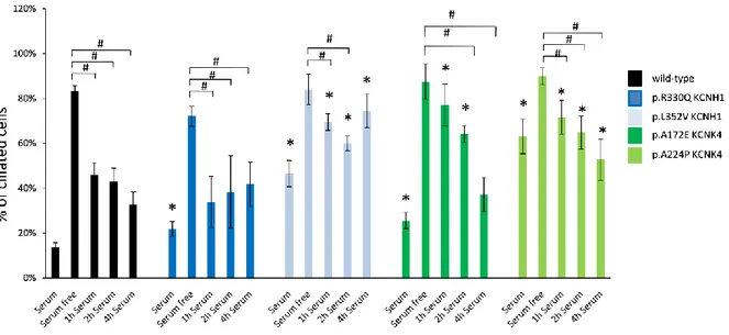
4.2 Characterization of the effect of KCNH1 and KCNK4 mutations on ciliogenesis
Ciliogenesis has been associated with cell cycle and control of cell proliferation (Malicki et al., 2017) therefore, as we disclosed an altered proliferation rate of mutated fibroblasts, we investigated a potential correlation between ciliation and the differential growth rate observed in ZLS and FHEIG fibroblasts. We measured ciliogenesis of mutant versus wild-type HDF using the serum starvation assay. Primary cilium was identified by immunofluorescence/confocal microscopy using antibodies against alpha acetylated-tubulin to label the microtubule axoneme in primary cilium, and CEP170 to label the basal body and the ciliated fractions in fibroblasts cultures. After 48h of serum starvation primary cilium was observed in most of cells
23 without any significant difference between wild-type and patients. Differently, before starvation there was a significant increase in the number of primary ciliated cells in cycling KCNH1 (p.Leu352Val and p.Arg330Gln) and in KCNK4 (p.Ala224Pro and p.Ala172Glu) patient-derived fibroblasts (Figure 10). Our data shows that all mutants studied display enhanced ability to promote cilia formation in cycling rather than in quiescent cells, compared to wild-type protein. Moreover, KCNH1 p.Leu352Val and KCNK4 p.Ala172Glu and p.Ala244Pro fibroblasts showed decreased ability to disassemble primary cilium after serum stimulation compared to wild-type fibroblasts.
Figure 10. Mutations of KCNH1 and KCNK4 impair ciliogenesis.
Control and patient’s fibroblasts were serum starved in serum free media to induce ciliogenesis and after 48h serum was reintroduced for 1, 2 and 4h to induce cilia disassembly. Percent of ciliated cells was counted using alpha acetylated tubulin/CEP170 as primary cilia markers. *P≤0.05 vs wild-type; #P≤0.05 vs serum free. Statistical significance was assessed using Student's t-test.
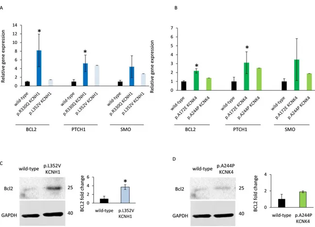
4.3 KCNH1 and KCNK4 variants affect SHH pathway
The primary cilium contains a highly specialised and compartmentalized molecular environment. For example, the Sonic hedgehog (SHH) signalling machinery is selectively localised to primary cilium structure.
In light of our previous results on cell proliferation and ciliogenesis, we sought to assess whether the SHH pathway was deregulated in the presence of mutant KCNH1 and KCNK4 proteins. We evaluated the basal expression levels of selected SHH target genes (BCL2, PTCH1 and SMO), in HDF and mutant KCNH1 and KCNK4 patient’s-derived fibroblasts, by quantitative real-time PCR. Interestingly, the basal expression levels of two key SHH-target genes, BCL2 and PTCH1, were significantly higher in p.Arg330Gln KCNH1 mutated fibroblasts compared to control, whereas SMO gene appeared to be deregulated without reaching statistical significances (Figure 11A). A comparable trend of PTCH1 and SMO transcripts expressions was observed in fibroblasts carrying p.Leu352Val mutation in KCNH1 (Figure 11A), which also showed a significantly over expression of BCL2 at the protein levels, as assessed by western blot analysis (Figure 11C).
24 Moreover, the basal transcript expression levels of both BCL2and PTCH1 were significantly higher in p.Ala172Glu KCNK4 mutated fibroblasts compared to control (Figure 11B). BCL2 expression appeared to be deregulated in p.Ala244Pro KCNK4 mutated fibroblasts levels without reaching statistical significances both at the RNA (Figure 11B) and protein levels (Figure 11D). Our results demonstrated a significant dysregulation of the SHH signaling in KCNH1 and KCNK4 mutant cells compared to controls.
Figure 11. The SHH pathway is deregulated in KCNH1 and KCNK4 patient’s fibroblasts.
RNA extracts from wild-type, KCNH1 and KCNK4 patient’s fibroblasts were analyzed by RT-qPCR using specific primers for human BCL2, PTCH1 and SMO genes using the ΔΔCt method (A, B). *P≤0.05. Statistical significance was assessed using Student's t-test. Proteins were extracted from wild-type, KCNH1 and KCNK4 patient’s fibroblasts. Protein lysates were subjected to western blotting analysis with the specified antibodies. Representative western blotting images and statistical analysis of BCL2 protein expression. Western GAPDH antibody was used for normalizations (C, D). Normalized values are means ± S.E.M. *P≤0.05. Statistical significance was assessed using Student's t-test.
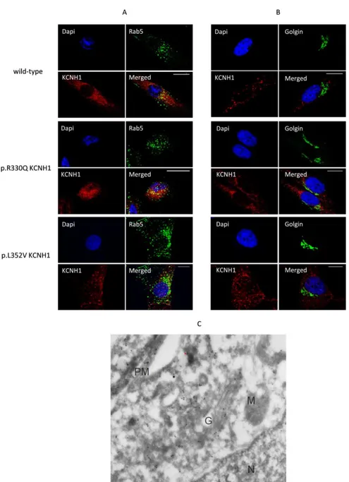
4.4 Characterization of the effect of KCNH1 mutations on KCNH1 sub-cellular localization
To evaluate whether the mutations in KCNH1 affect the sub-cellular localization of the channel, we performed immunofluorescence studies on primary skin fibroblasts of ZLS patients using specific sub-cellular compartments markers antibodies (Golgi apparatus and endosomes) and a commercially available KCNH1 antibody to stain KCNH1. Confocal laser-scanning microscopy analysis revealed cytoplasmic localization of KCNH1 with a reliable localization at the perinuclear and endosomal regions. In particular, as
25 shown in Figure 12A, KCNH1 showed partial overlap with the early endosome marker Rab5 in both wild-type and p.Arg330Gln mutated fibroblasts. On the contrary, we did not observe co-localization with Golgi marker (Figure 12B). A similar distribution was also observed in cells with the p.Leu352Val KCNH1 mutation.
Moreover, immunogold electron microscopy performed in hTERT RPE-1 cells revealed intracellular gold particles labeling KCNH1 in Golgi apparatus, endosomes and plasma membrane, consistently with immunofluorescence analyses (Figure 12C).
Figure 12. Co-localization of KCNH1 with endosome and Golgi markers in wild-type and patient’s fibroblasts.
A. Wild-type and patient’s fibroblasts were co-stained with anti-KCNH1 (red labelled) and anti Rab5 (green labelled). Nuclei (blue) were visualized by DAPI staining. Co-localization of KCNH1 and Rab5 is yellow (merged). Scale bar: 10 μm. B. Wild-type and patient’s fibroblasts were co-stained with anti-KCNH1 (red labelled) and anti Golgin (green labelled). Nuclei (blue) were visualized by DAPI staining. No specific co-localization was observed between KCNH1 and Golgin. Scale bar: 10 μm. C. Immunoelectron microscopy of 48h serum starved hTERT RPE-1 cells shows intracellular localization of KCNH1. Gold particles are observed in the Plasma membrane, Golgi apparatus and endosomes.N = Nucleus, PM=Plasma Membrane, G= Golgi apparatus, M=Mitochondrion.
26 4.5 Characterization of the effects of KCNH1 mutations on protein localization
Preliminary recent work related KCNH1 with primary cilium in hTERT RPE-1 and in mouse embryonic fibroblasts (Sánchez et al., 2016). However, the specific KCNH1 cilium localization and function have not been characterized. In order to characterize the link between KCNH1 and primary cilium and to test if major pathogenic mechanisms of mutants KCNH1 relies on cilium localization, we performed immunofluorescence experiments in hTERT RPE-1 cells, in human control fibroblasts and primary skin fibroblasts derived from patients with two KCNH1 missense mutations causing ZLS (Kortüm et al., 2015). We used alpha acetylated tubulin, CEP164 and EHD as specific ciliary markers. Acetylated tubulin is a protein associated with axoneme assembly, CEP164 is a basal body protein required for assembly of the primary cilium and EHD proteins regulate endocytic events and localize to the ciliary pocket.
As shown in Figure 13, wild-type KCNH1-stained vesicular rosettes were near the outermost boundary of the centrosome defined by CEP164. In particular, KCNH1 showed partial overlap with the distal appendage marker CEP164 (Figure 13A) and the ciliary pocket marker EHD (Figure 13B), suggesting a localization of KCNH1 in the ciliary pocket. No difference in proteins’ localization was observed between the wild-type and the p.Arg330Gln and p.Leu352Val missense mutants.
27 Figure 13. Localization of KCNH1 at the cilium base in wild-type and p.Arg330Gln and p.Leu352Val KCNH1- patients’ fibroblasts.
A. Wild-type and patient’s fibroblasts were starved for 48h prior to fixation and immunostaining procedures. Acetylated alpha tubulin (red) and CEP164 (pink) were used to mark the assoneme and the centrosome. Nuclei (blue) were visualized by DAPI staining. Zoomed-in areas in micrographs indicate localization of wild-type and mutant KCNH1 (green) proteins at the proximal end of the axoneme. Co-localization of KCNH1 and CEP164 is blank. Scale bar: 10 μm.B. Wild-type and patient’s fibroblasts were starved for 48h prior to fixation and immunostaining procedures. EHD was used to mark the ciliary pocket and nuclei (blue) were visualized by DAPI staining. Co-localization of KCNH1 (red) and EHD (green) is yellow. Scale bar: 10 μm.
The same approaches were used on hTERTRPE-1 cells, transfected with C-terminal Flag-tagged KCNH1. According to the results obtained in fibroblasts, wild-typeKCNH1 localized near the base of primary cilia, as well as p.Leu352Val and p.Arg330Gln mutant proteins (Figure 14).
28 Figure 14. Exogenous Flag-tagged KCNH1 localizes to the primary cilia.
hTERT RPE-1 cells were transfected with plasmid expressing KCNH1 flag-tagged wild type, p.Arg330Gln and p.Leu352Val mutant proteins. The staining was performed using anti-flag (green), anti-acetylated alpha tubulin (red) and anti-CEP164 (pink) antibodies. The panel shows localization of KCNH1 to the primary cilium base (white arrow). Scale bar: 10μm.
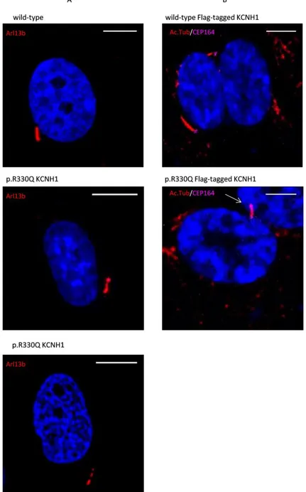
4.6 Characterization of the effect of KCNH1 mutations on primary cilium morphology and function
We evaluated morphology of primary cilium in control and ZLS patient’s fibroblasts. We characterized cilia morphology of 48h serum starved fibroblasts. A proportion of p.Arg330Gln fibroblasts showed a discontinuous axoneme, marked by a gap in Arl13b staining in all z-planes and bulbous tips (Figure 15A). Dysmorphic features were also observed in hTERT RPE-1 cell lines transfected with p.Arg330Gln Flag-tagged KCNH1 construct, which exhibited a bulbous projection at the tip (Figure 15B).
29
Figure 15. p.Arg330Gln KCNH1 mutation affects cilia morphology in quiescent cells.
A. Immunofluorescence microscopy of ciliated wild-type and p.Arg330Gln KCNH1 fibroblasts stained with antibodyto Arl13b (red). DNA was visualized with DAPI (blue). p.Arg330Gln fibroblasts show dysmorphic features (bulbous tip and discontinuous assoneme) compared to the control. Scale bar: 10 μm.
B. Immunofluorescence microscopy of ciliated hTERT RPE-1 cells transfected with plasmid expressing KCNH1 Flag tagged expressing wild-type and p.Arg330Gln mutant proteins. The staining was performing using anti-acetylated alpha tubulin (Ac.Tub, red) and CEP164 (pink) antibodies and DNA was visualized with DAPI (blue). p.Arg330Gln mutations leads to formation of bulbous ciliary tip. Scale bar: 10 μm.
In addition, structural abnormalities were detected in KCNH1 p.Leu352Val fibroblasts and hTERTRPE-1 cells transfected with p.Leu352Val Flag-tagged KCNH1, in which a portion of cells shows multiple cilia (Figure 16).
30 Figure 16. KCNH1 p.L352V variant leads to multiple cilia formation.
A. Immunofluorescence microscopy of ciliated wild-type and p.Leu352Val KCNH1 patient’s fibroblasts stained with antibodies to acetylated alpha tubulin (green) and CEP170 (red). DNA was visualized with DAPI (blue). ZLS patient’s fibroblasts exhibit multi ciliated cells. Scale bar: 10μm.
B. Immunofluorescence microscopy of ciliated hTERT RPE-1 cells transfected with plasmid expressing KCNH1 Flag tagged expressing wild type and p.Leu352Val mutant protein. The staining was performing using anti-Arl13b (red) antibody and DNA was visualized with DAPI (blue). hTERT RPE-1 cells transfected with p.Leu352Val Flag-tagged KCNH1 shows multiciliation. Scale bar: 10μm.
The structure anomalies of p.Arg330Gln cilia cells led to analyse the steady-state localization of the IFT172, a member of the IFT-B complex involved in the anterograde transport of cargo in the cilium (Gorivodsky et al., 2009). After 48h serum starvation, IFT172 was found almost exclusively at the base of the cilia in wild-type fibroblasts but in a proportion of p.Arg330Gln and p.Leu352Val fibroblasts at both the base and tip of the cilia (Figure 17). This bulbous morphology suggests impaired retrograde transport.
31 Figure 17. Impaired ciliary transport in ZLS patients’ fibroblasts.
Immunofluorescence microscopy of ciliated wild type and ZLS patient’s fibroblasts stained with IFT172 (green) and CEP170 (red, A) or Arl13b (red, B). DNA was visualized with DAPI (blue). Scale bar: 10μm.
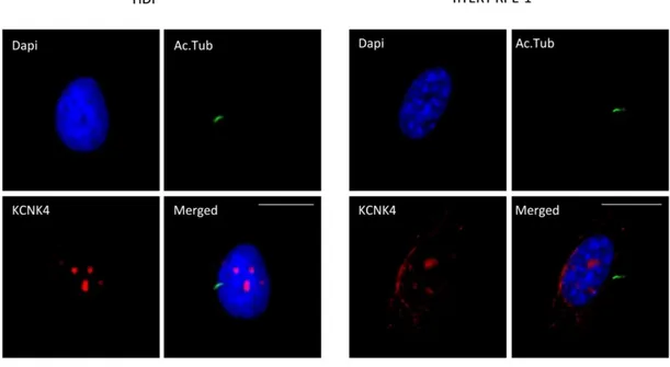
32 4.7 KCNK4 sub-cellular localization
Immunofluorescence assays were performed to eventually localize KCNK4 to the primary cilium, using alpha acetylated tubulin and KCNK4 specific antibody in HDF and hTERT RPE-1 cell lines.
As shown in Figure18 we did not observe localization of the K+ channel at the primary cilium. However, we could appreciate an unexpected nuclear localization of the protein in both HDF and hTERT RPE-1 cells, possibly indicating unrevealed roles of KCNK4.
Figure 18. Nuclear Localization of KCNK4.
Representative immunofluorescence microscopy images of HDF and hTERT RPE-1 cells stained with anti-KCNK4 and anti-alpha acetylated tubulin antibodies. The panel evidence a nuclear localization of KCNK4. Scale bar: 10μm.
To confirm this observation, we performed co-localization experiments with Lamin A/C, as a nuclear envelope marker, and Fibrillarin, as a nucleolus marker, using double staining with antibodies in wild-type HDF and hTERT RPE-1 cells. A significant level of co-localization was observed with Fibrillarin antibody in the nucleoli of HDF and hTERT RPE-1 cells (Figure 19).
33 Figure 19. Nucleolar localization of KCNK4 protein.
HDF and hTERT RPE-1 cells were stained with anti-KCNK4 (red) and anti- Lamin A/C or Fibrillarin (green) antibodies. DNA was visualized with DAPI (blue). KCNK4 co-localizes with nucleolar marker Fibrillarin. Scale bar: 10μm.
4.8 Localization of KCNK4 during cell cycle
We used the localization of Ki-67, a nuclear protein associated with cell proliferation and nucleoli, at the main cell cycle stages of hTERT RPE-1 cells, to evaluate KCNK4 localization during cell cycle. As shown in Figure 20A Ki-67 mainly localized in the nucleoli during the G1, S, and G2 phases and during late G2 and early prophase. When the nucleoli disassemble, the protein is diffusely distributed in the nucleoplasm. In contrast to cycling cells, quiescent cells do not express Ki-67 (Solovei et al., 2006). We performed co-immunofluorescence experiments with Ki-67 antibody, to recognize the different cell cycle phases of hTERT RPE-1 cells. As shown in figure 20B, we observed partially co-localization between KCNK4 and Ki-67 during the G1 phases and a completely co-localization during the S and G2 phases, according with previously
34 results showing its nucleolar localization. Interesting, the G0 cells, which did not express Ki-67, showed a strong accumulation of KCNK4 in defined nuclei foci.
Figure 20. Localization of KCNK4 during cell cycle phases.
A. Immunostaining of Ki-67 during cell cycle of HDF. Modified from Soloveiet al., 2006.
B. Representative immunofluorescence microscopy images of hTERT RPE-1 cells stained with anti-KCNK4 and anti-Ki67 antibodies. The panel shows a nucleoli localization of KCNK4 in G1, S and G2 phases and an accumulation in G0 phase. Nuclei were visualized with DAPI (blue). Scale bar: 10μm.
35
5. DISCUSSION
K+ channels are the most diverse group of ion channels. They control the K+ flux through the cell
membrane in response to multiple signals to modulate membrane potential and participate to many cell processes of both excitable and non-excitable cells. They are also key players in regulating cell excitability, cell volume, proliferation, apoptosis and cell cycle (Bauer et al., 2018).
Most functional studies on K+ channels focused on their electrophysiological properties. Notably, while electrophysiological studies were performed to shed light on the mechanisms underlying KCNH1 and KCNK4 mutations in ZLS and FHEIG, no functional data have been collected on the consequences of mutations on cell functions, i.e. localization, structure, and altered pathways. Based on these considerations, we explored the functional impact of two KCNH1 mutations associated with ZLS (p.Arg330Gln and p.Leu352Val; Kortüm et al., 2015) and two KCNK4 mutations associated FHEIG (p.Ala244Pro and p.Ala172Glu; Bauer et al., 2018) using cellular approaches.
The transmembrane potential has been reported as a cellular bioelectric parameter that influences the progression through the cell cycle. K+ channels have been implicated in the control of cell-cycle progression both through their influence on the membrane potential and through non-canonical permeation-independent mechanisms, showing remarkable cell cycle-dependent variations of expression and activity (Urrego et al., 2014). With the aim to investigate the functional impact of KCNH1 and KCNK4 mutations on cell cycle, we performed a proliferation assay by using primary fibroblasts as a cell model. Growth curve was carried out on control fibroblasts and on primary skin fibroblasts of patients with KCNH1-missense mutations p.Leu352Val causing ZLS (Kortüm et al., 2015), p.Arg330Glncausing ZLS (not published), SID (Bramswing et al., 2015) and SDDHS (Fukai et al., 2016) and with KCNK4-missense mutations (p.Ala244Pro and p.Ala172Glu) causing FHEIG (Bauer et al., 2018). We demonstrated that the mutations p.Arg330Gln in KCNH1 and p.Ala172Glu in KCNK4 did not impair cell proliferation. Differently, fibroblasts from patients carrying the p.Leu352Val substitution in KCNH1 and p.Ala244Pro in KCNK4 exhibited a very low proliferation rates compared to control fibroblasts, highlighting that both channels have a role in the regulation of cell proliferation. However, we showed that only one of the two analyzed mutations for both KCNH1 (p.Leu352Val) and KCNK4 (p.Ala244Pro) channels affect proliferation. One explanation could be that both KCNH1 p.Arg330Gln and KCNK4 p.Ala172Glu mutated proteins, when expressed in heterozygous state, may not be enough to observe a robust effect. Moreover, p.Leu352Val substitution map in the S5 helices and p.Arg330Gln in the voltage-sensing domain (S4 helix) of KCNH1 channel (Kortüm et al., 2015; Bramswig et al., 2015), and this different location would have a different impact on the proper cellular functions. In order to get better insight on this aspect and assess the functional role of K+ channels in cell proliferation we plan to perform additional experiments by using transfected cells overexpressing KCNH1 and KCNK4 carrying all mutations reported as pathogenetic (Kortüm et al., 2015; Bauer et al., 2018).
Additionally, it is known that KCNH1 activity is cell cycle regulated, suggesting that it is likely involved in fundamental cellular and developmental processes (Urrego et al., 2014). KCNH1 is considered a
bi-36 functional protein that not only regulates K+ flux but also intracellular signalling pathways, by mechanisms independent of K+ ion flux. The effect of KCNH1 on intracellular signalling was evident as the non-conducting mutants in open conformation fail to influence proliferation (Hegle et al., 2006). Structural analysis predicted that p.Leu352Val substitutions might affect the closed-open transition forming a tight hydrophobic cluster with other residues in the open structure which rearranges in the closed conformation (Kortüm et al., 2015). Moreover, electrophysiological studies have shown the functional consequences of the p.Leu352Val KCNH1 mutation on channel activity, which shifted in the activation threshold to more negative potentials and slower deactivation compared to the wild-type channel (Kortüm et al., 2015). Differently, no electrophysiological data have been collected for p.Arg330Gln KCNH1 variation, which may not have the same gain of function effect. Here, we hypothesized that p.Leu352Val KCNH1 channels display a more open conformation which results in failure of proliferation induction. The decreased proliferation rate of fibroblasts from patients carrying the p.Leu352Val substitution in KCNH1 and p.Ala244Pro in KCNK4 observed in our experiments suggests the occurrence of cell cycling defects introduced by these KCNH1 and KCNK4 missense mutations. To date, while several studies have demonstrated that KCNH1 plays an active role in cell cycle progression in both cancer and non-transformed cells, no data have been collected for KCNK4. Our results on mutated primary fibroblasts corroborate the role of KCNH1 as a mediator of proliferation of non-transformed cells and highlight a function for KCNK4 in regulation of cell proliferation.
Control of cell cycle has been associated with ciliogenesis (Malicki et al., 2017). In tissues, quiescent cells assemble a primary cilium, an antenna‐like sensory organelle that regulates multiple cell signalling pathways during development and in the adulthood (Morthorst et al., 2018). Cilia are dynamically regulated during cell cycle progression because assembly of the primary cilium requires a mature mother centriole: present in G0 and G1 and usually in S/G2 cells, but almost invariably resorbed before mitotic entry (Urrego et al., 2017). Mutations in genes that impair ciliary biogenesis, protein trafficking and function lead to a dysregulated signalling and cause diseases known as ciliopathies, which may affect nearly all organs during development or in adulthood (Urrego et al., 2017). It has been suggested that some clinical features of ZLS patients, as orofacial and digital abnormalities, together with severe mental retardation and morphological features, are at least in part reminiscent of ciliopathies (Sánchez et al., 2016). To evaluate whether KCNH1 (p.Arg330Gln and p.Leu352Val) mutations are associated with defects in assembly or disassembly of the primary cilium, we performed functional studies by using cell models.
Ciliogenesis assay was performed by immunofluorescence analysis in control and ZLS (KCNH1 p.Arg330Gln and p.Leu352Val) patients fibroblasts. We found a significant increase of cilia frequency in all the analyzed patients fibroblasts compared to control in cycling condition, before the induction of quiescent state by serum starvation, thus suggesting a functional role of KCNH1 in ciliogenesis. As K+ channels expression is not constant in all phases of the cell cycle, we might speculate that a significant fraction of mutant fibroblasts are in a different phase of cell cycle in cycling condition, possibly G0, compared to control fibroblasts. It was also observed that, upon reintroduction of serum, to induce cilium disassembly, the number of ciliated cells increase in fibroblasts with p.Leu352Val KCNH1 mutation. In