Jo
urnal o
f
Rad
io
lo
gy
Case Rep
orts
ww
w.Ra
diol
ogyC
ase
s.com
Rare lymphoid malignancies of the breast: report of
two cases illustrating potential diagnostic techniques
Orazio Schillaci
1, Laura Travascio
1, Annamaria Lacanfora
1*, Sara Ceccarelli
2, Giovanni Simonetti
21. Department of Biopathology and Diagnostic Imaging, University Tor Vergata, Rome, Italy
2. Department of Diagnostic Imaging and Interventional Radiology, Molecular Imaging and Radiotherapy, University Tor Vergata, Rome, Italy
* Correspondence: Annamaria Lacanfora, Department of Biopathology and Diagnostic Imaging, University Tor Vergata, Viale Oxford 81 00173 Rome, Italy
Radiology Case. 2012 Dec; 6(12):43-50 :: DOI: 10.3941/jrcr.v6i12.1194
ABSTRACT
Two cases of lymphoid malignancy involving the breast are herein presented. Both patients were admitted with a palpable breast mass. Ultrasound demonstrated hypoechoic, ill-defined lesions of the breast in both patients; mammogram also showed spiculated breast densities. Both patients underwent core biopsy, which revealed lymphomatous cells. Total-body evaluation was also performed by computed tomography and positron emission tomography/computed tomography revealing no other fluorodeoxyglucose-avid foci in the first case and supra and subdiaphragmatic disease in the second one.
CASE REPORT
Case 1
A 77-year-old Caucasian woman without comorbidity, except diabetes type II, asthma allergy and hypertension, was admitted with a palpable lesion localized between the right breast lower quadrants. A mammogram confirmed an increased density in the lower quadrants of the right breast [Fig.1 a,b]. A diagnostic ultrasound of the right breast was then performed which revealed two ill-defined, hypoechoic, heterogeneous lesions [maximum transverse diameter (mtd) 38 mm and 20 mm] between the right lower quadrants [Fig.2 a,b], corresponding to the mass noticed at the physical exam. Computed tomography [CT] of the chest, abdomen and pelvis confirmed the right breast densities and showed no lymphadenopathy or splenomegaly. The patient subsequently underwent an ultrasound-guided right breast core biopsy for cytological evaluation [Fig.3 a,b]. Large B-cell non-Hodgkin lymphoma with high Ki-67 growth fraction, increased levels of LDH was diagnosed. Staging evaluation by PET/CT [Fig.4] revealed high-level F-18 fluorodeoxyglucose uptake (SUVmax 17.5) within the bigger nodule localized at the lower quadrants of the right breast; no other sites of FDG-avidity were
identified. Ann Arbor stage was IE. The patient underwent four cycles of chemotherapy with Cyclophosphamide, Epyrubicina and Velbe [R-CHOP].
Case 2
A 69-year-old Caucasian woman with several medical co-morbidities [isteroannessiectomy, urinary incontinence and bladder prolapse, hypertension and dyslipidemia] was admitted with constitutional symptoms and axillary and cervical lymph nodes enlargement. A large B-cell non-Hodgkin lymphoma was diagnosed. The patient underwent six cycles of chemotherapy with Cyclophosphamide, Hydroxydaunorubicin, Oncovin, Prednisone and Rituximab [R-CHOP]. Six months later, the patient was readmitted with a palpable lump localized between the upper quadrants of the left breast. Mammogram and ultrasound revealed one defined, hypoechoic, 17 mm heterogeneous lesion in the left upper quadrants corresponding to the mass seen on physical exam [Fig.5]. The patient subsequently underwent an ultrasound-guided left breast core biopsy, for cytological evaluation [Fig.6]. A diagnosis of recurrence of non-Hodgkin lymphoma with secondary breast lymphoma, was made. Restaging evaluation by PET/CT confirmed an FDG-avid nodule CASE REPORT
Jo
urnal o
f
Rad
io
lo
gy
Case Rep
orts
ww
w.Ra
diol
ogyC
ase
s.com
(SUVmax 22) localized at the upper quadrants of the left breast. Other areas of pathologic FDG uptake were noticed in a paracaval node and in several multiple mesenteric nodes [Fig 7]. The patient is currently under a second line chemotherapy with ifosfamide gemcitabine, vinorelbine, and prednisone [IGEV] in order to undergo bone marrow transplant.
No distinct mammographic or ultrasonographic features have been described in literature to differentiate breast lymphoma from breast carcinoma. Imaging characteristics on mammography or ultrasonography are also nonspecific and indistinguishable from invasive ductal or lobular carcinoma. A sarcomatous mass and fibromatosis are also in the differential and cannot be readily distinguished from lymphoma. Fat necrosis is distinguished by showing fat signal centrally within the lesion (Table 2). Definitive diagnosis is made with core biopsy allowing treatment planning. Positron emission tomography/computed tomography with F-18 flourodeoxyglucose has gained value in diagnosis and monitoring therapeutic response of many cancers, including lymphoma. Breast lymphoma (Table 1) defined as lymphoma involving the breast has been historically classified into primary breast lymphoma [PBL] and secondary breast lymphoma [SBL], including respectively 0.04% to 0.5% of malignant breast neoplasms [1]. In 1972, Wiseman and Liao [2] first proposed criteria for PBL as follows: a) adequate pathologic specimens; b) mammary tissue and lymphomatous infiltrate in close association; c) no prior diagnosis of an extramammary lymphoma and d) no evidence of concurrent widespread disease except ipsilateral axillary lymph nodes. All those patients not meeting these four criteria are considered to have SBL. And, according to these criteria, all patients with PBL have localized disease with or without regional lymph node involvement [2]. PBL represents 1% of all non-Hodgkin's lymphomas NHL [3], that is known to include a broad variety of histologic types. The majority are B-cell lymphomas, and the most common type is diffuse large B-cell lymphoma, 40% to 70%; T-cell neoplasms are rare [4]. MALT-type lymphoma (extranodal marginal zone lymphoma of Mucosa-Associated Lymphoid Tissue) is a distinct subgroup of primary lymphoma of the breast with a reported incidence between 0% and 44% according to the series reported, and is characterized by indolent behaviour and good prognosis [4,5]. Burkitt's lymphoma, peripheral T-cell lymphoma, classical Hodgkin's lymphoma, and follicular lymphoma are less commonly noticed [4-6]. Burkitt's or Burkitt-like lymphoma, that involves the breast of a young pregnant or lactating woman, typically behaves aggressively. SBL may display a similar histological behavior with an increased frequency of follicular subtype lymphoma, though prognosis is still influenced by the primary lymphoma stage [7-11]. Prognosis of patients with breast lymphoma can range from 26 to 66% 5-years survival rates [12], with diffuse large B-cell type breast lymphoma displaying the worst prognosis. Also, the latter has a significant risk of contralateral breast involvement (5%) [13]. The Extra Nodal Lymphoma Study Group, ENLSG, also noted that young age and stage IIE disease are associated with poorer prognoses. Herein we report
2 cases of patients with palpable breast lesions, confirmed by mammography (Mx), ultrasonography (US) and CT, and then staged with F-18 FDG PET/CT. Mammary lumps, at first presentation, show (Table 2) as a stellate nodule or with typical BIRADS (Breast Imaging Reporting and Data System) calcification pattern on Mx [14], usually corresponding to a heterogeneous hypoechoic lesion on the US. Cytological sample is mandatory in nodules with those imaging features [14] to correctly plan a diagnostic and therapeutic approach. Patients herein presented were staged by means of F18 FDG PET/CT only, while patients with breast cancer also undergo bone scan.
The patients underwent four cycles of chemotherapy and were followed by PET/CT to evaluate response to treatment. Breast lymphoma treatment includes systemic chemotherapy (in most cases R-CHOP) [12-15], and local radiation therapy, because of their high chemo- and radio-sensitivity [16-18]. Kim et al. [18] reported that a short course of chemotherapy followed by local radiation therapy was a safe approach for stage IAE disease. On the other hand, a combination of full-course chemotherapy and local radiation therapy, or even bone marrow transplant, [16-18] has to be considered for stage IIAE disease and recurrent disease, respectively.
Correct histologic characterization of a suspect breast lesion is important, as diagnostic and therapeutic approach may change significantly. Lymphoma of the breast is rare, but has to be considered in the differential diagnosis when a breast mass is seen. The presented case highlights how PET-CT correctly staged and restaged an oncologic disease, influencing the therapeutic scheme.
1. Takemura A, Mizukami Y, Takayama T, Taniya T, Okumura H. Primary malignant lymphoma of the breast. Jpn J Radiol. 2009; 27:221-4. PMID: 19554416
2. Wiseman C, Liao KT. Primary lymphoma of the breast. Cancer. 1972; 29(6):1705-1712. PMID: 4555557
3. Harris NL, Jaffe ES, Diebold J, et al. World health organization classification of neoplastic diseases of the hematopoietic and lymphoid tissues: report of the clinical advisory Commitee meeting--Airlie House, Virginia. Mod Pathol. 1997; 13:193-207. PMID: 11920170
4. Lamovec J, Wotherspoon A, Jacquemier J. Malignant lymphoma and metastatic tumours. Tumors of the breast and female genital organs. World Health Organization classification of tumours. Lyon: IARC; 2003. pp. 107-109. PMID: 12647359
5. Gholam D, Bibeau F, El Weshi A, Bosq J, Ribrag V. Primary breast lymphoma. Leuk Lymphoma.2003; 44:1173-1178. PMID: 12916870
DISCUSSION
REFERENCES TEACHING POINT
Jo
urnal o
f
Rad
io
lo
gy
Case Rep
orts
ww
w.Ra
diol
ogyC
ase
s.com
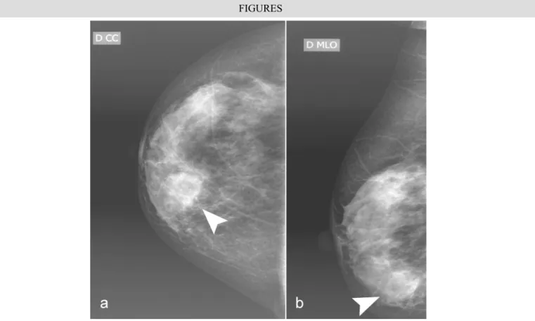
Figure 1: 77-year-old Caucasian woman with Large B-cell non-Hodgkin lymphoma of the right breast. Mammography of patient n 1. 1a: Right breast CC projection digital mammogram (Hologic Selenia Digital Mammography Unit). 1b: Right breast MLO projection digital mammogram . Images showing an ill-defined density in the retroareolar [1a] lower quadrants [1b] (arrows).
6. Wood NL, Coltman CA. Localized primary extranodal Hodgkin's disease. Ann Intern Med.1973; 78:113-118. PMID: 4565899
7. Martinelli G, Ryan G, Seymour JF, et al. Primary follicular and marginal-zone lymphoma of the breast: clinical features, prognostic features and outcome: A study by the International Extra Nodal Lymphoma Study Group. Ann Oncol.2009; 20(12):1993-9. PMID: 19570964
8. Giardini R, Piccolo C, Rilke F. Primary non-Hodgkin's lymphomas of the female breast.Cancer. 1992; 69:725-35. PMID: 1730123
9. Abbondanzo SL, Seidman JD, Lefkowitz M, Tavassoli FA, Krishnan J. Primary diffuse large B-cell lymphoma of the breast. A clinicopathological study of 31 cases. Pathol Res Pract.1996; 192(1):37-43. PMID: 8685040
10. Kuper Hommel MJ, Snijder S, Janssen-Heijnen ML, et al. Treatment and survival of 38 breast lymphomas: a population based study with clinical and pathological reviews. Ann Hematol. 2003; 82(7):397-404. PMID: 12764549
11. Gholam D, Bibeau F, Weshi El, Bosq J, Ribrag V. Primary breast lymphoma. Leuk Lymphoma. 2003; 44(7):1173-8. PMID: 12916870
12. Liu MT, Hsieh CY, Wang AY, et al. Primary breast lymphoma: a pooled analysis of prognostic factors and survival 93 cases. Ann Saudi Med. 2005; 25(4):288-93. PMID: 16212120
13. Ryan G, Martinelli G, Kuper-Hommel M, et al. Primary diffuse large B cell lymphoma of the breast: prognostic factors and outcomes of a study by the International Extra Nodal Lymphoma Study group. Ann Oncol. 2008; 19(2):233-41. PMID: 17932394
14. Kopans DB. Breast Imaging. Lippincott Williams and Wilkins 2007.pp.217-275
15. Babovic N, Jelic S, Jovanovic V. Primary non-Hodgkin lymphoma of the breast: is it possible to avoid mastectomy? J Exp Clin Cancer Res. 2000; 19:149-54. PMID:10965810 16. Domchek SM, Hecht JL, Fleming MD , Pinkus GS,
Canellos GP. Lymphomas of the breast: primary and secondary involvement. Cancer. 2002; 94(1):6-13. PMID: 11815954
17. Brogi E, Harris NL. Lymphomas of the breast: Pathology and clinical behavior. Semin Oncol.1999; 26:357-64. PMID: 10375092
18. Kim SH, Ezekiel MP, Kim RY. Primary lymphoma of the breast. Am J Clin Oncol. 1999; 22:381-83. PMID: 10440194
Jo
urnal o
f
Rad
io
lo
gy
Case Rep
orts
ww
w.Ra
diol
ogyC
ase
s.com
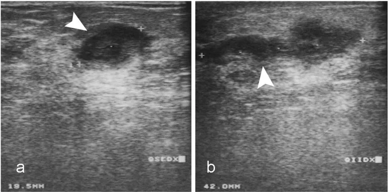
Figure 2: 77-year-old Caucasian woman with Large B-cell non-Hodgkin lymphoma of the right breast (a,b). US of the right breast, which reveals two intraparenchymal ill-defined hypoechoic heterogenous lesions (arrows) corresponding to a palpable mass on physical exam in the lower quadrants (respectively 19,5 and 45 mm).
PROTOCOL: iU22 with a probe center frequency of 9-12 MHz (Philips).
Figure 3: 77-year-old Caucasian woman with Large B-cell non-Hodgkin lymphoma of the right breast. The lymphoma contained medium to large monoclonal B cells positive for CD45, CD19, CD20, CD10, BCL-2, BCL-6 kappa+/lambda-. The lymphomatous lesions were all negative for CD5, CD10, and CD23. a). Lymphoid pattern of the lesion found in the right breast. b). Large B cell Lymphoma well defined on higher magnification x40.
Jo
urnal o
f
Rad
io
lo
gy
Case Rep
orts
ww
w.Ra
diol
ogyC
ase
s.com
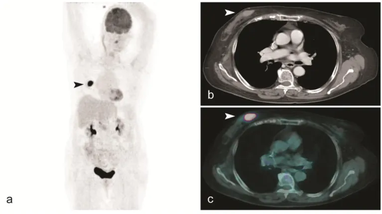
Figure 4: 77-year-old Caucasian woman with Large B-cell non-Hodgkin lymphoma of the right breast. In (a), the maximum intensity projection [MIP] of patient no. 1, which shows a single FDG-avid focus of activity (black arrow), corresponding to (b) a 27x 15 mm mammary nodule (SUVmax 17.5) in the right inner lower quadrant with high FDG uptake (c) in the axial CT and fused PET/CT images (white arrows). PROTOCOL: PET/CT system Discovery ST16, (GE Medical Systems, TN, USA) was used. A low dose CT scan was acquired for attenuation correction of PET images [80 mAs, 140 kVp, field of view (FOV) about 420-500mm, CT slice thickness 3.75]. After nonenhanced CT, total-body PET examination in the caudocranial direction, from upper thighs to vertex, was performed (two bed positions, 4 min per bed). Images were reconstructed using a standard iterative algorithm [ordered subsets expectation maximisation (OSEM)] after 25-30 min of acquisition. Contrast-enhanced CT scan [120-140 kVp, automatic milliamperage (limit 330-350mA), thickness 3.750 mm (reconstructed at 1.25 mm), acquisition mode 27.50/1.375:1, gantry rotation time 0.6 s, large FOV, matrix 512×512] was carried out with i.v. administration of nonionic iodinated contrast material (100-120 ml, 370 mgI/ml, at 2-2.5 ml/s), obtaining two successive stacks of scans. The first comprised the upper abdomen with a 30-s delay from the injection onset; the second extended from the neck to the pelvis with a 60-s delay.
Figure 5 (left): A 69-year-old Caucasian woman with non-Hodgkin lymphoma of the left breast. US which reveals an intraparenchymal ill-defined hypoechoic heterogenous lesion (arrow) corresponding to a palpable mass on physical exam in the upper left quadrants (17 mm)
PROTOCOL: iU22 with a probe center frequency of 9-12 MHz (Philips).
Jo
urnal o
f
Rad
io
lo
gy
Case Rep
orts
ww
w.Ra
diol
ogyC
ase
s.com
Figure 6: A 69-year-old Caucasian woman with non-Hodgkin lymphoma of the left breast. The lesion was a non-Hodgkin, grade II, germinal-center lymphoma of Bcell origin and exhibited a nodular pattern (> 75%) (a). More specifically, the nodules were formed by cells with morphology of centroblasts and centrocytes; the number of centroblasts did not exceed 15 per optical field (×40). The stroma exhibited hyalinization where the malignant lymphoid tissue was present. The malignant lymphoid cells were occasionally present within the fatty breast tissue, and a few entrapped mammary ducts were recognized within the sclerotic stroma. Immunohistochemically, the lymphoid cells were positive for CD20, bcl-2 (b) and CD10, whereas some CD10-positive cells were present outside the nodules. There was indication of kappa chain clonality. The Ki-67 proliferation marker was positive in 10% of cells.
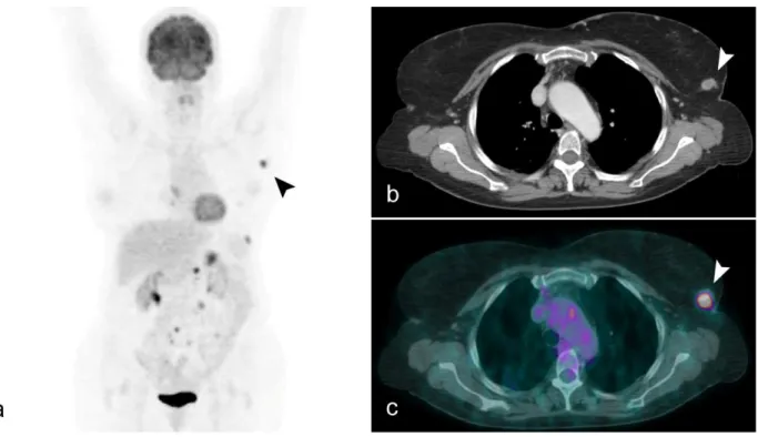
Figure 7: A 69-year-old Caucasian woman with non-Hodgkin lymphoma of the left breast. In (a) MIP of patient. no.2, showing pathologic FDG-uptake (SUVmax 22) in the 13x18 mm mammary nodule (black arrow) localized at the upper outer quadrant of the left breast. Other areas of pathologic FDG uptake were noticed in a paracaval node and in several multiple mesenteric nodes, better seen respectively in the axial CT and PET/CT fuse images (b-c) (white arrows). PROTOCOL: The PET/CT system Discovery ST16, (GE Medical Systems, TN, USA) was used. A low dose CT scan was acquired for attenuation correction of PET images [80 mAs, 140 kVp, field of view (FOV) about 420-500mm, CT slice thickness 3.75]. After nonenhanced CT, total-body PET examination in the caudocranial direction, from upper thighs to vertex, was performed (two bed positions, 4 min per bed). Images were reconstructed using a standard iterative algorithm [ordered subsets expectation maximisation (OSEM)] after 25-30 min of acquisition. Contrast-enhanced CT scan [120-140 kVp, automatic milliamperage (limit 330-350mA), thickness 3.750 mm (reconstructed at 1.25 mm), acquisition mode 27.50/1.375:1, gantry rotation time 0.6 s, large FOV, matrix 512×512] was carried out with i.v. administration of nonionic iodinated contrast material (100-120 ml, 370 mgI/ml, at 2-2.5 ml/s).
Jo
urnal o
f
Rad
io
lo
gy
Case Rep
orts
ww
w.Ra
diol
ogyC
ase
s.com
Breast fibromatosis Invasive ductal carcinoma
Fat necrosis Breast lymphoma Mammography Irregular dense mass
with spiculated margins
Irregular spiculated margins
Lucent center, peripheral rim of calcification
Ill-defined density
Ultrasound Hypoechoic, irregular shape, spiculated margins
Irregular hypoechoic shadowing mass
Acute phase: echogenic structure
Subacute phase: ill-defined complex cyst Late phase: calcified echogenic walls Ill-defined hypoechoic heterogeneous lesions PET/CT Low or no FDG avidity Typically high FDG avidity Typically high FDG avidity
Typically high FDG avidity
Table 2: Differential table for breast tumors (not all-embracing).
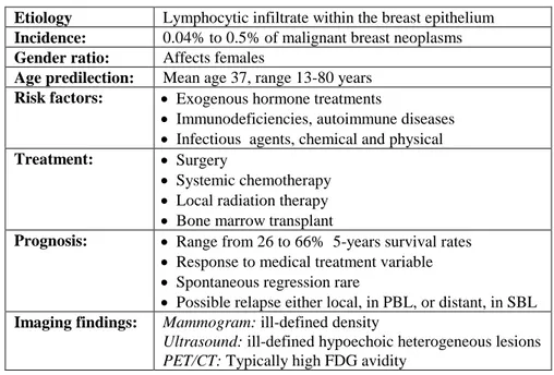
Etiology Lymphocytic infiltrate within the breast epithelium Incidence: 0.04% to 0.5% of malignant breast neoplasms Gender ratio: Affects females
Age predilection: Mean age 37, range 13-80 years Risk factors: Exogenous hormone treatments
Immunodeficiencies, autoimmune diseases
Infectious agents, chemical and physical Treatment: Surgery
Systemic chemotherapy
Local radiation therapy
Bone marrow transplant
Prognosis: Range from 26 to 66% 5-years survival rates
Response to medical treatment variable
Spontaneous regression rare
Possible relapse either local, in PBL, or distant, in SBL Imaging findings: Mammogram: ill-defined density
Ultrasound: ill-defined hypoechoic heterogeneous lesions PET/CT: Typically high FDG avidity
Jo
urnal o
f
Rad
io
lo
gy
Case Rep
orts
ww
w.Ra
diol
ogyC
ase
s.com
BIRADS: Breast Imaging Reporting and Data System CT: Computed Tomography
ENLSG: Extra nodal lymphoma Study Group F-18: Fluorine-18
FDG: Fludeoxyglucose
IGEV gemcitabine,vinorelbine, ifosfamide and prednisone MALT: mucosa-associated lymphoid tissue
MIP: Maximum Intensity Projection Mtd: Maximum transverse diameter Mx: Mammography
NHL: non-Hodgkin's lymphomas PBL: Primary breast lymphoma PET: Positron Emission Tomography
R-CHOP: Rituximab- Cyclophosphamide-
Hydroxydaunorubicin- Oncovin- Prednisone SBL: Secondary breast lymphoma
SUV: Standard Uptake Value
SUVmax: Maximun Standard Uptake Value US: Ultrasound
Breast; lymphoma; primary breast lymphoma; secondary breast lymphoma
This work was supported by grant from "Università di Roma Tor Vergata", Italy.
Online access
This publication is online available at:
www.radiologycases.com/index.php/radiologycases/article/view/1194
Peer discussion
Discuss this manuscript in our protected discussion forum at: www.radiolopolis.com/forums/JRCR
Interactivity
This publication is available as an interactive article with scroll, window/level, magnify and more features.
Available online at www.RadiologyCases.com Published by EduRad
www.EduRad.org ABBREVIATIONS
KEYWORDS